PREVALENCE OF ENTEROTOXIN-ENCODING GENES AMONG DIVERSE SHIGELLA STRAINS ISOLATED FROM PATIENTS WITH DIARRHEA,
SOUTHWEST IRAN
MOJTABAMOOSAVIAN1,2, SAKINEHSEYED-MOHAMMADI2,3, AHMAD FARAJZADEHSHEIKH1,2, SAEED KHOSHNOOD2,3, ARAMASAREHZADEGAN DEZFULI2, MORTEZA SAKI2,3*, GHOLAMREZAGHADERIAN2, FATEMEH SHAHI2,3, MAHTABABDI2
and FARIBAABBASI4
1Infectious and Tropical Diseases Research Center, Health Research Institute, Ahvaz Jundishapur University of Medical Sciences, Ahvaz, Iran
2Department of Microbiology, Faculty of Medicine, Ahvaz Jundishapur University of Medical Sciences, Ahvaz, Iran
3Student Research Committee, Ahvaz Jundishapur University of Medical Sciences, Ahvaz, Iran
4Department of Laboratory Sciences, Golestan Hospital, Ahvaz Jundishapur University of Medical Sciences, Ahvaz, Iran
(Received: 27 May 2018; accepted: 22 June 2018)
Shigella spp. are a major cause of bacillary dysentery, particularly among children in developing countries such as Iran. This study aimed to investigate the presence of two importantShigellaenterotoxins (ShET-1 and ShET-2), encoded by thesetandsengenes, respectively, by polymerase chain reaction (PCR) assay among Shigellaspecies isolated from children affected by shigellosis in Ahvaz, southwest of Iran. In this cross-sectional study, from June 2016 to April 2017, altogether 117Shigellaisolates were collected from fecal specimens of children aged<15 years with diarrhea in Ahvaz, southwest Iran. All isolates were identified by standard microbiological and molecular methods. The presence of enterotoxin genes was determined by PCR. The most prevalent isolate was Shigella flexneri (47.9%), followed byShigella sonnei(41%) andShigella boydii(11.1%), respectively.Shigella dysenteriaewas not detected in patients’samples. The frequencies ofset1A, set1B, and sengenes were 5.1% (6/117), 15.4% (18/117), and 76.9% (90/117), respectively. This study provides initial background on the prevalence and distribution of theShigella enterotoxin genes inShigella isolates in southwest of Iran. In addition, this study revealed a high prevalence ofsenenterotoxin gene inShigellaspecies.
Keywords: Shigella, diarrhea, Iran, enterotoxin, PCR,sen,set
*Corresponding author; E-mail:saki.mo@ajums.ac.ir
First published online September 11, 2018
Introduction
Shigellosis or bacillary dysentery caused by four Shigella species (Shigella flexneri, Shigella dysenteriae, Shigella boydii, and Shigella sonnei) is known as a main public health problem worldwide especially in low-hygienic regions [1–3]. Shigellosis is identified throughout different spectrum of clinical symptom from mild watery diarrhea to severe colitis. A major group affected by shigellosis are children under 5 years old [1, 4, 5]. Various epidemiological studies revealed that S. flexneri 2a and S. sonnei were predominant strains amongShigellaspp. in both developing and developed countries [6,7]. Based on the previous reports worldwide, 164.7 million people are annually infected by Shigella spp., in which 163 million of them are related to developing countries [8,9].
There are different virulence factors inShigellaspecies.Shigellaenterotoxin 1 (ShET1) and Shigella enterotoxin 2 (ShET2) are the two important virulence factors suggested to mediate early fluid secretion in the jejunum and cause of infection in the colon and create watery diarrhea due to shigellosis. The shared name is due to their similar properties as enterotoxins, as there is no homology between ShET1 and ShET2 [10].
ShET1 is encoded by set1A and set1B genes located on the Shigella chromosome and it is part of theShigellaIsland 1 (SHI-1), and mainly present in S. flexneri 2a isolates. The two subunits form a 55 kDa holo-AB-type toxin complex in an A1–B5 configuration. The holotoxin secretion mecha- nism is similar to that of the cholera holotoxin, via the secretory pathway and type-II secretion. ShET1 is associated with the watery phase of diarrhea [11, 12].
Another enterotoxin named ShET2 is encoded byospD3(sen), which is one of the threeospD genes found on the virulence invasion plasmid. This gene is present in all ofShigellaserotypes and participates in invasion to host epithelial cells [13].
Khuzestan province in the southwest of Iran is an endemic area forShigella infections in children [14]. Rapid diagnosis ofShigellastrains is an effective way to control and to decrease the rate of shigellosis outbreaks among the children.
Since using valid diagnostic techniques like polymerase chain reaction (PCR) is highly recommended [5], the aim of this study was to assess the prevalence of Shigellaspp. and the presence of two important ShET-1 and ShET-2 enterotoxin genes by PCR method amongShigellaisolates in children affected by shigellosis in Ahvaz, southwest Iran.
Materials and Methods Ethics
This study was approved by the ethics committee of Ahvaz Jundishapur University of Medical Sciences, Ahvaz, Iran (no.: IR.AJUMS.REC.1396.568).
Study design and specimen collection
This descriptive and cross-sectional study was conducted on 840 fecal specimens of children under 15 years old suffering from diarrhea during June 2016 and April 2017. All samples were obtained along with written consent from children’s parents. These patients were referred to the medical diagnostic labora- tories of the teaching hospitals of Abuzar and Golestan associated with the Ahvaz Jundishapur University of Medical Sciences in Ahvaz city, Iran. Patient informa- tion, such as age, gender, history of fever, vomiting, and bloody diarrhea, was recorded. Before antibiotics consumption, stool samples of all patients were collected in sterile containers and transferred to the Microbiology Department of Faculty of the Medicine for further investigation.
Isolation and identification ofShigella spp.
All specimens were cultured on MacConkey and Hektoen enteric (HE) agar (Merck, Germany), and then incubated at 37 °C for 24 h. Small green to bluish green colonies on HE and colorless colonies on MacConkey agar were selected and examined using standard biochemical and microbiological tests such as Triple Suger Iron agar (Merck, Germany), Lysine Iron agar (Merck, Germany), Simmon’s citrate (Merck, Germany), MR-VP, SH2 production, urease, indole production, and motility [15]. All of the isolates confirmed asShigellaspp. were stored at−80 °C in tripticase soy broth with 20% glycerol for further investiga- tions. Shigellaspecies were differentiated by PCR using specific primers.
DNA extraction
The boiling method was used to extract genomic DNA from Shigella isolates. A few bacterial colonies ofShigellastrains grown overnight on nutrient agar (Merck, Germany) were resuspended in microtubes containing 500 μl of Tris-EDTA buffer, then the microtubes were placed in Incublock microtube
incubators (Denville Scientific, USA) for 5 min at 95 °C, and then centrifuged at 14,000 rpm for 10 min at 4 °C. The supernatant was used as the DNA template in the PCR assays. The DNA quantity and quality were assessed using NanoDrop Spectrophotometer PROMO (Thermo Scientific, USA) and electrophoresis on 1.5% gel agarose, respectively [16].
PCR forShigella spp. differentiation and enterotoxin genes
Differentiation between Shigella spp. was performed by amplification of species-specific genes using primers for putative integrase for Shigella genus, rfc forS.flexneri, wbgZforS. sonnei, rfpBforS. dysenteriae, and hypothetical protein for S. boydii asdescribed in previous studies [17,18]. Afterward, the existence ofShigella enterotoxins was assessed by amplification of sequences associated withset1A, set1B, andsengenes according to previous method described by Cruz et al. [7].
PCR assay was performed in thermocycler (Eppendorf, Germany) using the 2×Master Mix (1.5 mM MgCl2, SinaClon, Iran). The primers oligonucleotides used in this study were obtained from Pishgam Co. (Iran). The reference strains of S. flexneri ATCC29903, S. sonnei ATCC25931, S. boydii ATCC8700, andS. dysenteriaeATCC13313 were used as positive control. The control positive for enterotoxins genes (set1A, set1B, and sen) was kindly donated by Dr. H. Hosseini Nave of the Department of Microbiology and Virology, School of Medicine, Kerman University of Medical Sciences, Kerman, Iran. The total volume of PCR reaction was 25μl prepared as follows: 12.5μl of 2×Master Mix, 1μl of 10 pM each primer, 1μl of template DNA, and distilled water to reach total volume of 25μl. The cycling program was as follows: 1 cycle at 95 °C for 5 min, 35 cycles at 954 °C for 60 s, varying annealing temperatures for each gene for 45 s, and 72 °C for 45 s, and afinal extension cycle at 72 °C for 5 min. The sterile deionized water was included with each PCR run as negative control. The primers’ sequences, annealing temperature, and PCR product size are shown in TableI.
PCR products electrophoresis
The PCR products were separated by electrophoresis (80 V, 40 min) using a 1% agarose gel (SinaClon, Iran) in 1×Tris/Borate/EDTA buffer. An amount of 5μl from each of the product was run on the agarose gel. Then, the gel was stained with ethidium bromide (0.5μg/ml) (SinaClon) and visualized by UV illuminator device (ProteinSimple, USA) and all images were saved on hard disk. A DNA marker of 100 bp (Sinaclon) was used for comparative analysis.
Results
In this study, a total number of 117Shigellaspecies were characterized from 840 fecal specimens by standard biochemical and microbiological tests. These specimens were collected from the children aged <15 years old suffering from diarrhea or dysentery during June 2016 and April 2017. Out of 840 cases, 660 (78.5%) were children under 10 years old. Considering the gender of patients, 308 (36.6%) were females and 532 (63.4%) were males.
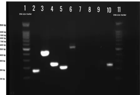
For the confirmation ofShigellagenus, PCR was carried out using specific primers for putative integrase gene (Figure 1) [17]. The results confirmed the presence of this gene in all 117 isolates. In addition, amplification of species- specific genes showed that most isolates belonged toS.flexneri(47.9%), followed byS. sonnei(41%) andS. boydii(11.1%), respectively (TableII).S. dysenteriae was not detected in any of the samples.
The distributions of theset1A, set1B, andsengenes in studiedShigellaspp.
are provided in TableII. The frequency ofsengene among 117Shigellastrains was most common with 76.9% (90/117), whereas theset1Aandset1Bgenes were found only in 6 (5.1%) and 19 (16.2%) isolates, respectively. Among the toxin producers, 17 (30.3%) S. flexneri strains, 4 (8.3%) S. sonnei, and 4 (30.6%) S. boydiiproduced ShET-1 (A or B subunit positive), respectively. None of the
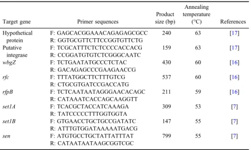
Table I.List of oligonucleotide primers used in PCR method
Target gene Primer sequences
Product size (bp)
Annealing temperature
(°C) References Hypothetical
protein
F: GAGCACGGAAACAGAGAGCGCC 240 63 [17]
R: GGTGCGTTCTTCCGGTGTTCTG Putative
integrase
F: TCGCATTTCTCTCCCCACCACG 159 63 [17]
R: CCGGATGTGTCTCGGGCAATC
wbgZ F: TCTGAATATGCCCTCTAC 430 60 [16]
R: GACAGAGCCCGAAGAACCG
rfc F: TTTATGGCTTCTTTGTCG 537 60 [16]
R: CTGCGTGATCCGACCATG
rfpB F: TCTCAATAATAGGGAACACAGC 211 59 [16]
R: CATAAATCACCAGCAAGGTT
set1A F: TCACGCTACCATCAAAGA 309 53 [7]
R: TATCCCCCTTTGGTGGTA
set1B F: GTGAACCTGCTGCCGATATC 147 55 [7]
R: ATTTGTGGATAAAAATGACG
sen F: ATGTGCCTGCTATTATTTAT 799 55 [7]
R: CATAATAATAAGCGGTCGC Note:PCR: polymerase chain reaction.
isolates were simultaneously positive for both set1Aand set1B genes. Further- more, thesengene was detected in 46 (82.1%)S.flexneri, 33 (68.7%)S. sonnei, and 11 (84.6%)S. boydii. Among the 117Shigellastrains, 20 (17%) isolates were positive for both thesetand thesengenes. Our results show that the prevalence of set1Agene was low in all the Shigellaspecies.
Discussion
The previous studies have indicated that humans and primates are only natural hosts ofShigellaspp. This bacterium with high adaptation with human is a major cause of bacillary dysentery throughout the world, and due to minimal infectious dose (less than 200 bacterial cells), transmission of disease is facilitated in areas with unsuicircumstances such as lack of sanitation [19, 20].
Molecular studies about Shigella enterotoxin genes are still insufficient worldwide and to our knowledge there are no published data on this subject in southwest of Iran. The assessment of Shigella virulence markers helps better understand its pathogenicity. In this study, we investigated Shigella isolates
Figure 1.PCR assay profile withShigellareference strains. Lane 1: 100 bp DNA marker; Lane 2:
positive control (putative integrase–Shigellagenus); Lane 3: positive control (wbgZ–S. sonnei);
Lane 4: positive control (hypothetical protein–S. boydii); Lane 5: positive control (rfpB– S. dysenteriae); Lane 6: positive control (rfc–S.flexneri); Lanes 7–9: negative control; Lane 10:
clinical sample (hypothetical protein–S. boydii); Lane 11: 100 bp DNA marker
TableII.Prevalenceoftheset1A,set1B,andsengenesinShigellaspp.isolatedfromchildrenwithdiarrhea SpeciesTotalno.of isolatesset1Ageneset1BgenesengeneNoneset1A+set1B+ sengenesset1A+sen genesset1B+sen genes S.flexneri56(47.9%)5(8.9%)12(21.4%)46(82.1%)8(14.3%)2(3.6%)3(5.3%)10(17.8%) S.sonnei48(41%)0(0%)4(8.3%)33(68.7%)12(25%)0(0%)0(0%)3(6.2%) S.boydii13(11.1%)1(7.6%)3(23%)11(84.6%)2(15.4%)0(0%)0(0%)2(15.4%) 117(100%)6(5.1%)19(16.2%)90(76.9%)22(18.8%)2(1.7%)3(2.5%)15(12.8%)
collected from children with diarrhea for the presence ofset1A, set1B, and sen enterotoxin genes.
Ourfindings showed thatS.flexneri(47.9%) is the most important cause of shigellosis among children in Ahvaz, southwest of Iran. Our results were in accordance with the previous studies in different parts of Iran, such as Babol, Abadan, and some countries like Brazil, China, Egypt, and India [7,21–24]. In addition, there are some studies that had reportedS. sonneias the most prevalent species. These differences may be related to higher hygiene in these regions [25].
In this study, we also assessed the presence of two chromosomal virulence genes (set1Aandset1B) and one plasmid virulence gene (sen) that encode shET1 and shET2, respectively. Both enterotoxins are significantly associated with bloody diarrhea [26].
This study and some studies in recent years are conflict with prior studies thatset1genes were almost exclusively found inS.flexneriserotypes 2 (2a and 2b) and rarely in other serotypes or species [26–29]. More studies in future may help the confirmation of SHI-1 island as location forset1gene in different species of Shigella.
We detectedset1genes in 30.3% ofS.flexneri, 8.3% ofS. sonnei, and 30.6%
ofS. boydii. Ranjbar [5] in Tehran showed that the prevalence ofset1gene was 66.6%, 21%, 50%, and 0% inS.flexneri, S. sonnei, S. boydii, andS. dysenteriae, respectively. According to Medeiros’s [30] results, 66.7% ofS.flexneri, 11.1% of S. sonnei, 25% ofS. boydii, and 0% ofS. dysenteriaecarriedset1genes. In the study by Roy et al. [31] in India, a total of 153Shigellaisolates were analyzed for the presence ofsetandsengenes. They foundsetgene only inS.flexneriisolates but not in other species andsengene was well distributed among all species [31].
In this study, both ShET-1 and ShET-2 were found in 26.7% ofS.flexneriisolates.
Similarly, Niyogi et al. [27] also reported that 26% ofS.flexneriisolates produced both ShET-1 and ShET-2.
Interestingly, most isolates had only one subunit of ShET-1, and B subunit was more frequent than A subunit (TableII). The contingency analysis in Cruz et al.’s [7] study showed that there is a relationship between existence ofset1B gene inShigellaisolates and dehydration symptoms in children. However, answer to the question about whether a single subunit would affect the pathogenicity of ShET-1 needs further study for verification.
In this study, the prevalence ofsengene was higher (78.7%) than other two genes that was in accordance with reports of the previous studies [12,30,32].
The conflict of various researches is likely because of the loss of the large plasmid that contains the gene in different Shigella species, differences in the distribution of species, and the sample size.
Conclusions
In this study, we afforded some baseline information about the distribution of some virulence genes in clinical strains of Shigella spp. in Ahvaz city, southwest Iran. We found that the prevalence of sen virulence gene is high amongShigella species in southwest Iran.
Acknowledgements
The authors would like to thank the children as well as their parents or guardians as they have agreed to participate in this study. This work was financially supported by Deputy Vice-Chancellor for research affair and Infectious and Tropical Diseases Research Center, Health Research Institute, Ahvaz Jun- dishapur University of Medical Sciences, Ahvaz, Iran (No.: OG-96125). MM and SS-M contributed equally to this work.
Conflict of Interest The authors declare no conflict of interest.
References
1. Ranjbar, R., Dallal, M. M. S., Talebi, M., Pourshafie, M. R.: Increased isolation and characterization ofShigella sonneiobtained from hospitalized children in Tehran, Iran. J Health Popul Nutr26, 426–430 (2008).
2. Bennish, M. L., Wojtyniak, B. J.: Mortality due to shigellosis: Community and hospital data. Rev Infect Dis13, S245–S251 (1991).
3. Dekker, J. P., Frank, K. M.: Salmonella, shigella, and yersinia. Clin Lab Med35, 225–246 (2015).
4. Niyogi, S. K.: Shigellosis. J Microbiol43, 133–143 (2005).
5. Ranjbar, R., Bolandian, M., Behzadi, P.: Virulotyping of Shigella spp. isolated from pediatric patients in Tehran, Iran. Acta Microbiol Immunol Hung64, 71–80 (2017).
6. Koppolu, V., Osaka, I., Skredenske, J. M., Kettle, B., Hefty, P. S., Li, J., Egan, S. M.:
Small-molecule inhibitor of theShigellaflexnerimaster virulence regulator VirF. Infect Immun81, 4220–4231 (2013).
7. Cruz, C. B. N., Souza, M. C. S., Serra, P. T., Santos, I., Balieiro, A., Pieri, F. A., Nogueira, P. A., Orlandi, P. P.: Virulence factors associated with pediatric shigellosis in Brazilian Amazon. Biomed Res Int2014, ID 539697 (2014).
8. Wang, Y. W., Watanabe, H., Phung, D. C., Tung, S. K., Lee, Y. S., Terajima, J., Liang, S. Y., Chiou, C. S.: Multilocus variable-number tandem repeat analysis for molecular typing and phylogenetic analysis ofShigellaflexneri. BMC Microbiol9, 278–288 (2009).
9. Li, S., Wang, J., Wei, X., Liu, Y., You, L., Luo, X., Tang, G., Sun, Q., Ye, C., Xu, J., Wang, D.: Molecular characterization ofShigella sonnei: An increasingly prevalent etiologic agent of shigellosis in Guizhou Province, Southwest of China. PLoS One11, e0156020 (2016).
10. Mattock, E., Blocker, A. J.: How do the virulence factors ofShigellawork together to cause disease? Front Cell Infect Microbiol7, 64 (2017).
11. Fasano, A., Noriega, F. R., Maneval, D. R., Chanasongcram, S., Russell, R., Guandalini, S., Levine, M. M.: Shigellaenterotoxin 1: An enterotoxin ofShigellaflexneri 2a active in rabbit small intestine in vivo and in vitro. J Clin Invest95, 2853–2861 (1995).
12. Yavzori, M., Cohen, D., Orr, N.: Prevalence of the genes forShigellaenterotoxins 1 and 2 among clinical isolates ofShigellain Israel. Epidemiol Infect128, 533–535 (2002).
13. Parsot, C., Ageron, E., Penno, C., Mavris, M., Jamoussi, K., D’Hauteville, H., Sansonetti, P., Demers, B.: A secreted anti-activator, OspD1, and its chaperone, Spa15, are involved in the control of transcription by the type III secretion apparatus activity inShigellaflexneri.
Mol Microbiol56, 1627–1635 (2005).
14. Pour, M. B. M. G., Shokoohizadeh, L., Navab-Akbar, F. T.: Analysis of clonal relationships amongShigellaspp. isolated from children with shigellosis in Ahvaz, Iran. J Paramed Sci 7, 45–51 (2016).
15. Tille, P.: Bailey & Scott’s Diagnostic Microbiology-E-Book. Elsevier Health Sciences, St. Louis, MO, 307–327 2015.
16. Li, S., Sun, Q., Wei, X., Klena, J. D., Wang, J., Liu, Y., Tian, K., Luo, X., Ye, C., Xu, J., Wang, D., Tang, G.: Genetic characterization of Shigella flexneri isolates in Guizhou Province, China. PLoS One10, e0116708 (2015).
17. Ojha, S. C., Yean, Y. C., Ismail, A., Banga, S. K. K.: A pentaplex PCR assay for the detection and differentiation ofShigellaspecies. BioMed Res Int2013, ID412370 (2013).
18. Kim, H. J., Ryu, J. O., Song, J. Y., Kim, H. Y.: Multiplex polymerase chain reaction for identification of shigellae and fourShigellaspecies using novel genetic markers screened by comparative genomics. Foodborne Pathog Dis14, 400–406 (2017).
19. Trevett, A. F., Carter, R. C., Tyrrel, S. F.: The importance of domestic water quality management in the context of faecal-oral disease transmission. J Water Health3, 259–270 (2005).
20. Hossain, M. A., Albert, M. J., Hasan, K. Z.: Epidemiology of shigellosis in Teknaf, a coastal area of Bangladesh: A 10-year survey. Epidemiol Infect105, 41–49 (1990).
21. Savadkoohi, R. B., Ahmadpour-Kacho, M.: Prevalence of Shigella species and their antimicrobial resistance patterns at Amirkola Children Hospital, North of Iran. Iran J Pediatr17, 118–122 (2007).
22. Jomezadeh, N., Babamoradi, S., Kalantar, E., Javaherizadeh, H.: Isolation and antibiotic susceptibility of Shigella species from stool samples among hospitalized children in Abadan, Iran. Gastroenterol Hepatol Bed Bench7, 218–223 (2014).
23. Sangeetha, A., Parija, S. C., Mandal, J., Krishnamurthy, S.: Clinical and microbiological profiles of shigellosis in children. J Health Popul Nutr32, 580–586 (2014).
24. Ranjbar, R., Afshar, D., Tavana, A. M., Najafi, A., Pourali, F., Safiri, Z., Sorouri Zanjani, R., Jonaidi Jafari, N.: Development of multiplex PCR for simultaneous detection of three pathogenicShigellaspecies. Iran J Public Health43, 1657–1663 (2014).
25. Mardaneh, J., Abbaspoor, S., Afrugh, P.: Prevalence ofShigellaspecies and antimicrobial resistance patterns of isolated strains from infected pediatrics in Tehran. Int J Enteric Pathog 1, 28–31 (2013).
26. Yaghoubi, S., Ranjbar, R., Dallal, M. M. S., Fard, S. Y., Shirazi, M. H., Mahmoudi, M.:
Profiling of virulence-associated factors inShigellaspecies isolated from acute pediatric diarrheal samples in Tehran, Iran. Osong Public Health Res Perspect8, 220–226 (2017).
27. Niyogi, S., Vargas, M., Vila, J.: Prevalence of thesat, setandsengenes among diverse serotypes of Shigella flexneri strains isolated from patients with acute diarrhoea. Clin Microbiol Infect10, 574–576 (2004).
28. Cristea, D., Oprea, M., Ciontea, A. S., Antohe, F., Usein, C. R.: Prevalence of virulence markers and pHS-2-like plasmids amongShigella sonnei and Shigellaflexneri isolates originating from shigellosis cases in Romania. Rev Romana Med Lab24, 103–110 (2016).
29. Sousa, M. Â. B., Mendes, E. N., Collares, G. B., Péret-Filho, L. A., Penna, F. J., Magalhães, P. P.: Shigella in Brazilian children with acute diarrhoea: Prevalence, antimicrobial resistance and virulence genes. Mem Inst Oswaldo Cruz108, 30–35 (2013).
30. Medeiros, P. H. Q. S., Lima, A. Â. M., Guedes, M. M., Havt, A., Bona, M. D., Rey, L. C., Soares, A. M., Guerrant, R. L., Weigl, B. H., Lima, I. F. N.: Molecular characterization of virulence and antimicrobial resistance profile ofShigellaspecies isolated from children with moderate to severe diarrhea in northeastern Brazil. Diagn Microbiol Infect Dis90, 198–205 (2018).
31. Roy, S., Thanasekaran, K., Roy, A. R. D., Sehga, S. C.: Distribution ofShigellaenterotoxin genes and secreted autotransporter toxin gene among diverse species and serotypes of Shigellaisolated from Andaman Islands, India. Trop Med Int Health2, 1694–1698 (2006).
32. Farfán, M., Garay, T., Prado, C., Filliol, I., Ulloa, M., Toro, C.: A new multiplex PCR for differential identification ofShigellaflexneriandShigella sonneiand detection ofShigella virulence determinants. Epidemiol Infect138, 525–533 (2010).