FAT BODY CULTURE SYSTEMS IN THE STUDY OF FROG DISEASES
ROBERT L. AMBORSKI DEPARTMENT OF MICROBIOLOGY
GRACE F. AMBORSKI
DEPARTMENT OF VETERINARY MICROBIOLOGY AND PARASITOLOGY
Louisiana State University- Baton Rouge, Louisiana
I. INTRODUCTION
A. Significance of the Problem
The decline of amphibian species is a world wide problem.
The pressures responsible for this decline can be identified in the natural environment as well as in the conditions of mainte- nance after the desired species have been collected. Thus Prestt er al. (1974) have indicated that loss of habitat, prιdation, road mortality, complications associated with hibernation, over collection, pollution and diseases all play roles in reducing the number of frogs in the natural environment. At the other end of the spectrum, Gibbs et al. (1971) suggested that once frogs have been collected and placed in water holding pens factors including overcrowding, lack of environmental temperature control, low oxy- gen level and lack of food contribute to poor health and increased
653
654 ROBERT L. AMBORSKI AND GRACE F. AMBORSKI
susceptibility to bacterial disease. It was noted in this report that in holding pens containing 20,000 to 30,000 frogs each, over half the frogs developed bacterial disease and died during one winter. The control of such diseases would make a significant contribution to the current frog problem.
B. The Major Bacterial Disease of Frogs
The most serious microbial threat to a frog's health is an epizootic form of bacterial septicemia usually accompanied by a cutaneous ulcerative condition, designated by Emerson and Norris
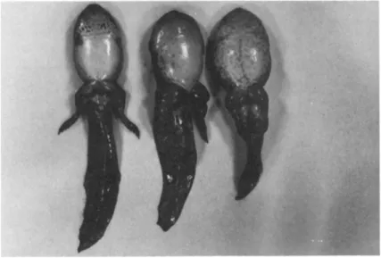
(1905) as Red-leg disease. This disease has destroyed both pond and laboratory populations of a variety of frog species. The disease in the adult frog is insidious sometimes lasting as long as six months before death. However the same disease in the tad- pole often results in massive overnight mortalities. Figure 1 presents a comparison between healthy and diseased tadpoles. The clinical signs of the disease include edema, hemorrhaging of the dermis from the mandible to the toes and the distension of the lymph sacs with a blood-tinged serous fluid, giving a bloated ap- pearance. In the case of the tadpole this latter effect is es- pecially pronounced in the femoral lymph sacs of the hind legs.
Numerous studies have been directed toward the identification of the bacteria associated with this disease. These efforts are summarized in Table I and indicate that at least thirteen gram negative species of six different genera can cause Red-leg dis- ease. As an approach to the study of the possible interactions between these bacteria we attempted to determine whether or not any of the bacterial isolates in Table I were invasive or demon- strated any tissue affinities. Using standard histological tech- niques and a tissue gram stain, tissues from diseased and healthy animals were screened for bacterial involvement. In no instance could gram negative bacteria be shown to be invasive in any of the frog tissues. However quite unexpectedly, bacteria were
FAT BODY CULTURE SYSTEMS 655
FIGURE 1 Bullfrog, Rana catesbeiana, tadpoles. Normal tad- pole CA) and diseased tadpoles CB).
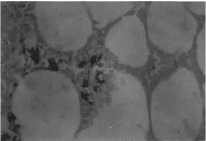
found to invade the fat body, and in every observed case these bacteria were gram positive. Figure 2 shows a histological sec- tion of frog fat body which demonstrates the presence of the gram positive bacterium. This organism has been isolated and through the determinations of substrate utilization, molar percent Guanine/Cytosine content of the DNA and cell wall analysis, this bacterium appears to be a species of Corynebacterium. As of yet all attempts to demonstrate the pathogenicity of this isolate have been negative. A major problem with this system lies in the fact that the Corynebacterium is a slow growing organism. Fur- thermore in those frogs which have died, the microbiological studies have been complicated by the presence of one or more of the gram negative species listed in Table I.
The inability to demonstrate the pathogenicity of the Cory- nebacterium or determine its contribution to the disease syndrome
(if any) led us on a search for other ways to look at this prob'-
656 ROBERT L. AMBORSKI AND GRACE F. AMBORSKI TABLE I History of the Attempts to Define
the Etiology of Red^leg Disease
Date Investigator Bacterial isolates
1898 Russell Bacillus hydrophilus fuscus (Aeromonas hydrophila) 1905 Emerson and Norris Bacillus hydrophilus fuscus
(Aeromonas hydrophila) 1942 Kulp and Borden Bacillus hydrophilus fuscus
(Aeromonas hydrophila)
1950 Miles Bacterium alkaligenes
(Alkaligenes faecalis)
1953 Kaplan Pseudomonas hydrophila
(Aeromonas hydrophila)
1963 Gibbs Aeromonas hydrophila
Citrobacter freundii 1966 Gibbs et al. Aeromonas hydrophila
Mima sp.
(Acinetobacter sp.) 1974 Glorioso et al. Aeromonas hydrophila
Aeromonas shigelloides Citrobacter freundii Flavobacterium sp.
Mima polymorpha
(Acinetobacter sp.) Proteus mirabilis Proteus morganii Proteus retgerii Proteus vulgaris
Pseudomonas aeruginosa Pseudomonas fluorescens Pseudomonas putida
lern. This report represents our attempts to develop a cell cul- ture system which would be applicable to further studies on the Corynebacteriurn.
FAT BODY CULTURE SYSTEMS 657
FIGURE 2 Gram stained histological section of a fat body from a diseased frog. Arrows indicate foci of gram positive bacteria. 172OX.
II. MATERIALS AND METHODS
A. Bacteria
The gram positive organism was isolated as previously de*- scribed and maintained on Ordal Earp agar (Glorioso et al., 1974).
Quantitative determinations were made by serial dilution and the spreader plate technique.
B. Frogs.
All animals were obtained from the Louisiana State University fisheries ponds in Ben Hur farms. The animals were maintained in flow through systems using dechlorinated tap water. Larvae (tad- poles) were maintained on a diet of wheat bran. Adults were fed on a diet of mosquitofish (Gambusia afinisl and crayfish (pro- cambarus clarkii]. The water temperature was regulated at 24 C and a photoperiod of 12L.12D was maintained. Blood samples were
658 ROBERT L. AMBORSKI AND GRACE F. AMBORSKI
collected weekly by heart puncture and screened for the presence of bacteria. Ë11 confirmed or suspect cases of bacteremia or Red- leg disease were removed from the study,
C. Organ culture
Animals were removed from their chambers and sacrificed by pithing. The ventral surface of each animal was washed with 10 percent formalin, and standard aseptic techniques were used to ex- pose the peritoneal cavity. The fat bodies, corpora adiposa, are attached to the anterior ends of both the ovaries and the testes, and are easily recognizable by their yellow color and their divi- sion into a number of finger-like bodies.
Fat bodies were aseptically removed and placed in sterile frog Ringer1s solution without antibiotics. Fat bodies were then fragmented and the fragments were allowed to soak for 30 minutes in the frog Ringer's solution to help remove blood cells. One half of the fragments were randomly selected and removed for bac- teriological determinations, and the remaining fragments were used to set up organ cultures. Each frog was processed separately and tissue samples were not mixed. The organ cultures were prepared by placing individual fragments measuring 5 to 10 mm in length on grids in plastic organ culture dishes (Bioquest, California) or by allowing fragments to float on the surface of the media. Frag- ments were removed at intervals and histological sections were prepared by standard techniques and stained for bacteria (Luna, 1968). All incubations were carried out at 24C.
D. Media
Eagle's minimal essential medium (MEM)r modified MEM equiva- lent to 30, 50, 70 and 90 percent MEM and Wolf and Quimby amphibi- an tissue culture medium was used in this study. Media and sera were purchased from Grand Island Biological (New York). Each
FAT BODY CULTURE SYSTEMS 659
medium was buffered with. 14 mM HEPES CN^2^hydroxyr*ethyl^pipera- zine^N1^ethane sulfonic acidl and did not contain antibiotics.
III. RESULTS
In a preliminary report, Amborski et al. (1974) indicated that frog fat bodies could be maintained in organ culture for periods up to 14 days before central necrosis was observed.
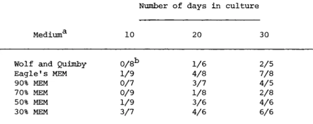
These observations have now been extended by using media with a reduced osmotic pressure as shown in Table II. The structural integrity of the organ culture system was maintained for periods of up to 30 days under the described conditions. Furthermore the histological patterns indicated that the free floating fragments gave results comparable to those observed in the fragments main- tained in the organ culture dishes. The concentration of the serum (1 to 10 percent) did not appear to be critical, and the source of the sera did not affect the results as fetal calf, bo- vine, equine, porcine and human sera gave comparable responses.
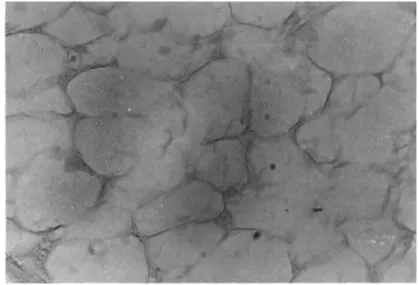
However one of the problems encountered during the long term incu- bations was microbial contamination as many frogs are parasitized with bacteria, yeasts, molds and ciliates. In addition, 30 per- cent of the fat bodies were discarded when the Corynebacterium was demonstrated in the tissue fragments of the apparently healthy donors. Figure 3 shows a typical fragment under floating condi- tions.
Fragments similar to those shown in Fig. 3 were exposed to 1 06 each of the Corynebacterium sp. by adding the bacteria direct- ly to the growth medium. After 24 hours the medium was removed and then each fragment was washed 3X with fresh medium and then incubated. Fragments were harvested daily and histological sec- tions were prepared and gram stained. In the presence of precip- itated stain, it was difficult to identify the Corynebacterium during the first four days of incubation, but after eight days of
660 ROBERT L. AMBORSKI A N D GRACE F. AMBORSKI
Number of days in culture
Mediuma 10 20 30
Wolf and Quimby Eagle's MEM 90% MEM 70% MEM 50% MEM 30% MEM
0/8b 1/9 0/7 0/9 1/9 3/7
1/6 4/8 3/7 1/8 3/6 4/6
2/5 7/8 4/5 2/8 4/6 6/6
aMedia supplemented with 10 percent fetal calf serum.
bRatio represents number of organ cultures showing central necrosis/total number of organ cultures observed.
incubation foci of bacteria were obvious. A histological section of an uninoculated culture is shown in Fig. 4, and a histological section prepared from an inoculated culture is shown in Fig. 5.
The clumping of the bacteria made it difficult to make any quanti- tative measurements on these cultures, but the Corynebacterium was readily isolated after eight days of culture. The amount of tissue destruction was apparently light, and it would appear that the action of the bacterium in the animal and in the culture sys- tem does not result in gross destruction of the fat body tissue.
As a result of some of our original observations, fragments of fat bodies were also set up as classical expiant cultures.
After an incubation period of 6 to 10 days, cells were observed around the periphery of the explants. However the continued out- growth was only observed in media containing 70 to 80 percent MEM and in the WQ medium, and under these conditions monolayers were observed to form after two weeks in culture. However it must be pointed out that the outgrowth did not present a uniform pattern, and even in cultures in media with a reduced osmolality, the ex- pansion of the initial outgrowth did not always occur. Typical
TABLE II Morphological Integrity of Fat Body Organ Cultures
FAT BODY CULTURE SYSTEMS 661
FIGURE 3 Frog fat body in organ culture. 50X.
FIGURE 4 Gram stained histological section of frog fat body organ culture after 13 days incubation. No bacteria added.
800X.
662 ROBERT L. AMBORSKI AND GRACE F. AMBORSKI
FIGURE 5 Gram stained histological section of frog fat body organ culture after 13 days incubation. Arrows indicate foci of gram positive bacteria. Bacteria were added five days after the organ culture was initiated. 800X,
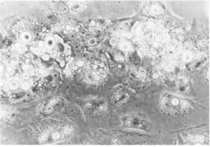
FIGURE 6 Adipocytes from expiant culture after eight days in culture, 700X.
FAT BODY CULTURE SYSTEMS 663
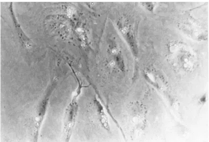
FIGURE 7 Adipocytes from expiant culture after 12 days in culture. 700X.
FIGURE 8 Adipocytes from expiant culture after 18 days in culture. 700X.
664 ROBERT L. AMBORSKI AND GRACE F. AMBORSKI
patterns of outgrowth, are shown in Figs. 6, 7 and 8. Figure 6 shows the outgrowth immediately adjacent to the expiant after 8 days in culture. Light refractile fat inclusions of varying size can be seen. It would appear that the loss of fat might involve the disruption of the large single inclusion into a number of smaller droplets. Even after 12 days in culture as shown in Fig.
7, the cells are still stellate and contain substantial amounts of fat. Finally as shown in Fig. 8, after 18 days in culture, the cells are more fibroblastic in shape, an intact nucleus can be observed and there is almost complete replacement of the fat by cytoplasm.
IV. DISCUSSION
A major problem in the study of Red-leg disease in frogs has been the large number of different bacteria which have been shown to contribute to the disease syndrome. This complexity has ef- fectively limited the studies on this syndrome and may reflect the same interactions which are involved in human infections caused by opportunistic pathogens. However in the case of the frog dis- ease, the observation by Van der Waaij et al. (1974) that the microbial flora of the frog could be substantially reduced by hibernating temperatures, and the present observations that the Corynebacterium sp. can infect fat in an organ culture system of- fers some new experimental approaches to understanding Red-leg disease.
The limits on the use of the frog fat body organ culture sys- tem appear to be the variability that we have observed in the cultural response of that tissue. Amphibians are essentially lean animals and do not have stores of subcutaneous fatty tissue as found in birds and mammals. The fat stored in the fat bodies are apparently necessary for maintaining the health and normal development of the gonads. There is thus a cyclic response in
FAT BODY CULTURE SYSTEMS 665
the deposition of fat and some of the variability observed in our studies may reflect differences in the stages of fat body devel- opment in the donor animal.
In general our results agree with the known response of am- phibian cells in culture CMonnickendam and Balls, 1973), and more
specifically there is agreement*with the known response of adi- pose cells in culture (Adebonojo, 1975).
ACKNOWLEDGMENTS
This work was supported by NIH Grant No. 5 P06 RR00635-04AR, The Louisiana State University Agriculture Experiment Station and The Department of Microbiology.
REFERENCES
Amborski, R. L., Glorioso, J. C. and Amborski, G. F. (1974). in
"Proc. Gulf Coast Regional Symp. Diseases of Aquatic Animals."
(R. L. Amborski, M. A. Hood a d R. R. Miller, eds.), pp. 217- 224. Center for Wetland Resources Louisiana State University, Baton Rouge, La.
Abebonojo, F. 0. C1975) . The Yale J. of Biol. and Med. 48, 9-16.
Emmerson, H. and Norris, C. (1905). J. Exptl. Med. 7, 32-58.
Gibbs, E. L., Gibbs, T. J. and Van Dyck, P. C. (1966). LaJb. Anim.
Care 16, 142-160.
Gibbs, E. L., Nace, G. W. and Emmons, M. B. (1971). Bioscience 21, 1027-1034.
Gibbs, E. L. (1973). Am. Zool. 13, 781-783.
Glorioso, J. C , Amborski, R. L., Amborski, G. F. and Culley, D.
D. (1974). Am. J. Vet. Res. 35, 1241-1245.
Kulp, W. L. and Borden, D. G. C1942). J. Bact. 44, 673-679.
Luna, L. G. C1968I. "Manual of Histological Staining Methods of
666 ROBERT L. AMBORSKI AND GRACE F. AMBORSKI
The Armed Forces Institute of Pathology," McGraw-Hill Book Co., New York.
Miles, Å, M. (1950). J. Gen. Microbiol. 4, 434-436.
Monnickendam, M. A. and Balls, M. (1973). Experientia 29, 1-17.
Prestt, I., Cooke, A. S. and Corbett, K. F. (1974). In "The Changing Flora and Fauna of Britain" (D. L. Haksworth, ed.) pp.
229-254. Academic Press, New York.
Rüssel, F. H. (1898). J. Am. Med. A. 30, 1442-1449.
Van der Waaij, D., Cohen, B. J. and Nace, G. W. (1974), Lab. Anim.
Sei. 24, 307-317,