Bioactivity-guided isolation of antiproliferative compounds from Centaurea jacea L.
Peter Forgo
a, István Zupkó
b, Judit Molnár
b, Andrea Vasas
a, György Dombi
c, Judit Hohmann
a,⁎
aDepartment of Pharmacognosy, University of Szeged, Eötvös u. 6, H-6720 Szeged, Hungary
bDepartment of Pharmacodynamics and Biopharmacy, University of Szeged, Eötvös u. 6, H-6720 Szeged, Hungary
cDepartment of Pharmaceutical Analysis, University of Szeged, Somogyi u. 4, H-6720 Szeged, Hungary
a r t i c l e i n f o a b s t r a c t
Article history:
Received 10 February 2012 Accepted in revised form 6 April 2012 Available online 17 April 2012
Bioassay-guided fractionation of the chloroform extract of Centaurea jaceaL. afforded the isolation of cirsiliol, apigenin, hispidulin, eupatorin, isokaempferide, axillarin, centaureidin, 6- methoxykaempferol 3-methyl ether, trachelogenin, cnicin, 4′-acetylcnicin and three aliphatic glucose diesters, including the new natural product 1β-isobutanoyl-2-angeloyl-glucose. The structures of the compounds were established on the basis of spectroscopic analyses (UV, MS and NMR). All compounds were isolated for the first time from this species. The compounds were evaluated for their tumour cell growth inhibitory activities on HeLa, MCF-7 and A431 cells. Different types of secondary metabolites (flavonoids, sesquiterpenes) were found to be responsible for the antitumour effects of the extracts; the highest activity was exerted by centaureidin, in addition to moderately active compounds (cirsiliol, isokaempferide, apigenin, hispidulin, cnicin and 4′-acetylcnicin).
© 2012 Elsevier B.V. All rights reserved.
Keywords:
Centaurea jacea Asteraceae Glucose esters Flavonoids Sesquiterpenes Antiproliferative activity
1. Introduction
The genus Centaurea (Asteraceae) comprises about 500 species, 221 of which are native to Europe[1]. The species of the genus are not widely applied in European folk medicine, though there are some references to their usage, most frequently for the treatment of ophthalmia (Centaurea calcitrapa,Centaurea cyanus), fever (Centaurea cyanus, Centaurea jacea, Centaurea solstitialis), gynaecological problems (C. cyanus), digestive com- plaints (C. calcitrapa,C. cyanus), wounds and dermatological complaints (C. calcitrapa,C. cyanus,C. jacea)[2]. In Hungarian traditional medicine, the most frequently used species is C. cyanus, presumably because of its wide-ranging geographical distribution. The majority of the HungarianCentaureaspecies have been poorly analysed from phytochemical and pharmaco- logical aspects. In a previous investigation the main components
ofC. arenariawere isolated by bioactivity-guided fractionation by our group and the antiproliferative activity of the compounds were evaluated[3].
In our screening programme for antiproliferative activity of Asteraceae species native to Hungary,Centaureaspecies (C. biebersteinii,C. jaceaandC. spinulosa) demonstrated high cell proliferation inhibitory activity against cervix adenocarci- noma (HeLa), skin epidermoid carcinoma (A431) and breast epithelial adenocarcinoma (MCF-7)[4,5]. The highest activity was recorded forC. jacea, whose chloroform extract, prepared from different plant parts (flowers, leaves and roots), signifi- cantly inhibited the growth of HeLa (57–86%), MCF-7 (44–64%) and A431 (43–69%) cellsin vitro. The antitumour constituents ofC. jaceawere not investigated earlier. The aim of the present work was the isolation and identification of the antiproliferative compounds from the aerial parts ofC. jaceausing bioactivity- guided fractionations.
C. jaceaL. (brown knapweed) is a perennial herb occurring widespread in Europe. This species has been used sporadically in traditional medicine (against fever and dermatological
⁎Corresponding author. Tel.: + 36 62 545558; fax: + 36 62 545704.
E-mail address:hohmann@pharm.u-szeged.hu(J. Hohmann).
0367-326X/$–see front matter © 2012 Elsevier B.V. All rights reserved.
doi:10.1016/j.fitote.2012.04.006
Contents lists available atSciVerse ScienceDirect
Fitoterapia
j o u r n a l h o m e p a g e : w w w . e l s e v i e r . c o m / l o c a t e / f i t o t e
complaints), but is regarded as bioindicator for monitoring of ozone effect. Previous studies revealed the presence of flavonoids (jaceidine[6], jacein[7], centaurein[8], jaceoside[9], 6-methoxykaempferol 3-methyl ether and its 7-O-glucoside [10]), sesquiterpenes (amarine, hydroamarine, melitensin and dihydromelitensin)[11], and cinnamic alcohol gluco- sides (syringin and coniferin)[12].
In the present study the dried plant materials were extracted with MeOH, and this extract was subjected to solvent–solvent partitioning, yielding n-hexane-, CHCl3- and aqueous MeOH- soluble phases. The CHCl3extract exerted substantial tumour cell growth inhibitory activity at 10μg/mL concentration primarily against HeLa cell line[4]. Therefore, the CHCl3extract was selected for a detailed phytochemical-pharmacological analysis in order to identify the antiproliferative constituents.
2. Experimental 2.1. General
Optical rotation was determined with a Perkin-Elmer 341 polarimeter. NMR spectra on solutions in CDCl3 were recorded on a Bruker Avance DRX 500 spectrometer at 500 MHz (1H) or 125 MHz (13C); the signals of the deuterated solvent were taken as reference. Two-dimensional (2D) experiments (1H,1H COSY, HSQC, HMBC and NOESY) were set up, performed and processed with the standard Bruker protocol. Polyamide was used for column chromatography (ICN). Silica gel plates were applied for analytical and preparative TLC (Merck 5717 and 5715). ESIMS was performed on an Applied Biosystems 3200 QTrap instrument in ion trap mode. For vacuum liquid chromatography (VLC), silica gel (60G, 15μm, Merck 11677) was applied. Rotation planar chromatography (RPC) was performed with a Chromatotron instrument (Model 8924, Harrison Research) on manually prepared silica gel (60 GF254, Merck 7730) plates. Preparative layer chromatography (PLC) was carried out on silica gel plates (20 × 20 cm 60GF254, Merck 5715). Separation was monitored at UV 254 nm. HPLC was carried out on a Waters instrument on a LiChrospher RP-18 (5μm, 250 × 4 mm, Merck) column.
2.2. Plant material
Aerial parts ofC. jaceawere collected in August 2010 in Budaörs, Hungary. Botanical identification of the plant material was performed by Dr. Tamás Rédei (Hungarian Academy of Sciences, Institute of Ecology and Botany, Vácrátót, Hungary) and a voucher specimen (no. 776) has been deposited at the Department of Pharmacognosy, University of Szeged.
2.3. Extraction and isolation
Dried and ground aerial parts of C. jacea(1.5 kg) were extracted with 20 L MeOH. After concentration, the extract (250.4 g) was dissolved in 1 L 50% aqueous MeOH, and solvent–solvent partition was performed first with 5 × 500 mL n-hexane, and then with 5 × 500 mL CHCl3. After evaporation, the CHCl3phase (62.5 g) was fractionated by CC on silica gel (940 g), using a gradient system ofn-hexane–acetone–MeOH (9:1:0, 8:2:0, 7:3:0, 6:4:0, 1:1:0, 50:50:3, 10:10:1, 5:5:1, 5:5:2 and 1:1:1, 500 mL each). Fractions with similar composition
were combined according to TLC monitoring, affording frac- tions F1–F21, which were tested for their antiproliferative effects. The most active compounds were concentrated in F11 (161.6 mg) [cell proliferation inhibition 94.04 ± 0.46% (HeLa);
46.39 ± 9.23% (MCF-7); 86.50 ± 1.62% (A431)], F13 (129.4 mg) [83.41± 1.52% (HeLa); 14.91± 2.48% (MCF-7); 51.88± 2.20%, (A431)] and F15 (198.5 mg) [96.12 ± 0.27% (HeLa); 65.24 ± 4.43% (MCF-7); 81.51 ± 0.59 % (A431)]. These fractions were separated by OCC on polyamide [800 mg (F11), 650 mg (F13) and 1.0 g (F15)], using a solvent system of MeOH–H2O (1:4, 2:3, 3:2 and 4:1, 50 mL each). Subfractions F11/8 and F11/9, obtained by polyamide CC of F11 with MeOH–H2O 3:2, were purified by preparative TLC, using cyclohexane–EtOAc–EtOH 50:50:3, which led to the isolation of trachelogenin (4) (16.3 mg). Subfractions F11/13 and F11/14, obtained with MeOH–H2O 3:2, were further fractionated by preparative TLC on silica gel, using toluene–EtOAc–HCOOH 5:4:1 to yield eupatorin (10) (8.6 mg). Subfractions F11/15–19, obtained by polyamide CC with MeOH–H2O 3:2, were separated by RPC with a gradient system of cyclohexane–EtOAc–EtOH (6:2:0, 6:3:0, 60:30:1, 60:30:2, 50:50:3 and 10:10:1). Subfractions 2 and 3, obtained with cyclohexane–EtOAc–EtOH 6:3:0, 60:30:1 and 60:30:2, were rechromatographed by preparative TLC with the solvent system CHCl3–MeOH 19:1, resulting in the isolation of isokaempferid (11) (6.1 mg, mp. 261–264 °C), hispidulin (9) (13.7 mg), 6-methoxykaempferol 3-methyl ether (14) (18.6 mg) and centaureidin (13) (20.2 mg). Finally, subfraction F11/20, obtained with MeOH–H2O 4:1, was purified by preparative TLC, using cyclohexane–EtOAc–EtOH 50:50:3, which led to the isolation of apigenin (7) (4.0 mg).
Fraction F13, which was eluted with n-hexane–acetone– MeOH 1:1:0, was separated on polyamide CC. From subfraction F13/1 eluted with MeOH–H2O 1:4 cirsiliol (8) (12.1 mg) was crystallised. The remaining material was further fractionated by preparative TLC on silica gel, using a mobile phase of CHCl3– MeOH 19:1, and finally it was separated by RP-HPLC, using MeOH–H2O 3:2, to yield 1β-isobutanoyl-2-angeloyl-glucose (1) (9.8 mg), 1β,2-diangeloyl-glucose (2) (10.0 mg) and 1β-(2- methylbutanoyl)-2-angeloyl-glucose (3) (11.0 mg). Subfraction F13/2–5, eluted with MeOH–H2O 1:4, were separated by RPC with a gradient system of cyclohexane–EtOAc–EtOH 50:50:3 to give 4′-acetyl-cnicin (6) (8.2 mg). Moreover, subfraction F13/6, obtained with MeOH–H2O 3:2, was purified by preparative TLC on silica gel with the mobile phase toluene–EtOAc–HCOOH 5:4:1, which led to the isolation of axillarin (12) (21.1 mg).
Fraction F15 was separated on polyamide CC. Subfrac- tions eluted with MeOH–H2O 1:4 and 2:3 were combined and subjected to RPC, using gradient solvent system of cyclohexane–EtOAc–EtOH (6:3:0, 60:30:1, 60:30:2, 60:30:3, 50:50:3, 10:10:1 and 5:5:1), to give pure cnicin (5) (168.2 mg, mp. 147–150 °C).
2.3.1. 1β-Isobutanoyl-2-angeloyl-glucose
(1): Colourless oil with [α]27D: +15 (0.1, CHCl3).1H NMR (CDCl3, 500 MHz) and13C NMR (CDCl3, 125 MHz) data, see Table 1. HR-ESIMS: m/z 334.2985 (calcd. 334.2984 for C15H26O8Na [M+Na]+).
2.3.2. 6-Methoxykaempferol 3-methyl ether
(14):1H NMRδH: (CD3OD): 3.71 (3H, s, 6-OCH3), 3.81 (3H, s, 3-OCH3), 6.20 (1H, s, H-8), 6.85 (2H, d,J= 8.8 Hz, H-3′,5′), 7.92
(2H, d,J= 8.8 Hz, H-2′,6′).13C NMRδC: 60.4 (3-OCH3), 60.5 (6- OCH3), 98.0 (C-8), 102.2 (C-10), 117.4 (C-3′,5′), 121.6 (C-1′), 131.0 (C-2′,6′), 135.9 (C-6), 138.2 (C-3), 152.8 (C-9), 155.4 (C- 5), 156.7 (C-2), 164.1 (C-7), 171.1 (C-4′), 178.7 (C-4).
2.4. Antiproliferative assay
Antiproliferative effects were measured in vitro on three human cell lines (HeLa, MCF-7 and A431) with the MTT ([3- (4,5-dimethylthiazol-2-yl)-2,5-diphenyltetrazolium bromide]) assay (Mosmann, 1983). Briefly, a limited number of human cancer cells (5000/well) were seeded onto a 96-well microplate and became attached to the bottom of the well overnight. On the second day of the procedure, the original medium was removed and 200μL MTT solution (5 mg/mL) was added. MTT was converted by intact mitochondrial reductase and precipi- tated as blue formazan crystals during a 4 h contact period. The medium was then removed, and the precipitated crystals were dissolved in 100μL DMSO during 60 min period of shaking.
Finally, the reduced MTT was assayed at 545 nm using a microplate reader, wells with untreated cells being taken as the control. Allin vitroexperiments were carried out on two microplates with at least five parallel wells. Doxorubicin and cisplatin were used as positive controls. Stock solutions of 10 mg/mL of the tested compounds and extracts were prepared with DMSO. The highest DMSO concentration (0.3%) of the medium did not have any significant effect on cell proliferation.
The dose–response curves of the compounds were fitted by means of the computer programme GraphPad Prism 4.0 (GraphPad Software, San Diego, CA, USA), and IC50values (the concentration at which the cell proliferation is 50% of the untreated control) were calculated.
3. Results and discussion
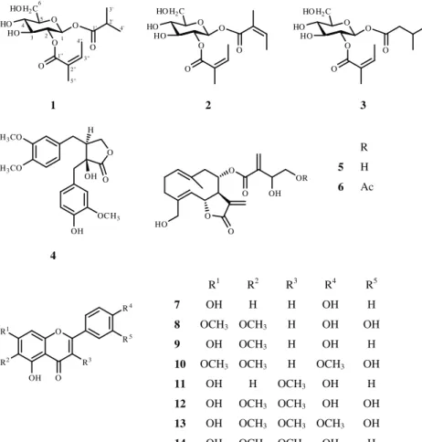
Dried and ground aerial parts ofC. jaceawere extracted with MeOH. After concentration, the extract was dissolved in 50% aqueous MeOH, and solvent–solvent partition was performed first withn-hexane and then with CHCl3. After evaporation, the CHCl3 phase was fractionated by OCC on silica gel, using a gradient system of n-hexane–acetone– MeOH. Fourteen compounds (1–14) were isolated from the highly and moderate active fractions through the use of VLC, CPC, preparative TLC and HPLC techniques. The compounds were identified by means of UV, ESIMS and NMR spectros- copy (1H-NMR, JMOD,1H,1H COSY, HSQC, HMBC and NOESY) and comparison of the spectral data with literature data.
Compound1was isolated as colourless oil with [α]27D+15 (CHCl3,c0.1). It was shown by HRESIMS to have the molecular formula C15H26O8(M= 332) through the presence of quasi- molecular ion peak at m/z 355.1363 [M+ Na]+ (calcd for C15H26O8Na 355.1369). The 1H and 13C NMR spectra of 1 revealed the presence of one isobutanoate group (δH2.56 sept, 1.13 d and 1.11 d;δC 175.4, 33.8, 18.8 and 18.3) and one angelate group (δH6.13 q, 1.95 d and 1.85 s;δC167.4, 127.0, 140.0, 20.4 and 15.9) (Table 1). The remaining signals indicated the presence of a diacylated glucose in the molecule. The positions of the ester groups were established via the HMBC experiment. The correlations of the carbonyl signal atδC175.4 (isobutanoyl CO) with the proton signals at δH5.72 (H-1) indicated the presence of the isobutanoyl group on C-1.
Similarly, the HMBC cross-peak of the carbonyl carbon signal atδC167.4 with the proton signal atδH5.02 (H-2) demon- strated the presence of the angeloyl group on C-2. All of the above evidence confirmed the structure of this compound as 1β-isobutanoyloxy-2-angeloyloxyglucose (1).
Table 1
NMR data of compounds1–3[CDCl3, 500 MHz (1H), 125 MHz (13C),δ(ppm), (J= Hz)].
1 2 3
Atom 1H 13C 13C 13C
1 5.72 d (8.2) 91.6 92.0 91.8
2 5.02 t (8.1) 72.5 72.5 72.2
3 3.74 m 75.2 75.4 75.0
4 3.74 m 70.1 70.2 69.8
5 3.52 m 76.3 76.3 76.2
6 3.91 brd (12.6), 3.88 brd (12.6) 61.5 61.5 61.3
1-O-acyl Isobutanoyl Angeloyl Isovaleroyl
1′ – 175.4 165.8 172.0
2′ 2.56 sept (7.0) 33.8 126.6 43.0
3′ 1.11 d (7.0) 18.8 141.4 25.3
4′ 1.13 d (6.9) 18.3 15.9 22.0
20.2 22.0
2-O-Acyl Angeloyl Angeloyl Angeloyl
1″ – 167.4 167.5 167.0
2″ – 127.0 127.1 127.0
3″ 6.13 q (6.8) 140.0 139.5 139.7
4″ 1.95 d (6.8) 15.9 15.8 16.0
5″ 1.85 s 20.4 20.4 20.3
OH 4.25 brs – – –
OH 4.1 brs – – –
OH 3.10 brs – – –
Compounds2and3were identified as 1β,2-diangeloyl- glucose (2) and 1β-(2-methylbutanoyl)-2-angeloyl-glucose (3), which were isolated previously only from Centaurea napifoliaandCentaurea diffusa[13,14]. Their 13C NMR data are published for the first time in this article (Table 1).
Compounds4–14were found to be identical in all of its characteristics, including NMR data, with trachelogenin (4) [15], cnicin (5)[3], 4′-acetylcnicin (6)[16], apigenin (7)[17], cirsiliol (8) [18], hispidulin (9) [19], eupatorin (10) [20], isokaempferide (11)[3,21], axillarin (12)[22], centaureidin (13) [23] and 6-methoxykaempferol 3-methyl ether (14) [24] (Fig. 1). Interestingly our bioassay directed isolation procedure led to the identification of other compounds than isolated earlier from this species with the only exception of 6-methoxykaempferol 3-methyl ether (14). Methoxylated flavones, such as cirsiliol (8), hispidulin (9), and the flavonol isokaempferid (11) were described previously from different Centaureaspecies[25]. Similarly, the germacranolide cnicin (5) was detected in someCentaureaspecies[26]. 4′-Acetyl-cnicin (6) is a structurally related analogue of amarine, isolated previously from the plant[27].
The isolated compounds were evaluated for their growth inhibitory activity on HeLa, MCF-7 and A431 cells (Table 2), and it emerged that flavones and sesquiterpenes exerted pronounced concentration-dependent effects. The highest activity was demonstrated by the flavonol centaureidin (13), with IC50 values of 0.08μM (HeLa), 0.13μM (MCF-7) and
0.35μM (A431). The extremely high cytotoxicity of centaur- eidin (13) was detected earlier by Beutler et al. in anin vitro screening in the NCI 60-cell line panel [28]. Interestingly, isokaempferide (11) and axillarin (12), the close analogues of centaureidin (13) were found to be less active, and only on HeLa cell line. Moreover, apigenin (7), cirsiliol (8), hispidulin (9), cnicin (5) and 4′-acetyl-cnicin (6) inhibited the prolif- eration of all three tumour cells significantly, while sugar esters (1–3) and trachelogenin (4) were found to be inactive against all the tested cell lines.
In summary, our bioactivity-guided isolations led to the conclusion that the strong inhibitory effect of the CHCl3
extract of C. jacea on the proliferation of cultured human tumour cell lines (HeLa, MCF7 and A431) may be attributed mainly to flavones and sesquiterpenoids. Predominantly, the content of the most active compound, centaureidin (13), determines the antitumour activity of the extract, with the other compounds (5–12,14) playing an additional role in this effect. Moreover, a favourable interaction between chemical- ly unrelated compounds (phenolics and sesquiterpenoids) could be suggested which is responsible for the overall antiproliferative action of the extract.
Acknowledgements
This work was supported by OTKA grant K72771, the New Hungary Development Plan projects TÁMOP-4.2.1/B-09/1/
O HO H2C HO
HO O O
O O
2 1 3 4
5 6
1' 2' 3'
4'
1'' 2''
3'' 4''
5''
O HO H2C HO
HO O O
O O
O HO H2C HO
HO O O
O O
1 2 3
O H3CO
H3CO
O
OC H3 OH
OH H
O HO
O O
O OH OR
4
O R1
OH O R3
R4
R2
R5
R
5 H
6 Ac
R1 R2 R3 R4 R5
7 OH H H OH H
8 OCH3 OCH3 H OH OH
9 OH OCH3 H OH H
10 OCH3 OCH3 H OCH3 OH
11 OH H OCH3 OH H
12 OH OCH3 OCH3 OH OH 13 OH OCH3 OCH3 OCH3 OH
14 OH OCH3 OCH3 OH H
Fig. 1.Structures of compounds isolated fromCentaurea jacea.
KONV-2010-0005 and TÁMOP-4.2.2/B-10/1-2010-0012, and Gábor Baross project OMFB-00339/2010. The authors thank to Tamás Rédei (Hungarian Academy of Sciences, Institute of Ecology and Botany, Vácrátót, Hungary) for the identification of the plant material and to Zoltán Kele (Department of Medical Chemistry, University of Szeged, Szeged, Hungary) for the mass spectrometry measurement. Andrea Vasas acknowledges the János Bolyai scholarship of the Hungarian Academy of Sciences.
Appendix A. Supplementary data
Supplementary data to this article can be found online at http://dx.doi.org/10.1016/j.fitote.2012.04.006.
References
[1] Tutin TB, Heywood VH, Valentine DH, Walters SM, Webb DA, editors. Flora Europaea, vol. 4. Cambridge–London–New York–Melbourne: Cambridge University Press; 1976.
[2] Hänsel R, Keller K, Rimpler H, Schneider G, editors. Hagers Handbuch der Pharmazeutischen Praxis, 4. Band. Berlin-Heidelberg: Springer-Verlag;
1992. p. 750–6.
[3] Csapi B, Hajdú Z, Zupkó I, Berényi Á, Forgo P, Szabó P, et al. Bioactivity- guided isolation of antiproliferative compounds fromCentaurea arenaria.
Phytother Res 2010;24:1664–9.
[4] Réthy B, Csupor-Löffler B, Zupkó I, Hajdú Z, Máthé I, Hohmann J, et al.
Antiproliferative activity of Hungarian Asteraceae species against human cancer cell lines. Part I. Phytother Res 2008;21:1200–8.
[5] Csupor-Löffler B, Hajdú Z, Réthy B, Zupkó I, Máthé I, Rédei T, et al.
Antiproliferative activity of Hungarian Asteraceae species against human cancer cell lines. Part II. Phytother Res 2009;23:1109–15.
[6] Farkas L, Hörhammer L, Wagner H. Endgültige Konstitutionsermittlung des Centaureins, eines Flavonglucosids ausCentaurea jaceaL. Tetrahedron Lett 1963;11:727–8.
[7] Farkas L, Hoerhammer L, Wagner H, Roesler H, Gurinak R. ACentaurea jaceaL. glükozidjainak vizsgálata I. Magy Kem Foly 1964;70:310–2.
[8] Farkas L, Hoerhammer L, Wagner H, Roesler H, Gurinak R. ACentaurea jaceaL. glükozidjainak vizsgálata II. Magy Kem Foly 1964;70:312–4.
[9] Wagner H, Hoerhammer L, Hoer R, Murakami T, Farkas L. Untersuchungen über die Glykoside von Centaurea jaceaL. III. Isolierung, Struktur und Synthese von 4,5,7-Trihydroxy-3,6-dimetoxy-flavon-7-mono-β-D- glucopyranoside. Tetrahedron Lett 1969;39:3411–4.
[10] Rosler H, Star AE, Mabry TJ. New 6-methoxyflavonols fromCentaurea jacea.
Phytochemistry 1971;10:450–1.
[11] Gonzalez AG, Bermejo J, Zaragoza T, Velazquez R. Chemistry of compounds 3. Sesquiterpene lactones fromCentaurea amaraL (amarin and dihydor- amarin). Analyt Quim C 1980;76:296–9.
[12] Plouvier V. The occurrence of syringoside in a number of botanical families. Compt Rend 1962;254:4196–8.
[13] Burno M, Fazio C, Paternostro MP, Díaz JG, Herz W. Sesquiterpene lactones and other constituents ofCentaurea napifolia. Planta Med 1995;61:374–5.
[14] Fortuna AM, de Riscala EC, Catalan CAN, Gedris TE, Herz W. Sesquiterpene lactones and other constituents ofCentaurea diffusa. Biochem Syst Ecol 2002;30:805–8.
[15] John LMD, Tinto WF. Revised13C-NMR assignments for the biologically active butyrolactone (−)-trachelogenin. J Nat Prod 1992;55:1313–4.
[16] Jakupovic J, Jia Y, Pathak P, Bohlmann F, King RM. Bisabolone derivatives and sesquiterpene lactones fromCentaureaspecies. Planta Med 1986;52:
399–401.
[17] Jung HA, Islam MDN, Kwon YS, Jin SE, Son YK, Park JJ, et al. Extraction and identification of three major aldose reductase inhibitors from Artemisia montana. Food Chem Toxicol 2011;49:376–84.
[18] Nagao T, Abe F, Kinjo J, Okabe H. Antiproliferative constituents in plants 10.
Flavones from the leaves ofLanata montevidensisBRIQ. and consideration of structure–activity relationship. Biol Pharm Bull 2002;25:875–9.
[19] Hase T, Ohtani K, Kasai R, Yamasaki K, Picheansoonthon C. Revised structure for hortensin, a flavonoid from Millingtonia hortensis. Phytochemistry 1995;40:287–90.
[20] Yam MF, Lim V, Salman JM, Ameer OZ, Ang LF, Rosidah N, et al. HPLC and anti-inflammatory studies of the flavonoid rich chloroform extract fraction ofOrtosiphon stamineusleaves. Molecules 2010;15:4452–66.
[21] Nakatani N, Jitoe A, Masuda T, Yonemori S. Constituents of Zingiberaceae.
Part II. Flavonoid constituents ofZingiber zerumbetSmith. Agric Biol Chem 1991;55:455–60.
[22] Barberá O, Marco JA, Sanz JF, Sánchez-Parareda J. 3-Methoxyflavones and coumarins fromArtemisia incanescens. Phytochemistry 1986;25:
2357–60.
[23] Long C, Sauleau P, David B, Lavaud C, Cassabois V, Ausseil F, et al. Bioactive flavonoids ofTanacetum parthenium. Phytochemistry 2003;64:567–9.
[24] Al-Dabbas MM, Kitahara K, Suganuma T, Hashimoto F, Tadera K. Antioxidant and α-amylase inhibitory compounds from aerial parts of Varhemia iphionoidesBoiss. Biosci Biotechnol Biochem 2006;9:2178–84.
[25] Hegnauer R. Chemotaxonomie der Planzen. VIII. Basel: Birkhäuser- Verlag; 1989.
[26] Tesevic V, Milosavljevic S, Vajs V, Janackovic P, Djordjevic I, Jadranin M, et al.
Quantitative analysis of sesquiterpene lactone cnicin in sevenCentaurea species wild-growing in Serbia and Montenegro using1H-NMR spectros- copy. J Serb Chem Soc 2007;72:1275–80.
[27] Bruno M, Pia Paternostro M, Gedris TE, Herz W. Sesquiterpene lactones and other constituents ofCentaurea nicaensis. Phytochemistry 1996;41:335–6.
[28] Beutler JA, Cardellina JH, Lin CM, Hamel E, Cragg GM, Boyd MR.
Centaureidin, a cytotoxic flavone fromPolymnia fruticosainhibits tubulin polymerisation. Biomed Chem Lett 1993;3:581–4.
Table 2
Antiproliferative effects (IC50) of the isolated compounds on different human tumour cell lines.
Compound IC50(μM)
HeLa MCF-7 A431
1β-Isobutanoyl-2-angeloyl-glucose (1) Inactive Inactive Inactive
1β,2-Diangeloyl-glucose (2) Inactive Inactive Inactive
1β-(2-Methylbutanoyl)-2-angeloyl-glucose (3) Inactive Inactive Inactive
Trachelogenin (4) Inactive Inactive Inactive
Cnicin (5) 34.50 ± 3.61 16.84 ± 1.99 17.86 ± 1.30
4′-Acetyl-cnicin (6) 6.76 ± 0.62 37.88 ± 1.52 31.28 ± 3.12
Apigenin (7) 10.64 ± 1.42 13.88 ± 2.38 12.34 ± 2.65
Cirsiliol (8) 10.96 ± 0.31 10.12 ± 1.14 12.30 ± 0.64
Hispidulin (9) 5.68 ± 0.31 37.97 ± 2.04 80.97 ± 3.72
Eupatorin (10) 29.79 ± 0.83 Inactive 52.50 ± 1.53
Isokaempferid (11) 15.05 ± 1.97 Negative Negative
Axillarin (12) 26.91 ± 2.70 Inactive Inactive
Centaureidin (13) 0.08 ± 0.004 0.13 ± 0.03 0.35 ± 0.05
6-Methoxykaempferol 3-methyl ether (14) 22.63 ± 2.42 40.76 ± 4.77 55.25 ± 4.59
Doxorubicin 0.15 ± 0.03 0.28 ± 0.01 0.15 ± 0.04
Cisplatin 12.34 ± 1.05 9.63 ± 0.75 2.84 ± 0.61