Article
Juncaceae Species as Promising Sources of Phenanthrenes:
Antiproliferative Compounds from Juncus maritimus Lam
Norbert Kúsz1,† , Dóra Stefkó1,†, Anita Barta1, Annamária Kincses2 , Nikoletta Szemerédi2 , Gabriella Spengler2 , Judit Hohmann1,3 and Andrea Vasas1,*
Citation: Kúsz, N.; Stefkó, D.;
Barta, A.; Kincses, A.; Szemerédi, N.;
Spengler, G.; Hohmann, J.; Vasas, A.
Juncaceae Species as Promising Sources of Phenanthrenes:
Antiproliferative Compounds from Juncus maritimusLam.Molecules2021, 26, 999. https://doi.org/10.3390/
molecules26040999
Academic Editor:
Owen M. McDougal Received: 20 January 2021 Accepted: 8 February 2021 Published: 13 February 2021
Publisher’s Note:MDPI stays neutral with regard to jurisdictional claims in published maps and institutional affil- iations.
Copyright: © 2021 by the authors.
Licensee MDPI, Basel, Switzerland.
This article is an open access article distributed under the terms and conditions of the Creative Commons Attribution (CC BY) license (https://
creativecommons.org/licenses/by/
4.0/).
1 Department of Pharmacognosy, University of Szeged, 6720 Szeged, Hungary; kusznorbert@gmail.com (N.K.);
stefko.dori@gmail.com (D.S.); bartaanita96@gmail.com (A.B.); hohmann.judit@szte.hu (J.H.)
2 Department of Medical Microbiology and Immunobiology, University of Szeged, Dóm tér 10,
6720 Szeged, Hungary; kincses.annamaria90@gmail.com (A.K.); szemeredi.nikoletta@med.u-szeged.hu (N.S.);
spengler.gabriella@med.u-szeged.hu (G.S.)
3 Interdisciplinary Centre of Natural Products, University of Szeged, Eötvös u. 6, 6720 Szeged, Hungary
* Correspondence: vasasa@pharmacognosy.hu; Tel.: +36-62-546-451
† These authors contributed equally to this work.
Abstract: Juncaceae family represents an abundant source of phenanthrenes. In continuation of our work aiming at the isolation of biologically active compounds from Juncaceae species, Juncus maritimusLam. was subjected to phytochemical and pharmacological investigations. The isolation process was carried out by using combined extraction and chromatographic methods. The structures of the obtained chemical compounds were elucidated by spectroscopic analysis, including HRESIMS, 1D (1H,13C-JMOD), and 2D (1H-1H-COSY, HSQC, HMBC, NOESY) NMR spectra. Four new [maritins A–D (1–4)] and seven known phenanthrenes (5–11) were isolated from the plant, of which two (4and11) are phenanthrene dimers composed of effusol monomers. Maritin C (3) has an unusual 4,5-ethanophenanthrene skeleton most likely produced by biosynthetic incorporation of a vinyl group into a cyclohexadiene ring. Compounds1–11were tested for their antiproliferative activity on seven human tumor cell lines (HeLa, HTM-26, T-47D, A2780, A2780cis, MCF-7, KCR) and one normal cell line (MRC-5) using the 3-(4,5-dimethylthiazol-2-yl)-2,5-diphenyltetrazolium bromide (MTT) assay. The dimeric phenanthrenes showed strong antiproliferative activity against T-47D cells with IC50values of 9.1 and 6.2µM, respectively.
Keywords:Juncus maritimus; maritins A–D; phenanthrene dimers; dihydrophenanthrene
1. Introduction
Phenanthrenes, a small group of aromatic secondary metabolites, have recently gained considerable attention due to their structural diversity and promising pharmacological prop- erties. To date, various phenanthrene derivatives have been described from plant species belonging to the Annonaceae, Aristolochiaceae, Cannabaceae, Combretaceae, Euphorbiaceae, Dioscoreaceae, Lauraceae, Malpighiaceae, Orchidaceae, Stemonaceae, and Juncaceae fami- lies [1]. Phenanthrenes are reported to possess a wide range of biological activities including pronounced cytotoxic, antiproliferative, and apoptosis induction effects [2,3].
Juncus maritimusLam. is a perennial halophyte herb native to coastal salt marshes regu- larly flooded with seawater. In the Algerian, Moroccan, and Tunisian folk medicines, prepa- rations of the plant have long been used as analgesic, antiseptic, and anti-inflammatory remedies to treat various ailments, such as infections of the urinary and reproductive systems, injuries, wounds, and skin diseases [4]. The rhizomes ofJ. maritimusare also rec- ommended for insomnia [5]. However, only a few studies investigated the phytochemical constituents of this plant. In one study, the dichloromethane partition of methanol extract obtained from the rhizomes ofJ. maritimusexerted strong antiviral activity. Bioactivity- guided fractionation led to the identification of the known phenanthrene, dehydrojuncusol,
Molecules2021,26, 999. https://doi.org/10.3390/molecules26040999 https://www.mdpi.com/journal/molecules
Molecules2021,26, 999 2 of 11
as a novel inhibitor of hepatitis C (HCV) replication [6]. Dehydrojuncusol interfered with the function of nonstructural protein NS5A, an essential component of the viral life cycle targeted by many antiviral agents in the treatment of HCV [7]. A recent article described the isolation of the known effusol fromJ. maritimus,which showed significant in vitro antifungal activity against the common wheat pathogenZymoseptoria tritici[8].
The dichloromethane leaf extract of the plant displayed enhanced free radical scavenging activity in a ferric reducing antioxidant power assay [9]. These results clearly demonstrate thatJ. maritimusis worthy of further phytochemical analysis.
As part of our ongoing research program, we describe here the isolation and structure determination of four new [maritins A–D (1–4)] and seven known (5–11) phenanthrenes from the methanol extract ofJ. maritimus. The antiproliferative activity of the isolated phenanthrenes was investigated on seven human cancer cell lines (HeLa, HTM-26, T-47D, A2780, A2780cis, MCF-7, KCR) and one normal cell line (MRC-5).
2. Results
Dried aerial parts ofJ. maritimuswere ground and extracted with MeOH at room temperature. After concentration, the extract was dissolved in 50% aqueous MeOH, and solvent–solvent partition was performed withn-hexane, chloroform (CHCl3), and ethyl acetate (EtOAc). The CHCl3phase was separated and purified with a combination of different chromatographic methods (column chromatography (CC), vacuum liquid chromatography (VLC), medium pressure liquid chromatography (MPLC), preparative thin-layer chromatography (TLC), and HPLC) to afford 11 compounds (Figure1).
Molecules 2021, 26, x 2 of 11
phytochemical constituents of this plant. In one study, the dichloromethane partition of methanol extract obtained from the rhizomes of J. maritimus exerted strong antiviral activity. Bioactivity-guided fractionation led to the identification of the known phenanthrene, dehydrojuncusol, as a novel inhibitor of hepatitis C (HCV) replication [6].
Dehydrojuncusol interfered with the function of nonstructural protein NS5A, an essential component of the viral life cycle targeted by many antiviral agents in the treatment of HCV [7]. A recent article described the isolation of the known effusol from J. maritimus, which showed significant in vitro antifungal activity against the common wheat pathogen Zymoseptoria tritici [8]. The dichloromethane leaf extract of the plant displayed enhanced free radical scavenging activity in a ferric reducing antioxidant power assay [9]. These results clearly demonstrate that J. maritimus is worthy of further phytochemical analysis.
As part of our ongoing research program, we describe here the isolation and structure determination of four new [maritins A–D (1–4)] and seven known (5–11) phenanthrenes from the methanol extract of J. maritimus. The antiproliferative activity of the isolated phenanthrenes was investigated on seven human cancer cell lines (HeLa, HTM-26, T-47D, A2780, A2780cis, MCF-7, KCR) and one normal cell line (MRC-5).
2. Results
Dried aerial parts of J. maritimus were ground and extracted with MeOH at room temperature. After concentration, the extract was dissolved in 50% aqueous MeOH, and solvent–solvent partition was performed with n-hexane, chloroform (CHCl3), and ethyl acetate (EtOAc). The CHCl3 phase was separated and purified with a combination of different chromatographic methods (column chromatography (CC), vacuum liquid chromatography (VLC), medium pressure liquid chromatography (MPLC), preparative thin-layer chromatography (TLC), and HPLC) to afford 11 compounds (Figure 1).
(1) (2) (3) (4)
(5) (6) (7) (8)
(9) (10) (11)
Figure 1. Structures of phenanthrenes (1–11) isolated from Juncus maritimus.
The structure determination was carried out by extensive spectroscopic analysis using 1D (1H- and 13CJMOD) and 2D (1H-1H COSY, HSQC, HMBC, and NOESY) NMR Figure 1.Structures of phenanthrenes (1–11) isolated fromJuncus maritimus.
The structure determination was carried out by extensive spectroscopic analysis using 1D (1H- and13CJMOD) and 2D (1H-1H COSY, HSQC, HMBC, and NOESY) NMR and HRMS spectroscopy and comparison of the spectral data with published literature values.
Compound1(maritin A) was isolated as a yellow amorphous solid. Its HRESIMS provided the molecular formula C18H18O3through the presence of a peak atm/z281.1183 [M−H]−(calcd. for C18H17O3, 281.1178). The1H-NMR spectrum displayed signals of
twoortho-coupled aromatic methines (δH7.13 d and 6.63 d,J= 8.4 Hz), an aromatic proton singlet (δH6.92), two methylenes (δH2.76 m and 2.68 m, each 2H), an oxymethylene (δH 4.79 s, 2H), a methyl group (δH 2.21 s, 3H), and a vinyl moiety (δH 6.90 dd,J= 17.4 and 10.9 Hz;δH5.65 dd,J= 17.4 and 1.2 Hz;δH5.18 dd,J= 10.9 and 1.2 Hz) (Table1, Figure S1).
The 18 carbon resonances observed in the 13C-JMOD NMR spectrum, including two oxygen-bearing sp2carbons atδC155.1 and 155.3, were attributable to a pentasubstituted phenanthrene derivative.
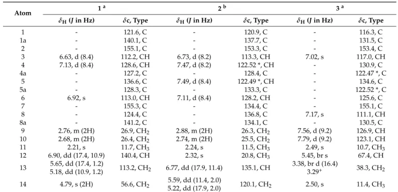
Table 1.1H (500 MHz) and13C (125 MHz) NMR data of compounds1–3.
Atom 1a 2b 3a
δH(Jin Hz) δc, Type δH(Jin Hz) δc, Type δH(Jin Hz) δc, Type
1 - 121.6, C - 120.9, C - 116.3, C
1a - 140.1, C - 137.7, C - 131.5, C
2 - 155.1, C - 153.3, C - 153.4, C
3 6.63, d (8.4) 112.2, CH 6.73, d (8.2) 113.3, CH 7.02, s 117.0, CH
4 7.13, d (8.4) 128.6, CH 7.47, d (8.2) 122.52 *, CH - 130.9, C
4a - 127.2, C - 128.4, C - 122.47 *, C
5 - 136.6, C 7.49, d (8.4) 122.49 *, CH - 134.6, C
5a - 128.3, C - 133.3, C - 122.52 *, C
6 6.92, s 113.0, CH 7.11, d (8.4) 128.2, CH - 125.6, C
7 - 155.3, C - 134.4, C - 155.1, C
8 - 124.4, C - 136.8, C 7.17, s 111.1, CH
8a - 141.2, C - 134.1, C - 130.5, C
9 2.76, m (2H) 26.9, CH2 2.88, m (2H) 26.3, CH2 7.56, d (9.2) 126.9, CH
10 2.68, m (2H) 26.4, CH2 2.74, m (2H) 25.5, CH2 7.79, d (9.2) 123.1, CH
11 2.21, s 11.7, CH3 2.24, s 11.5, CH3 2.49, s 10.7, CH3
12 6.90, dd (17.4, 10.9) 140.4, CH 2.32, s 20.8, CH3 5.45, br s 67.4, CH
13 5.65, dd (17.4, 1.2)
5.18, dd (10.9, 1.2) 113.2, CH2 6.77, dd (17.9, 11.4) 135.1, CH 3.38, br d (16.4)
3.29+ 38.3, CH2 14 4.79, s (2H) 56.6, CH2 5.59, dd (11.4, 2.0)
5.22, dd (17.9, 2.0) 120.1, CH2 2.50, s 11.4, CH3
ameasured in methanol-d4;bmeasured in CDCl3; * interchangeable signals;+overlapped with residual H2O signal.
The1H-1H COSY correlations defined three sequences of correlated protons, namely, –CH2–CH2– (H2-9–H2-10), –CH=CH2(H-12–H-13a and H-13b), and –CH=CH– (H-3–H-4) fragments (Figure2). The structure of compound 1was assembled with the aid of an HMBC experiment. Heteronuclear long-range correlations of H-3 and H2-10 with C-4a (δC127.2), H-4, H-6, and H2-9 with C-5a (δC 128.3), H2-9, H2-10, and H2-14 with C-8a (δC141.2), as well as of H-4, H2-9, H2-10, and H3-11 with C-1a (δC140.1) established a 9,10-dihydrophenanthrene skeleton. HMBC correlations from H-3, H-4, and H3-11 to C-2 (δC155.1), and from H-6 and H2-14 to C-7 (δC155.3) suggested that compound1contains two hydroxy groups at the positions of C-2 and C-7. The location of the H3-11 methyl group at C-1 was dictated by its HMBC correlations with C-1, C-1a, and C-2. The two- and three- bond correlations between H2-14 (δH4.79), C-7, C-8 (δC124.4), and C-8a demonstrated that the freely rotating hydroxymethyl substituent is attached to C-8. The location of the vinyl moiety at C-5 (δC136.6) was confirmed by the H-6/C-12 and H-13/C-5 HMBC correlations.
The NOE cross-peaks between H-4/H-12, H-13a/H-6, H2-9/H2-14, and H2-10/H3-11 were consistent with the proposed structure of1, as shown in Figure2.
Molecules2021,26, 999 4 of 11
Molecules 2021, 26, x 4 of 11
Figure 2. Key HMBC (H→C) and 1H-1H COSY (–) interactions of maritin A (1).
Compound 2 (maritin B) was obtained as a white amorphous solid. Its molecular formula was deduced to be C18H18O based on the protonated molecule in the HRESIMS at m/z [M + H]+ 251.1429 (calcd. for C18H19O, 251.1430). The 1H-NMR spectrum contained signals of two pairs of ortho-coupled aromatic protons (δH 7.47 d and 6.73 d, J = 8.2 Hz; 7.49 d and 7.11 d, J = 8.4 Hz), two methylenes (δH 2.88 m and 2.74 m, each 2H), a vinyl substituent (δH 6.77 dd, J = 17.9 and 11.4 Hz; δH 5.59 dd, J = 11.4 and 2.0 Hz; δH 5.22 dd, J = 17.9 and 2.0 Hz), and two methyl groups (δH 2.32 s and 2.24 s, each 3H) (Table 1). The HMBC correlations from H3-11 (δH 2.24) to C-1 (δC 120.9), C-1a (δC 137.7), and C-2 (δC 153.3), and further correlations between H-3 (δH 6.73), H-4 (δH 7.47), and C-2 showed that a methyl and a hydroxy group are situated on the adjacent carbons C-1 and C-2, respectively. The locations of another methyl (δH 2.32) and a vinyl substituent at C-7 and C-8, respectively, were apparent from the HMBC correlations H3-12/C-6, H3-12/C-7, H3- 12/C-8, H-6/C-8, H2-9/C-8, and H-14/C-8. Further heteronuclear correlations were detected between H-3, H-5 (δH 7.49), H2-10 (δH 2.74), and C-4a (δC 128.4), H-4, H-6 (δH 7.11), H2-9 (δH
2.88), and C-5a (δC 133.3), and from H-5, H2-9, and H2-10 to C-8a (δC 134.1). The NOE cross- peaks H-6/H3-12, H3-12/H-13, H2-9/H-14b, and H2-10/H3-11 supported the proposed structure of compound 2.
Separation of the plant extract yielded compound 3 (maritin C) as an orange amorphous solid. According to a peak of the deprotonated molecule at m/z 279.1027 [M − H]− in the HRESIMS data, the molecular formula C18H16O3 (calcd. for C18H15O3, 279.1021) was assigned to 3. The 1H-NMR spectrum exhibited two aromatic methines coupled with each other (δH 7.79 and 7.56 d, J = 9.2 Hz), two aromatic singlets (δH 7.17 and 7.02), two methyl groups (δH 2.50 s and 2.49 s, each 3H), and signals of an oxymethine (δH 5.45, br s) and a saturated methylene (δH 3.38 and 3.29, each 1H). The 1H-1H COSY spectrum afforded two structural elements, the aforementioned –CH=CH– (δH 7.79 and 7.56) and a –CH(OR)–
CH2– fragment (δH 5.45, 3.38, and 3.29). The proton signals at δH 7.02 (H-3) and δH 2.49 (H3- 11) gave HMBC correlations with a downfield shifted, nonprotonated carbon displayed at δC 153.4, while the aromatic singlet at δH 7.17 (H-8) and the methyl group at δH 2.50 (H3- 14) gave HMBC correlations to a carbon resonating at δC 155.1. Thus, it was deduced that this phenanthrene bears hydroxy groups at C-2 and C-7. The two methyls were placed onto C-1 and C-6 on the basis of the corresponding H3-11/C-1, H3-11/C-1a, H3-11/C-2, H3- 14/C-5, H3-14/C-6, and H3-14/C-7 HMBC correlations. Further long-range correlations from H-9 (δH 7.56) to C-1a, C-5a, and C-8, as well as from H-10 (δH 7.79) to C-1, C-4a, and C-8a established a phenanthrene skeleton with an aromatic ring B. Considering the HMBC cross-peaks of H-13a (δH 3.38) with C-3 (δC 117.0), C-4 (δC 130.9), C-4a (δC 122.5), and C-5 (δC 134.6), it was clear that a vinyl group was incorporated into an oxygen-substituted cyclohexadiene ring. From a biosynthetic point of view, compound 3 was likely formed from a dehydrojuncusol precursor through the modification of its vinylic double bond, followed by a ring closure between C-4 and C-13. The depicted structure of maritin C was corroborated by NOE cross-peaks between H-3/H-13a and b, H-12/H3-14, H-8/H-9, and H- 10/H3-11. The specific optical rotation of 3 was recorded as zero, therefore, it was isolated as a racemic mixture.
Compound 4 (maritin D) has the molecular formula C34H30O4 compatible with its protonated molecule at m/z 503.2203 [M + H]+ (calcd. for C34H31O4, 503.2222) in the
Figure 2.Key HMBC (H→C) and1H-1H COSY (–) interactions of maritin A (1).
Compound2(maritin B) was obtained as a white amorphous solid. Its molecular formula was deduced to be C18H18O based on the protonated molecule in the HRESIMS at m/z[M + H]+251.1429 (calcd. for C18H19O, 251.1430). The1H-NMR spectrum contained signals of two pairs ofortho-coupled aromatic protons (δH7.47 d and 6.73 d,J= 8.2 Hz;
7.49 d and 7.11 d,J= 8.4 Hz), two methylenes (δH 2.88 m and 2.74 m, each 2H), a vinyl substituent (δH 6.77 dd,J = 17.9 and 11.4 Hz;δH 5.59 dd, J= 11.4 and 2.0 Hz; δH 5.22 dd,J= 17.9 and 2.0 Hz), and two methyl groups (δH2.32 s and 2.24 s, each 3H) (Table1).
The HMBC correlations from H3-11 (δH 2.24) to C-1 (δC120.9), C-1a (δC137.7), and C-2 (δC153.3), and further correlations between H-3 (δH6.73), H-4 (δH7.47), and C-2 showed that a methyl and a hydroxy group are situated on the adjacent carbons C-1 and C-2, respectively. The locations of another methyl (δH 2.32) and a vinyl substituent at C-7 and C-8, respectively, were apparent from the HMBC correlations H3-12/C-6, H3-12/C-7, H3-12/C-8, H-6/C-8, H2-9/C-8, and H-14/C-8. Further heteronuclear correlations were detected between H-3, H-5 (δH 7.49), H2-10 (δH2.74), and C-4a (δC128.4), H-4, H-6 (δH 7.11), H2-9 (δH2.88), and C-5a (δC133.3), and from H-5, H2-9, and H2-10 to C-8a (δC134.1).
The NOE cross-peaks H-6/H3-12, H3-12/H-13, H2-9/H-14b, and H2-10/H3-11 supported the proposed structure of compound2.
Separation of the plant extract yielded compound3(maritin C) as an orange amor- phous solid. According to a peak of the deprotonated molecule atm/z279.1027 [M−H]− in the HRESIMS data, the molecular formula C18H16O3(calcd. for C18H15O3, 279.1021) was assigned to3. The1H-NMR spectrum exhibited two aromatic methines coupled with each other (δH7.79 and 7.56 d,J= 9.2 Hz), two aromatic singlets (δH7.17 and 7.02), two methyl groups (δH2.50 s and 2.49 s, each 3H), and signals of an oxymethine (δH5.45, br s) and a saturated methylene (δH3.38 and 3.29, each 1H). The1H-1H COSY spectrum afforded two structural elements, the aforementioned –CH=CH– (δH7.79 and 7.56) and a –CH(OR)–CH2– fragment (δH5.45, 3.38, and 3.29). The proton signals atδH7.02 (H-3) andδH2.49 (H3-11) gave HMBC correlations with a downfield shifted, nonprotonated carbon displayed atδC 153.4, while the aromatic singlet atδH7.17 (H-8) and the methyl group atδH2.50 (H3-14) gave HMBC correlations to a carbon resonating atδC155.1. Thus, it was deduced that this phenanthrene bears hydroxy groups at C-2 and C-7. The two methyls were placed onto C-1 and C-6 on the basis of the corresponding H3-11/C-1, H3-11/C-1a, H3-11/C-2, H3-14/C-5, H3-14/C-6, and H3-14/C-7 HMBC correlations. Further long-range correlations from H-9 (δH 7.56) to C-1a, C-5a, and C-8, as well as from H-10 (δH 7.79) to C-1, C-4a, and C-8a established a phenanthrene skeleton with an aromatic ring B. Considering the HMBC cross-peaks of H-13a (δH3.38) with C-3 (δC117.0), C-4 (δC130.9), C-4a (δC122.5), and C-5 (δC134.6), it was clear that a vinyl group was incorporated into an oxygen-substituted cyclohexadiene ring. From a biosynthetic point of view, compound3was likely formed from a dehydrojuncusol precursor through the modification of its vinylic double bond, followed by a ring closure between C-4 and C-13. The depicted structure of maritin C was corroborated by NOE cross-peaks between H-3/H-13a and b, H-12/H3-14, H-8/H-9, and H-10/H3-11. The specific optical rotation of3was recorded as zero, therefore, it was isolated as a racemic mixture.
Compound4 (maritin D) has the molecular formula C34H30O4 compatible with its protonated molecule atm/z503.2203 [M + H]+(calcd. for C34H31O4, 503.2222) in the HRESIMS data. The 34 carbon signals displayed in the13C-JMOD NMR spectrum suggested that compound4is a phenanthrene dimer (Figure S19). The1H-NMR spectrum, combined with homonuclear1H-1H COSY correlations, showed the presence of two vinyl groups (H-12–H2- 13:δH6.96 dd,J= 17.4 and 10.9 Hz;δH5.67 d,J= 17.4 Hz;δH5.23 d,J= 10.9 Hz; H-120–H2-130: δH6.64 dd,J= 17.3 and 11.4 Hz;δH5.33 dd,J= 17.3 and 0.9 Hz;δH4.78 d,J= 11.4 Hz), a –CH=CH– (H-3–H-4:δH7.38 and 6.83 d,J= 8.4 Hz) and two –CH2–CH2– structural portions (H2-9–H2-10:δH2.68 m and 2.78 m, each 2H; H2-90–H2-100:δH2.63 m and 2.64 m, each 2H), and two methyls (H3-11:δH2.28 s; H3-110:δH2.30 s, each 3H) in4(Table2).
Table 2.1H (500 MHz) and13C (125 MHz) NMR data of compound4in methanol-d4.
Effusol Monomer OH-2 Effusol Monomer
Atom δH(Jin Hz) δc, Type Atom δH(Jin Hz) δc, Type
1 - 127.7, C 10 - 123.4, C
1a - 140.8, C 10a - 133.5, C
2 - 154.4, C 20 - 144.6, C
3 6.83, d (8.4) 117.8, CH 30 - 156.6#, C
4 7.38, d (8.4) 128.7, CH 40 6.66, s 116.1, CH
4a - 131.4, C 40a - 127.06 *, C
5 - 137.9, C 50 - 137.4, C
5a - 126.7, C 50a - 126.98 *, C
6 6.88, d (2.2) 113.8#, CH 60 6.68, br s 113.8#, CH
7 - 157.2, C 70 - 156.6#, C
8 6.69 d (2.2) 115.1, CH 80 6.61, br s 115.0, CH
8a - 142.1, C 80a - 141.7, C
9 2.68, m (2H) 31.4, CH2 90 2.63#, m (2H) 31.6, CH2
10 2.78, m (2H) 26.7, CH2 100 2.64#, m (2H) 26.1, CH2
11 2.28, s 12.4, CH3 110 2.30, s 12.0, CH3
12 6.96, dd (17.4, 10.9) 140.1, CH 120 6.64 dd (17.3, 11.4) 140.2, CH 13 5.67, d (17.4)
5.23, d (10.9) 114.2, CH2 130 5.33, dd (17.3, 0.9)
4.78, d (11.4) 113.7, CH2
# overlapping signals; * interchangeable signals.
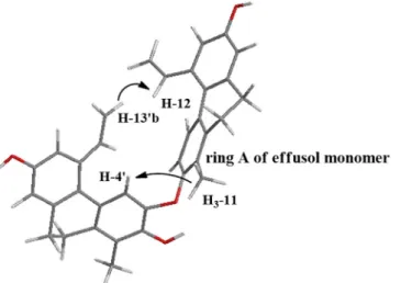
Two pairs ofmeta-coupled aromatic protons (H-6 and H-8: δH 6.88 d and 6.69 d, J= 2.2 Hz; H-60and H-80:δH6.68 br s and 6.61 br s) were also identified via weaker4JH-H (W-type) COSY cross-peaks and three-bond HMBC correlations between the corresponding methine groups. Further analysis of the HMBC correlations unambiguously determined that4is comprised of two monomers of a known 9,10-dihydrophenanthrene, effusol, which was also isolated as an individual compound (6) from the plant. Taking into account the HMBC correlations from H-40and H3-110to C-20(δC144.6), it was concluded that oxygen atoms are connected to both of the vicinal carbons C-20and C-30(δC156.6). Although no HMBC correlations were observed between the monomers, NOE cross-peaks H-40/H3-11 and H-130b/H-12 indicated the close proximity of these protons, and consequently implied that the monomers must be attached through an ether bond between C-2/C-20or C-2/C-30. In order to determine the exact structure, energy-minimized structures were generated for each of the hypothetical compounds by using the MM2 force field method. A minimum energy conformation (Figure3) provided by molecular dynamics calculations was in good agreement with the aforementioned NOE correlations and suggested that the ether bond was formed between C-2/C-30. The proposed structure was further confirmed by the significantly shielded nature of H-40and vinyl resonances H-120–H2-130compared to H-4 and H-12–H2-13 of the other monomer (Table2).
Molecules2021,26, 999 6 of 11
Molecules 2021, 26, x 6 of 11
Figure 3. Calculated molecular structure of maritin D (4). The arrows indicate diagnostic NOESY correlations. Note that the markedly upfield shifted H-4′ and H-12′–H2-13′ are situated in the shielding cone of ring A.
This phenomenon was likely caused by the anisotropic effect of aromatic ring A since H-4′ and the vinyl moiety H-12′–H2-13′ are located in the shielding cone of ring A. In case of the presence of a C-2/C-2′ linkage, H-4′ and H-12′–H2-13′ would be located too far from ring A and, therefore, their chemical shifts would be less affected by the aromatic ring current effects. Considering the above findings, the structure of 4 was formulated as depicted in Figure 1.
Besides the new compounds maritins A–D (1–4), seven known phenanthrenes, namely, juncusol (5) [10], effusol (6) [10], 2,7-dihydroxy-1-methyl-5-aldehyde-9,10- dihydrophenanthrene (7) [11], 2,7-dihydroxy-1,8-dimethyl-5-vinyl-9,10- dihydrophenanthrene (8) [10], juncunol (9) [10], jinflexin A (10) [12], and effususin A (11) [13], were also isolated from J. maritimus. Their structures were identified by 1D and 2D NMR spectroscopy, and by comparison of the 1H and 13C NMR chemical shift values with literature data. All compounds but effusol (7) are described for the first time from J.
maritimus. Moreover, the 1H and 13C NMR assignments of jinflexin A in methanol-d4 were not reported previously.
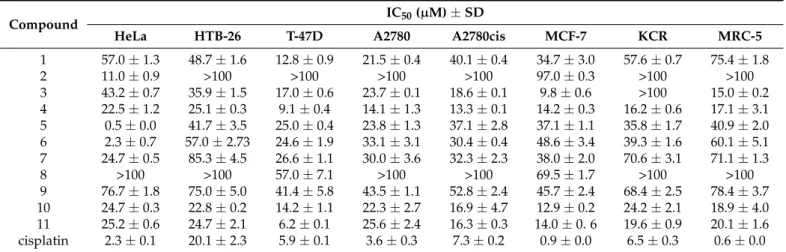
The obtained phenanthrenes 1–11 were tested for their antiproliferative activity against seven human tumor cell lines (HeLa, HTM-26, T-47D, A2780, A2780cis, MCF-7, KCR) and one normal human fetal lung fibroblast (MRC-5) cell line using the 3-(4,5- dimethylthiazol-2-yl)-2,5-diphenyltetrazolium bromide (MTT) assay with cisplatin as a positive control (Table 3). Among the tested compounds, dimeric phenanthrenes (4 and 11) built up by effusol monomers showed substantial antiproliferative activity against all cell lines investigated. The highest activities were detected on T-47D ductal carcinoma cells (IC50 9.1 µM for 4 and 6.2 µM for 11, respectively) for both compounds. No significant differences were observed between the effects of dimers on different cell lines.
Table 3. Antiproliferative activity (IC50) of the tested phenanthrenes 1–11.
Compound IC50 (µM) ± SD
HeLa HTB-26 T-47D A2780 A2780cis MCF-7 KCR MRC-5 1 57.0 ± 1.3 48.7 ± 1.6 12.8 ± 0.9 21.5 ± 0.4 40.1 ± 0.4 34.7 ± 3.0 57.6 ± 0.7 75.4 ± 1.8
2 11.0 ± 0.9 >100 >100 >100 >100 97.0 ± 0.3 >100 >100
3 43.2 ± 0.7 35.9 ± 1.5 17.0 ± 0.6 23.7 ± 0.1 18.6 ± 0.1 9.8 ± 0.6 >100 15.0 ± 0.2 4 22.5 ± 1.2 25.1 ± 0.3 9.1 ± 0.4 14.1 ± 1.3 13.3 ± 0.1 14.2 ± 0.3 16.2 ± 0.6 17.1 ± 3.1 5 0.5 ± 0.0 41.7 ± 3.5 25.0 ± 0.4 23.8 ± 1.3 37.1 ± 2.8 37.1 ± 1.1 35.8 ± 1.7 40.9 ± 2.0 6 2.3 ± 0.7 57.0 ± 2.73 24.6 ± 1.9 33.1 ± 3.1 30.4 ± 0.4 48.6 ± 3.4 39.3 ± 1.6 60.1 ± 5.1 7 24.7 ± 0.5 85.3 ± 4.5 26.6 ± 1.1 30.0 ± 3.6 32.3 ± 2.3 38.0 ± 2.0 70.6 ± 3.1 71.1 ± 1.3
8 >100 >100 57.0 ± 7.1 >100 >100 69.5 ± 1.7 >100 >100
Figure 3.Calculated molecular structure of maritin D (4). The arrows indicate diagnostic NOESY correlations. Note that the markedly upfield shifted H-40and H-120–H2-130 are situated in the shielding cone of ring A.
This phenomenon was likely caused by the anisotropic effect of aromatic ring A since H-40and the vinyl moiety H-120–H2-130are located in the shielding cone of ring A. In case of the presence of a C-2/C-20 linkage, H-40 and H-120–H2-130 would be located too far from ring A and, therefore, their chemical shifts would be less affected by the aromatic ring current effects. Considering the above findings, the structure of4was formulated as depicted in Figure1.
Besides the new compounds maritins A–D (1–4), seven known phenanthrenes, namely, juncusol (5) [10], effusol (6) [10], 2,7-dihydroxy-1-methyl-5-aldehyde-9,10-dihydrophenanthrene (7) [11], 2,7-dihydroxy-1,8-dimethyl-5-vinyl-9,10-dihydrophenanthrene (8) [10], juncunol (9) [10], jinflexin A (10) [12], and effususin A (11) [13], were also isolated fromJ. maritimus.
Their structures were identified by 1D and 2D NMR spectroscopy, and by comparison of the1H and13C NMR chemical shift values with literature data. All compounds but effusol (7) are described for the first time fromJ. maritimus. Moreover, the1H and13C NMR assignments of jinflexin A in methanol-d4were not reported previously.
The obtained phenanthrenes1–11were tested for their antiproliferative activity against seven human tumor cell lines (HeLa, HTM-26, T-47D, A2780, A2780cis, MCF-7, KCR) and one normal human fetal lung fibroblast (MRC-5) cell line using the 3-(4,5-dimethylthiazol- 2-yl)-2,5-diphenyltetrazolium bromide (MTT) assay with cisplatin as a positive control (Table3). Among the tested compounds, dimeric phenanthrenes (4and11) built up by effusol monomers showed substantial antiproliferative activity against all cell lines investi- gated. The highest activities were detected on T-47D ductal carcinoma cells (IC509.1µM for4and 6.2µM for11, respectively) for both compounds. No significant differences were observed between the effects of dimers on different cell lines.
T-47D cells were the most sensitive to the phenanthrenes, but four compounds (3,4, 10, and11) displayed remarkable antiproliferative activity (IC50< 20µM) against A2780cis and MCF-7 cells, too. Maritin B (2), juncusol (5), and effusol (6) were effective only against the HeLa cells (with respective IC50values of 11.0, 0.5, and 2.3µM). Maritin A (1) and compound8 differ from each other in their substitution at the position of C-8. Based on the experimental data, it seems that replacement of the C-8 methyl group in8to a hydroxymethyl moiety present in 1 leads to a drastic increase in the antiproliferative activity. Effusol (6) and compound7also have similar chemical structures, the difference between them involves the presence of a C-5 vinyl group in6and a formyl moiety at the same position in7. No significant differences were observed upon comparison of the activities of these phenanthrenes except for the HeLa cell line. Interestingly, effusol (6) containing a C-7 hydroxy function displayed much stronger antiproliferative activity on several tumor cell lines than juncunol (9), which has a C-7 methyl substituent. This
finding suggests that the presence of a polar hydroxy substituent on C-7 is probably more favorable for the antiproliferative effects than its methyl counterpart. Compounds7–9 were weakly effective on all cell lines tested. Although the structure of8closely resembles that of jinflexin A (10) (the two phenanthrenes contain a vinyl and a methoxyethyl moiety at C-5, respectively), substantial differences were found in their antiproliferative profiles:
compound8had no effects on any of the cell lines investigated, while10exerted activity against all seven tumor cell lines. The presence of a methoxyethyl moiety instead of a vinyl group at C-5 appears to enhance the antiproliferative activity of phenanthrenes.
Unfortunately, the tested phenanthrenes showed no selectivity except for compound1that exhibited a less antiproliferative effect on MRC-5 normal lung fibroblasts compared to the other cancer cell lines. The other compounds considerably inhibited the proliferation of MRC-5 cells, too.
Table 3.Antiproliferative activity (IC50) of the tested phenanthrenes1–11.
Compound IC50(µM)±SD
HeLa HTB-26 T-47D A2780 A2780cis MCF-7 KCR MRC-5
1 57.0±1.3 48.7±1.6 12.8±0.9 21.5±0.4 40.1±0.4 34.7±3.0 57.6±0.7 75.4±1.8
2 11.0±0.9 >100 >100 >100 >100 97.0±0.3 >100 >100
3 43.2±0.7 35.9±1.5 17.0±0.6 23.7±0.1 18.6±0.1 9.8±0.6 >100 15.0±0.2 4 22.5±1.2 25.1±0.3 9.1±0.4 14.1±1.3 13.3±0.1 14.2±0.3 16.2±0.6 17.1±3.1 5 0.5±0.0 41.7±3.5 25.0±0.4 23.8±1.3 37.1±2.8 37.1±1.1 35.8±1.7 40.9±2.0 6 2.3±0.7 57.0±2.73 24.6±1.9 33.1±3.1 30.4±0.4 48.6±3.4 39.3±1.6 60.1±5.1 7 24.7±0.5 85.3±4.5 26.6±1.1 30.0±3.6 32.3±2.3 38.0±2.0 70.6±3.1 71.1±1.3
8 >100 >100 57.0±7.1 >100 >100 69.5±1.7 >100 >100
9 76.7±1.8 75.0±5.0 41.4±5.8 43.5±1.1 52.8±2.4 45.7±2.4 68.4±2.5 78.4±3.7 10 24.7±0.3 22.8±0.2 14.2±1.1 22.3±2.7 16.9±4.7 12.9±0.2 24.2±2.1 18.9±4.0 11 25.2±0.6 24.7±2.1 6.2±0.1 25.6±2.4 16.3±0.3 14.0±0. 6 19.6±0.9 20.1±1.6 cisplatin 2.3±0.1 20.1±2.3 5.9±0.1 3.6±0.3 7.3±0.2 0.9±0.0 6.5±0.3 0.6±0.0
3. Materials and Methods
3.1. General Experimental Procedures
Optical rotation was determined in CHCl3at ambient temperature, using a Perkin- Elmer 341 polarimeter (PerkinElmer, MA, USA). NMR spectra were recorded in CDCl3, methanol-d4, and dimethyl sulfoxide-d6on a Bruker Avance DRX 500 spectrometer (Bruker, MA, USA) at 500 MHz (1H) and 125 MHz (13C-JMOD). The signals of the deuterated solvents were taken as references. The chemical shift values (δ) were given in ppm and coupling constants (J) are expressed in Hz. The high-resolution MS spectra were acquired on a Thermo Scientific Q-Exactive Plus Orbitrap mass spectrometer (Thermo Fisher Scien- tific, MA USA) equipped with ESI ion source in positive ionization mode. The resolution was over 1 ppm. The data were acquired and processed with MassLynx software (Waters, MA, USA). All solvents used for chromatographic separations and purification steps were analytical or HPLC grade (VWR Ltd., Szeged, Hungary).
For vacuum liquid chromatography (VLC), silica gel (silica gel GF254, 15µm, Merck) and reversed-phase silica (LiChroprep RP-18, 40-63µm, Merck) were used. Medium- pressure liquid chromatography (MPLC) was performed by a Combi Flash Rf+ Lumen instrument (Teledyne ISCO, NE, USA) on a reversed-phase RediSep Rf HP Gold (50 g) column. Preparative thin-layer chromatography (prep. TLC) was performed on silica gel plates (TLC silica gel 60 F254, Merck, Darmstadt, Germany), and on reversed-phase silica gel plates (TLC silica gel 60 RP-18 F254S, Merck). Sephadex LH-20 (25–100µm, Sigma- Aldrich, Budapest, Hungary) was used for gel filtration. HPLC was carried out on a Waters Millipore instrument with UV detection at 254 nm over normal- (Kinetex Luna Silica, 3µm, 150×4.6 mm, Phenomenex Inc., CA, USA) and reversed-phase (Kinetex 5µm C18, 150×4.6 mm and LiChrospher RP-18, 5µm, 250×4 mm) columns.
Molecules2021,26, 999 8 of 11
3.2. Plant Material
Juncus maritimusLam. (whole plants, 2.2 kg) was collected in June 2018, near Vir (coordinates: 44◦31080.7400N; 15◦05072.0000E) (Croatia), and identified by one of the authors, LászlóBakacsy (Department of Plant Biology, University of Szeged, Szeged, Hungary). A voucher specimen (No. 884) has been deposited at the Herbarium of the Department of Pharmacognosy, University of Szeged, Szeged, Hungary.
3.3. Extraction and Isolation
The plant material (aerial part) was air-dried (2.2 kg) at room temperature. Thereafter, it was ground and percolated with 40 L methanol at room temperature. After evapora- tion, the extract was dissolved in 50% aqueous methanol, and repetitive solvent—solvent partition was performed with 6×0.5 Ln-hexane, 10×0.5 L chloroform, and 5×0.5 L EtOAc. The concentrated chloroform-soluble fraction (32 g) was separated by vacuum liquid chromatography (VLC) on silica gel with a gradient system of cyclohexane—EtOAc—
MeOH [from 98:2:0 to 1:1:1 (1500 mL/eluent); volume of collected fractions was 150 mL].
This separation yielded 14 fractions (A-N). The fractions were combined according to their TLC patterns.
All major fractions were purified by Sephadex LH-20 gel chromatography using CH2Cl2—MeOH (1:1) as eluent. Fraction B/2 was separated by normal-phase HPLC under gradient conditions, using cyclohexane–EtOAc (19:1 to 9:1 in 10 min and 9:1 to 65:35 in 1 min; flow rate 1.5 mL/min) as mobile phase to obtain compounds5(tR= 8.3 min, 1.2 mg) and2(tR= 10.4 min, 2.9 mg). Purification of fractions D/4 and D/5 by preparative TLC afforded compounds6(4.3 mg) and7(3.4 mg).
Fraction E was chromatographed by reversed-phase MPLC using MeOH–H2O (from 8:2 to 1:0). Subfraction E/1 was then further purified by reversed-phase HPLC under gra- dient conditions, using MeOH–H2O (from 45:55 to 82:18 in 10 min; flow rate 1.2 mL/min) as mobile phase to yield compound10(tR= 5.6 min, 2.4 mg). Subfraction E/2 was sepa- rated by preparative TLC on silica gel using cyclohexane–EtOAc–EtOH (20:10:1) as solvent system to yield compounds9(3.5 mg) and3(4.5 mg).
Fractions H/3 and I/4 were combined (HI/3-4) because of their similar chemical composition and were purified by reversed-phase MPLC using a stepwise gradient solvent system composed of MeOH–H2O (from 8:2 to 1:0). Subfraction HI/3-4/1 was separated by preparative TLC on silica gel using cyclohexane–EtOAc–EtOH (20:10:1) as eluent to isolate compound8(10.4 mg). HI/3-4/1/2 was purified by reversed-phase HPLC under gradient conditions, using MeOH–H2O (from 45:55 to 82:18 in 10 min; flow rate 1.2 mL/min) as mobile phase, and compound1(tR= 9.0 min, 5.6 mg) was isolated. Subfraction HI/3- 4/7 was further fractionated by normal-phase HPLC under gradient conditions, using cyclohexane–EtOAc (from 80:20 to 65:35 in 12 min; flow rate 1.7 mL/min) as mobile phase, to afford compound11(tR= 13.2 min, 2.3 mg). Subfraction HI/3-4/9 was separated by preparative TLC on silica gel using an isocratic cyclohexane–EtOAc–EtOH (60:30:3) eluent, and then HI/3-4/9/3 was purified by reversed-phase HPLC under gradient conditions, using acetonitrile–H2O (from 56:44 to 70:30 in 10 min; flow rate 1.2 mL/min) as mobile phase to yield compound4(tR= 7.5 min, 2.0 mg).
Maritin A (1)
Yellow amorphous solid; for1H- and13C-JMOD NMR (in methanol-d4) data, see Table1; HRESIMSm/z281.1183 [M−H]−(calcd. for C18H17O3, 281.1178).
Maritin B (2)
White amorphous solid; for1H- and13C-JMOD NMR (in CDCl3) data, see Table1;
HRESIMSm/z[M + H]+251.1430 (calcd. for C18H19O, 251.1430).
Maritin C (3)
Orange amorphous solid; [α]25D0 (c0.1, MeOH); for1H-and13C-JMOD NMR (in methanol-d4) data, see Table1; HRESIMSm/z279.1027 [M−H]− C18H16O3(calcd. for C18H15O3, 279.1021).
Maritin D (4)
Yellow amorphous solid; for1H- and13C-JMOD NMR (in methanol-d4) data, see Table2; HRESIMSm/z[M + H]+503.2203 (calcd. for C34H31O4, 503.2222).
Jinflexin A (10):
1H-NMR (500 MHz, methanol-d4): δH 6.67 (1H, d, J= 8.3 Hz; H-3), 6.87 (1H, d, J= 8.3 Hz; H-4), 6.86 (1H, s; H-6), 2.87 (1H, m; H-9a), 2.37 (1H, m; H-9b), 2.88 (1H, m;
H-10a), 2.38 (1H, m; H-10b), 2.22 (3H, s; H3-11), 4.85 (1H, overlaps with residual H2O signal; H-12), 1.57 (3H, d,J= 6.2 Hz; H3-13), 2.19 (3H, s; H3-14), 2.92 (3H, s; 12-OCH3).13C NMR (125 MHz, methanol-d4):δC121.8 (C-1), 140.5 (C-1a), 154.9 (C-2), 112.4 (C-3), 127.5 (C-4), 127.4 (C-4a), 129.3 (C-5a), 138.4 (C-5), 111.5 (C-6), 155.1 (C-7), 121.4 (C-8), 139.8 (C-8a), 27.6 (C-9), 26.7 (C-10), 11.8 (C-11), 76.5 (C-12), 23.4 (C-13), 11.8 (C-14), 55.7 (12-OCH3).
3.4. Antiproliferative Assay 3.4.1. Cell Lines
Breast cancer cell line MCF-7 (ATCC®® HTB-22) and the drug-resistant subline of the human breast cancer MCF-7 (ECACC 86012803; KCR) were purchased from LGC Promochem (Teddington, UK). Both cell lines were cultured in Eagle’s Minimal Essential Medium (EMEM, containing 4.5 g/L glucose) supplemented with a non-essential amino acid mixture, a selection of vitamins, and 10% heat-inactivated fetal bovine serum. In every third passage, 0.56µg/mL doxorubicin was added to the medium in order to maintain the ABCB1 (P-glycoprotein) expression in KCR cells. A2780 human ovarian cancer cell line (ECACC European Collection of Authentical Cell Culture, Sigma Cat. no. 93112519) and the cisplatin-resistant human ovarian cancer cell line A2780cis (ECACC European Collection of Authentical Cell Culture, Sigma Cat. no. 93112517) were purchased from Merck KGaA (Darmstadt, Germany). The human ovarian cancer cell lines were cultured in RPMI 1640 medium supplemented with 10% heat-inactivated fetal bovine serum. The RPMI 1640 medium of the cisplatin-resistant cell line A2780 was supplemented with 1µM cisplatin.
HeLa (ATCC®CCL-2™) human cervix carcinoma, HTB-26 breast adenocarcinoma, T-47D (ATCC®HTB-133™) ductal carcinoma, and MRC-5 human embryonal lung fibroblast cell lines (ATCC®CCL-171) were purchased from LGC Promochem (Teddington, UK). The cells were cultured in Eagle’s Minimal Essential Medium (EMEM, containing 4.5 g/L glucose) supplemented with a non-essential amino acid mixture, a selection of vitamins, and 10%
heat-inactivated fetal bovine serum. HTB-26 cell line was cultured in RPMI 1640 medium supplemented with 10% heat-inactivated fetal bovine serum. T-47D cells were cultured in RPMI 1640 medium supplemented with 10% heat-inactivated fetal bovine serum, 2 mM L-glutamine, 1 mM Na-pyruvate, and 100 mM Hepes. All of the cells were incubated at 37◦C, in a 5% CO2, 95% air atmosphere.
3.4.2. Antiproliferative Assay
The antiproliferative effect of the compounds was determined on the human breast (MCF-7, KCR, T-47D, HTB-26), cervical (HeLa), and ovarian (A2780, A2780cis) cancer cells, and on MRC-5 (human embryonic lung fibroblast) cell line. The adherent cells were cultured in 96-well flat-bottomed microtiter plates, using EMEM supplemented with 10%
heat-inactivated fetal bovine serum or RPMI 1640 supplemented with 10% heat-inactivated fetal bovine serum, respectively. The density of the cells was adjusted to 6×10³ cells in 100µL per well, the cells were seeded for 24 h at 37◦C, 5% CO2, then the medium was removed from the plates and fresh medium (100µL per well) was added to the cells.
The effects of increasing concentrations of compounds on cell proliferation were tested in 96-well flat-bottomed microtiter plates. The compounds were diluted in the appropriate medium, the dilutions of compounds were performed in separate plates and then added to the cells. The starting concentration of the compounds was 100µM, and two-fold serial dilution was performed (concentration range: 100-0.19µM). The culture plates were incubated at 37◦C for 72 h; at the end of the incubation period, 20µL of MTT (thiazolyl blue tetrazolium bromide, Sigma) solution (from a stock solution of 5 mg/mL) was added to each well. After incubation at 37◦C for 4 h, 100µL of sodium dodecyl sulfate (SDS) (Sigma)
Molecules2021,26, 999 10 of 11
solution (10% in 0.01 M HCI) were added to each well and the plates were further incubated at 37◦C overnight. Cell growth was determined by measuring the optical density (OD) at 540/630 nm with Multiscan EX ELISA reader (Thermo Labsystems, Cheshire, WA, USA).
Mean IC50values were obtained by best fitting the dose-dependent inhibition curves in GraphPadPrism5 program (GraphPad Software version 5.00 for Windows, San Diego, CA, USA) from four parallel experiments for each cell line. Results are expressed in terms of IC50, defined as the inhibitory dose that reduces the proliferation of the cells exposed to the tested compounds by 50% [14].
4. Conclusions
Eleven phenanthrenes, including four new ones (1–4), were isolated from the methano- lic extract ofJ. maritimus. All compounds but effusol (7) were reported for the first time from the plant. Some of the new phenanthrenes possess interesting structural features. The ob- tained compounds are substituted with hydroxy, methyl, hydroxymethyl, and vinyl groups.
In cases of three compounds, the C-5 vinyl groups of biosynthetic intermediates were either incorporated into a cyclohexadiene ring (maritin C,3) of a rare 4,5-ethanophenanthrene scaffold or further modified into a formyl (7) or a methoxyethyl (10) substituent. The new phenanthrene maritin D (4) contains two effusol monomers attached to each other through a C-2–C-30ether bond, which resulted in the formation of a unique diaryl ether skeleton.
The two isolated dimers (4and11) displayed substantial antiproliferative activity against all investigated cell lines. For both compounds, the highest activities (comparable to the positive control cisplatin) were detected on T-47D ductal carcinoma cells. In general, T-47D cells were the most sensitive to the phenanthrenes, but some of the isolated compounds (e.g., maritin C (3) on MCF-7 cells; maritin B (2), juncusol (5), and effusol (6) on HeLa cells) exerted outstanding inhibitory potential against other malignant cell lines, too. An assess- ment of the results of our pharmacological evaluation allowed us to gain a deeper insight into structure-antiproliferative activity relationships of naturally occurring phenanthrenes.
Supplementary Materials:Supplementary materials are available online, Figures S1–S26.
Author Contributions:Investigation: A.B., A.K., N.S.; D.S. performed the extraction and isolation;
N.K. and J.H. performed the spectral analysis and structure determination; D.S., A.K., N.K. and G.S.
performed the antiproliferative assay; A.V. conceived and designed the experiments; N.K. and A.V.
wrote the manuscript. All authors have read and agreed to the published version of the manuscript.
Funding:This research was funded by theÚNKP-20-4 (first author N.K.) andÚNKP-18-3-I (first author D.S.)—New National Excellence Program of the Ministry for Innovation and Technology from the source of the National Research, Development and Innovation Funds, grant NTP-NFTO-19-B-0208, EFOP 3.6.3-VEKOP16-2017-00009, Economic Development and Innovation Operative Programme GINOP-2.3.2-15-2016-00012, GINOP-2.3.2-15-2016-00020, and grant 20391-3/2018/FEKUSTRAT of the Ministry of Human Capacities, and the National Research, Development and Innovation Office, Hungary (NKFIH; K128963).
Acknowledgments:The authors would like to thank LászlóBakacsy (Department of Plant Biology, University of Szeged) for the identification of the plant.
Conflicts of Interest:The authors declare no conflict of interest. The funders had no role in the design of the study; in the collection, analyses, or interpretation of data; in the writing of the manuscript, or in the decision to publish the results.
Sample Availability:Not availability.
References
1. Tóth, B.; Hohmann, J.; Vasas, A. Phenanthrenes: A promising group of plant secondary metabolites.J. Nat. Prod.2018,81, 661–678.
[CrossRef] [PubMed]
2. Bisoli, E.; Freire, T.V.; Yoshida, N.C.; Garcez, W.S.; Queiróz, L.M.M.; Matos, M.D.F.C.; Perdomo, R.T.; Garcez, F.R. Cytotoxic phenanthrene, dihydrophenanthrene, and dihydrostilbene derivatives and other aromatic compounds fromCombretum laxum.
Molecules2020,25, 3154. [CrossRef] [PubMed]
3. Zhang, Y.; Zhang, Q.; Xin, W.; Liu, N.; Zhang, H. Nudol, a phenanthrene derivative fromDendrobium nobile, induces cell cycle arrest and apoptosis and inhibits migration in osteosarcoma cells.Drug Des. Dev. Ther.2019,13, 2591–2601. [CrossRef] [PubMed]
4. Lakhdari, W.; Dehliz, A.; Acheuk, F.; Mlik, R.; Hammi, H.; Doumandji-Mitiche, B.; Gheriani, S.; Berrekbia, M.; Guermit, K.;
Chergui, S. Ethnobotanical study of some plants used in traditional medicine in the region of Oued Righ (Algerian Sahara).J.
Med. Plants Stud.2016,4, 204–211.
5. El-Shamy, A.I.; Abdel-Razek, A.F.; Nassar, M.I. Phytochemical review ofJuncus L.genus (Fam. Juncaceae).Arab. J. Chem.2015, 8, 614–623. [CrossRef]
6. Sahuc, M.-E.; Sahli, R.; Rivière, C.; Pène, V.; Lavie, M.; Vandeputte, A.; Brodin, P.; Rosenberg, A.R.; Dubuisson, J.; Ksouri, R.; et al.
Dehydrojuncusol, a natural phenanthrene compound extracted fromJuncus maritimus, is a new inhibitor of Hepatitis C virus RNA replication.J. Virol.2019,93, e02009-18. [CrossRef] [PubMed]
7. Pan, T.-C.; Lo, C.-W.; Chong, W.M.; Tsai, C.-N.; Lee, K.-Y.; Chen, P.-Y.; Liao, J.-C.; Yu, M.-J. Differential proteomics reveals discrete functions of proteins interacting with hypo-versus hyper-phosphorylated NS5A of the Hepatitis C virus.J. Proteome Res.2019, 18, 2813–2825. [CrossRef] [PubMed]
8. Sahli, R.; Rivière, C.; Siah, A.; Smaoui, A.; Samaillie, J.; Hennebelle, T.; Roumy, V.; Ksouri, R.; Halama, P.; Sahpaz, S. Biocontrol activity of effusol from the extremophile plant,Juncus maritimus, against the wheat pathogenZymoseptoria tritici.Environ. Sci.
Pollut. Res.2017,25, 29775–29783. [CrossRef] [PubMed]
9. Rodrigues, M.J.; Gangadhar, K.N.; Zengin, G.; Mollica, A.; Varela, J.; Barreira, L.; Custódio, L.Juncaceaespecies as sources of innovative bioactive compounds for the food industry:In vitroantioxidant activity, neuroprotective properties and in silico studies.Food Chem. Toxicol.2017,107, 590–596. [CrossRef] [PubMed]
10. Greca, M.; Fiorentino, A.; Mangoni, L.; Molinaro, A.; Monaco, P.; Previtera, L. 9,10-dihydrophenanthrene metabolites fromJuncus effususL.Tetrahedron Lett.1992,33, 5257–5260. [CrossRef]
11. Wang, Y.-G.; Wang, Y.-L.; Zhai, H.-F.; Liao, Y.-J.; Zhang, B.; Huang, J.-M. Phenanthrenes fromJuncus effususwith anxiolytic and sedative activities.Nat. Prod. Res.2012,26, 1234–1239. [CrossRef] [PubMed]
12. Tóth, B.; Liktor-Busa, E.; Kúsz, N.; Szappanos,Á.; Mándi, A.; Kurtán, T.; Urbán, E.; Hohmann, J.; Chang, F.-R.; Vasas, A.
Phenanthrenes fromJuncus inflexuswith antimicrobial activity against methicillin-resistantStaphylococcus aureus.J. Nat. Prod.
2016,79, 2814–2823. [CrossRef]
13. Stefkó, D.; Kúsz, N.; Barta, A.; Kele, Z.; Bakacsy, L.; Szepesi,Á.; Fazakas, C.; Wilhelm, I.; Krizbai, I.A.; Hohmann, J.; et al.
Gerardiins A–L and structurally related phenanthrenes from the halophyte plantJuncus gerardiiand their cytotoxicity against triple-negative breast cancer cells.J. Nat. Prod.2020,83, 3058–3068. [CrossRef]
14. Bacher, F.; Wittmann, C.; Nove, M.; Spengler, G.; Mar´c, M.A.; Enyedy, E.A.; Darvasiova, D.; Rapta, P.; Reiner, T.; Arion, V.B.
Novel latonduine derived proligands and their copper(ii) complexes show cytotoxicity in the nanomolar range in human colon adenocarcinoma cells andin vitrocancer selectivity.Dalton Trans.2019,48, 10464–10478. [CrossRef] [PubMed]