USE OF NONSPECIFIC ESTERASE STAIN
Glenn A. Miller Page S. Morahan
INTRODUCTION
Esterases catalyze the hydrolysis of a large number of uncharged carboxylic esters:
O O
II II
R -O-O-R + HOH > R -C +
R2~~°
HOH
Esterase activity has, over the years, been used in conjunc- tion with such activities as adherence and phagocytic tests to identify and quantitate mononuclear phagocytes (1). Es- terase activity is generally demonstrated by using a-naphthyl acetate or butyrate as substrates. The naphthol liberated by the enzyme is coupled with a diazonium salt to give an in- soluable, brightly colored, azo dye (2,3).
METHODS FOR STUDYING Copyright © 1981 by Academic Press, Inc.
MONONUCLEAR PHAGOCYTES 3 6 7 All rights of reproduction in any form reserved.
ISBN 0-12-044220-5
Most esterase-staining procedures have employed cells fixed as a smear on glass slides or as cytocentrifuged prepa- rations ( 2 - 6 ) . The adaptation of Koski et al. (1) using α-naphthyl butyrate is generally reproducible and reliable;
however, it does require cell smears, freshly prepared reagents, and a minimum of 1 hr to complete the staining pro- cedure. Recently, modifications have been made to permit the esterase identification of mononuclear phagocytes while the cells are suspended in a fluid medium (7, 8 ) . These pro- cedures were originally developed for use in the Technicon automated analysis Hemalog D machine. The procedure that we shall detail is the latter and has been used as an aid for the identification of monocytes and macrophages in the studies of human, rat, and mouse cells (7 - 1 1 ) .
II. REAGENTS
All materials used in the assay are available premixed from Technicon Instruments Corporation, Tarrytown, New York and include the following:
Solution A, Sodium nitrate, 1% w/v (T01-0530) Solution B, Basic fuchsin, 1% w/v (T01-0529)
Solution C, Sodium cacodylate, 7% (buffer and serum lipase inhibitor, T01-0678)
Solution D, a-naphthyl butyrate (T01-0680) Solution E, Saponin formalin fixative (T01-1679)
These reagents are available from the Technicon Corporation, are stable at room temperature and have a shelf life of about 1 yr.
Note: Recently the formulas for the various Technicon monocyte staining reagents became available (11a). It is now possible either to use stable reagents that are available com- mercially or that may be prepared in the laboratory as follows : Solution A (Mononitrite) (for 1 liter contains)
Water, deionized
Sodium nitride, 10.00 gm
(adjust to pH 8 with 0.05 H HC1) Solution B (Mono dye) (for 1 liter)
Water, deionized
Sulfuric acid, 41.20 gm
Pararosaniline hydrochloride, 10.00 gm Solution C (Mono buffer) (for 1 liter)
Water, deionized
Sodium cacodylate, 70.00 gm
Solution D (Mono sub) (for 1 liter) Diethylene glycol
a-Naphthyl butyrate, 2.45 gm Methanol 200 ml
30% Brij 35 (polyxyethylene lauryl ether) in water, 58 ml
Solution E (Mono fix) (for 1 liter) Water, deionized
Diethylene glycol, 478.50 gm Formaldehyde (35%), 50 ml Sodium acetate, 10.90 gm Eserine sulfate, 0.584 gm
We must emphasize, however, that our experience stems ex- clusively from the use of the commercially available reagents.
III. PROCEDURE
(1) In a 12 x 75 m m disposable glass tube (Tube A) 6 drops of solution A (nitrate) and 6 drops of solution B (dye) are mixed for 1 m i n to form the unstable hexazonium pararosanilin.
(2) T o tube A add 18 drops of solution C (buffer) and 3 drops o f solution D (substrate). Set the tube aside.
(3) Add to a second 12 x 75 m m tube (tube B) 2 drops o f the test cell suspension at about 1 07 cells/ml. T h e cell con- centration is not critical but should be sufficient to permit easy counting.
(4) To tube B also add 2 drops of fetal calf serum to p r e - serve the osmotic balance of the solution and 4 drops of solu- tion E (fixative). Mix for 40 sec. Since fixation has been shown to influence the esterase activity o f a cell population, do not m i x longer than 40 sec.
(5) Add the contents of tube B to tube A, m i x well and incubate in a 37°C water bath for 12 m i n .
(6) Fill the wells of a heraacytometer slide, add a cover- slip, and count the cells which are stained deeply red.
IV. CALCULATION OF DATA
Generally 200 cells are counted in each sample to deter- mine the percentage of esterase-staining cells.
V. CRITICAL COMMENTS
Typical Reactions
Nonspecific esterase-staining mononuclear phagocytes are easily distinguished due to the red or reddish brown staining pattern produced in esterase-positive cells. Cells that do not have nonspecific esterase activity appear unstained.
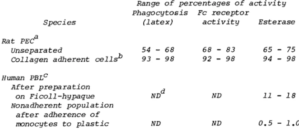
There is generally a good correlation between such markers as phagocytosis, adherence, presence of prominent lysosomes by acridine orange fluorescence (12), and the presence of IgG-Fc receptors in esterase containing cells. Representative results are presented in Table I.
In Table II is shown the percentage of esterase-staining cells in various species and under various treatment procedures.
There is good correlation between increases in the number of cells with staining activity and elicitation procedures.
Our experience, however, has indicated that human monocytes stain more intensely than rat macrophages that stain more in- tensely than mouse peritoneal macrophages. Mouse thioglycol- late-elicited peritoneal macrophages stained more intensely than
TABLE I. Correlation of Esterase Activity with Various Macrophage Markers
Range of percentages of activity Phagocytosis Fc receptor
Species (latex) activity Esterase Rat PECa
Unseparated
Collagen adherent cells ,b 54 - 68 93 - 98
NDd
68 - 83 92 - 98
ND
65 - 75 94 - 98
11-18 Human PBL^
After preparation on Ficoll-hypaque Nonadherent population
after adherence of
monocytes to plastic ND ND 0.5 - 1.0
aCorynehacterium parvum-activated peritoneal exudate cells.
bPeritoneal macrophage population after adherence to col- lagen, removal of nonadherent cells, and recovery of adherent cells (8).
cPeripheral blood lymphocytes.
^ND, Not determined.
TABLE II. Species and Treatment Procedure, Variation in Esterase Staining
Esterase positive cells (%)
Species Resident Proteose Thioglycollate C. parvum
Human* 13-18 NDd ND ND
Ratb 45 -55 60 - 70 ND 65 - 75
Mouse0 20 - 30 ND 42-58 17-28
a
Ficoll-Hypaque separated peripheral blood.
^Peritoneal exudate cells of Lewis, Brown Norray or Sprague
c
Peritoneal exudate cells of CDl mice.
^ND, Not determined.
resident or Corynebacterium parvum macrophages; positive cells in the latter groups often appear pink rather than brightly dark red. With some mouse macrophage preparations, the Koski et al. (1) method may be more reliable. When investigators from the Technicon Corporation tested peripheral blood of vari- ous species for esterase activity according to the method used for human blood, they concluded that only human and rabbit mono- cytes stained well, while staining of mouse, rat, guinea pig, and bovine monocytes was less reliable (Cremins et al., unpub- lished observations). In our hands peritoneal macrophages of any type, elicited mouse and rabbit alveolar macrophages, and rabbit splenic macrophages, have all stained deep red.
It is important to reemphasize, however, that no single method will be optimum in identifying mononuclear phagocytes in all situations. Methods aimed at different properties of these cells will need to be tested in order to determine which is most reliable for the particular case; often more than one method will need to be routinely used.
B. Advantages and Disadvantages
The assay system as indicated is relatively simple to set up and should be followed as indicated in Section III. It is important to allow the hexazonium pararosanilin to form before dilution with buffer, and fixation should not be permitted to proceed too long. The availability of commercially available reagents that are stable at room temperature is a major con- venience of the method. The use of cells in suspension is also an advantage.
The major disadvantage of the method is the price of the
reagents. They are available from the Technicon Corporation
only in 3.8 liter quantities each and in the price range of
$750.00, with more than half of the price being attributed to the substrate. Since such small quantities are involved in the assay, a single laboratory would not use liter quantities in several years. The availability of Hemalog D machines in various cities make the reagents available locally in many areas and negotiations can be made to acquire the small quanti- ties of the reagents that are needed.
C. Specificity of the Reaction for Mononuclear Phagocytes
Recently, a number of reports have appeared indicating that localized granules of esterase reaction product, in addition to being found in mononuclear phagocytes, may also be found in mature, resting T cells (2, 13 - 18) and in null cells (2).B cells and lymphocytes with high-affinity Fc receptors for IgG termed L lymphocytes) and activated T cells appeared to be negative. There is one report, however, of activity in some B cells with EAC rosetting capacity (19). The various positive- ly staining cell types, however, may still be differentiated from each other due to the variety of staining patterns seen.
Mononuclear phagocytes generally show a dotlike granular stain- ing pattern all over the cytoplasm. T cells show a single granule or several localized granules near the membrane, and null cells show scattered granular staining patterns (2, 13 - 18). The identification of cells other than those of the mono- cyte-macrophage group by esterase activity involved an altera- tion of the various procedures. Time of incubation, fixation procedure, and type and quantity of substrate all seem impor- tant, and results vary substantially from one procedure to an- other. It would not be surprising for other lymphoid cell types to demonstrate positivity as variations of the procedure evolve.
REFERENCES
1. I. R. Koski, D. G. Poplack, and R. M. Blaese. A non- specific esterase stain for the identification of raono- cytes and macrophages. In "In Vitro Methods in Cell- Mediated and Tumor Immunity" (B. R. Bloom and J. R. David, eds.), pp. 359-362. Academic Press, New York, 1976.
2. K. E. Higgy, G. F. Burns, and F. G. J. Hayhoe. Discrimi- nation of B, T, and Null lymphocytes by esterase cytochem- istry. J. Haematol. 18: 437-448, 1977.
3. C. Y. Li, K. W. Lam, and L. T0 Yam. Esterases in human leucocytes. J. Histochem. Cytochem. 21: 1-12, 1973.
4. L· T. Yam, C. Y. Li, and W. H. Crosby. Cytochemical iden- tification of monocytes and granulocytes. Am. J. Clin.
Pathol. 55: 283-290, 1971.
5. H. Braunstein. Esterase in leukocytes. J. Histochem.
Cytochem. 7: 202, 1959.
6. L. Rozenszajn, M. Leiberich, D. Shoham, and J. Epstein.
The esterase activity in megaloblasts, leukaemic, and normal haemopoetic cells. Br. J. Haematol. 14: 605-610, 1968.
7. H. Ansley and L. Ornstein. Enzyme histochemistry and dif- ferential white cell counts on the Technicon Hemalog D.
Adv. Automated Anal. 1: 437-446, 1971.
8. S. B. Tucker and R. E. Jordan. Rapid identification of monocytes in a mixed mononuclear cell preparation. J.
Immunol. Methods 14: 267-269, 1977.
9. M. W. Campbell, M. M. Sholley, and G. A. Miller. Macro- phage heterogeneity in tumor resistance: Cytostatic and cytoxic activity of Corynebacterium parvum-activated pro- teose peptone elicited rat macrophages against Moloney sarcoma tumor cells. Cell. Immunol. 50: 153-168, 1980.
10. G. A. Miller, M. W. Campbell, and J. L. Hudson. Separa- tion of rat peritoneal macrophages into functionally dis- tinct subclasses by centrifugal elutriation. J. Reticulo- endothel. Soc. 27: 167-174, 1980.
11. P. S. Morahan and A. M. Kaplan. Antiviral and antitumor functions of activated macrophages. In "Immune Modulation and Control of Neoplasia by Adjuvant Therapy" (M. A.
Chirigos, e d . ) , pp. 447-457. Raven Press, New York, 1978.
11a. C. Lawrence and R. Grossman. Simple butyrate esterase stain for monocytes. Stain Technol. 54: 321-323, 1980.
12. A. C. Allison. Fluorescence microscopy of lymphocytes and mononuclear phagocytes and the use of silica to eliminate the latter. In "In Vitro Methods in Cell-Mediated and Tumor Immunity: (B. R. Bloom and J. R. David, eds.), pp.
395-404. Academic Press, New York, 1976.
13. J. Mueller, G. Brun de Re, H. Buerki, H. U. Keller, M. W. Hess, and H. Cottier. Nonspecific acid esterase activity: A criterion for differentiation of T and B lymphocytes in mouse lymph nodes. Eur. J. Immunol. 5:
270-274, 1975.
14. J. Mueller, H. U. Keller, G. Brun de Re, H. Buerki, and M. W. Hess. Nonspecific esterase activity in T cells.
In "Advances in Experimental Medicine and Biology, Immune Reactivity of Lymphocytes, Development, Expression, and Control: (M. Feldman and A. Globerson, eds.), pp. 117-122.
Academic Press, New York, 1976.
15. A. Ranki. Nonspecific esterase activity in human lympho- cytes: Histochemical characterization and distribution among major lymphocyte subclasses. Clin. Immunol. Immu- nopathol. 10: 47-58, 1978.