RESEARCH PAPER
Tetralone derivatives are MIF tautomerase inhibitors and attenuate macrophage activation and amplify the hypothermic response in endotoxemic mice
Janos Garaia , Marcell Krekob , LaszloOrfi} b , Peter Balazs Jakusc , Zoltan Rumbusd , Patrik Keringerd , Andras Garamid , Eszter Vamosc , Dominika Kovacsc , Viola Bagone Vantusc , Balazs Radnaic and Tamas Lorandc
aDepartment of Pathophysiology, Institute for Translational Medicine, University of Pecs, Medical School, Pecs, Hungary;bDepartment of Pharmaceutical Chemistry, Semmelweis University, Budapest, Hungary;cDepartment of Biochemistry and Medical Chemistry, University of Pecs, Medical School, Pecs, Hungary;dDepartment of Thermophysiology, Institute for Translational Medicine, University of Pecs, Medical School, Pecs, Hungary
ABSTRACT
Macrophage migration inhibitory factor (MIF) is a pro-inflammatory cytokine playing crucial role in immun- ity. MIF exerts a unique tautomerase enzymatic activity that has relevance concerning its multiple func- tions and its small molecule inhibitors have been proven to block its pro-inflammatory effects. Here we demonstrate that some of theE-2-arylmethylene-1-tetralones and their heteroanalogues efficiently bind to MIF’s active site and inhibit MIF tautomeric (enolase, ketolase activity) functions. A small set of the syn- thesised derivatives, namely compounds (4), (23), (24), (26) and (32), reduced inflammatory macrophage activation. Two of the selected compounds (24) and (26), however, markedly inhibited ROS and nitrite production, NF-jB activation, TNF-a, IL-6 and CCL-2 cytokine expression. Pre-treatment of mice with com- pound (24) exaggerated the hypothermic response to high dose of bacterial endotoxin. Our experiments suggest that tetralones and their derivatives inhibit MIF’s tautomeric functions and regulate macrophage activation and thermal changes in severe forms of systemic inflammation.
ARTICLE HISTORY Received 1 February 2021 Revised 30 March 2021 Accepted 6 April 2021 KEYWORDS MIF; MIF inhibitors; 2- arylmethylene-1-tetralones;
tautomerase; inflammation
Introduction
Macrophage migration inhibitory factor (MIF) was the first repre- sentative of those polypeptide immune mediators that have been grouped later as “cytokines”1,2. Since its description 50 years ago MIF has accumulated a bewildering variety of immune and non- immune functions3. MIF has been also considered to be a missing link between inflammation and tumorigenesis4.
MIF structure shares no homology with other known cytokines.
Its structural relatives are the mammalian enzyme,D-dopachrome- tautomerase (DDT)5, and the prokaryotic enzymes: chorismate mutase, 5-carboxymethyl-2-hydroxymuconate-isomerase (CHMI), trans- and cis-3-chloroacrylic acid dehalogenase (CaaD and cis- CaaD, respectively), and 4-oxalocrotonate-tautomerase (4-OT)6,7. MIF exists in a homotrimeric form of 12.4 kD monomers. In each monomer, two a-helices are packed against a four-stranded b-sheet8. There are reports, however, of the occurrence of a dimeric form of MIF9 as well as of other oligomerisation states10 and also of heteromers11.
More than twenty years ago Rorsman et al. reported that recom- binant MIF catalyses the tautomerisation of the non-naturally occur- ring D-dopachrome, transforming the coloured compound to the colourless dihydroindole carboxylic acid (DHICA)12, however, L-dopa- chrome methyl ester (a melanin precursor) have also been found a suitable substrate. This was soon followed by the identification of phenylpyruvate andp-hydroxyphenylpyruvate as alternate substrates for the tautomeric activity of MIF13. In the homotrimeric MIF, the
catalytic site is located between each of two adjacent monomers.
Acidic pKa of the N-terminal proline of each MIF monomer is thought to play a crucial role in the keto-enol tautomerisation reac- tion7. A direct link between cytokine activity and tautomerase cata- lytic site of MIF has been reported14, although a“true”endogenous small molecule ligand has yet to be found. Blocking of endogenous MIF by a small molecule such as“ISO-1”and neutralisation of MIF by anti-MIF antibodies or by plant-derived MIF inhibitors reduces the manifestations of inflammatory conditions such as type II collagen- induced arthritis, immunologically induced kidney disease, experi- mental autoimmune encephalomyelitis, experimental allergic neuritis, immunoinflammatory diabetes, experimental autoimmune myocardi- tis, irradiation-induced acute pneumonitis, sepsis, and ischemia–re- perfusion injury15–18. Therefore, inhibitors of MIF tautomerase hold promise for prospective clinical use in many pathologic conditions19. Development of individualised therapy targeting MIF in these condi- tions is expected based on the human genetic data supporting the role of high-expression MIF alleles in the clinical severity and end- organ complications of a number of inflammatory disorders20.
Molecular modelling techniques have been used lately to find
“designer”inhibitors of promising potential21–23. The reported MIF enzyme inhibitors include long-chain fatty acids, E-2-fluoro-p- hydroxycinnamate, dopachrome analogues, tryptophan and tyro- sine derivatives24, or coumarin- and chromenone derivatives19,25.
A large variety of natural compounds show inhibitory effect of the MIF enzyme including e.g. ellagic acid26. According to previ- ous results, caffeic acid proved to be an efficient inhibitor of MIF CONTACTBalazs Radnai balazs.radnai@aok.pte.hu Department of Biochemistry and Medical Chemistry, University of Pecs, Medical School, Pecs, Hungary.
Supplemental data for this article can be accessedhere.
ß2021 The Author(s). Published by Informa UK Limited, trading as Taylor & Francis Group.
This is an Open Access article distributed under the terms of the Creative Commons Attribution License (http://creativecommons.org/licenses/by/4.0/), which permits unrestricted use, distribution, and reproduction in any medium, provided the original work is properly cited.
2021, VOL. 36, NO. 1, 1357–1369
https://doi.org/10.1080/14756366.2021.1916010
tautomerase when using either dopachrome27 or phenylpyru- vate28 as the substrate. Phenylpyruvate tautomerase was also inhibited by curcumin28, a substance known long for its anti-- inflammatory and cancer chemopreventive properties–a constitu- ent of the spice turmeric29. Therefore, the selection of our potential MIF inhibitors was based on the unsaturated ketone – structural motif–found in several natural or synthetic MIF inhibi- tors such as coumarines and chromenones and cinnamates such as caffeic acid, curcumin, andN-acetyl-p-benzoquinone19. In add- ition, according to our previous studies, some tetralone derivatives proved to be efficient MIF inhibitors30.
Based on these findings, we have focused on the group of cyc- lic a,b-unsaturated ketones to study their inhibitory effects towards the MIF tautomerase activity. To find compounds with good inhibitory effect, we have previously selected some different model compounds as 2-arylidenecycloalkanones, 2,5-diarylidene- cyclopentanone and few 2-arylidene-1-tetralones30. The enol-keto tautomeric conversion of phenylpyruvate (ketonase reaction) was investigated by spectrophotometric method. From the substances investigated, several proved to be good inhibitors of the tauto- merase activity of the MIF enzyme.
This paper deals with a large molecular library of E-2-aryl- methylene-1-tetralones and their hetero-analogues related to nat- urally occurring flavonoids. Some representatives of this latter group have previously been proven as potent MIFtautomerase inhibitors28,31. At this end, potential analogies between the bio- logical roles of flavonoids and melanins (or precursors thereof) deserve particular attention32 concerning possible implication of MIF in (neuro-) melanogenesis33.
The molecule library we have used here shows large structural diversity. We have chosen cyclic ketones of different size and with different heteroatoms as skeletal compounds, and applied various aryl side chains to find an optimal lead compound. We have var- ied the size of the ketone ring, the aromatic substituent C-ring (with electron withdrawing and electron donating groups), and also changed the type of the B-ring (see Figure 1 and Table 1).
Through in silico methods, we aimed to identify the key interac- tions required for MIF inhibitory activity. In addition, we tested the biological activity of the best tautomerase inhibitors from the molecular library in lipopolysaccharide-(LPS)-induced macrophages and also in LPS-treated mice.
Material and methods Reagents
All reagents were purchased from Sigma-Aldrich (St. Louis, MO) unless otherwise indicated. A molecular library of 38 structurally related known compounds has been studied. Based on their struc- tural features, this library could be further divided into two sub- groups (Table 1). The group I consists of a 1-indanone derivative (1), substituted arylmethylene–tetralones (2, 4–18), heteroaryl–me- thylene–tetralones (19–26) and a benzosuberone derivative (3).
Group II contains chromanones and their thioanalogues (27–38). All of the compounds investigated here have been prepared by base catalysed aldol condensation, the conditions (temperature, solvent
and catalyst) of the synthetic method were a little bit different. See the detailed methods below. (Their structure is depicted in Supplementary material’s Figure S1 and in Table 1; the synthesis method is in Ref. [34].) Scheme 1 describes the general synthetic method. The preparation of the E-2-phenylmethylene-1-indanone (1), E-2-phenylmethylene-1-benzosuberone (3) and E-2-arylmethy- lene-1-tetralones (2, 4–26) was carried out at room temperature in ethanol. The synthesis of theE-3-arylmethylenechroman-4-ones and E-3-arylmethylene-1-thiochroman-4-ones (27–36) was performed at 140C with piperidine as a catalyst without solvent. The 4-thiochro- manone-1-oxide (36) and1,1-dioxide (37) was obtained according to literature methods35. All of the compounds were purified by recrystallisation from methanol and with column chromatography.
Their structural characterisation is based on FT-IR methods and pre- viously published NMR data36–53. Thus, all of the test compounds studied here have been published elsewhere and characterised, see the references above. All of the compounds were proved asE-iso- mer based on the NMR measurements54.
Tautomerase assay
The enol-keto tautomeric conversion of phenylpyruvate (ketonase reaction) was assessed at room temperature by monitoring the decrease in absorbance at 288 nm on a dual path Shimadzu 2100 UV spectrophotometer (Shimadzu, Kyoto, Japan) according to Taylor et al.55with minor modifications. Briefly, the 0.72 ml reaction mixture consisted of the enzyme at a final concentration of 0.4mg/
ml and the substrate in a 50 mM sodium phosphate buffer (pH ¼ 6.5). The phenylpyruvate substrate was dissolved fresh in absolute ethanol to yield a final concentration of 100mM. The reaction was monitored for 75 s at room temperature. A recombinant human MIF (from ATGen Co. Ltd., Seoul, Republic of Korea) was used as the enzyme source. Inhibitors were dissolved fresh in ethanol or DMSO. Ethanol or DMSO did not affect the enzyme reaction at the amounts applied. The compounds were diluted to achieve final concentrations of 200, 100, 50, 20, 5, 1, and 0.5mM in the reaction mixture. All data presented have been derived from consonant measurements repeated at least three times. As reference inhibitor caffeic acid (ketonase and enolase IC50: 0.5 and 2.0mM respectively) was used28. The areas under the curves obtained with and without the inhibitors were used to compare the conversion rates of the substrate. The IC50 value – the concentration of the given com- pound required to achieve 50% inhibition56 –was obtained by a nonlinear curve fitting (Sigma Plot 2000, SPSS Inc., Chicago, IL, USA) on the data obtained with the different concentrations of the inhibitor using the equation: f¼minþ(maxmin)/(1þ(x/EC50)nHill).
Molecular modelling
Molecular modelling was applied as a means of gathering informa- tion on the structure’s binding mode. Glide docking was conducted on various MIF-ligand crystallographic structures in Extra Precision mode without constraints. Dockings with core restrictions using two types of docking output structures were examined to assess the favoured binding mode. Covalent docking was used to visualise the hypothesised adduct formation with Pro1. Molecular Dynamics simulations were used to assess the stability of the reversible com- plexes and explore the nature of the binding site interactions.
The crystallographic structure of MIF in complex with an inhibitor was obtained from Protein Data Bank (PDB 6B1K). All calculations were carried out with the modules of Schrodinger Suites 2019€ –2 (Schr€odinger, LLC, New York, NY) in Maestro. The protein was pre- pared by adding hydrogens and missing side chains. Water Figure 1. General formula of the test compounds(1–38).
molecules, sulphate ions and glycerol molecules were removed, as well as ligands in the active sites formed by chains A–B and B–C.
Hydrogen bonds were optimised at pH ¼ 7.4, followed by Impref minimisation using OPLS3e force field. The 3D structures of the ligands were determined by LigPrep at pH¼7.4 using OPLS3e force field. The grid box for docking was centred on the original ligand between chains A and C with automatic ligand size. Initial Glide docking experiments were run without any pharmacological con- straints. Type I constrained docking was run with a tolerance of 0.1 Å, while in Type II constrained docking, the tolerance was set to 2 Å. Docking scores and MM/GBSA calculation results of reversible complexes in kcal/mol are summarised in Table 2. The model was validated by docking 3-(3,4-dihydroxyphenyl)-7-hydroxy-4H-chromen-
4-one (RCSB ligand ID 47X, co-crystallized in MIF PDB entry 3L5R) with extra precision and without using constraints. Four viable poses were found, all of them similar to the binding mode observed in 3L5R, with docking scores ranging between 9.975 and 10.533 kcal/mol. Ligand RMSD values ranging from 0.7428 to 1.6388 Å for all heavy atoms were calculated, while values between 0.3643 and 0.7292 Å were calculated for the 7-hydroxy-4H-chromen- 4-one core residing in the solvent inaccessible binding pocket of MIF. RMSD values for the ligand were calculated after binding site alignment considering residues within 5 Å range of the ligand, and 3L5R was used as the reference structure in its native form.
Covalent dockings were run in CovDock within Maestro. The following custom covalent mechanism protocol was created for this purpose:
LIGAND_SMARTS_PATTERN 1,[C]¼[C]-[C]¼[O]
RECEPTOR_SMARTS_PATTERN 2,[C]-[N;H1,-1]
CUSTOM_CHEMISTRY ("<1>",("charge",0,1)) CUSTOM_CHEMISTRY ("<1>j<2>",("bond",1,(1,2))) CUSTOM_CHEMISTRY ("<2>=[C]-[C]¼[O]",("bond",1,(1,2)))
Pro1A is present in the conjugate acid form, and was manually deprotonated before initiating covalent dockings. The same core Table 1. Inhibitor activity of the test compounds (1–38) in the enol-keto and keto-enol tautomeric conversion of phenylpyruvate.
Compounds X Ar
Inhibition of ketonase (IC50,lM)
Inhibition of enolase (IC50,lM)
1 – Phenyl 82.8 ± 16.5 20.2 ± 3.9
2 CH2 Phenyl 127 ± 20 113 ± 16
3 (CH2)2 Phenyl 84.0 ± 15.3 129 ± 19
4 CH2 40- CH3-phenyl 16.5 ± 3.4 2290 ± 343
5 CH2 40-OCH3-phenyl 57.0 ± 14.1 33.4 ± 8.8
6 CH2 40-Cl-phenyl 59.1 ± 9.7 42.8 ± 3.1
7 CH2 20-Cl-phenyl 2640 ± 406 458 ± 73
8 CH2 40-Br-phenyl 52.2 ± 11.5 232 ± 54
9 CH2 30-Br-phenyl 7340 ± 1024 3350 ± 424
10 CH2 40-F-phenyl 72.4 ± 3.4 42.1 ± 4.3
11 CH2 20,60-(Cl)2-phenyl 58.9 ± 10.3 35.5 ± 5.9
12 CH2 30,40-(Cl)2-phenyl 2620 ± 696 91.5 ± 9.0
13 CH2 20,40-(Cl)2-phenyl 16.9 ± 4.2 301 ± 42
14 CH2 40-COOH-phenyl 187 ± 32 37.7 ± 16.3
15 CH2 40-(NCH3)2--phenyl 90.2 ± 15.1 495 ± 101
16 CH2 40-CN-phenyl 146 ± 17 43.2 ± 12.4
17 CH2 30,40-(OCH3)2-phenyl 585 ± 94 302 ± 63
18 CH2 30,40-(OCH2O)-phenyl 50.7 ± 6.0 213 ± 31
19 CH2 20-Furyl 57.7 ± 12.6 25.4 ± 4.7
20 CH2 20-Thienyl 54.0 ± 5.5 52.3 ± 7.6
21 CH2 20-Pyrrolyl 142 ± 31 125 ± 39
22 CH2 N-Methyl-20-pyrrolyl 23.8 ± 3.8 27.8 ± 5.6
23 CH2 30-Indolyl 58.8 ± 10.6 2.89 ± 0.75
24 CH2 20-Pyridyl 5.63 ± 0.94 28.6 ± 7.4
25 CH2 30-Pyridyl 20.3 ± 5.5 28.6 ± 6.0
26 CH2 40-Pyridyl 21.0 ± 6.9 209 ± 77
27 O Phenyl 267 ± 59 1740 ± 483
28 O 40- CH3-phenyl 102 ± 19 3620 ± 829
29 O 30- CH3-phenyl 56.1 ± 8.8 443 ± 112
30 O 40-OCH3-phenyl 96.0 ± 29.9 425 ± 70
31 O 30-OCH3-phenyl 97.0 ± 20.7 205 ± 32
32 O 20-OCH3-phenyl 4.31 ± 1.34 1260 ± 159
33 O 40-Br-phenyl 311 ± 77 1890 ± 438
34 O 20-Br-phenyl 98.3 ± 16.0 3200 ± 887
35
O
O
419 ± 113 –
(Because of low solubility data are not available)
36 S Phenyl 208 ± 42 633 ± 108
37 SO Phenyl 94.6 ± 25.4 193 ± 59
38 SO2 Phenyl 160 ± 38 151 ± 27
Scheme 1. General synthetic method for the preparation of the title compounds 1–38. Reagents and conditions (i) base as NaOH or piperidine, temperature:
ambient temperature or 140C.
constraint settings were applied as to the Glide docking experi- ments. The tolerance was set to 2 Å in both instances. The pose prediction docking mode was used. Docking scores and MM/GBSA calculation results of covalent complexes in kcal/mol are summar- ised inTable 2.
Molecular dynamics simulations ranging between 1.2 and 175 ns were applied to a selected group of the compounds with- out using constraints. All processes were initiated using Desmond in Maestro. Output structures of a docking experiment with Type I core constraints were used as starting points. SPC solvent model was used and box boundaries were adjusted to a distance of 10 Å from the protein–ligand complexes in each axis. Net positive charge of the protein was neutralised by chloride ions. 0.15 M NaCl was added to the systems in each instance. Simulations were carried out using NPT conditions, at a constant temperature of 300 K and pressure of 1 atm, with Nose–Hoover chain thermostat (1 ps relaxation time) and MTK barostat (2 ps relaxation time) methods. The cut-off radius for coulombic interactions was set to 9 Å. The systems were relaxed before simulation using the default relaxation process of Desmond57. The stability of (24) in the active site was explored through a 175 ns simulation with 25 ps record- ing intervals, and the Thermal MMGBSA script available in Schr€odinger was used to obtain multiple binding free energy val- ues for the complex, sampling every 100th frame of the
simulation. The 1.2 ns simulation of (24) was also conducted with TIP3P water model to explore the nature of interactions on the ps scale in a different MD setup.
Cell culture and treatments
In cell culture experiments, we used RAW264.7 mouse monocyte/
macrophage cell line (ECACC, Salisbury, UK; passage number:
8–15) and RAW-BlueTM cells (Invivogen, Toulouse France, passage number: 10–14). RAW264.7 and RAW-BlueTM cells were cultured in 5% CO2 at 37C in endotoxin-tested Dulbecco’s Modified Eagle’s Medium (high glucose, 4.5 g/L, 2 mM L-glutamine; Corning, Corning Incorporated Costar, Corning, NY) supplemented with 10% FBS. For culturing RAW-BlueTMcells, we also added 100mg/ml normocin and 200mg/ml zeocin to the medium. The day before the experiments, cells were plated onto 24- or 96-well plates and cultured overnight.
Then medium was replaced to fresh one and cells were induced by 0.1mg/ml or 1mg/ml lipopolysaccharide (LPS; E. coli, 0127:B8; Sigma- Aldrich, Budapest, Hungary). The freshly synthesised tetralone deriva- tives were dissolved in DMSO and applied in 20mM concentration as a pre-treatment, added 30 min before LPS induction. To exclude the effects of vehicle, both CTRL and LPS-treated cells received the same amount of DMSO.
Table 2. Glide XP docking scores and CovDock affinity scores of the compounds using type I and type II docking constraints.
Reversible Covalent
Type I Type II Type I Type II
docking score (XP)
MM/GBSA
dG Bind docking score (XP)
MM/GBSA
dG Bind cdock affinity
MM/GBSA
dG Bind cdock affinity
MM/GBSA dG Bind
1 8.499 54.71 8.519 57.99 8.693 61.64 6.249 37.79
2 8.132 50.83 7.819 55.61 8.233 34.65 6.685 36.06
3 – – 6.134 28.08 7.312 24.51 6.981 36.11
4 8.213 52.36 7.034 42.78 8.188 35.80 6.140 47.58
5 8.210 53.75 4.216 46.44 8.141 37.61 5.607 24.69
6 8.135 52.37 7.225 38.32 8.269 45.84 6.819 27.79
7 8.309 55.48 8.360 51.57 8.045 50.49 7.748 6.62
8 8.193 51.5 6.560 39.76 8.046 36.68 6.685 43.60
9 8.052 53.51 5.437 36.13 8.183 37.85 6.176 34.18
10 8.204 50.87 6.788 53,12 8.626 41.35 6.855 34.79
11 8.236 57.97 7.176 20.02 7.897 42.24 7.804 46.15
12 7.926 52.70 6.785 44.62 8.186 40.35 6.525 48.47
13 8.278 57.06 7.075 42.91 8.024 52.93 7.111 32.16
14 8.209 51.36 – – 8.301 40.16 5.758 29.96
15 8.221 52.12 4.795 2.46 8.369 37.02 6.363 47.81
16 8.281 51.63 6.784 41.76 7.457 45.26 6.289 43.93
17 8.369 54.02 – – 8.464 45.79 5.367 44.99
18 8.143 50.91 6.521 41.23 8.197 46.33 6.542 41.71
19 7.935 53.14 7.953 57.54 7.890 41.16 6.452 44.63
20 8.063 54.70 7.505 54.38 8.308 34.01 6.735 32.73
21 8.019 51.92 7.732 54.85 8.000 28.22 7.221 34.68
22 7.929 54.38 6.722 49.94 7.823 39.63 6.360 18.93
23 8.200 50.17 6.152 36.16 8.344 39.62 6.480 35.48
24 8.227 55.40 7.714 60.28 8.504 42.35 7.072 33.41
25 8.093 50.90 6.606 53.61 8.256 41.14 7.077 35.23
26 8.206 50.79 6.501 54.63 8.601 40.48 6.242 32.12
27 8.425 56.44 6.836 56.88 8.533 48.12 6.750 34.59
28 8.289 58.79 7.046 44.12 8.837 48.69 6.608 33.40
29 8.196 58.01 6.252 43.46 8.228 49.88 7.206 28.65
30 8.315 58.44 4.182 47.12 8.667 50.57 5.632 36.95
31 8.132 54.03 4.606 41.29 8.560 51.83 6.300 34.00
32 8.500 61.06 6.732 15.06 8.626 45.56 6.887 9.84
33 8.341 57.21 6.865 48.23 7.404 50.21 6.634 39.94
34 8.195 54.00 8.28 43.08 8.134 44.49 7.360 27.69
35 – – – – – – – –
36 7.700 37.08 6.352 48.78 7.495 25.07 6.765 27.60
37 – – – – –5.623 –26.39 –6.764 –32.26
38 – – – – –5.975 12.70 – –
Calculated binding energies are provided for both, the reversible and covalent MIF-ligand complexes.
ROS and nitrite production
To measure the amount of reactive oxygen species we seeded RAW264.7 cells onto 96-well plates in a density of 105cells/well.
Cells were treated with the tetralones as a pre-treatment and they were induced with 1mg/ml LPS for 24 h. Then 2mM dihydrorhod- amine123 (DHR123; Life Technologies, Carlsbad, CA) fluorescent dye was added and incubated for an additional 2 h. Fluorescent intensity of the dye (excitation 490 nm/emission 510–570 nm) was measured with Glomax Multi Detection System (PromegaVR, Madison, WI, USA).
For nitrite measurements, we applied the same culturing condi- tions, treatments, and equipment as described above. After 24 h of incubation, 50ml medium was removed and added to equal amount of Griess reagent (Sigma-Aldrich, St. Louis, MO, USA) in 96-well plate.
The optical density was measured at 550 nm wavelength.
NF-jB activation
The detection of NF-jB activation was accomplished by using RAW-BlueTM cells. RAW-BlueTM cells are genetically modified RAW264.7 macrophages, which are able to secrete embryonic alkaline phosphatase (EAP) upon LPS induction and following NF- jB activation. The levels of EAP can be examined using QUANTI- BlueTMdetection medium.
RAW-BlueTM cells were seeded into 96-well plate at a density of 105cells/well. Cells were treated with tetralones (pre-treatment, 20mM) and with 1mg/ml LPS. 24 h after LPS treatment 20ml medium was removed and incubated with 100ml QUANTI-BlueTM detection medium (Invivogen; Toulouse, France) in 96-well plates.
The optical density was measured at 600 nm.
Cytokine production
For cytokine concentration measurements, RAW264.7 cells were cultured in 24-well plates at a density of 5105 cells/well and treated with tetralones (pre-treatment, 20mM) and with 1mg/ml LPS for 24 h. TNF-a, IL-6 and CCL-2 levels were determined from the culturing media by using Ready-Set-Go ELISA kits (eBioscience, San Diego, CA, USA) for TNF-aand IL-6 detection and mouse CCL- 2 uncoated ELISA-kit (Invitrogen, Vienna, Austria) for CCL-2 deter- mination. ELISA-kits were applied as provided by the protocol of the manufacturer, the optical density was measured at 450 nm.
Mouse thermometry studies
Thermophysiological experiments were performed in 24 C56BL/6 adult male mice. Experimental procedures were approved by the Animal Research Review Committee of the University of Pecs, Medical School (Permit number: BA02/2000–6/2018). At the time of the experiments, the mice weighed 28 ± 4 g. Animals were housed in temperature-controlled rooms on a 12/12 h light/dark cycle. Standard rodent chow and tap water were availablead libi- tum. All experimental procedures were carried out according to the European Communities Council Directive of 2010/63/EU under protocols approved by the Institutional Animal Use and Care Committee of the University of Pecs. Mice were anaesthetised with an intraperitoneal (i.p.) injection of ketamine-xylazine (81.7 and 9.3 mg/kg, respectively) and received antibiotic prophylaxis intramuscularly (gentamycin, 6 mg/kg). During the surgery, mice were kept on a heating pad (PECO Services Ltd., Brough, UK), and then to prevent postsurgical hypothermia, the animals were allowed to recover from anaesthesia in a temperature-controlled
chamber (model MIDI F230S; PL Maschine Ltd., Tarnok, Hungary) set to 31C. Each mouse was implanted intraperitoneally with a miniature biotelemetry transmitter (G2 E-Mitter series; Starr Life Sciences Corp., Oakmont, PA) to record abdominal temperature (Tab) and general locomotor activity. The latter has been shown to play an important thermoregulatory role in mice58–60. The transmitter was inserted into the abdominal cavity through a small midline incision and was fixed to the left side of the abdom- inal wall with suture. The surgical wound was closed and the mice were returned to their home cages. The mice were allowed a 2–3 weeks post-surgical recovery period before an experiment.
Telemetry receivers (model ER-4000; Starr Life Sciences Corp., Oakmont, PA, USA) were positioned in a temperature-controlled room, and the home cages of mice were placed on top of the receivers, as described earlier59. The ambient temperature of the room was set to 26C, which is near the lower end of the thermo- neutral zone for mice61. On the day of an experiment, compound (24) at 1.6 mg/kg or its vehicle (10% DMSO in saline) was adminis- tered intraperitoneally in bolus (3.3 ml/kg). Thirty minutes later, either LPS (5.0 mg/kg) fromEscherichia coli0111:B4 (Sigma-Aldrich, St. Louis, MO) or saline was intraperitoneally injected to the mice.
Thermoregulatory responses were compared by two-way ANOVA followed by Fisher LSDpost hoctest, as appropriate, using Sigma Plot version 11.0 (Systat Software, San Jose, CA, USA) with the level of significance set at p<0.05. The data are reported as means ± SEM.
Results
Tautomerase assay
First the inhibitory effect on the ketonase reaction is discussed. As regards the structure–activity relationship, the size of the cyclic ketone ring has an impact on the inhibitory effect (see Table 1).
From the phenyl substituted compounds, the five-membered (1) and the seven-membered (3) showed almost identical inhibitory effect (IC50¼82.8 and 84.0mM, respectively), while the six-mem- bered (2) exerted a lower activity (IC50¼127mM). The substituted derivatives of (2) containing both homoaromatic and hetero- aromatic side chain were examined.
Summarising the results of the measurements studying the possible inhibitors of ketonase reaction, the best inhibitors are the compounds with a nonpolar aromatic side chain, such as the methyl derivative (4), theortho,para-dichloro compound (13) and the polar 2-pyridyl derivative (24). Analysis of the structure–activ- ity relationship is presented later on in more detail.
The inhibition of the enolase activity was also investigated. In the group of the compounds with a homocyclic skeleton, some good inhibitors can be found, such as five-membered phenyl derivative (1) (IC50¼20.2mM). Compounds with a heteroaryl side chain produce the highest bioactivity. From the five-membered derivatives the indolyl- (23, IC50¼2.89mM), the furyl derivative (19, IC50¼25.4mM) and the N-metylpyrrolyl compound (22, IC50¼27.8mM) are the best inhibitors. As for the six-membered ones, two pyridyl derivatives (24 and25, IC50¼28.6mM for both) exerted the best activity, while the 4-pyridyl derivative (26) proved to be a weaker enolase inhibitor. Comparing the enolase and ketonase activity and focussing on the selectivity, some of our compounds acted as good ketonase inhibitors having high select- ivity, such as (4) (IC50enolase/IC50ketonase ¼ 13.9), (13) (IC50enolase/IC50ketonase ¼18), (26) (IC50enolase/IC50ketonase
¼10), while the substance (23) (IC50ketonase/IC50enolase ¼20) behaved as a strong and selective enolase inhibitor.
Molecular modelling Docking experiments
The majority of MIF inhibitors target the active site where enzym- atic reactions take place. The protein is known to form homo- trimers, in which binding sites are formed by each of two adjacent chains. As represented in the Protein Data Bank entry 3IJJ, 4-hydroxyphenylpyruvate contacts with Pro1, Lys32 and Ile64 of monomer A, and Tyr95 and Asn97 of monomer C. Available structures of MIF in complex with various inhibitors reveal the importance of these residues in ligand binding62. Like the series of compounds presented in this paper, many MIF inhibitors con- tain more than one aromatic ring63, suggesting the extensive involvement ofp-interactions.
Reversible docking
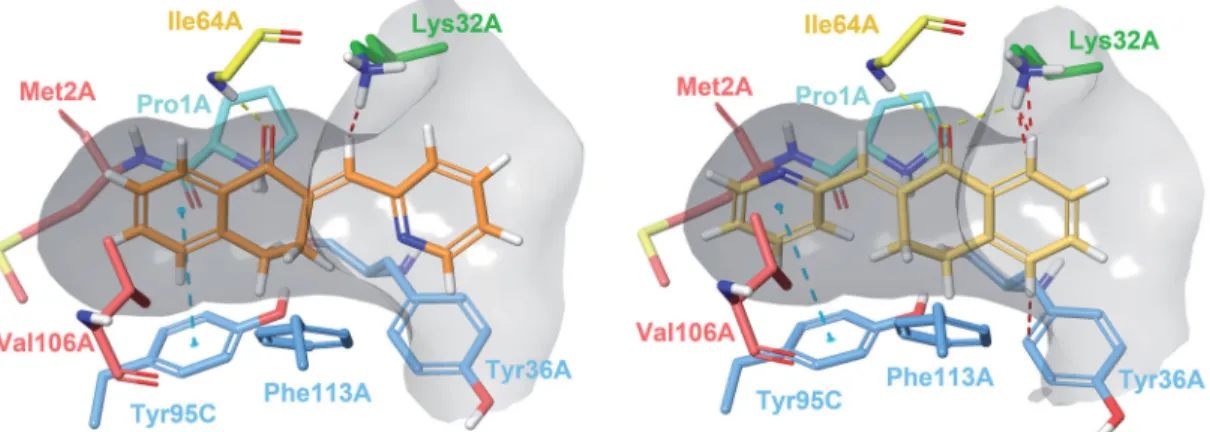
Docking experiments revealed two possible types of binding mode (Figure 2). In Type I complexes, the A and B rings occupy the binding pocket. The ketone oxygen forms a hydrogen bond with the peptide NH of Ile64A. The aromatic ring interacts with Tyr95C throughp–p stacking. The catalytic Pro1A and Lys32A are in the proximity of the methylene bridge. The aryl or heteroaryl group bonds with Lys32A, Tyr36A and Phe113A through hydro- phobic andp-interactions. Such binding mode can be observed in the PDB entry 3L5R of a 3-phenylchromen-4-one compound64. In type II binding mode, where the ligand is horizontally flipped, the C-ring interacts with Tyr95C, the ketone oxygen with Ile64A and Lys32A, and the A ring with Tyr36A. A similar binding mode of a structurally related covalent inhibitor can be observed in the PDB entry 4Z1U65. After visual analysis and consideration of docking scores, we propose Type I binding mode to be more favourable.
Covalent adduct formation
In the reversible docking models, the position of the electrophilic methylene bridge relative to Pro1A hints at the possibility of a covalent mechanism. Such catalytic proline residues reportedly possess the ability to mediate enzymatic conversions through covalent catalysis66. NAPQI has been shown to bind covalently to Pro1, while exhibiting non-covalent inhibitory activity67. The cova- lent mechanism of a series of MIF inhibitors has also been verified by X-ray crystallography and enzymatic assays65, where both cova- lent and non-covalent tautomerase inhibitory activity have been attributed to“compound 12”, visible in PDB entry 4Z1U65.
Both Type I and Type II constraints were used for covalent docking to assess whether any of the two determined binding poses would be favourable for a covalent reaction to occur between Pro1A and the methylene carbon. Covalent adduct for- mation was successful for most molecules, with the exception of (35) and (38) due to steric clashes. Covalent docking affinity and MM/GBSA scores indicate that the reaction is more likely to hap- pen in Type I binding mode (Table 2). Even though an increased number of steric clashes with Asn97C, Tyr95C and Pro1A are vis- ible, minimisation of the output structure results in a reasonable covalent adduct (Figure 3).
Molecular dynamics
We conducted MD simulations to reveal binding characteristics obscured by using a rigid protein structure for docking. The H- bond between Ile64A and the ketone oxygen is a crucial contact.
Thep–pstacking between the A ring and Tyr95C is a T-type stack- ing interaction. Weak hydrophobic contacts with Val106A and Met2A are present. The arylmethylene substituent is exposed to Figure 2. Type I (left) and Type II (right) binding modes of tetralones in the active site of MIF, identified through reversible docking experiments. Carbons of (24) are coloured orange (Type I) and yellow (Type II). Residues are coloured according to their type of interaction with the ligand: yellow residues form H-bonds, blue residues formp–pstacking and green form cation-pinteractions, red residues are hydrophobic, and the catalytic Pro1 is coloured cyan. The grey area is the inner surface of the binding site. Red dashed lines indicate steric clashes.
Figure 3. Compound 24covalently bound to Pro1A of MIF in Type I binding mode. The H-bond with Ile64A is represented by the yellow, and thep–pstacking with Tyr95C by the blue dashed line. The docking output structure was minimised to avoid clashes due to the rigid receptor model. Active site residues involved in reversible binding are shown. Some atoms are hidden for clearer view of the covalent adduct.
the solvent (see ligand RMSF in Supplementary material, Figure S2) and forms a sandwich-typep–pstacking with Tyr36A. Rotation of the C-ring is hindered by constant interactions with Lys32A, Tyr36A and Phe113A (Supplementary material,Figure S3). A steric clash may occur for 3-substituted phenylmethylene-tetralones such as the inactive analogue (9). The protonated amino group of Lys32A is capable of forming a cation-p bond with the C-ring, especially of heteroaryl derivatives (22–26). The interaction is less pronounced for phenyl compounds, such as (4) and (13), and vir- tually absent from models of (9). All three interactions can be pre- sent concurrently. A water molecule can also be involved in ligand binding, where the catalytic Pro1A and the adjacent Tyr36A interacts with the ligand through a water bridge. The hydrogen of the water molecule occasionally engages in close contact with the methylene carbon or interacts with heteroatoms and halogen sub- stituents of the C ring.
The importance of the interaction is most pronounced in the case of (24), where the 2-pyridyl nitrogen is adequately positioned to maintain a constant water bridge interaction (Figure 4).
Consequently, a stable complex forms between (24) and MIF, where no major conformational change is observed for 65 ns. The presence of interactions in percentage of frames are: Ile64A(H-bond)
¼ 65%; Tyr95C(p–p) ¼ 64%, Lys32A(cation–p) ¼ 69%, Pro1A(water bridge)¼ 91%, Tyr36A(water bridge)¼ 83%. Additionally,p–p interac- tions with Tyr36A and Phe113A were observed in 18% of the frames for both residues. The most prominent weak hydrophobic contacts are formed with Met2A, Tyr36A, Val106A and Phe113A and are present throughout the entirety of the simulation. The averaged free binding energy was calculated at 59.32 kcal/mol (max¼ 51.19, min¼ 68.75; SD¼3.40) for this phase. Minor pro- tein and ligand movements at 65 ns result in (24) assuming a min- imally altered, but stable position until 138.79 ns. In this phase of roughly 73 ns in length, H-bond interaction with Ile64A diminish to 21% as it forms weak hydrophobic contacts instead, and p–p interaction with Tyr95C falls to 2%. The water bridge interaction becomes even more pronounced with 94% and 90% presence rates for Pro1A and Tyr36A. Lys32A and Phe113A continue to interact with thep–electron system of the C ring (60% and 32%).
Free binding energies for this phase were calculated at an average of 54.82 kcal/mol (max¼ 46.95, min=-63.18; SD ¼ 3.88). At 138.79 ns Trp108A disrupts binding as it intrudes the active site, which results in cessation of most H-bond andp interactions, and the distancing of the methylene carbon from Pro1A as well. As a result, (24) undergoes a major positional shift, and onlyp–pstack- ing interactions of the C-ring with Phe113A and Trp108A are vis- ible with 41% and 27% frequencies. Weak hydrophobic contacts with Phe49C and Tyr95C also develop. The complex remains unchanged until the end point of the 175 ns simulation (see Supplementary material Figures S4–S7 for RMSD plot and active site residue contributions).
In simulation of (24) using TIP3P water model, the phenolic oxygen of Tyr95C acts as the H-acceptor instead of Tyr36A; such model describing an enolate intermediate was published previ- ously68. We have also identified a potential chloride ion binding motif at the active site. Chloride binding in MIF may be a remnant of a lost dehalogenase function. Such enzymatic activity is exhib- ited by structurally related bacterial tautomerases CaaD and cis- CaaD, of which the former also possesses a phenylpyruvate tauto- merase activity similar to that of MIF6.
Structure–activity relationship
Both aromatic rings interact with active site residues. The ideal size for the B ring is either 5 or 6 atoms without any substituents; larger ring systems are less favourable. The 1-tetralone ring is preferred over 4-chromanones, and thiochromanone analogues have even weaker effect. The ortho-substitution of the C ring, as well as switching an ortho-carbon to nitrogen, is favourable for a water bridge interaction with Pro1A to occur. The heterocyclic nitrogen of (24) and, to a lesser extent, the 2-chloro substituent of (13), are able to bind a water molecule. For the 3-pyridyl and 4-pyridyl ana- logues (25) and (26), or the 3-bromo substituted (9), no persistent water bridge interaction is observed. Certain para-substituents are partially solvated, or can interact with the phenolic hydroxyl of Tyr36A. Some may also stabilise the molecule and prevent changes in binding pose.Meta-substituents seem to prevent tighter binding, possibly due to steric clashes with proximal residues. Changing in the phenyl group meta- or para-carbon to nitrogen, also enhances inhibitory activity, although to a lesser extent compared to the 2- pyridyl analogue. Changing the phenyl ring to a 5-membered het- erocycle mostly increases activity, although not for unsubstituted pyrrolyl derivative (21), that shows somewhat weaker effect com- pared to the phenyl analogue. Electron rich derivatives such as (23) are more likely to interact with the active site lysines, while elec- tron-deficient rings are less likely to do so.
Macrophage activation
Tetralone derivatives reduce ROS and NO production in LPS- induced macrophages
MIF has been involved for long in the pathomechanism of inflam- mation including macrophage activation. One of the most import- ant signs of inflammatory macrophage induction is the production of reactive oxygen and nitrogen species, like superoxide (O2) and nitrogen monoxide (NO)69. Therefore, we measured ROS and nitrite (NO rapidly oxidises to NO2in the medium of cells) production of LPS-induced and tetralone derivatives-treated RAW264.7 macro- phage cells. 24 h after LPS treatment we detected a significant, approximately3-fold increase in ROS production compared to the basal level of vehicle-treated control cells (Figure 5(A)). Three out of the five selected compounds slightly inhibited ROS production. To Figure 4. Interactions between the active site residues and compound24as it
maintains its binding conformation. Both an active site-bound water molecule and a chloride ion are visible. Hydrogen bonds are represented by yellow,p–p stackings by blue, cation-p interactions by green and ionic bonds by purple dashed lines. The grey area represents the inner surface of the binding site.
Some atoms of active site residues are not shown for clearer view on interactions.
be more precise, we detected at about20% decrease in ROS con- centrations of (24)-, (26)- and (32)-treated, LPS-activated cells. In contrast to these (4) failed to modify ROS level and surprisingly, (23) induced a slight increase in it. Nitrite concentrations were found to be considerably increased (20-fold) by LPS treatment (Figure 5(B)), however in the media of (24)-, (26)- and (32)-treated active macrophages, we detected markedly lower nitrite concentra- tions (50%). Interestingly, compounds (4) and (23) could also inhibit nitrite production, but to a lower extent.
Majority of tetralone derivatives diminish NF-jB activation in macrophages
NF-jB is an inflammatory transcription factor, which can be acti- vated by PAMPs, like LPS via TLR470 or even by oxidative stress71 and induces proinflammatory protein expression. LPS induced a dramatic increase (40-fold) in the transcriptional activity of NF- jB compared to DMSO-control (Figure 6). We found slight, but statistically significant inhibitory effect in the case of (24), (26), (23) and (4) treatments, in relation to the LPS-activated cells.
Compound (32) could not show any reducing effect on NF- jB activation.
Tetralone derivatives modulate cytokine and chemokine produc- tion in RAW264.7 cells
NF-jB participates in the protein expression of many inflammatory cytokines, like TNF-a, IL-672and chemokines like, CCL273. Levels of TNF-a, IL-6 and CCL2 were measured in the media of activated
macrophages upon tetralone treatments. LPS induced a powerful increase (5–20-fold) in the concentrations of all investigated cytokines and chemokines (Figure 7). (24), (26) and (32) reduced TNF-a production, but (23) and (32) failed to do so (Figure 7(A)).
In case of IL-6 all of the five selected tetralone compounds could diminish IL-6 production, but (24), (26) and (32) had the most dominant effect (Figure 7(B)). CCL2 levels were also negatively modified by all of the investigated tetralones, which showed com- parable inhibitory efficacy (Figure 7(C)).
Thermophysiology
The administration of compound (24) or its vehicle on its own did not cause any change in the abdominal temperature (Tab) and locomotor activity of the mice. On the contrary, the applied high dose (5.0 mg/kg i.p.) of LPS in a near subthermoneutral environ- ment caused marked hypothermia and hypokinesis in vehicle-pre- treated mice (Figure 8), as expected74. Compared to saline, LPS caused a significant drop in Tab of vehicle-pre-treated mice from 8 to 22 h post-administration (p<0.05), with the biggest inter- group difference of 2.3C at 13–14 h (p<0.001). The LPS-induced hypothermia was brought about, at least in part, by suppressed locomotor activity, which was lower in LPS-treated than in saline- treated mice throughout the experiment and was significantly dif- ferent between the groups at 11 h (p<0.05) and from 13 to 22 h post-administration (p<0.001) (Figure 8). Pre-treatment of the mice with compound (24) exaggerated and prolonged the LPS- induced decrease in Tab. In compound (24)-pre-treated mice the LPS hypothermia was significant from 7 to 24 h post-administra- tion, compared to saline-treated mice, with the biggest intergroup difference of 4.1C at 14 h post-administration (p<0.001).
Importantly, the decrease of Tab in response to LPS was signifi- cantly more pronounced in mice pre-treated with compound (24) than in vehicle-pre-treated mice between 10 and 24 h post-LPS administration (p<0.05). The LPS-induced decrease in locomotor activity could be also observed throughout the experiment after pre-treatment with compound (24), and did not differ from what was seen in vehicle-pre-treated mice. However, it should be noted that the locomotor activity was reduced to near-zero level in both LPS-treated groups, thus an exaggeration of the response (i.e. a further decrease) to compound (24) was hardly possible.
Figure 5. Tetralone derivatives reduce ROS and nitrite production. RAW264.7 cells were pre-treated with tetralones for 30 min and they were induced by 1lg/ml LPS for 24 h. (A) ROS production was measured by using 2lM dihydrorhodamine 123 fluorescent dye (fluorescent intensity was measured at 490 nm [excitation]/
510–570 nm [emission] wavelengths). (B) Nitrite production was evaluated by applying Griess reagent (optical density was measured at 550 nm). Data are pre- sented as means ± SD in percentage of LPS-treated group;n¼36 (pooled data of 3 independent experiments with 6 parallels); Student’sttest,pvalues<0.05 were considered significant.p<0.05;p<0.01;p<0.001; n.s.¼non- significant.
Figure 6. The effect of tetralone derivatives on NF-jB production. NF-jB activa- tion in macrophages was evaluated by using RAW-BlueTM cells. Macrophages were pre-treated with tetralones for 30 min and they were induced by 1lg/ml LPS for 24 h. The activity of secreted embryonic alkaline phosphatase was meas- ured by using QUANTI-BlueTMdetection medium. The optical density was meas- ured at 600 nm. Data are presented as means ± SD in percentage of LPS-treated group;n¼36 (pooled data of three independent experiments with six paral- lels); Student’s ttest,pvalues<0.05 were considered significant.p<0.001;
n.s.¼non-significant.
Discussion
As regards the mechanism of action of the test compounds, we suppose, similarly to other authors, the importance of the N-ter- minal proline. The role of this basic amino acid in the mechanism of MIF was discussed by some researchers75. Mc Lean et al.
studied hydroxyquinoline derivatives as efficient MIF inhibitors76. Applying crystallographic methods they were able to detect the covalent adduct formed in the reaction of Pro1 and an intermedi- ate unsaturated ketone (“quinone methide”). Similar observations were made by Orita et al. also using hydroxyquinolines25.
Our test compounds, the cyclic a,b-unsaturated ketones are well known alkylators reacting with nucleophiles (amines, thiols) in the b-position in a Michael addition. They show a preferential affinity towards the thiols77. Thus, we suggest a Michael addition with Pro1 for our test compounds. This type of synthetic product has been described by Shankar et al. when reacting enones with
piperidine78. The corroboration of the proposed mechanism is under progress.
Concerning potential anti-inflammatory activity of the tetralone analogues reported here, it is noteworthy that certain herbs used against inflammatory conditions are rich in tetralone substances79,80.
Next, we intended to give a picture of the biological efficacy of five preferred tetralone derivatives, namely (4), (23), (24), (26) and (32). These compounds were selected based on their excellent tautomerase inhibitor activity, i.e. they are potent MIF inhibitors (see Table 1). The most efficient compounds, namely (24):
(IC50¼5.63mM for ketonase activity) or (32): (IC50¼4.31mM for ketonase activity) are even more powerful pharmacological inhibi- tors, than the well-known and widely accepted ISO-1 (14.41mM)23. Since a strong association between tautomerase inhibition and attenuation of inflammatory macrophage activation exists81, and other MIF inhibitors were previously found to possess anti-inflam- matory effects in LPS-induced RAW264.7 macrophages82, we decided to use this model in our investigations as well.
All of the chosen tetralone derivatives were found to have anti-inflammatory potencies in activated macrophages, but two of them (24and26) showed remarkably powerful effects. These two compounds inhibited ROS and NO production, NF-jB activation, Figure 8. The effect of compound 24 on thermoregulation and locomotor activ- ity in mice. The effects of compound 24 (or vehicle) pre-treatment at30 min on the abdominal temperature (top) and locomotor activity (bottom) of mice treated with lipopolysaccharide (LPS) or saline at time zero (doses indicated). Pre- treatment with compound 24 significantly exaggerates the hypothermic response to LPS, while it does not further reduce the already near-zero level of locomotor activity in LPS-treated mice. The number of mice was 8 (n¼8) in each group.
Data are presented as means ± SD.
Figure 7. Cytokine production of activated macrophages: (A) TNF-a; (B) IL-6; (C) CCL-2. LPS-activated RAW264.7 cells were pre-treated with tetralones for 30 min and they were induced by 1lg/ml LPS for 24 h. Cytokine concentrations were measured with ELISA-kits, optical density was measured at 450 nm. Data are pre- sented as means ± SD in percentage of LPS-treated group;n¼6; Student’sttest;
p values < 0.05 were considered significant. p<0.01;p<0.001; n.s. ¼ non-significant.