Kidney International, Vol. 44 (1993), pp. 1251—1258
Effects of rhGH and rhIGF-1 on renal growth and morphology
OTTO MEHLS, TOMASZ IRZYNJEC, EBERHARD RITz, STAFFAN EDEN, GABOR KovAcs, GÜNTER KLAUS, JURGEN FLOEGE, and GERHARD MALL
Departments of Pediatrics, Internal Medicine and Pathology, University of Heidelberg, Germany; Silesian School of Medicine, Katowice, Poland; Department of Physiology, University of Goteborg, Goteborg, Sweden; and Institute of Pathophysiology, Semmelweis University,
Budapest, Hungary
Effects of rhGH and rhIGF-1 on renal growth and morphology It is known that in rodents recombinant human growth hormone (rhGH) and recombinant human insulin-like growth factor (rhIGF-l) increase renal mass. It is uncertain, however, whether renal mass increases in proportion to body growth, or whether renal growth is stimulated selectively. In 120 to 150 g female Sprague-Dawley rats, we measured the effects of rhGH and rhIGF-l and their combination by the following parameters: kidney weight/body weight ratio, DNA/protein ratio, mRNA of GH receptor and of IGF-l, mitosis index and PCNA (by immunohistology), zonal architecture and glomerular diameter by mi- cromorphometry. Both rhGH and rhIGF-l dose-dependently increased renal weight and body weight over vehicle treated controls. With rhGH, liver dry weight/body weight ratio increased, but kidney dry weight!
body weight ratio remained unchanged (0.99 0.06x i0 vs. 1.02
0.07in vehicle controls). In contrast, a significant increase of kidney dry weight/body weight ratio was seen in rats treated with rhIGF- 1 (1.3
0.21 x l0). Addition of high doses of rhGH to high doses of rhIGF-1 caused no further increase of the ratio despite a significant further increase of body weight. rhGH increased the abundance of renal GH receptor mRNA (0.46 0.32amolljsg DNA vs. 0.08 0.07in controls) and of IGF-1 mRNA (1.35 0.5pg/tg DNA vs. 0.35 0.17),whereas no change was seen with IGF-l treatment. rhGH and rhIGF-1 increased kidney DNA/protein ratio, mitoses and PCNA expression in various renal structures. Further stimulation of mitoses by rhGH was seen even after subtotal nephrectomy, which was associated with markedly stim- ulated baseline proliferation of renal cells. The results document that rhGH increases renal weight in proportion to body weight while rhIGF-l increases it out of proportion to body weight. Both peptides increase renal glomerular and tubular cell proliferation and renal DNA/protein ratio. This observation points to a major role of hyper- plasia in renal weight gain of young female animals treated with the peptides. The size of glomeruli increased, but the increment was in proportion to body weight. Glomeruloscierosis was not detected even after 60 days of treatment with rhGH or with rhIGF-1.
Acromegaly is associated with reversible nephromegaly and increased glomerular filtration rate (GFR) [1—3]. Furthermore, it is known that extractive growth hormone (GH) [4] or recombi- nant OH [5] and insulin-like growth factor (IGF)-1 [6] raise GFR.
Recombinant human (rh) GH promotes growth in uremic animals [7—9] and uremic children [10], and is therefore widely used for the treatment of growth retardation in children with renal failure. Concern has been raised that stimulation of GFR
Received for publication October 5, 1992 and in revised form July 12, 1993 Accepted for publication July 13, 1993
© 1993 by the International Society of Nephrology
and renal growth might accelerate progression of renal failure, since both hyperfiltration [11] and glomerular enlargement [12]
are important determinants of progression of renal failure.
Although renal failure is not a known consequence of acromeg- aly [13], this concern is well founded since glomeruloscierosis occurs in transgenic mice expressing the OH gene [14, 15].
It has been shown that renal weight is increased in transgenic mice expressing OH [14, 16], in acromegalic rats bearing GH producing hypophyseal tumors [17], and rats treated with rhGH [18—20]. It is currently unknown, however, whether rhGH and rhIGF-l have specific renotropic effects, that is, whether renal growth outstrips body growth, or whether they stimulate renal growth in proportion to body growth. The present study was carried out to address this issue.
Methods Animals
Female Sprague-Dawley (SD) rats (Ivanovas Co., Kisslegg/
Allgau, Germany), weighing 120 to 150 g, were used for the experiments I to 8. Female rats were chosen because a better response to growth hormone in female rats had previously been documented by others [8]. One week prior to the study, the animals were transferred to single cages at constant room temperature (24°C) and humidity (70%) on a 12 hour on/12 hour off light cycle. The diet contained 13800 kJ/kg, 0.95% calcium, 0.8% phosphorus, 500 lU/kg vitamin D3, and 18% protein (wt/wt). Experimental animals and controls had free access to food (Altromin C 1000 diet, Altromin Co., Lage/Lippe, Ger- many) and deionized water.
The experiments I to 8 were not contemporaneous. In each experiment, animals were assigned to the experimental or control groups using random numbers.
In experiment 8, the animals were subjected to two-stage subtotal nephrectomy (NX) or sham operation as described previously [7, 21]. Subtotal nephrectomy of the left kidney was performed one week prior to surgical removal of the right kidney. At this moment, the animals had a mean body weight of 136 g. Control animals (Co) were sham-operated (renal decap- sulation). The control group was pair fed as previously de- scribed [7].
Recombinant peptides
rhGH and rhIGF- I were administered subcutaneously twice daily in doses indicated under Protocols. rhGH(Genotropin) 1251
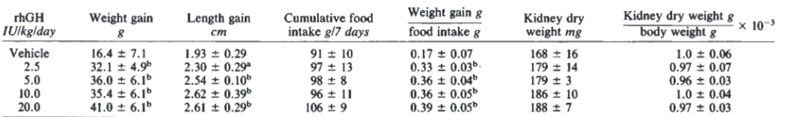
Table 1. Dose dependent effect of rhGH for seven days on growth in SD rats rhGH
lU/kg/day
Weight gain
g Length gain
cm
Cumulative food intake g/7 days
Weight gain g
food intake g Kidney dry weight mg
Kidney dry weight g body weight g
Vehicle 16.4 7.1 1.93 0.29 91 10 0.17 0.07 168 16 1.0 0.06
2.5 32.1 4.9" 2.30 0.29a 97 13 0.33 0.03" 179 14 0.97 0.07
5.0 36.0 61b 2.54 010b 98 8 0.36 0.04" 179 3 0.96 0.03
10.0 35.4 6.1" 2.62 0.39" 96 11 0.36 186 10 1.0 0.04
20.0 41.0 6.1" 2.61 0.29" 106 9 0.39 0.05" 188 7 0.97 0.03
N =6animals per group.
Significant difference between vehicle group and experimental group ( P < 0.05, b p < 0.01) Table 2. Organ weight/body weight ratio under rhGH treatment with
increasing duration of treatment
Treatment duration
Kidney dry weight g
body weight g x 10 Heart dry weight g body weight g X 10
10 IU 10 IU
days rhGH/kg/day Vehicle rhGH/kg/day Vehicle
0 1.19 0.05 0.98 0.04
4 1.11 0.04 1.14 0.05 0.98 0.05 0.95 0.08 8 1.03 0.05 1.11 0.04 0.90 0.05 1.02 0.13
11 1.10 0.20 1.10 0.08 0.94 0.03 0.94 0.04
21 1.25 0.12 1.24 0.06 0.95 0.06 0.95 0.05
60 0.88 0.06 0.88 0.06 0.70 0.04 0.77 0.07
N =7 animals per group.
was provided by Kabi Pharmacia Peptide Hormones (Stock- holm, Sweden), rhIGF-l by Ciba-Geigy Co. (Basel, Switzer- land; CGP 35' 126), and by Kabi Pharmacia Peptide Hormones (Stockholm, Sweden; CH/B/60229—51).
Protocols
Experiment 1: Dose dependent effects of rhGH for seven days on growth in SD rats (Table 1). Five groups of healthy animals (N =6animals per group) were treated with increasing doses of rhGH ranging from 0 to 20 lU/kg/day given in two divided doses s.c. for a period of seven days.
Experiment2: Time course of the effect oflOIUrhGH/kg/day on growth in SD rats (Table 2). Five groups of healthy animals (N =7animals per group) were treated with 10 IU rhGH/kg day s.c. in two divided doses for various time intervals ranging from four to 60 days.
Experiment 3: Dose- and time-dependent effects of rhIGF-1 on growth in SD rats (Table 3). Six groups of healthy animals (N
= 7 animals per group) were treated with increasing doses rhIGF-l ranging from 0 to 16 mg/kg/day in two divided doses s.c. for a period of seven days. Five animals were treated with 3 mg rhIGF-1/kg/day for 60 days.
Experiment 4: Effects of concomitant treatment with rhGH (20 lU/kg/day) and rhIGF-1 (8 mg/kg/day) for 14 days on growth in SD rats (Table 4). The number of animals was 10 for the three treatment groups (20 IU rhGH/kg/day; 8 mg rhIGF-l/kg/day; 20 IU rhGH + 8 mg rhIGF-l/kg/day) and for the control (vehicle) group. Peptides and vehicle were injected in two divided doses twice daily s.c.
Experiment 5: Effect of rhGH (JO lU/kg/day) and of rhIGF-1 (3 mg/kg/day) forfive days on GHR mRNA and IGF-1 mRNA in kidney and liver tissue (Table 5). The number of animals was seven for the vehicle and treatment groups. Peptides and vehicle were injected in two divided doses twice daily s.c.
Experiment 6: Effects of 10 IU rhGH/kg/day for 14 days on renal morphology in SD rats (Table 6). Nine animals were studied in the treatment and control (vehicle) groups, respec- tively. The animals were injected with 10 IU rhGH/kg/day s.c.
in two divided doses twice daily. At the end of the experiment, the kidneys were fixed by perfusion technique for analysis of renal architecture and glomerular stereology.
Experiment 7: Effect of treatment with rhGH (10 lU/kg/day) or rhIGF-J (4 mg/kg/day) for five days on renal DNA/protein ratio in SD rats (Table 7). The number of animals was seven for the treatment and for the control (vehicle) groups. The animals were injected with 10 IU rhGH/kg/day or 4 mg rhIGF-1/kg/day, respectively. The peptides were administered s.c. in two di- vided doses twice daily.
Experiment 8: Effect of treatment with rhGH (10 lU/kg/day) or rhIGF-1 (4 mg/kg/day) for 4 days on mitoses and PCNA expres- sion of tubular and glomerular cells (Table 8). The number of animals was seven for the treatment groups [renal intact animals and subtotally nephrectomize (NX) animals treated with rhGH or with rhIGF-1] and the control groups (renal intact animals and subtotally NX animals treated with vehicle). All groups had free access to food. The renal intact animals were pair fed to subtotally NX animals. The animals received 5 i.p. injections of colchicme (Demecolcin, Serva Co., Heidelberg, Germany) or vehicle at 30 minute intervals prior to the end of the experiment in the morning hours (yielding a total colchicine dose of 800 .ig). Kidneys were excised under anesthesia and fixed in 5% buffered formalin. A second set of animals was not pretreated with coichicine prior to death. Renal tissue was fixed in methyl Camoy's solution and processed for detection of PCNA.
Analytical techniques
In vivo measurements. Body weight was measured during the afternoon in non-fasting animals. Food intake was measured daily and food conversion ratio (weight gain per food intake) was calculated [8]. Nose to tail tip distances were measured in anaesthesized animals under complete muscle relaxation as described previously [21]. Blood pressure was measured by tail plethysmography as described previously [21].
Organ weight. The animals were sacrificed by aortic puncture under intramuscular anesthesia (30 mg/kg Ketanest, Park Davis Co., Berlin, Germany and 0.3 mg/kg Valium, Hoffmann La Roche, Grenzach-Wyhlen, Germany). Organs were weighed before and after desiccation (24 hr; 80°C in the presence of desiccant). Kidney volume was quantitated by measuring the volume of water displaced after submersion.
Biochemistry and hormonal measurements. Serum biochem- istry was analyzed using a multichannel Technicon Autoana- lyzer (Technicon Instruments, Tarrytown, New York, USA).
Mehls et a!: Renotropic effects of rhGH and rhlGF-1 1253
Table 3. Dose dependent effect of rhIGF- 1 for seven days on growth in SD rats
rhIGF-1 Weight gain Length gain Cumulative Weight gain g
dney
Kidney dry weight g> lo-
mg/kg/day
g
cm food intake g food intake g dry weight mg body weight gVehicle 25.6 3.0 2.81 0.30
117 9
0.21 0.03158 6
1.06 0.061 33.0 4.1" 3.33 0.28a
109 9
0.25 O.O4170 8
1.09 0.042 34.3 50b 3.80 0.70" 132 13 0.26 0.04a 175 12 1.10 0.04
4 35.4 5.3" 3.84 040b 104 10 0.34 005b 180 6 1.13 0.03k
8 39.6 6.9k' 4.04 0.16" 113 12 0.35 0.05" 189 14 1.15 0.08''
16
40.2 33b
4.20 02b 112 9 0.36 0.03" 193 16 1.18 0.08"N =7animals per group
Significant difference between vehicle group and experimental group (P< 0.05; bP< 0.01)
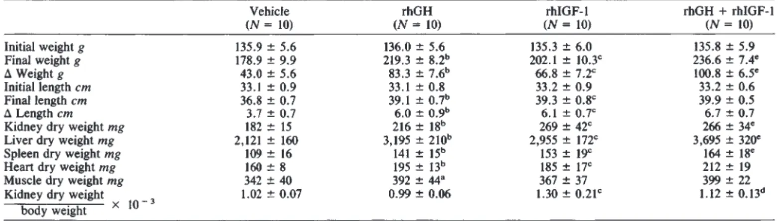
Table 4. Effect of concomitant treatment with rhGH (20 lU/kg/day) and rhIGF-l (8 mg/kg/day) for 14 days on growth in SD rats
Vehicle rhGH rhIGF-l rhGH + rhIGF-1
(N =10) (N =10) (N = 10) (N =10)
Initial weight g 135.9 5.6 136.0 5.6 135.3 6.0 135.8 5.9
Final weight g 178.9 9.9 219.3 8.2" 202.1 l0.3c 236.6 7.4°
A Weight g
43.0 5.6
83.3 7.6" 66.8 7.2c 100.8 6.5°Initial length cm 33.1 0.9 33.1 0.8 33.2 0.9 33.2 0.6
Final length cm 36.8 0.7 39.1 0.7"
39.3 0.8
39.9 0.5A Length cm 3.7 0.7
6.0 09'
6.1 0.7c 6.7 0.7Kidney dry weight mg 182 15 216 18" 269 42C 266 34°
Liver dry weight mg 2,121 160 3,195 210" 2,955 172C 3,695 320°
Spleen dry weight mg 109 16 141 15'' 153 19C
164 18
Heart dry weight mg
160 8
195 13b 185 17C 212 19Muscle dry weight mg
342 40
392 44°367 37 399 22
Kidney dry weight 1.02 0.07 0.99 0.06 1.30 0.21c 1.12 0.13"
body weight X 10
a p< 0.05, bP< 0.01, rhGH vs. vehicle P < 0.01, rhIGF-1 vs. vehicle
dP< 0.05; e p < 0.01, rhGH + rhIGF-1 vs. rhGH
Table 5. Effect of 3 mg rhIGF-1/kg/day or 10 IU rhGH/kg/day for five days on growth hormone receptor (GHR) mRNA and IGF-l
mRNA in kidney and liver
Vehicle rhGH rhIGF-l
Kidney
IGF-1 mRNA 0.35 0.17 1.35 0.5° 0.6 0.33
pg//2g DNA
GHR mRNA 0.08 0.07 0.46 0.32° 0.16 0.14
amol/sg DNA Liver
IGF-l mRNA 30.7 12.5 43.0 11.05 30.9 4.94
pg/pg DNA
GHR mRNA 3.28 0.55 2.69 0.43 3.3 0.43
amol/pg DNA
N =7 animals per group.
P < 0.001, rhGH vs. vehicle and rhIGF-1 (ANOVA)
Creatinine was determined by a kinetic method according to Jaffé without deproteinization; the within assay coefficient of variation was <3%. Measurements of rhGH, rhIGHF-1 and of rat GH were done by RIA techniques as described earlier [10, 18].
Chemical analysis of renal tissue.Renaltissue was frozen in liquid nitrogen and powdered. Total protein was measured according to Lowry. DNA was measured using a modified method according to Blin and Stafford [22]. In brief, tissue frozen in liquid nitrogen was powdered, 100 mg were mixed with the lysis buffer (0.075 M NaCI, 0.024 M ETDA, 0.02 M SDS, 1 M NaHPO4) and extracted with 1:1 phenol-chloroform
Table 6. Effect of 10 IU rhGH/kg/day for 14 days on renal morphology in SD rats
Vehicle
(N=18)
rhGH
(N=18)
Body weight g 176 8.2 199 7.9
Kidney volume° cm3 1.04 0.06 1.18 0.08"
Cortex volume % of total kidney 78.4 2.6 78.4 2.4 volume
Width of cortex ÷ medulla mm 8.60 0.75 8.80 0.54 Width of outer + inner medulla mm 6.10 0.71 6.30 0.56 Width of inner medulla mm 4.20 0.50 4.40 0.47 Glomerular area pin2
Glomerular (3D) diameter pin
84S1 430 137.2 3.5
8951 321"
140.5 2.8"
a After perfusion fixation bp< 0.05 vehicle vs. rhGH
and 24:1 chloroform-isoamylalcohol (vol/vol). After precipita- tion with isopropanolol, DNA was quantitated photometrically.
Solution hybridization for GH receptor mRNA and IGF-1 mRNA in renal and liver tissue. (a) GH-receptor mRNA probe.
A pT7T3 18 U vector carrying a 560 base pair BamHI fragment [23] was linearized with EcoRI cleaving in the cloning box of the vector, and labeled ([32P]UTP or [35S]UTP) cRNA was generated with T3 polymerase (Promega, Madison, Wisconsin, USA) under the conditions indicated by the manufacturer.
(b) IGF-1 mRNA probe. The radioactive probe was prepared as described by Melton et a! [24]. In brief, the DNA clone used was
Table 7. DNA/protein ratio of kidney in rhGH and IGF-l treated SD rats
Vehicle
rhGH 10 lU/kg/day
rhIGF-l 4 mg/kg/day
Bodyweightg 141±6 159±6 143±4
Weightf5 days g 22.9 2.6
40.4 5.9
25.6 2.5Kidney dry weight mg
136 8 143 8
144 15left kidney
Protein content mg
47.2 4.6
53.9 8.0 58.5 49a right kidneyDNA content pg 1344 395 3031 923° 3432 692k DNA/protein 30.1 10.6 58.8 23.4° 59.0 l2.6a
ratio pg/mg
N =7animals per group; duration of treatment 5 days.
aP< 0.05 difference treatment group vs. vehicle
a 153 bp genomic subclone of pSP64 in both orientations of mouse IGF-l corresponding to exon 3 (by analogy with human IGF-l).
Analyses of cDNA clones for IGF-1 indicate that two forms of IGF- 1 mRNA could exist [25]. Thestructure of the probe used in this study would allow detection of both forms of IGF-l mRNA.
GH receptor (GHR) mRNA was quantified by solution hybrid- ization as previously described [26]. In brief, the tissue was homogenized in a buffer containing 1% NaDodSO4, 20 mM Tris- HCI (pH 7.5), and 4 mrvi EDTA and digested with protemase-K (200 zg/ml) at 35°C for 35 minutes. Total nucleic acids (TNA) were then extracted with phenol-chloroform. The TNA samples were prepared from liver and kidney tissue from individual rats.
TNA samples were hybridized at 70°C for 24 hours in 0.06 mM NaCI, 20 mM Tns-HC1 (pH 7.5), 4 mM EDTA, 0.1%
NaDodSO4, 10 mrs dithiothrol, and 25% formamide (vol/vol) with a "S-labeled GH-receptor cRNA probe in a volume of 40 td. The samples were then treated with 40 pg RNase A and 2 pg RNase T1 in the presence of 100 jg herring sperm DNA for 45 minutes at 37°C in a volume of I ml. Protected probe was precipitated with 100 .d trichioracetic acid (6 mol/liter), col- lected on glass-fiber filters (GF/C, Whatman International Ltd., Maidstone, UK) and counted in a scitillation counter. A tissue TNA preparation, originally compared with an in vitro tran- scribed OH-receptor mRNA, was used to generate a standard curve. The standard curve was linear between 0.1 and 16 amol.
The DNA content of the samples was analyzed [27], and 10 to 45 pg DNA in samples were assayed. Within this range, the hybridization signal paralleled the standard curve. In all assays, the lowest concentration of GH receptor mRNA measured was well above the lowest point of the standard curve. Each TNA sample was analyzed in duplicate. The results are expressed as the amount of GHR mRNA per DNA (amolIpg). The coefficient of variation was 6% within and between experiments.
The solution hybridization assay for quantification of IGF- 1 mRNA was described earlier [28]. It contained principally the same steps as the assay for quantification of GHR mRNA. The hybridization signal was compared with that of a tissue standard curve originally compared with known amounts of the synthetic 153 nucleotide mRNA strand, as described previously [29].
Results are expressed as the amount of IGF- 1 mRNAIDNA (pg/sg). Each sample was analyzed in duplicate. The within- assay coefficient of variation (C.V.) was less than 20% in the range of 40 to 400 amol of the RNA standard. The between-
assay C.V. was estimated by repeated analysis of the same TNA preparation and was 23%.
Mitosis counts in renal tissue and immunoperoxidase stain- ing. (a)Counting of tubular cell mitoses in renal tissue. HE stained 8 js sections of paraffin embedded tissue were used as described before [30]. Mitoses were counted at 400 x magnifi- cation using a Zeiss Co. Integrationsplatte 11100/25. Mitoses were counted in 50 fields each in the cortex, outer stripe, inner stripe and inner medulla using a systematic random sampling technique.
(b)Immunoperoxidase staining for counting glomerular cell prolferation. Four micrometer sections of methyl Carnoy's fixed renal tissue were processed by an indirect immunoperox- idase technique as previously described [31]. The proliferating nuclear antigen (PCNA), which is expressed by actively prolif- erating cells [32], was detected using l9A2 (Coulter Electronics, Hialeah, Florida, USA), a murine 1gM monoclonal antibody against human PCNA. For all tissues, negative controls con- sisted of substitution of the primary antibody with equivalent concentrations of an irrelevant munne monoclonal antibody. In each kidney, over 40 cross sections (range 40 to 80) of consec- utive cortical glomeruli containing more than 20 discrete capil- lary segments each were evaluated by a person who was unaware of the origin of the slides. Mean values per kidney were then calculated for the number of proliferating (PCNA+) cells per glomerular cross section.
Renal histology and renal morphometry. For histological evaluation of glomerulosclerosis and tubulointerstitial lesions in experiment 2 and experiment 3, the kidneys were fixed in formalin stained with hematoxylin/eosin and analyzed by light microscopy.
For morphometric studies, the viscera were fixed by retro- grade vascular perfusion at a pressure of 110 mm Hg after
catheterization of the distal abdominal aorta. Before fixation, the vascular system was flushed, with a dextran solution (Rheomacrodex'; Schiwa Co., Glandorf, Germany) containing 0.5 g/liter procaine HC1. The vena cava inferior was incised to drain the blood.
The kidney was dissected into 1 mm thick slices cut perpen- dicularly to the interpolar axis. The specimen was embedded in paraffin and cut into 4 sm thick slices and then stained with hematoxylin/eosin. For area measurements, a semiautomatic image analyzing system (Videoplan, Kontron Co., Eching, Germany) was used. At a magnification of x 10, the area fraction of the cortex and medulla was determined and volume density and total volume were calculated as described [33]. At a magnification of x 400, the mean area of glomerular profiles was measured. Data are given as mean cross sectional area of glomeruli. The diameters were determined by identifying the inner edges of the thin parietal layer of cells forming Bowman capsule in every sixth consecutive glomerular section. Assum- ing a spherical size of glomeruli, the true three-dimensional diameter (stereological diameter) was obtained from the size distribution of diameters according to Weibel [33].
Statistics
Dataare given as X SD or as mean (range). Results were evaluated using Wilcoxon's test for random samples or paired differences and by ANOVA.
Mehis et a!:Renotropic effects of rhGH and rhlGF-1 1255
Table 8. Effect of rhGH and of rhIGF-I on mitoses in tubular cells of SD rats
Treatment Renal status
Number of mitoses/mm2
Distal tubule Proximal tubule Total tubules
Vehicle intact 8.3 3.9 10.0 5.4 18.3 8.0
IOIUrhGH/kg/day intact 12,2 5,Ø 17.7 6.9t 29.9
Vehicle subtotal NX 140.9 29.7 209.8 45.6 350.7 67.8
10 IU rhGH/kg/day subtotal NX 168.0 22.2a 325.6 30.3'' 493.6 38.6a
Vehicle intact 8.3 1.9 12.8 2.5 21.1 4.2
4 mg rhIGF-1/kg/day intact 13.0 2.2k' 20.3 44a 55b
Vehicle subtotal NX 130.3 13.7 267.3 14.8 397.6 25.8
4 mg rhIGF-lIkg/day subtotal NX 163.3 153b 349.8 338b 513.4 437b
Duration of treatment was 4days.
N=7 animals per group.
a P < 0.05 and bP< 0.01 rhGH vs. vehicle or rhIGF-l vs. vehicle
Results
Dose-dependent effects of rhGH for seven days on growth in SD rats
Short term s.c. administration of 2.5 to 20.0 IU of rhGH/kg/day in two divided doses caused significant increments in weight and length with inconsistent effects on cumulative food intake, but caused a signfficant increase of the food conversion ratio, that is, weight gain per food intake (Table 1). There were no significant effects on systolic blood pressure (vehicle 110 5 mm Hg, 20 lU/kg/day 112 4mm Hg), serum creatinine (0.31 0.06 vs. 0.32 0.06 mgldl) or serum phosphate (2.41 0.15 vs. 2.46 0.12 mmol/liter). Serum concentration of rhGH (by human GH RIA) 12 hours after the last injection of 5 IU rhGH/kg (which had been administered every 12 hr) was 13.9 5.2 ng/ml. (The assay measured < 0.17 ng/ml in rats without rhGH injection). Concom- itant measurements of rat GH showed that endogenous produc- tion of GH was suppressed (by specific rat assay 0.9 0.6 ng/ml vs. 10.2 7.3 ng/ml without rhGH treatment). There was some increase of IGF-l serum concentration measured with RIA adapted for rats [181: 479 64 ng/ml without versus 654 87 ng/ml with 10 IU rhGHlkg/day. Kidney dry weight increased with increasing doses of rhGH, but the kidney/body weight ratio was not significantly changed by rhGH.
Time course of the effect of rhGH on growth in SD rats Ten international units of rhGH/kg/day by twice daily s.c.
injections caused a progressive increment of cumulative weight gain and length gain above the vehicle treated controls (Table 2).
As in experiment 1, there was a consistent increase in the food conversion ratio, but no significant increase of the kidney dry weight or heart dry weight/body weight ratio despite treatment for up to 60 days. The organ weight/body weight ratios changed with time both in the experimental and in the control groups. Renal histology did not demonstrate the development of glomeruloscle- rosis or tubulointerstitial lesions, even after 60 days.
Dose-and time-dependent effect of rhIGF-1 on growth in SD rats
rhIGF-1had similar actions on weight gain, length gain, and food conversion ratio as rhGH (Table 3). There were no effects on systolic blood pressure, serum creatinine and serum phos- phate (data not given). Serum concentration of (exogenous) rhIGF-1 measured with human RIA [10] 12 hours after the last
injection of 4 mg rhIGF-1/kg (given every 12 hours) was 932 102 ng/ml versus 208 29 ng/ml invehicle injected animals.
rhIGF-1 not only caused a significant increase of kidney dry weight, but, in contrast to rhGH, also caused a dose dependent increase of the kidney dry weight/body weight ratio.
After 60 days of treatment with rhIGF-1, kidney weight/body weight ratio was 0.91 0.05 in rhIGF-1 treated rats and 0.88 0.05 in controls (N = 5 per group). Renal histology did not demonstrate the development of glomerulosclerosis or tubu- lointerstitial lesions, even after 60 days of rhIGF-l treatment.
Effect of concomitant treatment with rhGH and rhIGF-1 for 14 days on growth in SD rats
To test whether rhGH and rhIGF-l have additive effects, we used the doses of rhGH (20 lU/kg/day) and of rhIGF-l (8 mg/kg/day) which had caused maximal effects on weight gain in the previous experiments 1 and 2 (Table 4). In comparison to treatment with rhGH, the combined administration of the two peptides caused a significantly greater weight gain and also a small but significant (P < 0.05) further increase in length gain.
The combined administration caused a further significant rise of liver, spleen and of kidney dry weight, but no further increment of heart and muscle dry weight. While the dry weight/body weight ratio was not further increased for heart and muscle, spleen dry weight/body weight ratio (x 10—s) tended to increase with rhGH + rhIGF-1 (0.69 0.06) versus rhGH alone (0.64 0.05). The same was noted for liver dry weight/body weight ratio (x l0—) (15.6 0.82 vs. 14.6 0.9).
Consistent with the observations in Tables 1 and 3, rhIGF-1, but not rhGH, caused a significant increase in kidney dry weight/body weight ratio. Administration of rhGH together with IGF-1 caused no further increase.
Development of glomerulosclerosis again was not seen in any of the treatment groups.
Effectsof rhGH and rhIGF-1 on GHR mRNA and IGF-1
mRNAin kidney and liver tissue
Treatment with 10 IU rhGH/kg/day significantly increased the GHR mRNA in the kidney, but not in the liver (Table 5). At the same time, an increase of IGF- 1 mRNA was noted which again was only significant within the kidney. Treatment with 3 g rhIGF-l/kg/day did not cause a significant increase of GHR mRNA or IGF-l mRNA in kidney and liver.
Effects of rhGH on renal morphology
Mter treatment of female SD rats with 10 IU rhGH/kg/day by twice daily s.c. injections for 14 days, kidney volume by Archimedian principle was significantly greater in rhGH-treated animals (Table 6). In contrast, the kidney volume/body weight ratio was not significantly different (vehicle 0.99 0.06cm3/g;
rhGH 0.97 0.07 cm3/g), but the ratio is based on perfusion fixed renal tissue. The proportion of kidney volume comprised of cortex was unchanged. An increase of zonal width and of glomerular area was noted in rhGH-treated animals. The per- cent increase of the respective mean values was comparable to the mean percent increase of kidney volume (+ 10%) and of body weight (+ 12%).
Effect of treatment with rhGH or rhIGF-1 for five days on renal DNA/protein ratio in SD rats
Treatment of female SD rats with submaximal doses of rhGH or rhIGF-1 respectively for five days caused an (insignificant) increase of kidney dry weight, while protein content increased significantly (but marginally) together with a striking increase in DNA content (Table 7). In parallel, an increase of DNA/protein ratio was noted for rhGH as well as for rhIGF-1 treated animals.
The results suggest that renal growth was accounted for more by hyperplasia than by hypertrophy.
Effect of treatment with rhGH or rhIGF-1 for five days on mitosis and PCNA expression of tubular and glomerular cells
in SD rats
In renal intact animals, 10 IU rhGH/kg for four days caused a significant increase in the number of mitoses demonstrable in SD rats treated with colchicine for three hours prior to the end of the experiment (Table 8). A significant increase was seen both in distal and proximal tubular epithelial cells.
When the baseline number of mitoses was raised by prior subtotal nephrectomy before administration of rhGH, a further increment of mitoses was still noted after rhGH. The results observed with 4 mg rhIGF-1/kg/day were comparable.
To validate the colchicine method with an independent approach, in a second set of animals the expression of prolifer- ating nuclear antigen (PCNA) was determined using a murine 1gM monoclonal antibody against human PCNA. The number of PCNA positive nuclei per glomerular cross section was significantly increased (P =0.008;ANOVA) both after rhGH (3.08 0.53) andrhIGF-1 (2.95 0.41)compared to vehicle controls (2.2 0.55).
Discussion
The salient features of the present study are the observation that both rhGH and rhIGF- 1 increase renal weight. The in- crease of renal weight following administration of rhGH was in proportion to the increase in body weight. In contrast, admin- istration of rhIGF-l either alone or in combination with rhGH increased the renal weight/body weight ratio, suggesting some selective stimulation of renal growth. After administration of rhGH, the glomerular volume increased in proportion to the increase in body weight. Administration of rhGH and of rhIGF-1 up to 60 days did not cause glomerulosclerosis or tubulointerstitial changes in female animals.
This systematic analysis extends previous observations of Guler et al in hypophysectomized rats, which documented that rhGH increased renal weight in proportion to body weight [34].
Our study shows that rhGH, in doses which are close to those maximally stimulating body growth, does not significantly affect the renal and other organ weight/body weight ratio in hypoph- ysis intact young female SD rats on ad libitum intake of food.
Protein intake has a known stimulatory effect on renal growth and GFR [12]. The possibility that the effect of rhGH was mediated by changes in protein intake was not formally ex- cluded by a pair feeding experiment, but we found no significant increase of food intake. This is in agreement with previous data of other [20] and our own groups [8, 18]. In contrast to previous studies [35], the mortality in our animals was zero. Most of our experiments lasted for one to two weeks. For longer experi- ments, one important potential limitation of the efficacy of (foreign) rhGH or rhIGF-l to rats is antibody production. For rhGH this is known to occur in female SD rats (A. Skottner- Lundin, Kabi Pharmacia, Peptide Hormones, Stockholm, per- sonal communication) after two to three weeks of administra- tion. In accordance with this, after three weeks the body weight increment in human peptide hormone treated animals became identical with that in solvent treated animals (experiments 2 and 3) and growth curves ran parallel, but the initial growth advan- tage was preserved throughout the experiment.
It is not excluded that more prolonged effective treatment with rhGH would progressively increase the kidney weightl body weight ratio in parallel to what has been reported for transgenic mice expressing GH [15, 36, 37]. But even in transgenic mice expressing GH this finding has not been re- ported uniformly [16, 38]. One difficulty of measuring the kidney weight/body weight ratio in transgenic mice is the fact that body weight increases rapidly during the first weeks of life, but decreases continuously during the last weeks before death.
Therefore, different results will be obtained at different stages of the experiment. This has not been worked out systemati- cally. It can also not be excluded that a vastly greater amount of GH produced by the transgenic animals accounts for the different results. In transgenic mice expressing human GH, serum hGH levels up to 900,000 ng/ml have been reported [15].
Inthis respect, it is interesting that the serum concentration of rhGH in SD rats treated with 10 IU rhGH/kg/day ranged between 3.3 and 20 ng/m1 12 hours after the last injection. This is comparable to the serum concentrations in rhGH treated children with chronic renal failure (mean 20 3.4ng/ml 12 hr after injection of 4 IU rhGH/m2; [13]).
In striking contrast to rhGH, rhIGF- 1 dose-dependently increased the renal weight/body weight ratio above the ratio in vehicle controls. This result extends the previous findings of Guler et al [34] in hypophysectomized rats and demonstrates greater proportional stimulation of renal growth even in hy- pophysis-intact rats. This result is remarkable since rhIGF-l increased heart and muscle weight without increasing heart and muscle weight/body weight ratio. In contrast, spleen and liver weight/body weight ratios were slightly increased, illustrating considerable target organ selectivity.
The present finding of a selective renotrophic effect of IGF-1 is in agreement with previous observations in nitrogen re- stricted rats [39] and in subtotally nephrectomized rats [40]. We did not specifically address the issue concerning the effects of
Mehis et a!: Renotropic effects of rhGH and rhIGF-1 1257 rhIGF-l on renal growth after subtotal nephrectomy. In pilot
experiments it had proven extremely difficult to ablate a stan- dardized amount of renal tissue. Our results show, however, that the effect of IGF-1 on renal growth does not require prestimulation of renal growth by ablation.
The kidney is a known target organ for both GH and IGF- 1.
GH receptors [41] and IGF-1 receptors [20, 42—44] have been demonstrated and IGF-1 is locally produced in the kidney, particularly when renal growth is stimulated, such as after ablation [43, 45, 46], during diabetes mellitus [43] and in hypersomatotrophic states [47]. The reasons why rhGH and rhIGF-1 act differently on renal growth are unknown. It is of note, however, that addition of rhGH to rhIGF-1 (Table 2) failed to increase the kidney weight/body weight ratio above that seen in animals given rhIGF-l alone. A significant further increment in weight gain was noted, however. Interestingly enough, rhGH increased the GHR mRNA and IGF-l mRNA in the kidney. These findings would be compatible with the notion that the effects of IGF-1 are mediated, at least in part, by pathways different from those mediating the effects of rhGH.
The finding that rhGH differently stimulated GHR mRNA and IGF-1 mRNA in the kidney and in the liver points to organ selectivity, and deserves further investigation. It has been reported that rhGH stimulates GHR mRNA in the liver of hypophysectomized rats [48]. However, studies in hypophysis- intact rats have not been reported to date.
We considered that an unchanged renal weight/body weight ratio after rhGH does not necessarily exclude potential changes of renal architecture, such as the redistribution of the relative width of different zones as seen after glucagon or vasopressin which causes a disproportional increase of medullary width [49]. The present study, however, showed no significant change of the relative widths of the different renal zones in rhGH treated animals. Glomeruli are enlarged in acromegalic patients
[31andtransgenic mice expressing GH [14], IGF-1- [14, 38] or GH-releasing factor [14, 50]. Potential effects on glomerular size are of interest, since an increase in glomerular size may be permissive for the development of glomerulosclerosis. Indeed, glomerulosclerosis was seen in transgenic mice expressing GH, but this was not seen in transgenic mice expressing IGF-1 [14].
In our animals on short-term rhGH treatment, glomerular size increased, but in proportion to the body weight gain, so that the ratio glomerular volume/body weight did not change. We em- phasize that glomerulosclerosis or tubulointerstitial lesions were not seen in our study in female rats, even after prolonged (60 days) administration of rhGH or rhIGF-1 in doses per body weight which were considerably in excess of what is adminis- tered to uremic children [101. The interpretation of these findings must be cautious in view of antibody formation against rhGH and rhIGF-1 as discussed above. On the other hand, the development of glomeruloscierosis has been reported in male SD rats treated with rhGH for 28 weeks [35] in similar doses. It is known that male rats are more prone to the development of glomerulosclerosis than female rats. Whether this explains the difference of results requires further studies.
Renal growth after ablation involves mainly hypertrophy [51], although in younger animals hyperplasia does also contribute [52, 53]. To address the relative contribution of hypertrophy and hyperplasia to the renal growth response to rhGH and rhIGF- 1, respectively, we examined the renal DNA/protein ratio and the
proportion of renal cells undergoing proliferation and mitosis.
Both methods suggested a major contribution of renal cell prolif- eration, indicating that in these young female animals the increment in renal weight mainly involves hyperplasia at least within the time frame of the study. The results in the young animals cannot necessarily be extrapolated to older animals.
With respect to rhGH treatment of children with chronic renal failure, concern has been raised that such treatment accelerates progression of renal failure by promoting the gene- sis of glomerulosclerosis. Although our experiments do not directly bear on this issue, the results are reassuring in that they indicate that rhGH (i) fails to stimulate renal growth and glomerular volume out of proportion to the increase in body weight, and (ii) does not cause glomerulosclerosis in experi- ments of two months duration. Different effects on kidney growth should be expected if one considers the use of rhIGF-1 alone or in combination with rhGH for the treatment of stunting and GH insensitivity [54] in chronic renal failure.
Acknowledgments
The study was supported by Kabi Pharmacia Peptide Hormones, Stockholm, Sweden (kindly providing rhGH and rhIGF-l), and by Ciba-Geigy Co., Basel, Switzerland (kindly providing rhIGF-1). The authors are indebted to Carola Mayer, Axel Schauer, and Stefan Worgall for technical assistance and Prof. R. Waldherr, Institute of Pathology, University of Heidelberg, for histological evaluation of the kidneys. The secretarial help of Mrs. R. Greiffenhagen is acknowledged.
Reprint requests to Otto Mehis, M.D., Division of Pediatric Neph- rology, University Children's Hospital, Im Neuenheimer Feld 150, D-6900 Heidelberg, Germany.
References
1. BARNEi-rHL,PERLY AM, HEINBECKER P: Influence of eosinophil cells of hypophysis on kidney function. Proc Soc Exp Biol 52:114, 1943
2. FALKHEDEN T, WIcKB0M I: Renal function and kidney size follow- ing hypophysectomy in man. Ada Endocrinol (Copenh) 48:348—
354, 1965
3. GERSHBERG H, HEINEMANN HO, STUMPF HH: Renal function studies and autopsy report in a patient with gigantism and acromeg- aly. J Clin Endocrinol Metab 17:377—385, 1957
4. SANDAHL CHRISTIANSEN J, GAMMELGAARD J, Oitsiov H, ANDERSEN AR, TELMER 5, PARVING HH: Kidney function and size in normal subjects before and during growth hormone administra- tion for one week. Eur J Clin Invest 11:487—490, 1981
5. HIRSCHBERG R, RAAB H, BERGAMO R, KOPPLE JD: The delayed effect of growth hormone on renal function of humans. Kidney In!
35:865—870, 1989
6. GULER HP, ECKARDT KU, ZAPF J, BAIJER C, FROESCH ER:
Insulin-like growth factor I increases glomerular filtration rate and renal plasma flow in man. Acta Endocrinol (Copenh) 121:101—106,
1989
7. MEHLS 0, RITZ E: Skeletal growth in experimental uremia. Kidney mt 24 (Suppl 15):S53—S62, 1983
8. MEHLS 0, RITz E, HUNZIKER EB, EGGLI P. HEINRICH U, ZAPF J:
Improvement of growth and food utilization by human recombinant growth hormone in uremia. Kidney In! 33:45—52, 1988
9. NAKANO M, KAINER G, FOREMAN J, D.uir' K, CHAN JC: The effect of exogenous rat growth hormone therapy on growth of uremic rats fed an 8% protein diet. Pediatr Res 26:204—207, 1989 10. ToNSHOFF B, MEHLS 0, HEINRICH U, BLUM WF, RANKE MB,
SCHAUER A: Growth-stimulating effects of recombinant human growth hormone in children with end-stage renal disease. J Pediatr
116:561—566, 1990
11. YOSHIDA Y, Foo A, IcHIKAwA I: Glomerular hemodynarnic changes vs. hypertrophy in experimental glomerular sclerosis.
Kidney mt 35:654—660, 1989
12. BRENNERBM,MEYER TW, HOSTErFER TH: Dietary protein intake and the progressive nature of kidney disease: The role of hemody- namically mediated glomerular injury in the pathogenesis of pro- gressive glomerular sclerosis in aging, renal ablation, and intrinsic renal disease, N Engi J Med 307:652—659, 1982
13. TONSHOFF B, HEINRICH U, MEHLS 0: How safe is the treatment of uraemic children with recombinant human growth hormone? Pedi- air Nephrol 5:454—460, 1991
14. Doi T, STRIKER Li, QUAIFE C, Coin-i FG, PALMITER R, BEHR- INGER R, BRINSTER R, STRIKER GE: Progressive glomerulosclero- sis develops in transgenic mice chronically expressing growth hormone and growth hormone releasing factor but not in those expressing insulin like growth factor-I. Am J Pathol 131:398—403,
1988
15. WANKE R, HERMANNSW,F0LGER S. WOLF E, BREMG:Acceler- ated growth and visceral lesions in transgenic mice expressing foreign genes of the growth hormone family: An overview. Pediatr Nephrol 5:513—521, 1991
16. HAMMER RE, BRINSTER RL, PALMITER RD: Use of gene transfer to increase animal growth. Cold Spring Harbor Symp Quant Biol 50:379—387, 1985
17. KAWAGUCHI H, ITOH K, MORI H, HAYASHI Y, MAKINO S: Renal pathology in rats bearing tumour-secreting growth hormone. Pedi- atr Nephrol 5:533—538, 1991
18. KOVACS G, FINE RN, WORGALL 5, SCHAEFER F, HUNZIKER EB, SKOTTNER-LINDUN A, MEHLS 0: Growth hormone prevents ste- roid-induced growth depression in health and uremia. Kidney mt 40:1032—1040, 1991
19. MILLER SB, HANSEN VA, HAMMERMAN MR: Effects of growth hormone and IGF-I on renal function in rats with normal and reduced renal mass. Am J Physiol 259:F747—F751, 1990
20. POWELL DR, ROSENFELD RG, HINTZ RL: Effects of growth hormone therapy and malnutrition on the growth in rats with renal failure. Pediatr Nephrol 2:424—430, 1988
21. MEHLS 0, RITZ E, GILL! G, SCHMIDT-GAYK H, KREMPIEN B, KOURIST B, WESCH H, PRAGER P: Skeletal changes and growth in experimental uremia. Nephron 18:288—300, 1977
22. BLIN N, STAFFORD DW: A general method for isolation of high molecular weight DNA from eukaryontes. Nuci Acids Res 3:2303—
2308, 1976
23. MATHEWS LS, ENGBERG B, NORSTEDT G: Regulation of rat growth hormone receptor gene expression. J Biol Chem 264:9905—9910, 1989 24. MELTON DA, KRIEG PA, REBAGLIATI MR, MANIATIS T, ZINN K, GREEN MR: Efficient in vitro synthesis of biologically active RNA and RNA hybridization probes from plasmids containing a bacte- riophage SP:6 promoter. Nuci Acids Res 12:7035—7056, 1984 25. ROTWEIN P: Two insulin-like growth factor I messenger RNAs are
expressed in human liver. Proc Nail Acad Sd USA 83:77—81, 1986 26. DURNAM DM, PALMITER RD: A practical approach for quantitating specific mRNAs by solution hybridization. Anal Biochem 131:385—
393, 1983
27. LABARCAC,PAIGEN P: A simple, rapid and sensitive DNA assay procedure. Anal Biochem 102:344-352, 1980
28. VIKMAN K, ISGAARD J, EDEN S: Growth hormone regulation of insulin-like growth factor-I mRNA in rat adipose tissue and isolated rat adipocytes. J Endocrinol 131:139—145, 1991
29. ISGAARD J, NILss0N A, VIKMAN K, ISAKSS0N OGP: Growth hormone regulates the level of insulin-like growth factor-I mRNA in rat skeletal muscle. J Endocrinol 120:107—112, 1988
30. MATTHIAS 5, BUsCH R, MERKE J, MALL G, RITZ E: Effects of l,25(OH)2D3 on compensatory renal growth. Kidney mt 40:212—
218, 1991
31. JOHNSON RJ, GARCIA RL, PRITZL P, ALPERS CE: Platelets mediate glomerular cell proliferation in immune complex nephritis induced by anti-mesangial cell antibodies in the rat. Am J Patho! 136:369—
374, 1990
32. KURKI P, VANDERLAAN M, DOLBEARE F, GRAY J, TAN EM:
Expression of proliferating cell nuclear antigen (PCNA)/cyclin during the cell cycle. Exp Cell Res 166:209—219, 1986
33. WEIBEL ER: Stereological Methods. I. Practical Methods for Biological Morphometry, London, Academic Press, 1979 34. GULER HP, ZAPF J, SCHEIWILLER E, FROESCH ER: Recombinant
human insulin-like growth factor I stimulates growth and has distinct effects on organ size in hypophysectomized rats. Proc Nat!
Acad Sci USA 85:4889—4893, 1988
35. ALLEN DB, Fooo A, EL-HAYEK R, LANGHOUGH R, FRIEDMAN AL: Effects of prolonged growth hormone administration in rats with chronic renal insufficiency. Pediatr Res 31:406—410, 1992 36. DILLEY RJ, SCHWARTZ SM: Vascular remodeling in the growth
hormone transgenic mouse. Circ Res 65:1233—1240, 1989
37. SHEA BT, HAMMER RE, BRINSTER RL: Growth allometry of the organs in giant transgenic mice. Endocrino!ogy 121:1924-1930, 1987 38. QUAIFE Ci, MATHEWS LS, PINKERT CA, HAMMER RE, BRINSTER
RL, PALMITER RD: Histopathology associated with elevated levels of growth hormone and insulin-like growth factor I in transgenic mice. Endocrinology 124:40—48, 1989
39. Toass FM, KNOWLES SE, OWENS PC, READ LC, CHANTLER CS, GARGOSKY SE, BALLARD FJ: Effects of full length and truncated insulin-like growth factor-I on nitrogen balance and muscle protein metabolism in nitrogen-restricted rats. J Endocr 128:97—lOS, 1991 40. MARTIN AA, TOMAS FM, OWENS PC, KNOWLES SE, BALLARD FJ,
READ LC: IGF-I and its variant, des(1—3)IGF-I, enhance growth in rats with reduced renal mass. Am J Physiol 261 :F626—F633, 1991 41. HAMMERMAN MR, ROGERS SA: Distribution of IGF receptors in
the plasma membrane of proximal tubular cells. Am I Physiol
253:F841—F847, 1987
42. HAMMERMANNMR:The growth hormone-insulin-like growth fac- tor axis in kidney (editorial review). Am I Physiol 257:F503—F5l4,
1989
43. FLYVBJERG A, THORLACIUS-USSING 0, NAERAA R, INGERSLEV J, ORSKOV H: Kidney tissue somatomedin C and initial renal growth in diabetic and uninephrectomised rats. Diabeto!ogia 31:310-314,
1988
44. PILLION Di, HASKELL JF, MEEZAN E: Distinct receptors for insulin-like growth factor I in rat renal glomeruli and tubules. Am J Physiol 255:E504-E512, 1988
45. LAJARA R, ROTWEIN P, BORTZ JD, HANSEN VA, SADOW JL, BETTS CR: Dual regulation of insulin-like growth factor I expres- sion during renal hypertrophy. Am I Physiol 257:F252—F261, 1989 46. STILES AD, SOSENKO RS, D'ERCOLE AJ, SMITH BT: Relation of kidney tissue somatomedin-C/insulin-like growth factor Ito post- nephrectomy renal growth in rat. Endocrinology 117:2397—2401,
1985
47. MILLER SB, ROTWEIN P, BORTZ JD, BECHTEL PJ, HANSEN VA, ROGERS SA, HAMMERMAN MR: Renal expression of IGF I in hypersomatotropic states. Am I Physiol 259:F251—F257, 1990 48. BICK T, AMIT T, BARKEY R, HERTZ P, YOUDIM MBH, HOCHBER
Z: The interrelationship of growth hormone (GH), liver membrane GH receptor, serum GH-binding protein activity, and insulin-like growth factor I in the male rat. Endocrinology 126:1914—1920, 1990 49. BANKIR L, FISCHER L, FISCHER S, JUKKULA K, SPECHT HC, Kiuz
W: Adaptation of the rat kidney to altered water intake and urine concentration. Pflugers Arch 412:42—59, 1988
50. HAMMER RE, BRINSTER RL, ROSENFELD MG, EVANS RM, MAYO
KE: Expression of human growth hormone-releasing factor in transgenic mice results in increased Somatic growth. Nature 315:
413—416, 1985
51. FINE L: The biology of renal hypertrophy. Kidney mt 29:619—634, 1986
52. HEINE WD, STOCKER E: Regeneration of kidney parenchyma under normal and pathological conditions. Beitr Pathol 145:98—99, 1972
53. WESSON LG: Compensatory growth and other growth responses of the kidney. Nephron 51:149—184, 1989
54. BLUM WF, RANKE MB, KIETZMAN K, TONSHOFF B, MEHL5 0:
Growth hormone resistance and inhibition of somatomedin activity by excess of insulin-like growth factor binding protein in uremia.
Pediatr Nephrol 5:539—534, 1991