ACCESS
Metrics & More Article Recommendations*
sı Supporting InformationABSTRACT:
Although tyrosine kinase inhibitors (TKIs) have revolutionized cancer therapy in the past two decades, severe drawbacks such as strong adverse e
ffects and drug resistance limit their clinical application. Prodrugs represent a valuable approach to overcoming these disadvantages by administration of an inactive drug with tumor-speci
fic activation. We have recently shown that hypoxic prodrug activation is a promising strategy for a cobalt(III) complex bearing a TKI of the epidermal growth factor receptor (EGFR). The aim of this study was the optimization of the physicochemical properties and enhancement of the stability of this compound class. Therefore, we synthesized a series of novel derivatives to investigate the in
fluence of the electron-donating properties of methyl substituents at the metal-chelating moiety of the EGFR inhibitor and/or the ancillary acetylacetonate (acac) ligand. To understand the e
ffect of the di
fferent methylations on the redox properties, the newly synthesized complexes were
analyzed by cyclic voltammetry and their behavior was studied in the presence of natural low-molecular weight reducing agents.
Furthermore, it was proven that reduction to cobalt(II) resulted in a lower stability of the complexes and subsequent release of the coordinated TKI ligand. Moreover, the stability of the cobalt(III) prodrugs was investigated in blood serum as well as in cell culture by diverse cell and molecular biological methods. These analyses revealed that the complexes bearing the methylated acac ligand are characterized by distinctly enhanced stability. Finally, the cytotoxic activity of all new compounds was tested in cell culture under normoxic and various hypoxic conditions, and their prodrug nature could be correlated convincingly with the stability data. In summary, the performed chemical modi
fications resulted in new cobalt(III) prodrugs with strongly improved stabilities together with retained hypoxia-activatable properties.
■
INTRODUCTIONThe epidermal growth factor receptor (EGFR) belongs to the family of receptor tyrosine kinases, a group of proteins that are responsible for numerous signal transduction processes in the human body (e.g., cell growth, differentiation, and metabo- lism).
1Hence, an overexpression of the EGFR can be observed in various types of solid tumors, including those of lung, head and neck, ovary, breast, and colon.
2,3Especially in non-small-cell lung cancer (NSCLC), which is still one of the leading causes of cancer-related deaths worldwide, the EGFR is overexpressed in at least 50% of the patients.
4Moreover,
“activating mutations
”of the EGFR protein have been observed in
≤20% of the patients, which results in a permanent activation of this signaling pathway.
5As such, cancer cells are highly dependent on the respective growth signals and the development of EGFR inhibitors as targeted therapeutics has been of great interest over the past two decades. As a result of this intensive research, several small-molecule or antibody inhibitors targeting the EGFR have been clinically developed mainly for NSCLC treatment.
6The mode of action of low-molecular weight EGFR tyrosine kinase inhibitors (TKIs) is the (ir)reversible binding into the ATP-binding pocket, which hampers the activation of the downstream signaling [e.g., phosphorylation of extracellular
signal-regulated kinases (ERKs)].
7The clinically approved EGFR TKIs comprise ge
fitinib (Iressa, 2003), erlotinib (Tarceva, 2004), afatinib (Gilotrif, 2013), and osimertinib (Tagrisso, 2017), which are all used for the treatment of NSCLC.
6In addition, erlotinib (in combination with gemcitabine) is approved for advanced and metastatic pancreatic cancer.
8Unfortunately, besides the rapid development of drug resistance, EGFR- targeting TKIs in clinical application found their limitations in insu
fficient tumor accumulation and induction of side e
ffects such as severe papulopustular skin rashes, gastrointestinal-related adverse events, or fatigue.
9It is noteworthy that the intensity of these observed
“on-target
”adverse e
ffects directly correlates with therapy response.
10,11Thus, patients su
ffering from the most severe side e
ffects (and consequently most likely to have to discontinue therapy) are the
Received: October 16, 2020 Published: November 21, 2020 Downloaded via Eva Anna Enyedy on December 8, 2020 at 15:40:58 (UTC). See https://pubs.acs.org/sharingguidelines for options on how to legitimately share published articles.
ones who would bene
fit most from EGFR inhibitor treatment.
10Because adverse e
ffects usually arise from a lack of tumor speci
ficity, the use of prodrug systems is a promising approach to overcoming these drawbacks. Anticancer prodrugs are de
fined as inactivated (nontoxic) derivatives of drugs, which ideally release their active moiety at the desired site of action (e.g., tumors) by speci
fic activation.
12Cancer tissue distinguishes itself from the healthy surroundings in different ways.
13One well-researched example is the occurrence of hypoxic areas in solid tumors caused by insu
fficient blood supply based on their uncontrolled and fast growth.
14,15To exploit these tumor characteristics, several substance classes of hypoxia-activated prodrugs such as nitroaromatics, quinones, transition metal complexes [especially cobalt(III) systems], and aromatic N- oxides have been developed. Some of these compounds [e.g.,
tirapazamine, TH-302 (evofosfamide), and apaziquone] have already been investigated even in clinical phase III studies;
however, no representative has reached clinical approval so far.
16Notably, for the large class of existing TKIs, only a few attempts have been made to convert them into prodrugs that can be activated under hypoxic conditions.
17−20With regard to metal-based drugs, cobalt complexes can be used as attractive prodrug systems due to their adjustable redox potential and well-established coordination chemistry.
21−23Most important for this prodrug system is the kinetic inertness of octahedral cobalt(III) complexes, whereas (after a one- electron reduction) the cobalt(II) state is labile with fast ligand exchange processes.
21,24Applying this mechanism to the hypoxic environment of a tumor, the inactive prodrug will undergo an irreversible reduction in the hypoxic tissue with
Figure 1.Proposed mechanism of the hypoxia-activated cobalt(III) prodrug system. In healthy tissue (left), the cobalt(III) complex is too bulky tofit into the ATP-binding pocket of the EGFR and is therefore biologically inactive. In the hypoxic environment of the tumor (right) an irreversible reduction takes place. This results in the release of the TKI ligand with formation of cobalt(II) species {[Co(H2O)6]2+and mixed acac/H2O complexes} and subsequent inhibition of EGFR-downstream signaling.Scheme 1. Chemical Structures of EGFR Inhibitor Ligands L and MeL as well as Cobalt(III) Complexes Co(acac)
2L
+,
Co(Meacac)
2L
+, Co(acac)
2MeL
+, and Co(Meacac)
2MeL
+subsequent ligand release. In contrast, in healthy tissues the complex is stable, preventing the ligand from exerting its biological activity. It is noteworthy that Ware et al. already showed that in the case of monodentate aziridine ligands, the cobalt(II) complexes do not have su
fficient stability under normoxic conditions. Consequently, bidentate chelating ligands are preferred for the design of novel prodrug complexes.
21We have recently successfully developed the
first cobalt(III)-based prodrug [Co(acac)
2L
+] for a new EGFR inhibitor (denoted as L)
17(Figure 1). Notably, the potential of this new compound class could be observed in several cancer cell models in vitro and demonstrated encouraging results in vivo using xenograft tumor models in mice. However, subsequent investigations showed only moderate stability of the complex toward reduction in blood serum. Consequently, the aim of this study was to further improve this substance class by decreasing the cobalt redox potential leading to higher stability. Therefore, we synthesized several novel derivatives, evaluated their properties (electro- chemical potential, interaction with natural reducing agents, and serum stability), and correlated them with their cytotoxic activity against cancer cell lines.
■
RESULTS AND DISCUSSIONSynthesis and Characterization.
To design an EGFR inhibitor that can coordinate to cobalt(III), we used in our previous study the typical quinazoline ring of most approved EGFR inhibitors but modi
fied the 6 position by introducing an ethylenediamine (
“en
”) type metal-binding moiety [L (Scheme 1)].
17Reaction with Na[Co(acac)
2(NO
2)
2] (acac = acetylacetonate) yielded the cobalt(III) EGFR inhibitor ternary complex Co(acac)
2L
+. To develop derivatives with lower redox potentials and consequently higher expected (blood plasma) stabilities, we followed two strategies: (1) introduction of an electron-donating methyl group at the
“en
”moiety (MeL) and/
or (2) using methylacetylacetone (Meacac) instead of acac as the ancillary ligand.
MeL was synthesized using N-(3-bromophenyl)quinazoline- 4,6-diamine, N-Boc-(methylamino)-acetaldehyde, and sodium cyanoborohydride. After deprotection with HCl, MeL could be obtained in
∼80% yield as a dihydrochloride salt. The cobalt(III) complexes were synthesized by reaction of Na[Co- (acac)
2(NO
2)
2] or Na[Co(Meacac)
2(NO
2)
2] with L or MeL in
a water/methanol mixture in the presence of activated charcoal following a procedure described by Denny et al.
21(Scheme 1).
Finally, addition of brine to the reaction mixture resulted in the precipitation of the complex as a chloride salt. Afterward, the crude product was puri
fied by reversed-phase high-performance liquid chromatography (HPLC) (without addition of acids to the eluent to avoid a counter ion exchange). The novel compounds were characterized by mass spectrometry,
1H and
13
C one- and two-dimensional nuclear magnetic resonance (NMR) spectroscopy, and elemental analysis.
Of note are especially the NMR spectra of the cobalt(III) complexes. In the case of Co(acac)
2L
+17and Co(Meacac)
2L
+, two independent signal sets can be observed, belonging to two pairs of diastereomers (Figure S1 and ref
17). The twostereogenic centers originate from the propeller chirality of the complex itself and the anilinic NH group. In the case of methylated ligand MeL, an additional stereocenter is formed,
25,26resulting in four sets of signals for complexes Co(acac)
2MeL
+and Co(Meacac)
2MeL
+(Figures S2 and S3).
The presence of these isomers can be easily seen in the
1H NMR spectra, where depending on the complex, two or four separated singlets of the NH group appear (Figure S4). However, especially in the aromatic area and within the
“en
”bridge the protons tend to overlap. Therefore, an exact assignment of all NMR signals could not be achieved (see
Experimental Section).Isolation of pure diastereomers by reversed-phase HPLC was not possible, because the product peaks could not be separated (also a change in the gradient or solvent and the addition of formic acid did not result in su
fficient separation).
Fluorescence Properties.
In our previous work,
17we showed that EGFR inhibitor ligand L possesses distinct
fluorescence properties with an emission maximum at 455 nm upon irradiation at 370 nm. This
fluorescence is completely quenched by coordination to the cobalt(III) ion [Co(acac)
2L
+].
Therefore, we also examined the
fluorescence of MeL and the novel complexes in phosphate-buffered saline (PBS) at pH 7.40.
The three-dimensional (3D) spectrum of MeL (Figure 2A) is
similar in intensity and maxima (
λem= 455 nm, and
λex= 365
nm) compared to L. In agreement to our previous data, the
fluorescence of all cobalt(III) complexes is negligible, most
probably due to the metal center, resulting in extremely fast
intersystem crossing rates in the excited state
27[Figure 2B for
Figure 2.(A) 3D full excitation−emission landscape of MeL(Rayleigh scattering of first and second order appears as diagonal ridges). (B) Fluorescence emission spectra at aλexof 365 nm ofMeL,Co(acac)2MeL+, andCo(Meacac)2MeL+(the peaks at 420 nm are caused by Raman scattering28). All measurements were performed in PBS at pH 7.40 (30μM ligand, 30μM complex, and 25.0°C).Co(acac)
2MeL
+and Co(Meacac)
2MeL
+]. Therefore, we could exploit the
fluorescence properties for stability studies of the complexes.
Lipophilicity.
The
first physicochemical property we investigated was the lipophilicity of the complexes, because it is a critical parameter for passing biological membranes.
Distribution coe
fficients (D
7.4) presented in
Table 1were determined by the traditional shake-
flask method in an n-octanol/bu
ffered aqueous solution at physiological pH. All compounds containing an EGFR inhibitor ligand (L or MeL) show highly lipophilic character, despite the positive charge of the complexes. As expected, the methylated compounds are more lipophilic than Co(acac)
2L
+(Table 1). However, all complexes still show good water solubility. The active EGFR inhibitor ligand L itself proved to be highly lipophilic (logD
7.40= 1.86).
29Therefore, the lipophilic character of the complexes is mainly based on the coordinating EGFR inhibitor ligand and just slightly modulated by the attachment of the cobalt- (methyl)acetylacetonato fragment [the simple model complex Co(acac)
2en
+is very hydrophilic with a log D
7.40value of
−
1.86]. The highly lipophilic character of the complexes apparently contradicts the relatively good water solubility (0.5−1.0 mM in water or PBS). Variation of the composition of the aqueous phase in the case of Co(acac)
2L
+revealed a strong dependence of the distribution coe
fficient on the ion content of the aqueous solution (see footnote of
Table 1). Thisphenomenon refers to ion pair formation taking place between the single positively charged complex and anions like Cl
−and H
2PO
4−present in the buffer system. Distribution coefficients determined in 20 mM phosphate and 0.1 M KCl probably represent best the lipophilicity of the complexes under physiological conditions.
These results are in line with HPLC measurements, where the retention time of the complexes increases with the number of methyl substituents resulting in the following order: Co- (acac)
2L
+< Co(acac)
2MeL
+< Co(Meacac)
2L
+<
Co(Meacac)
2MeL
+.
Cyclic Voltammetry.
As the reduction process is crucial for the activation of cobalt(III)-based prodrug systems, the redox properties of the complexes were investigated to elucidate the e
ffects of ligand methylation at di
fferent positions. Cyclic voltammetry measurements were performed in aqueous solution at pH 7.40 (10 mM phosphate bu
ffer with 0.1 M KCl). The voltammograms at a scan speed of 30 mV/s showed a single irreversible cathodic peak in the range of 4
−120 mV versus the normal hydrogen electrode (NHE), which can be assigned to the reduction of cobalt(III) to cobalt(II) (Figure 3).
Also, at a much higher scan speed of 1000 mV/s, the redox processes were still completely irreversible (Figure S5).
Compared to reference compound Co(acac)
2L
+with a cathodic peak at 62 mV versus NHE, the methylation of the acac ligand Co(Meacac)
2L
+resulted in the desired lower cathodic peak potential at 4 mV versus NHE. This trend is also in agreement with literature data of cobalt(III) prodrug systems containing nitrogen mustard ligands.
21In contrast to the expectations, methylation of the
“en
”moiety of the EGFR inhibitor ligand Co(Meacac)
2MeL
+did not further decrease the reduction potential but resulted in a slightly higher cathodic potential (17 mV vs NHE). Analogously, Co(acac)
2MeL
+also showed an increased potential (120 mV vs NHE) compared to that of Co(acac)
2L
+(62 mV vs NHE).
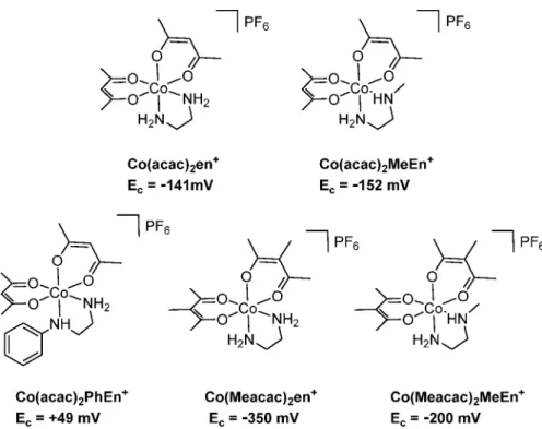
Notably, the reduction potential of reference compound Co(acac)
2en
+was distinctly lower with a value of
−141 mV versus NHE even compared to the most promising representa- tive of the EGFR inhibitor-containing complexes, Co- (Meacac)
2L
+, at 4 mV versus NHE. To verify that the adjacent aromatic moiety of the quinazoline is responsible for this shift and that methylation of the
“en”moiety indeed increases the redox potential, additional model complexes were synthesized (Figure 4): (1) Co(acac)
2PhEn
+to determine the in
fluence of the phenyl moiety and (2) Co(Meacac)
2en
+, Co(acac)
2MeEn
+, and Co(Meacac)
2MeEn
+to con
firm the shifts of the cathodic peak potential as a result of the di
fferent methylations (MeEn = N-methylethylenediamine; PhEn = N-phenylethylenediamine).
The synthesis of the complexes followed a similar procedure as described above, and the only di
fference was the use of ammonium hexa
fluorophosphate for precipitation of the complexes (see details in the
Experimental Section).Indeed, cyclic voltammetric measurements performed for Co(acac)
2PhEn
+showed a strong shift of the cathodic peak potential to 49 mV in comparison to that of Co(acac)
2en
+at
−
141 mV (Figure 4). This can be explained by the electron- withdrawing e
ffect of the phenyl ring, which distinctly increases the redox potential of the cobalt center. With regard to the methylations at the
“en
”versus the acac moiety, the model complexes con
firmed (even more pronounced) the tendencies observed for the EGFR inhibitor-containing complexes [except Co(acac)
2MeEn
+with a slightly lower E
cvalue]. The introduction of the methylated acac moiety led to a signi
ficantly
Table 1. Distribution Coe
fficients (
D7.40) of the Complexes at pH 7.40 [
T= 25.0
°C, and
I= 0.1 M (KCl)]
complex logD7.40
Co(acac)2L+a 1.59±0.06
Co(Meacac)2L+ 2.24±0.04
Co(acac)2MeL+ 2.05±0.17
Co(Meacac)2MeL+ >2.7
Co(acac)2en+ −1.86±0.05
L 1.86±0.0329
aDistribution coefficients measured under different conditions: logD
=−0.55±0.01 (water, pH∼6.5),−0.09±0.03 [20 mM phosphate buffer (pH 7.40)], and 2.22±0.03 [20 mM phosphate buffer and 0.58 M KCl (pH 7.40)].
Figure 3. Cyclic voltammograms of Co(acac)2L+, Co(Meacac)2L+, Co(acac)2MeL+, andCo(Meacac)2MeL+in 10 mM phosphate buffer (pH 7.40) (1.5 mM complex,I= 0.10 M KCl, scan rate of 30 mV/s, 25.0
°C). Potentials are referenced to the NHE.
decreased reduction potential, whereas the methylation of the
“
en
”ligand showed the opposite e
ffect. Therefore, in the case of Co(Meacac)
2MeEn
+, the in
fluence of the methylated acac moiety is widely annihilated by the methylation of
“en
”.
Notably, all reduction processes were completely irreversible independent of the scan speed (30
−1000 mV/s). In the literature, the proposed mechanism for cobalt(III) prodrugs usually comprises that in healthy tissues, which are provided with a su
fficient supply of oxygen, and after the reduction to cobalt(II), an immediate re-oxidation step occurs, regenerating the inert cobalt(III) complex.
23,30However, at the same time for most of the cobalt(III) complexes in the literature, the redox process in an aqueous solution is completely irreversible.
31−34This is in line with pulse radiolysis studies also revealing that the re-oxidation rates under normoxic conditions are too slow.
35Instead, competition between oxygen and the cobalt(III) complexes for one-electron reductants was suggested. Interest- ingly, despite this irreversibility, e.g., cobalt(III) nitrogen
mustard complexes showed promising hypoxia selective anticancer activity.
32In particular, Ware et al. tried to optimize the electrochemical properties of cobalt(III) prodrug systems, resulting in (partially) reversible complexes with highly hypoxia selective activity against cancer cells.
36Unfortunately, their lead compound did not show signi
ficant activity in a mouse model.
However, as our compound class, which displays irreversible electrochemical properties, also possesses hypoxia-dependent anticancer activity in a mouse model,
17this indicates that the irreversibility of the reduction process does not interfere with the in vivo e
ffectiveness. Therefore, it is currently unknown how the exact mechanism of the hypoxic selectivity of cobalt(III) complexes works and which electrochemical properties are ideal.
Aqueous Solution Stability of the Formed Cobalt(II) Complexes.
After reductive activation of the cobalt(III) prodrugs, it is essential to investigate whether the formed cobalt(II) complexes indeed can release the coordinated targeting ligand. For this reason, we selected two ternary
Figure 4.Cobalt(III) model complexes synthesized to investigate the effect of the methyl and phenyl substitution at the“en”and/or acac moiety.Ecis the cathodic peak potential vs NHE of the cobalt complexes measured at a scan rate of 30 mV/s in 10 mM phosphate buffer (pH 7.4).Table 2. Proton Dissociation Constants (
Ka) of Hacac as well as Fully Protonated MeEn and PhEn Together with the Overall Stability Constants (
β) for the Binary and Ternary Cobalt(II) Complexes Determined by pH-Potentiometric Measurements
aacac (A) MeEn (B) PhEn (B)
pKa1 8.80±0.01b pKa1 7.04±0.02 1.85±0.01c
pKa2 − pKa2 9.98±0.01 9.34±0.01c
LogβValues of the Binary Complexesc
[Co(II)L]+ 5.05±0.02b [Co(II)L]2+ 5.26±0.01 3.63±0.05
[Co(II)L2]0 8.66±0.05b [Co(II)L2]2+ 9.15±0.02 6.75±0.09
[Co(II)L3]− − [Co(II)L3]2+ 12.94±0.04 −
LogβValues of the Ternary Complexes
[Co(II)AB]+ 9.82±0.05 8.03±0.15
[Co(II)A2B]0 12.73±0.09 12.08±0.09 [Co(II)AB2]+ 13.20±0.12 11.29±0.13
aIn ternary (mixed-ligand) complexes,“A”denotes acac and“B”denotes MeEn or PhEn [25.0°C;I= 0.1 M (KCl)].bReported data for the cobalt(II) acac system: pKa1= 8.83, logβ[CoL] = 5.10, and logβ[CoL2] = 9.08 (25°C;I= 0.1 M NaClO4).40cpKavalues of 1.76±0.03 and 9.42
±0.03, and 1.73±0.03 and 9.37±0.03, determined by ultraviolet−visible and spectrofluorometric titrations, respectively.
systems as models to study the potential liberation of the (N,N) ligand from the mixed-ligand cobalt complex upon reduction.
Thus, the aqueous stability of several cobalt(II) complexes was investigated under strictly anaerobic conditions. Due to the limited water solubility of cobalt(II) chloride complexes with L, model ligands with better solubility, namely, MeEn and PhEn, were used. First, deprotonation processes of the acetlyacetone (Hacac) and the fully protonated MeEn and PhEn ligands were followed at pH 2.0
−11.5 (Table 2). The calculated pK
afor Hacac of 8.80 is in good agreement with literature data (pK
a= 8.82) reported at I = 0.1 M NaClO
4.
37The two pK
avalues of MeEn [pK
a1= 7.04 (MeNH
2+); pK
a2= 9.98 (NH
3+)]
correspond well to the deprotonation constants of the fully protonated form of
“en
”,
38,39but the pK
a1dramatically di
ffers from that of PhEnH
22+(pK
a1= 1.85; pK
a2= 9.34). The latter model ligand is quite comparable to EGFR inhibitor L (pK
a1<
1.0; pK
a2= 9.21) investigated in our previous work.
29Following the determination of the stability constants for the binary complexes (Table 2), overall stability constants for the mixed-ligand complexes formed in the cobalt(II)
−acac
−MeEn and cobalt(II)
−acac
−PhEn ternary systems were calculated on the basis of pH-potentiometric titrations. Three types of complexes were formed, [CoAB]
+, [CoA
2B], and [CoAB
2]
+, where
“A
”denotes acac and
“B
”refers to MeEn or PhEn.
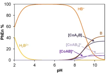
Concentration distribution curves in
Figure 5were computed using the stability constants of the 1:2:1 cobalt(II)
−acac
−PhEn composition that corresponds to the reduced form of Co(II)- (acac)
2L
0. On the basis of these data, negligible amounts (<1%) of PhEn are coordinated to cobalt(II) at pH 7.4. For the 1:2:1 cobalt(II)
−acac
−MeEn system,
∼11% are still coordinated to cobalt(II) at pH 7.4 (Figure S6). However, it has to be mentioned that these calculations were performed for 1 mM cobalt(II) (where the titrations were performed), and the amount of the intact ternary complex further decreases with lower concentrations [e.g., in the latter system at pH 7.4 and 0.1 mM cobalt(II), the coordinated MeEn fraction is only
∼2%].
As a conclusion, these measurements strongly support the assumption that after reduction of the cobalt(III) complexes, the respective EGFR-targeting ligand completely dissociates, which enables subsequent inhibition of the EGFR-downstream signaling.
Interaction with Natural Low-Molecular Weight Reducing Agents.
Next, we investigated if common bio-
logically relevant low-molecular weight reducing agents can reduce Co(acac)
2L
+or Co(Meacac)
2L
+. Glutathione (GSH) and reduced nicotinamide adenine dinucleotide (phosphate) (NADH/NADPH) are responsible for the redox equilibrium in the cytosol, while ascorbic acid (AA) is found in both extra- and intracellular space. Among them, NADH is the strongest reducing agent (formal potential at pH 7.0 of
−0.32 V forNAD
+/NADH
41), followed by GSH (
−0.24 V for GSSG/GSH at pH 7.0
41). For AA, usually 0.06 V for dehydro-
L-ascorbate/
AA at pH 7.0 is reported.
42However, recent literature also suggests even higher values in the range of 0.35
−0.50 V.
43,44GSH is produced in cells at concentrations (1
−10 mM) at least one order of magnitude higher than those of NADH (30
−100
μM).
45,46The interaction of AA, GSH, and NADH at pH 7.40 (phosphate buffer) was investigated exemplarily for Co- (acac)
2L
+and Co(Meacac)
2L
+. The reaction was followed by ultraviolet
−visible (UV
−vis) and
fluorometric detection at 25
°
C with a 10-fold excess of reducing agent. Spectro
fluorometry was proven to be an e
ffective technique for monitoring this redox reaction, because the free EGFR inhibitor ligand L is highly
fluorescent, while its cobalt(III) complex has negligible emission (vide supra).
17Accordingly, an increase in the emission intensity is expected upon reduction and concomitant release of the free ligand.
Figure 6indicates practically no reduction process of Co(Meacac)
2L
+in the presence of 10 equiv of GSH, and no signi
ficant amount of free L appeared in samples even after 24 h. The same tendency was observed by UV
−vis detection (data not shown). For Co(acac)
2L
+, again no liberation of the active ligand could be detected within 24 h.
AA and NADH were also not able to reduce the two complexes even after 24 h. These data show that the complexes are highly stable in aqueous solution and biologically relevant low- molecular weight non-enzymatic reducing agents are not responsible for the reduction of the cobalt(III) complexes. For other hypoxia-activatable drugs like tirapazamine and its second- generation analogue SN30000, P450 oxidoreductases (POR) have been proposed as proteins responsible for reduction.
47However, Ware and co-workers
36and Wilson and co-workers
48showed for di
fferent types of cobalt(III) complexes that the cytotoxicity did not change in cells overexpressing POR
Figure 5.Concentration distribution diagram for the 1:2:1 cobalt(II)−
acac−PhEn system. A = acac; B = PhEn [1 mM cobalt(II);I= 0.10 M (KCl); 25.0°C].
Figure 6. Fluorescence emission spectra of Co(Meacac)2L+ in the presence of 10 equiv of GSH followed for 24 h. The dashed spectrum corresponds to the emission spectrum of free EGFR inhibitorL[ccomplex
= 15μM;cfree ligand= 15μM;λEX= 350 nm; pH 7.40 (10 mM phosphate buffer and 0.1 M KCl); 25.0°C].
compared to the parental cell line. Therefore, the actual reducing molecules or reductase(s) are still unknown.
Serum Stability.
The data presented in the previous sections indicate that even in the presence of low-molecular weight reducing agents the cobalt(III) complexes are completely stable. Therefore, we wanted to investigate if this is still true in a more elaborate biological environment like blood serum. To this end, the four EGFR inhibitor complexes were dissolved in 50 mM phosphate puffer and mixed in a 1:10 ratio with fetal calf serum (FCS; bu
ffered with 150 mM phosphate bu
ffer to keep a stable pH of 7.4) to a
final concentration of 50
μM. The samples were incubated at 37
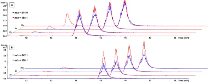
°C and after 0, 2, 6, 24, and 26 h extracted with acetonitrile and measured by HPLC-MS. The extracted ion mass chromatogram of Co(acac)
2L
+clearly showed the time- dependent release of the free EGFR inhibitor (Figure 7A; red peak at 11.8 min, m/z 358.1). The intact complex could be observed at 14.2 min (blue peak, m/z 614.0). The red signal at the same retention time (Figure 7A; 14.2 min, m/z 358.1) is a mass spectrometry artifact and belongs to the free ligand generated during ionization of the complex. In contrast, the mass spectra of Co(Meacac)
2L
+incubated in serum revealed much smaller amounts of the released ligand (Figure 7B).
The same trend was observed for Co(acac)
2MeL
+and Co(Meacac)
2MeL
+(Figure S7). Therefore, in this experiment, a distinct increase in the stability in the presence of the Meacac ligand could be observed in both complexes with
∼85% intact compound after serum incubation for 26 h at 37
°C. The stability of these complexes was much higher than that of Co(acac)
2L
+and Co(acac)
2MeL
+, having only 43% and 50%
intact compound after 26 h, respectively (Figure 8). In general, these results are in accordance with the cyclic voltammetry measurements, where Co(Meacac)
2L
+and Co(Meacac)
2MeL
+showed the lowest redox potential of the four complexes.
Together, this indicates a strong in
fluence of the methylation of acac for the stability of cobalt(III) prodrugs; however, no bene
ficial e
ffect was seen in the case of methylation of the
“en
”moiety.
Biological Investigations. Evaluation of the Complex Stability in the Presence of Cells under Normoxic Conditions.
As a next step, we addressed the question of whether the new derivatives are also more stable than Co(acac)
2L
+in the
presence of cells under normoxic cell culture conditions (medium containing 10% serum at 37
°C, 21% O
2, and 5%
CO
2). Therefore, the stability of the cobalt(III) complexes was monitored indirectly by microscopy as well as
flow cytometry in A431 cells by exploiting the
fluorescence of released ligands L and MeL. As shown in
Figure 9, the stability of the compoundsbearing a Meacac ligand was distinctly increased compared to those of the acac derivatives. Consequently, while in case of the acac drugs most of the ligand was already released after 24 h (clearly visible by the blue
fluorescence of the cells), the micrographs of the Meacac compounds remained widely unchanged.
This also could be con
firmed by subsequently performed
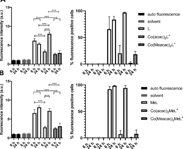
flow cytometry investigations (Figure 10). In detail, the evaluation of the
fluorescence intensities showed that the cobalt complexation led to a 1.9
−2.6-fold reduced mean
fluorescence and a 4
−90-
Figure 7.Time-dependent stability of (A)Co(acac)2L+and (B)Co(Meacac)2L+incubated in FCS at 37°C (pH 7.4, 150 mM phosphate buffer) and analyzed by HPLC−mass spectrometry (depicted are the extracted ion mass chromatograms). Due to the different ionization properties, the intensities of the free ligand (m/z358.1) and cobalt(III) complexes (m/z614.0 or 642.1) cannot be directly compared.Figure 8.Stability measurements of Co(acac)2L+,Co(Meacac)2L+, Co(acac)2MeL+, andCo(Meacac)2MeL+incubated in FCS at 37°C (pH 7.4, 150 mM phosphate buffer) and analyzed by mass spectrometry over a period of 26 h. They-axis shows the relative ratio of the integrated peak areas of the intact complex over time (in percent) compared to the area at the starting point (0 h).
fold reduced number of ligand-positive cells at the 6 h time point. This is in good agreement with the already published data on Co(acac)
2L
+.
17However, after incubation for 24 h 100% of cells treated with the two acac complexes became ligand- positive, resulting in a mean
fluorescence similar to that of the samples treated with the free ligand. In contrast, in the samples of the Meacac-containing group, the mean
fluorescence was much less a
ffected, as the percentage of ligand-positive cells increased only from 2.2% to 18.4% and from 1% to 7.5%, in the cases of Co(Meacac)
2L
+and Co(Meacac)
2MeL
+, respectively.
This indicates that in contrast to the acac-auxiliary ligand, the cobalt(III) complexes bearing a Meacac moiety are highly stable under normoxic cell culture conditions for more than 1 day.
To evaluate if cobalt complexation also prevents EGFR inhibition under normoxic conditions, Western blot analyses of the EGFR phosphorylation at position Y1068 (activating phosphorylation) as well as the activation of the EGFR downstream protein ERK 1/2 were performed. To ensure an EGFR-dependent signaling, A431 cells were serum-starved for 24 h, incubated with the indicated cobalt(III) drugs at three different concentrations for 2 h, and stimulated by EGF for 10 min prior to protein isolation (Figure 11). In agreement with our stability data, the Meacac ligand set e
fficiently prevented the release of both L and MeL, indicated by the distinctly weakened
EGFR-inhibitory potential of Co(Meacac)
2L
+and Co- (Meacac)
2MeL
+in comparison to Co(acac)
2L
+and Co- (acac)
2MeL
+. The potent EGFR inhibition by free MeL and L under these conditions is shown in
Figure S8and ref
17,respectively.
Hypoxia-Dependent Cytotoxicity of the Cobalt Complexes.
Finally, we wanted to evaluate whether the Meacac coordination sphere also a
ffects the hypoxic activation of the complexes. To characterize the activity of the free EGFR inhibitor ligands, prior to the assessment of their anticancer activity in cell culture, the EGFR-inhibitory potential was investigated in a cell-free kinase inhibition assays in the presence of a 10-fold excess of ATP. The results showed that the methylation at the terminal amino group slightly reduced the EGFR inhibition potential from an IC
50value of 0.29 nM for L to a value of 0.41 nM for MeL (Figure
S9).Subsequently, MTT-based cytotoxicity assays of the com-
plexes in comparison to the respective metal-free ligands were
performed under di
fferent O
2levels (21%, 5%, 1%, or 0.1%)
(Table 3 and
Figure 12). As expected, the two EGFR inhibitorligands L and MeL did not show distinct differences in their
anticancer activity in normoxic versus the di
fferent hypoxic
conditions. However, MeL was approximately twice as e
ffective
as L. This is interesting, as the kinase assay mentioned above did
Figure 9.Fluorescence microscopic measurements indicating the release of the ligand from the different cobalt(III) complexes. Release of (A)Land (B)MeLfrom the different cobalt(III) complexes under normoxic cell culture conditions (37°C, 21% O2, and 5% CO2) using UVfluorescence microscopy. A431 cells were incubated with 10μM drugs for 6 or 24 h. Images are overlays of representativefluorescence and differential interference contrast microscopies (10×objective) of the different treatments processed by ImageJ software.Figure 10.Release of (A)Lor (B)MeLfrom the indicated cobalt(III) complexes under normoxic cell culture conditions (37°C, 21% O2, and 5%
CO2) byflow cytometry. A431 cells were incubated with 10μM drugs for 6 or 24 h, and the fold change influorescence intensity (left, after normalization withfluorescence intensity of the cells) and the percent offluorescence-positive cells (right) were evaluated using Diva Software and GraphPad Prism. Statistical significance was calculated via two-way analysis of variance with a multiple-comparison test and Bonferroni correction with p< 0.001 (***).
Figure 11.Impact of new cobalt(III) complexes on the EGFR signaling cascade (pEGFR, pERK 1/2) under normoxic conditions. A431 cells were grown in medium with or without FCS and treated with the indicated drug for 2 h. After EGFR stimulation with 50 ng/mL EGF for 10 min, cells were harvested, lysated, and further analyzed by Western blotting. The ratios of pEGFR or pERK 1/2 levels of the treated samples (after normalization to the loading controlβ-actin) to the levels of the control (−FCS and +EGF) are given below the respective bands.
not show a higher EGFR inhibitor potency for this new inhibitor, probably indicating a shift in the target kinase spectrum.
In good agreement with our previous study,
17the formerly investigated Co(acac)
2L
+exhibited an
∼2-fold weaker anti- cancer activity under normoxia (Table 3) than under reduced oxygen conditions (
≤1%). Also in the case of Co(acac)
2MeL
+, the cobalt complex was
∼2-fold less active than the respective free ligand. However, under hypoxic conditions, Co- (acac)
2MeL
+did not show full MeL activity even under the lowest oxygen levels of 0.1%. In contrast, the two Meacac- containing cobalt(III) complexes both showed IC
50values of
>50
μM under normoxic conditions and also weak hypoxia with 5% O
2did not enhance the activity to a relevant extent.
However, the reduction of oxygen levels to 1% signi
ficantly (p <
0.001) increased the cytotoxic activity of the two prodrugs, resulting in IC
50values of
∼25 and∼32μM forCo(Meacac)
2L
+and Co(Meacac)
2MeL
+, respectively. In the case of Co- (Meacac)
2L
+, a further decrease in the O
2levels from 1% to 0.1% generated similar results. In contrast, for Co- (Meacac)
2MeL
+, the presence of 0.1% O
2induced an additional signi
ficant (p < 0.05) improvement in drug e
fficacy (IC
50value
of
∼20
μM) compared to that for 1% O
2hypoxia, resulting in a 2.9-fold increase in cytotoxicity compared to that under normoxic conditions.
It is well-known that cobalt(II) ions have some biological e
ffects like an upregulation of the expression of the hypoxia inducible factor (HIF).
49Furthermore, a possible e
ffect could also arise from iron(III) binding of released Meacac.
50However, we already investigated in our previous work
17CoCl
2as well as the complexes [Co(II)(acac)
2en] and [Co(III)(acac)
2en]PF
6with a simple ethylenediamine ligand without an EGFR-binding moiety. No signi
ficant cytotoxic activity could be observed against A431 cells under both normoxia and hypoxia. Therefore, we can widely exclude a contribution of Co(II) ions or released acac (and subsequent iron chelation) on the anticancer activity of EGFR inhibitor-bearing cobalt(III) prodrugs.
■
CONCLUSIONSDespite the revolutionizing e
ffect they have had on cancer therapy, TKIs are limited in their clinical application due to severe side e
ffects and rapid development of drug resistance.
Hence, the design of tumor-speci
fically activated prodrugs is an important strategy for reducing these adverse e
ffects. We Table 3. IC
50Values of L and MeL in Comparison to the Respective Cobalt(III) Prodrugs against A431 Cancer Cells after Treatment for 72 h under Di
fferent O
2Levels (21% to 0.1%)
aIC50(μM±SD)
drug normoxia hypoxia with 5% O2 hypoxia with 1% O2 hypoxia with 0.1% O2
L 12.0±1.3 12.8±0.9 12.7±2.0 13.7±2.9
MeL 6.9±1.8 8.7±1.2 5.5±0.4 5.4±0.8
Co(acac)2L+ 22.9±5.7b 18.7±4.0b 13.4±0.3 7.2±1.4
Co(Meacac)2L+ 51.9±9.4c,e 45.5±2.7c 25.3±3.9 23.5±5.1
Co(acac)2MeL+ 15.1±1.6 13.5±3.2 12.5±3.1 11.9±3.5
Co(Meacac)2MeL+ 58.6±4.4c,e 55.2±1.4c 32.0±2.9d 19.9±1.0
erlotinib 13.3±4.7 nd 14.4±4.0 nd
aValues are given as means±SD of at least three independent experiments performed in triplicate. Statistical significance, between the drugs under normoxic and different hypoxic conditions, was calculated via one-way analysis of variance with a multiple-comparison test and Bonferroni correction.bp< 0.01 compared to hypoxia with 0.1% O2.cp< 0.001 compared to hypoxia with 1% and 0.1% O2.dp< 0.05 compared to hypoxia with 0.1% O2.ep< 0.001 compared to the corresponding ligand and Co(acac)2X+derivative under normoxia.
Figure 12.Cytotoxic activity of the indicated compounds against A431 cancer cells. The incubation time of the compounds on the cells was 72 h under normoxic and three different hypoxic conditions (5%, 1%, or 0.1% O2). Values are given as means±the standard deviation of one representative experiment performed in triplicate.
the cytotoxic activity of all compounds under normoxia versus hypoxia revealed that the complexes with distinctly higher stability still possess promising hypoxia-activatable properties.
However, the IC
50values at 0.1% O
2after 72 h were signi
ficantly higher compared to those of the free EGFR inhibitor ligands.
More in-depth studies are now needed to evaluate the underlying reasons for this e
ffect. Finally, a balance between su
fficient stability of the complex and tumor-speci
fic release of the EGFR inhibitor ligand is needed. However, only evaluation in future in vivo experiments will reveal whether Co- (Meacac)
2MeL
+is a promising candidate with both high stability and activity.
■
EXPERIMENTAL SECTIONMaterials and Methods.All solvents and reagents were obtained from commercial suppliers. They were, unless stated otherwise, used without further purification. Anhydrous MeOH and tetrahydrofuran over molecular sieves were bought from Fisher Chemicals. The precursors Na[Co(acac)2(NO2)2] and Na[Co(Meacac)2(NO2)2] were obtained following the protocol of Denny et al.21 Co(acac)2en+, Co(acac)2L+, and L were synthesized according to our previous publication.17
For all HPLC measurements, Milli-Q water (18.2 MΩcm, Merck Milli-Q Advantage, Merck, Darmstadt, Germany) was used. Preparative RP-HPLC was performed on an Agilent 1200 Series system controlled by Chemstation software. As the stationary phase, either a XBridge BEH C18 OBD Prep Column (130 Å, 5μm, 19 mm×250 mm) or an Atlantis T3 OBD Prep Column (100 Å, 10μm, 19 mm×250 mm), each from Waters Corp., was used. The general procedure included a flow rate of 17.06 mL/min, an injection volume of≤10 mL, and a column temperature of 25°C. Milli-Q water and acetonitrile (ACN) without addition of acids were used as eluents unless stated otherwise.
Stability and kinetic experiments were analyzed on an Agilent 1260 Infinity system using a Waters Atlantis T3 column (150 mm×4.6 mm) coupled to a Bruker amaZon SL ESI mass spectrometer. If not stated otherwise, water (containing 0.1% formic acid) and ACN (containing 0.1% formic acid) were used as eluents with a gradient of 1% to 99%
ACN within 29 min. Elemental analyses were performed by the Microanalytical Laboratory of the University of Vienna on a Perkin Elmer 2400 CHN Elemental Analyzer. Electrospray ionization (ESI) mass spectra were recorded on a Bruker amaZon SL ion trap mass spectrometer in positive and/or negative mode by direct infusion.
High-resolution mass spectra were recorded on a Bruker maXis UHR ESI time-of-flight mass spectrometer. Expected and experimental isotope distributions were compared.1H and13C NMR one- and two- dimensional spectra were recorded in DMSO-d6 with a Bruker FT- NMR AV NEO 500 MHz spectrometer at 500.10 (1H) and 125.75 (13C) MHz at 298 K or a Bruker FT-NMR AVIII 600 MHz spectrometer at 600.25 MHz (1H) and 150.93 MHz (13C). Chemical shifts (parts per million) were referenced internally to the solvent
solvent was removed, and the yellow residue was extracted in 600 mL of EtOAc and washed with 400 mL of 1 M HCl, saturated NaHCO3, and brine. The organic phase was separated, dried over Na2SO4, and concentrated in vacuo. The crude product was purified via flash chromatography (15:1 dichloromethane/MeOH). Yield: 1.48 g (65%).
1H NMR (500.1 MHz, DMSO-d6):δ1.19 (s, 6H), 1.39 (s, 3H), 1.41 (s, 3H), 2.77−2.93 (m, 2H), 3.42−3.50 (m, 2H), 6.31 (d,J= 8.0 Hz, 1H), 7.91−7.31 (m, 3H), 7.35 (t, 1H), 7.56 (d,J= 2.0 Hz, 1H), 7.91 (d,J= 2.0 Hz, 1H), 8.17 (s, 1H), 8.39 (s, 1H), 9.34 (d,J= 6.0 Hz, 1H).
N4-(3-Bromophenyl)-N6-[2-(methylamino)ethyl]quinazoline-4,6- diamine Dihydrochloride (MeL).To a solution oftert-butyl [2-({4- [(3-bromophenyl)amino]quinazolin-6-yl}amino)ethyl](methyl)- carbamate (1.48 g, 3.12 mmol) in EtOH (30 mL) was added concentrated HCl (1.25 mL, 40.0 mmol), and the reaction mixture was refluxed for 3 h. The solution was cooled to room temperature and stored overnight at 4°C. The yellow precipitate wasfiltered off, washed with EtOH, and driedin vacuo. Yield: 1.11 g (80%).1H NMR (600.25 MHz, DMSO-d6):δ2.61 (t, 3H, H23), 3.15 (m, 2H, H21), 3.67 (m, 2H, H20), 7.19 (s, 1H, H18), 7.45 (t, 3H, H16), 7.48−7.54 (m, 2H, H2, H15), 7.77 (d, 1H,J= 2.0 Hz, H3), 7.88 (d, 1H,J= 2.0 Hz, H11), 8.00 (s, 1H, H6), 8.13 (s, 1H, H13), 8.78 (s, 1H, H8), 9.09 (s, 2H, H22), 11.50 (s, 1H, H17).13C NMR (150.93 MHz, DMSO-d6): δ32.45 (C23), 38.93 (C21), 46.20 (C20), 98.57 (C6), 115.42 (C5), 120.73 (C3), 121.00 (C14), 123.71 (C11), 126.53 (C2), 127.23 (C13), 128.72 (C15), 130.41 (C16), 130.88 (C4), 138.67 (C12), 146.30 (C8), 148.57 (C1), 158.23 (C10). MS: calcd for [C17H18BrN5]+, 372.08; found, 372.08. Anal. Calcd for C17H18BrN5·2HCl (Mr= 445.18 g/mol): C, 45.86; H, 4.53; N, 15.73. Found: C, 45.75; H, 4.22; N, 15.47.
Bis(3-methyl-2,4-pentanedionato) N6-(2-Aminoethyl)-N4-(3- bromophenyl)quinazoline-4,6-diamine Cobalt(III) Chloride [Co- (Meacac)2L+].Na[Co(Meacac)2(NO2)2] (84.4 mg, 0.22 mmol) was dissolved in H2O (1.6 mL) and MeOH (4.5 mL).L(100 mg, 0.23 mmol) was dissolved in H2O (1 mL), neutralized with NaOH (1.65 mL, 0.28 M in MeOH), and subsequently added to the cobalt precursor solution together with activated charcoal (64 mg). The resulting mixture was stirred for 1 h at room temperature,filtered through Celite, and washed with small amounts of a MeOH/H2O mixture (1:1). Brine
(30 mL) was added to thefiltrate, and the resulting solution was left at 4
°C overnight. The formed green precipitate wasfiltered offthe next day.
The crude product (250 mg of a dark green solid) was purified by RP- HPLC (Xbridge, H2O/MeOH, isocratic 57:43, without formic acid or TFA to avoid counter ion exchange). Yield: 67 mg (44%). The ratio of the two isomers was 1:0.36.
Shifts of the main isomer.1H NMR (500.1 MHz, DMSO-d6):δ1.00 (s, 3H, CH3, Meacac), 1.65 (s, 3H, CH3, Meacac), 1.88 (s, 3H, CH3, Meacac), 2.05−2.06 (m, 6H, CH3, Meacac), 2.35 (s, 3H, CH3, Meacac), 2.67−2.97 (m, 3H, CH2, en), 3.60−3.69 (m, 1H, CH2, en), 5.28−5.37 [m, 1H, CH, NH2(en)], 5.73−5.80 [m, 1H, CH, NH2(en)], 7.36−7.43 (m, 2H, ph), 7.52 (dd, 1H,J= 9 Hz,J= 2 Hz, quin), 7.67 (d, 1H,J= 9 Hz, quin), 7.82−7.89 (m, 1H, ph), 7.93 [d, 1H, NH(en)], 8.10 (s, 1H, ph), 8.14−8.17 (m, 1H, quin), 8.65−8.70 (m, 1H, quin), 10.12 (s, 1H, NH).13C NMR (125.75 MHz, DMSO-d6): δ 13.8 (CH3, Meacac), 14.9 (CH3, Meacac), 26.1 (2C, CH3, Meacac), 26.5 (2C, CH3, Meacac), 41.8 (CH2, en), 50.9 (CH2, en), 98.3 (Cq, Meacac), 100.5 (Cq, Meacac), 113.4 (CH, quin)*, 121.2 (Cq, ph), 121.3 (CH, ph), 124.7 (CH, ph), 126.5 (2C, CH, quin + ph), 130.0 (CH, quin)*, 130.6 (CH, ph), 140.6 (Cq, ph), 142.8 (Cq, quin), 153.7 (CH, quin)*, 157.0 (Cq, quin), 186.2 (Cq, acac), 186.9 (Cq, acac), 187.3 (Cq, acac), 187.9 (Cq, acac).
Shifts of the minor isomer.1H NMR (500.1 MHz, DMSO-d6):δ1.77 (s, 3H, CH3, Meacac), 1.78 (s, 3H, CH3, Meacac), 1.99 (s, 3H, CH3, Meacac), 2.05−2.06 (m, 3H, CH3, Meacac), 2.20 (s, 3H, CH3, Meacac), 2.34 (s, 3H, CH3, Meacac), 2.67−2.97 (m, 3H, en), 3.72−
3.80 (m, 1H, en), 5.28−5.37 [m, 1H, NH2(en)], 5.73−5.80 [m, 1H, NH2(en)], 7.18 (dd, 1H,J= 9 Hz,J= 2 Hz, quin), 7.36−7.43 (m, 2H, ph), 7.46 [m, 1H, NH(en)], 7.71 (d, 1H,J= 9 Hz, quin), 7.82−7.89 (m, 1H, ph), 8.14−8.17 (m, 1H, ph), 8.65−8.70 (m, 2H, quin), 10.25 (s, 1H, NH).13C NMR (125.75 MHz, DMSO-d6):δ14.7 (CH3, Meacac), 14.9 (CH3, Meacac)*, 25.9 (CH3, Meacac), 26.6 (CH3, Meacac), 26.8 (CH3, Meacac), 26.7 (CH3, Meacac), 51.2 (CH2, en)*, 52.2 (CH2, en)*, 100.4 (Cq, Meacac), 100.6 (Cq, Meacac), 114 (2C, CH, quin)*, 121.3 (CH, ph)*, 124.7 (CH, ph)*, 126.5 (2C, CH, -quin + ph)*, 129.3 (CH, quin)*, 130.6 (CH, ph)*, 153.7 (CH, quin)*, 187.7 (Cq, Meacac), 188.2 (Cq, Meacac).
MS: calcd for [C28H34BrCoN5O4]+, 642.11; found, 642.28. Anal.
Calcd for C28H34BrClCoN5O4·2H2O (Mr= 714.92 g/mol): C, 47.04;
H, 5.36; N, 9.80. Found: C, 46.85; H, 5.09; N, 9.68.*Detected only in two-dimensional (2D) NMR.
B i s ( 2 , 4 - p e n t a n e d i o n a t o ) N4- ( 3 - B r o m o p h e n y l ) - N6- [ 2 - (methylamino)ethyl]quinazoline-4,6-diamine Cobalt(III) Chloride [Co(acac)2MeL+].Na[Co(acac)2(NO2)2] (83.6 mg, 0.23 mmol) was dissolved in H2O (1.6 mL) and MeOH (1.6 mL).MeL(105 mg, 0.24 mmol) was dissolved in H2O (1.1 mL), neutralized with NaOH (1.9 mL, 0.25 M in MeOH), and subsequently added to the cobalt complex solution with activated charcoal (55.2 mg). The resulting mixture was stirred for 1 h at room temperature,filtered through Celite, and washed with small amounts of MeOH. Brine (6.1 mL) was added to thefiltrate, and the resulting solution was extracted with dichloromethane (3×10 mL). The organic phase was separated, dried with Na2SO4, and evaporated. The crude product (115 mg of a dark green solid) was purified by RP-HPLC (H2O/ACN, 30−42% ACN, 26 min, without formic acid or TFA to avoid counter ion exchange). Yield: 63 mg (40%). The ratio of the four isomers (A:B:C:D) was 1:0.42:0.33:0.20.
Shifts of the main isomer A.1H NMR (500.1 MHz, DMSO-d6):δ 1.56 (s, 3H, CH3, acac), 1.77 (d, 3H,J= 6 Hz, CH3, MeEn), 2.01 (s, 3H, CH3, acac), 2.02 (s, 3H, CH3, acac), 2.30 (s, 3H, CH3, acac), 2.58−3.04 (m, 3H, CH2, MeEn), 3.68−3.92 (m, 1H, CH2, MeEn), 4.79 (s, 1H, CH, acac), 5.67−5.75 (m, 1H, CH, acac), 6.74 [m, 1H, NH(en)], 7.33−7.43 (m, 2H, CH, ph), 7.46 (dd, 1H, CH,J= 8 Hz,J= 2 Hz, quin), 7.61−7.73 (m, 1H, CH, quin), 7.83−7.94 (m, 1H, CH, ph), 8.13 (t, 1H, CH, ph), 8.23−8.27 (m, 1H, CH, quin), 8.29−8.38 [m, 1H, NH(en)], 8.62−8.70 (m, 1H, CH, quin), 10.13 (s, 1H, NH).13C NMR (125.75 MHz, DMSO-d6):δ26.2 (CH3, acac), 26.4 (CH3, acac), 26.5 (CH3, acac), 26.6 (CH3, acac), 37.5 (CH3, MeEn), 50.8 (CH2, MeEn), 53.1 (CH2, MeEn), 96.4 (CH, acac), 98.8 (CH, acac), 115.7 (Cq, quin)*, 121.6 (Cq, ph), 121.8 (CH, ph), 125.3 (CH, ph), 127.0 (CH, ph), 127.5 (CH, quin), 130.9 (CH, quin), 131.0 (CH, ph), 141.1 (Cq,
ph), 142.7 (Cq, quin), 148.5 (Cq, quin)*, 157.5 (Cq, quin), 189.6 (Cq, acac), 189.7 (Cq, acac), 190.0 (Cq, acac), 190.9 (Cq, acac).
Shifts of isomer B.1H NMR (500.1 MHz, DMSO-d6):δ1.68 (s, 3H, CH3, acac), 2.06 (d, 3H,J= 6 Hz, CH3, MeEn), 2.03 (s, 3H, CH3, acac), 2.11 (s, 3H, CH3, acac), 2.26 (s, 3H, CH3, acac), 2.58−3.04 (m, 3H, CH2, MeEn), 3.68−3.92 (m, 1H, CH2, MeEn), 5.55 (s, 1H, CH, acac), 5.67−5.75 (m, 1H, CH, acac), 6.18 [m, 1H, NH(en)], 7.29 (dd, 1H, CH,J= 8 Hz,J= 2 Hz, quin), 7.33−7.43 (m, 2H, CH, ph), 7.61−7.73 (m, 1H, CH, quin), 7.83−7.94 [m, 2H, CH, ph + NH(en)], 8.16 (m, 1H, CH, ph), 8.62−8.70 (m, 1H, CH, quin), 8.70−8.74 (m, 1H, CH, quin), 10.23 (s, 1H, NH).13C NMR (125.75 MHz, DMSO-d6):δ26.5 (CH3, acac), 26.8 (CH3, acac), 26.9 (CH3, acac), 27.0 (CH3, acac), 35.8 (CH3, MeEn), 49.8 (CH2, MeEn), 53.5 (CH2, MeEn), 98.3 (2C, CH, acac), 115.7 (CQ, quin)*, 116.1 (CH, quin)*, 121.6 (Cq, ph), 121.7 (CH, ph), 125.2 (CH, ph), 126.9 (CH, ph), 127.5 (CH, quin), 129.6 (CH, quin), 131.0 (CH, ph), 141.3 (Cq, ph), 142.5 (Cq, quin), 148.5 (Cq, quin)*, 157.8 (Cq, quin), 189.0 (Cq, acac), 189.7 (Cq, acac), 189.8 (Cq, acac), 190.1 (Cq, acac).
Shifts of isomer C.1H NMR (500.1 MHz, DMSO-d6):δ1.76 (s, 3H, CH3, acac), 1.90 (d, 3H,J= 6 Hz, CH3, MeEn), 1.99 (s, 3H, CH3, acac), 2.07 (s, 3H, CH3, acac), 2.26 (s, 3H, CH3, acac), 2.58−3.04 (m, 3H, CH2, MeEn), 4.16−4.27 (m, 1H, CH2, MeEn), 5.51 (s, 1H, CH, acac), 5.67−5.75 (m, 1H, CH, acac), 6.87 [m, 1H, NH(en)], 7.25 (dd, 1H, CH,J= 8 Hz,J= 2 Hz, quin), 7.33−7.43 (m, 2H, CH, ph), 7.61−7.73 (m, 1H, CH, quin), 7.83−7.94 [m, 1H, NH(en)], 7.98 (d, 1H,J= 9.0 Hz, CH, ph), 8.16 (m, 1H, CH, ph), 8.62−8.70 (m, 1H, CH, quin), 8.98 (s, 1H, CH, quin), 10.33 (s, 1H, NH).13C NMR (125.75 MHz, DMSO-d6): δ 26.5 (CH3, acac)*, 26.7 (CH3, acac)*, 27.0 (CH3, acac)*, 27.1 (CH3, acac)*, 36.6 (CH3, MeEn)*, 52.9 (CH2, MeEn)*, 51.1 (CH2, MeEn)*, 98.1 (CH, acac), 114.4 (CH, quin)*, 115.7 (Cq, quin)*, 117.0 (CH, quin)*, 121.6 (Cq, ph)*, 122.0 (CH, ph), 125.6 (CH, ph), 127.0 (CH, ph)*, 127.5 (CH, quin), 129.6 (CH, quin), 131.0 (CH, ph), 141.3 (Cq, ph), 142.1 (Cq, quin)*, 148.5 (Cq, quin)*, 189.6 (Cq, acac), 190.1 (Cq, acac), 190.4 (Cq, acac).
Shifts of isomer D.1H NMR (500.1 MHz, DMSO-d6):δ1.71 (s, 3H, CH3, acac), 1.88 (s, 3H, CH3, acac), 2.19 (d, 3H, J = 6 Hz, CH3, -MeEn), 2.58−3.04 (m, 3H, CH2, MeEn), 3.68−3.92 (m, 1H, CH2, MeEn), 4.75 (s, 1H, CH, acac), 5.67−5.75 (m, 1H, CH, acac), 6.18 [m, 1H, NH(en)], 7.33−7.43 (m, 2H, CH, ph), 7.61−7.73 (m, 1H, CH, quin), 7.83−7.94 (m, 1H, CH, ph), 8.23−8.27 (m, 1H, CH, ph), 8.29− 8.38 [m, 1H, NH(en)], 8.62−8.70 (m, 1H, CH, quin), 8.70−8.74 (m, 1H, CH, quin), 10.21 (s, 1H, NH).13C NMR (125.75 MHz, DMSO- d6):δ26.5 (CH3, acac)*, 26.7 (CH3, acac)*, 35.4 (CH3, MeEn), 53.5 (CH2, MeEn)*, 96.5 (CH, acac)*, 98.6 (CH, acac)*, 115.7 (Cq, quin)*, 121.6 (Cq, ph)*, 122.1 (CH, ph), 125.4 (CH, ph), 127.0 (CH, ph)*, 127.5 (CH, quin), 143.4 (Cq, quin)*, 189.3 (Cq, acac)*, 189.5 (Cq, acac)*, 131.0 (CH, ph).
MS: calcd for [C27H32BrCoN5O4]+, 628.10; found, 628.26. Anal.
Calcd for C27H32BrClCoN5O4·2H2O (Mr= 700.89 g/mol): C, 46.27;
H, 5.18; N, 9.99. Found: C, 46.24; H, 5.05; N, 9.77.*Detected only in 2D NMR.
Bis(3-methyl-2,4-pentanedionato) N4-(3-Bromophenyl)-N6-[2- (methylamino)ethyl]quinazoline-4,6-diamine Cobalt(III) Chloride [Co(Meacac)2MeL+]. Na[Co(Meacac)2(NO2)2] (85.6 mg, 0.21 mmol) was dissolved in H2O (1.6 mL) and MeOH (1.2 mL).MeL (100 mg, 0.23 mmol) was dissolved in H2O (1 mL), neutralized with NaOH (1.8 mL, 0.25 M in MeOH), and then added with activated charcoal (52.5 mg) to the cobalt precursor solution. The resulting mixture was stirred for 1 h at room temperature andfiltered through Celite, which was washed with small amounts of MeOH. Brine (5.8 mL) was added to thefiltrate, and the resulting solution was extracted with dichloromethane (3×10 mL). The organic phase was separated, dried with Na2SO4, and evaporated. The crude product (100 mg of a dark green solid) was purified by RP-HPLC (H2O/ACN, 30− 42% ACN, 26 min, without formic acid or TFA to avoid counter ion exchange). Yield: 37.6 mg (24%). The ratio of the four isomers (A:B:C:D) was 1:0.34:0.23:0.19.
Shifts of the main isomer A.1H NMR (500.1 MHz, DMSO-d6):δ 1.00 (s, 3H, CH3, Meacac), 1.57 (s, 3H, CH3, Meacac), 1.67−1.72 (m, 3H, CH3, MeEn), 1.90 (s, 3H, CH3, Meacac), 2.08 (s, 3H, CH3,
![Table 1. Distribution Coe ffi cients ( D 7.40 ) of the Complexes at pH 7.40 [ T = 25.0 ° C, and I = 0.1 M (KCl)] complex log D 7.40 Co(acac) 2 L + a 1.59 ± 0.06 Co(Meacac) 2 L + 2.24 ± 0.04 Co(acac) 2 MeL + 2.05 ± 0.17 Co(Meacac) 2 MeL + >2.7 Co(acac) 2 e](https://thumb-eu.123doks.com/thumbv2/9dokorg/851004.44877/4.938.484.849.804.1062/table-distribution-coe-cients-complexes-complex-meacac-meacac.webp)