Short Report
Pathogenic and targetable genetic alterations in 70 urachal adenocarcinomas
Henning Reis 1,4, Kristan E. van der Vos2, Christian Niedworok3, Thomas Herold1,4, Orsolya Modos5, Attila Szendr}oi5, Thomas Hager1, Marc Ingenwerth1, Dani€el J. Vis2, Mark A. Behrendt6,7, Jeroen de Jong8, Michiel S. van der Heijden2,9, Benoit Peyronnet10, Romain Mathieu10, Marcel Wiesweg11, Jason Ablat12, Krzysztof Okon13, Yuri Tolkach14, David Keresztes5, Nikolett Nagy5, Felix Bremmer15, Nadine T. Gaisa16, Piotr Chlosta13, Joerg Kriegsmann17, Ilona Kovalszky18, Jozsef Timar19, Glen Kristiansen14, Heinz-Joachim Radzun15, Ruth Kn€uchel16, Martin Schuler4,11, Peter C. Black12, Herbert R€ubben3, Boris A. Hadaschik3,4, Kurt Werner Schmid1,4, Bas W.G. van Rhijn6, Peter Nyirady5and Tibor Szarvas3,5
1Institute of Pathology, West German Cancer Center, University of Duisburg-Essen, University Hospital Essen, Essen, Germany
2Division of Molecular Carcinogenesis, Netherlands Cancer Institute - Antoni van Leeuwenhoek Hospital, Amsterdam, Netherlands
3Department of Urology, West German Cancer Center, University of Duisburg-Essen, University Hospital Essen, Essen, Germany
4German Cancer Consortium (DKTK), Partner Site University Hospital Essen, Essen, Germany
5Department of Urology, Semmelweis University, Budapest, Hungary
6Department of Surgical Oncology (Urology), Netherlands Cancer Institute - Antoni van Leeuwenhoek Hospital, Amsterdam, Netherlands
7Department of Surgery, Division of Urology, University Hospital of Basel, Basel, Switzerland
8Department of Pathology, Netherlands Cancer Institute - Antoni van Leeuwenhoek Hospital, Amsterdam, Netherlands
9Department of Medical Oncology, Netherlands Cancer Institute - Antoni van Leeuwenhoek Hospital, Amsterdam, Netherlands
10Department of Urology, University of Rennes, Rennes, France
11Department of Medical Oncology, West German Cancer Center, University of Duisburg Essen, University Hospital Essen, Essen, Germany
12Vancouver Prostate Centre, University of British Columbia, Vancouver, BC, Canada
13Department of Pathomorphology, Jagiellonian University, Cracow, Poland
14Institute of Pathology, University of Bonn, Bonn, Germany
15Institute of Pathology, University of G€ottingen, G€ottingen, Germany
16Institute of Pathology, RWTH Aachen University, Aachen, Germany
17Center for Histology, Cytology and Molecular Diagnostics Trier, Trier, Germany
18First Institute of Pathology and Experimental Cancer Research, Semmelweis University, Budapest, Hungary
19Second Department of Pathology, Semmelweis University, Budapest, Hungary
Key words:urachal cancer, molecular genetics, targeted therapy, colorectal cancer, urothelial carcinoma
Abbreviations:5-FU: 5-Fluorouracil; BRAF: v-Raf murine sarcoma viral oncogene homolog B; CISH: chromogen ISH; CRC: colorectal cancer (colorectal adenocarcinoma); FFPE: formalin-fixed paraffin embedded; EGFR: epidermal growth factor receptor; ERBB2: erb-B2 receptor tyrosine kinase 2; FGFR: fibroblast growth factor receptor; FISH: fluorescence ISH; HER2: human epidermal growth factor receptor 2; IHC: immunohis- tochemistry; ISH: in situ hybridization; KRAS: v-Ki-ras2 Kirsten rat sarcoma viral oncogene homolog; MAPK: mitogen-activated protein kinase;
MET: tyrosine-protein kinase met/hepatocyte growth factor receptor; MLH1: mutL homolog 1; MMR(d): DNA mismatch repair (deficiency);
MSH2: mutS protein homolog 2; MSH6: mutS homolog 6; MSI (-h): microsatellite instability (-high); NGS: next-generation sequencing; NKI:
Netherlands Cancer Institute; NRAS: neuroblastoma RAS viral oncogene homolog; OS: overall survival; PD-L1: programmed death-ligand 1;
PDGFRA: platelet derived growth factor receptor alpha; PI3K: phosphoinositide 3-kinase; PIK3CA: phosphatidylinositol-4,5-bisphosphate 3-kinase catalytic subunit alpha; PMS2: postmeiotic segregation increased 2; RAF: rapidly accelerated fibrosarcoma; RFS: recurrence-free survival; SCC:
squamous cell carcinoma; SNP: single nucleotide polymorphism; TCGA: the cancer genome atlas; TERT: telomerase reverse transcriptase; TP53:
tumor protein p53; TPS: tumor proportion score; UC: urothelial carcinoma; UrC: urachal cancer; WHO: World Health Organization Additional Supporting Information may be found in the online version of this article.
H.R. and K.E.v.V. contributed equally to this work.
Conflict of Interest/Financial Disclosure:TH received speaker’s fees from MSD, BMS, Roche, and Chugai. MS is a consultant for Astra Zeneca, Boehringer Ingelheim, BMS, Novartis, Roche and received honoraires for CME-presentations from AbbVie, Alexion, Boehringer Ingelheim, BMS, Celgene, Lilly, MSD, Novartis, Pierre Fabre and received funding (to institution) from Boehringer Ingelheim, BMS, and Novartis.
Grant sponsor:Janos Bolyai Research Scholarship of the Hungarian Academy of Sciences;Grant sponsor:National Research, Development and Innovation Office;Grant number:NVKP_16-1–2016-004
DOI:10.1002/ijc.31547
History:Received 29 Oct 2017; Accepted 28 Mar 2018; Online 19 Apr 2018
Correspondence to: Henning Reis, West German Cancer Center, University of Duisburg-Essen, Institute of Pathology, University Hospital Essen, Hufelandstr. 55, 45147 Essen, Germany, Tel.:149 201 723 3310, Fax:149 201 723 5926, E-mail: Henning.Reis@uk-essen.de
Tumor Markers and Signatures
International Journal of Cancer
Urachal cancer (UrC) is a rare but aggressive malignancy often diagnosed in advanced stages requiring systemic treatment.
Although cytotoxic chemotherapy is of limited effectiveness, prospective clinical studies can hardly be conducted. Targeted therapeutic treatment approaches and potentially immunotherapy based on a biological rationale may provide an alternative strategy. We therefore subjected 70 urachal adenocarcinomas to targeted next-generation sequencing, conductedin situand immunohistochemical analyses (including PD-L1 and DNA mismatch repair proteins [MMR]) and evaluated the microsatellite instability (MSI) status. The analytical findings were correlated with clinicopathological and outcome data and Kaplan-Meier and univariable/multivariable Cox regression analyses were performed. The patients had a mean age of 50 years, 66% were male and a 5-year overall survival (OS) of 58% and recurrence-free survival (RFS) of 45% was detected. Sequence variations were observed inTP53(66%),KRAS(21%),BRAF(4%),PIK3CA(4%),FGFR1(1%),MET(1%),NRAS(1%), andPDGFRA(1%).
Gene amplifications were found inEGFR(5%),ERBB2(2%), andMET(2%). We detected no evidence of MMR-deficiency (MMR- d)/MSI-high (MSI-h), whereas 10 of 63 cases (16%) expressed PD-L1. Therefore, anti-PD-1/PD-L1 immunotherapy approaches might be tested in UrC. Importantly, we found aberrations in intracellular signal transduction pathways (RAS/RAF/PI3K) in 31% of UrCs with potential implications for anti-EGFR therapy. Less frequent potentially actionable genetic alterations were additionally detected inERBB2(HER2),MET,FGFR1, andPDGFRA. The molecular profile strengthens the notion that UrC is a distinct entity on the genomic level with closer resemblance to colorectal than to bladder cancer.
Introduction
Urachal cancer (UrC) is a rare but aggressive cancer. It is derived from the urachus, an embryological structure extend- ing from the allantois to the fetal bladder. The urachus usu- ally degenerates to form a fibromuscular cord running from the dome of the urinary bladder to the umbilicus, where it forms the median umbilical ligament. Incomplete obliteration occurs in up to 32% of adults but is usually microscopic and asymptomatic.1
Approximately 90% of UrC are adenocarcinomas that show remarkable histomorphological similarities with both primary bladder adenocarcinoma and colorectal adenocarci- noma (CRC), which represent the main differential diagnostic entities.2The correct differentiation of these entities is impor- tant, as therapeutic options differ. While localized UrC is usually treated by partial cystectomy anden bloc removal of the umbilical ligament and umbilicus, primary bladder ade- nocarcinoma usually requires radical cystectomy, and CRC growing into the bladder requires either additional surgical management of the primary tumor or palliative management.
A significant proportion of UrC cases require further ther- apy due to recurrent and/or metastatic disease in 21–48%.2 This calls for effective systemic therapy regimes, while radio- therapy has only a limited role.3Chemotherapy has tradition- ally been chosen in analogy to urothelial bladder cancer due to its close anatomical relation. However, non-bladder cancer
inspired regimens including 5-Fluorouracil (5-FU) like in CRC show more favorable characteristics in advanced UrC.2
In addition, recent molecular data and positive results from the first targeted therapies point towards therapeutic similarities between UrC and CRC. Like in CRC, oncogenic signaling in UrC appears to employ preferably the epidermal growth factor receptor (EGFR)/mitogen-activated protein kinase (MAPK) and phosphoinositide 3-kinase (PI3K) path- ways. Moreover, anti-EGFR agents such as cetuximab seem to exert positive effects.4–6
To date, genetic data on UrC still is scant or reported only in few cases.4,5,7–12 In addition, data on non-adenocarci- noma UrC is completely lacking. We therefore analyzed a large cohort of urachal adenocarcinomas in a targeted sequencing approach supplemented with in situ and protein expression analyses including evaluation of PD-L1 expression and evalua- tion of the MMR/MSI status. In addition, a small subset of non-adenocarcinoma UrCs was also analyzed. We aimed to gain more insight in potentially targetable molecular alterations of UrC to eventually facilitate targeted therapy and/or immuno- therapy decision-making.
Material and Methods
Cohorts and clinicopathological data
Due to the rarity of UrC, an international, multi-institutional approach was chosen and two cohorts were created.
What’s new?
Urachal cancer (UrC) is a rare but aggressive cancer. In this study, the authors analyzed a number of genes and proteins that might be altered in UrC. They found no evidence of unusual mismatch repair (MMR) or microsatellite instability (MSI) status.
However, they did observe aberrations in PD-L1-status, and in intracellular signal-transduction pathways (RAS/RAF/PI3K), as well as alterations in ERBB2 (HER2), MET, FGFR1, and PDGFRA. These results might provide a basis for targeted and/or immu- notherapeutic approaches.
Tumor Markers and Signatures
The first cohort included formalin-fixed paraffin embed- ded (FFPE) urachal adenocarcinoma samples from 41 patients from nine centers: University Hospital Essen (Ger- many),4Semmelweis University Budapest (Hungary), Univer- sity of G€ottingen (Germany), University Hospital/RWTH Aachen University (Germany), University of Bonn (Ger- many), Jagiellonian University Cracow (Poland), University Hospital of Rennes (France), Institute of Pathology of Trier (Germany), and the Vancouver Prostate Center (Canada).
Additionally, four cases of non-adenocarcinoma histology (n 5 2: squamous cell carcinoma (SCC); n 5 1: urothelial carcinoma (UC), n 5 1: undifferentiated carcinoma) were available and analyzed.
The second, national Dutch cohort included FFPE samples from 29 patients with urachal adenocarcinomas from the Netherlands Cancer Institute (NKI) in Amsterdam (The Netherlands).
Diagnosis of urachal cancer was established after consider- ation of multi-disciplinary results. Histopathology was evalu- ated in accordance with (adapted) WHO-criteria13–15 and review of available material was conducted. Data was requested from cooperating institutions using a standardized 42 parame- ter datasheet.
The first international multi-institutional cohort was analyzed at the University Hospital Essen (Institute of Pathology) and the second national Dutch cohort at the Netherlands Cancer Insti- tute (NKI) in Amsterdam.
Clinicopathological data was collected retrospectively by chart review and from epidemiological databases. Clinical follow-up of patients varied by tumor stage and individual institutional rou- tines. Overall survival (OS) was defined as the time from diag- nosis to death from any cause. Recurrence-free survival (RFS) was defined as the time to occurrence of metastatic disease and/
or local recurrence. The study is in accordance with the princi- ples embodied in the Declaration of Helsinki. It was approved before start of experiments by the ethics committee of the Medi- cal Faculty of the University of Duisburg-Essen (16–6902-BO) and included collection of external tumor samples for analyses.
The Translational Research Board of the Netherlands Cancer Institute – Antoni van Leeuwenhoek Hospital (CFMPB310) additionally approved analysis of the Dutch cohort.
Targeted next-generation sequencing (NGS)
Customized NGS panels were used to cover important known actionable driver mutations/alterations from CRC, UC and additional main cancer entities (15/13 gene panels; Support- ing Information Tables S1–S3).
A detailed description of sample processing, DNA extrac- tion, sequencing and data analysis can be found in Support- ing Information Document S1. In brief, in both cohorts representative FFPE tumor tissue blocks were selected and DNA was extracted. After target enrichment, libraries were sequenced on an Illumina MiSeq platform (first cohort, Essen) and Illumina Hiseq 2500 platform (second cohort, Amsterdam) followed by mapping to the human genome
(version hg19), data filtering and reporting of all non-benign, non-SNP variants found in Cosmic and/or Clinvar (selection of pathogenic alterations).
In situhybridization (ISH) assays
In cases with evidence for gene amplification detected by NGS (fold change:3.5) in the first cohort, the findings were validated by ISH (ERBB2: chromogen ISH (CISH), EGFR/
MET: fluorescence ISH [FISH]) followed by immunohisto- chemistry (IHC). Further details can be found in Supporting Information Document S1.
Immunohistochemistry
p53-IHC analyses were done from available FFPE material from the cohort of Essen (n512). PD-L1 analyses were per- formed using clone 22C3 and MMR-status was determined by MLH1-, MSH2-, MSH6, and PMS2-IHC. MMR-IHC results of a subset of cases (n 5 12) has been published.16 Due to the limited amount of tissue available, further IHC- assays (HER2, EGFR, and c-MET) were restricted to cases with signs of gene amplification in NGS- and ISH-analyses.
Further details can be found in Supporting Information Doc- ument S1 and Table S4.
Microsatellite instability (MSI) analyses
In brief, multiplexed PCRs were performed using five pri- mers. MSI-h was defined as the presence of two or more unstable microsatellite markers. Further details can be found in Supporting Information Table S5.
Statistical analyses
Statistical analyses were conducted with SPSS (v23; IBM, Armonk, NY). The Chi square-test,ttest or Spearman corre- lation was used when appropriate. Kaplan-Meier survival analyses with the log-rank test were performed to assess the impact of selected variables on OS and RFS. In addition, uni- variable/multivariable Cox regression analyses were con- ducted (inclusion criteria for multivariable analysis: p0.05 in univariable analysis).p-values0.05 were assumed statis- tically significant, while p-values 0.1 were designated as trends. Genetic alterations were visualized using the Onco- Printer (v.1.0.1) and MutationMapper (v1.0.1) tools.17,18
Results
Clinicopathological and prognostic characteristics of urachal adenocarcinomas
Detailed clinicopathological and survival data of the 70 ana- lyzed urachal adenocarcinomas are shown in Tables 1 and 2.
Both cohorts were well-balanced in terms of clinicopathologi- cal characteristics.
Data on follow-up was available in 64 of the 70 patients (91.4%) with urachal adenocarcinomas. The median follow-up was 49 months (range: 2–212 months). The 5-year OS and RFS rates were 58% and 45% respectively, with a median OS of 109 months (SE: 29.9) and median RFS of 50 months (SE: 13.0).
Tumor Markers and Signatures
Genomic alterations in urachal adenocarcinomas
Detailed information is shown in Figure 1, Supporting Infor- mation Document S1, Figure S1, and Table S2.
Overall, 73 pathogenic mutations and four gene amplifica- tions were detected in the 70 analyzed urachal adenocarcinomas.
At least one genomic alteration was found in 55 of 70 patients (79%). All reported mutations were predicted to be pathogenic.
Missense mutations were the most common type of alter- ation (n 5 64), followed by truncating mutations (n 5 4), and nonsense mutations (n 5 2). There was one deletion, one insertion and one splice-site mutation.
Two of the four patients with gene amplifications were female (50%), three were intestinal type UrC (EGFR-ampli- fied: n52, ERBB2-amplified:n5 1), one was a signet ring
Table 1.Clinicopathological data in relation to mutational data
Table 1 n(%)
Any mutation MAPK/PI3K alterated
Yes (n) Yes % p Yes (n) Yes % p
Sex Female 24 (34) 18 75 0.920 9 38 0.433
Male 46 (66) 35 76 13 28
Total 70 53 76 22 31
Age median (min, max) 50 (24, 78) – – –
UrC type Intestinal 30 (43) 24 80 0.499 10 33 0.846
Mucinous 26 (37) 18 69 8 31
NOS 5 (7) 4 80 1 20
Signet ring 4 (6) 4 100 1 25
Mixed 5 (7) 3 60 2 40
Total 70 53 76 22 31
Signet ring cells Yes 11 (17) 10 91 0.202 3 27 0.725
No 55 (83) 40 73 18 33
Total 66 50 76 21 32
Sheldon I 0 (0) 0 0 0.165 0 0 0.609
II 8 (12) 5 63 2 25
IIIA 28 (41) 20 71 10 36
IIIB 5 (7) 3 60 1 20
IIIC 1 (2) 1 100 1 100
IIID 1 (2) 1 100 0 0
IVA 5 (7) 4 80 1 20
IVB 20 (29) 17 85 5 25
Total 68 51 75 20 29
Mayo I 17 (28) 10 59 0.073 4 24 0.877
II 19 (31) 14 74 8 42
III 5 (8) 4 80 1 20
IV 20 (33) 17 85 5 25
Total 61 45 74 18 30
LN status N0 28 (48) 19 68 0.321 9 32 0.942
N1 12 (21) 10 83 4 33
No LND 18 (31) 15 83 5 28
Total 58 44 76 18 31
Dist. met. M0 42 (68) 31 74 0.355 13 31 0.795
M1 20 (32) 17 85 5 25
Total 62 48 77 18 29
Mutational data is separately displayed for cases with any detected mutation and for cases with MAPK/PI3K-pathway activation (mutations in intra- cellular signal transduction pathways). Detailed clinicopathological data separately displayed for both cohorts can be found in Supporting Informa- tion Table S3.
Abbreviations: UrC, urachal cancer, LN, lymph node, Dist. met., distant metastasis, NOS, not otherwise specified, LND, lymph node dissection.
Tumor Markers and Signatures
cell type UrC (MET-amplified; Supporting Information Fig. S2) and all were alive at last follow-up.
Activating MAPK/PI3K-alterations were common events in urachal adenocarcinomas with 22 mutations in K-/NRAS, BRAF or PIK3CA affecting 22 of 70 patients (31%; Fig. 1,
Supporting Information Fig. S1). These alterations were mutually exclusive, whereas in 16 of 22 cases (73%) a con- comitant TP53 mutation was detected. All mutations affected functional regions with predicted pathogenicity (Supporting Information Fig. S1).
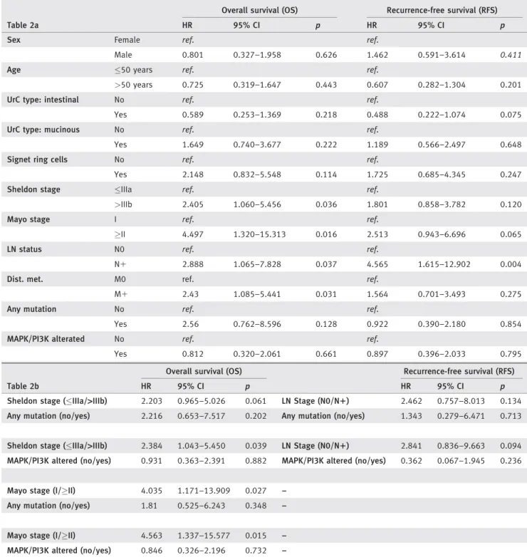
Table 2.Univariable (a) and multivariable (b) Cox-analyses in the total cohort of urachal adenocarcinomas
Table 2a
Overall survival (OS) Recurrence-free survival (RFS)
HR 95% CI p HR 95% CI p
Sex Female ref. ref.
Male 0.801 0.327–1.958 0.626 1.462 0.591–3.614 0.411
Age 50 years ref. ref.
>50 years 0.725 0.319–1.647 0.443 0.607 0.282–1.304 0.201
UrC type: intestinal No ref. ref.
Yes 0.589 0.253–1.369 0.218 0.488 0.222–1.074 0.075
UrC type: mucinous No ref. ref.
Yes 1.649 0.740–3.677 0.222 1.189 0.566–2.497 0.648
Signet ring cells No ref. ref.
Yes 2.148 0.832–5.548 0.114 1.725 0.685–4.345 0.247
Sheldon stage IIIa ref. ref.
>IIIb 2.405 1.060–5.456 0.036 1.801 0.858–3.782 0.120
Mayo stage I ref. ref.
II 4.497 1.320–15.313 0.016 2.513 0.943–6.696 0.065
LN status N0 ref. ref.
N1 2.888 1.065–7.828 0.037 4.565 1.615–12.902 0.004
Dist. met. M0 ref. ref.
M1 2.43 1.085–5.441 0.031 1.564 0.701–3.493 0.275
Any mutation No ref. ref.
Yes 2.56 0.762–8.596 0.128 0.922 0.390–2.180 0.854
MAPK/PI3K alterated No ref. ref.
Yes 0.812 0.320–2.061 0.661 0.897 0.396–2.033 0.795
Table 2b
Overall survival (OS) Recurrence-free survival (RFS)
HR 95% CI p HR 95% CI p
Sheldon stage (IIIa/>IIIb) 2.203 0.965–5.026 0.061 LN Stage (N0/N1) 2.462 0.757–8.013 0.134 Any mutation (no/yes) 2.216 0.653–7.517 0.202 Any mutation (no/yes) 1.343 0.279–6.471 0.713
Sheldon stage (IIIa/>IIIb) 2.384 1.043–5.450 0.039 LN Stage (N0/N1) 2.841 0.836–9.663 0.094 MAPK/PI3K altered (no/yes) 0.931 0.363–2.391 0.882 MAPK/PI3K altered (no/yes) 0.362 0.067–1.945 0.236
Mayo stage (I/II) 4.035 1.171–13.909 0.027 –
Any mutation (no/yes) 1.81 0.525–6.243 0.348 –
Mayo stage (I/II) 4.563 1.337–15.577 0.015 –
MAPK/PI3K altered (no/yes) 0.846 0.326–2.196 0.732 –
OS-related multivariable analysis models were calculated for Sheldon and Mayo stages separately (bothp<0.05 in univariable analysis). As both staging systems include information on status of lymph nodes and distant metastasis, these factors were excluded from OS-related models. For RFS, only lymph node status exhibited apvalues<0.05 in univariable analyses. Therefore, RFS models have been calculated separately including lymph node status.
Abbreviations: HR, hazard ratio, CI, confidence interval, LN, lymph node, Dist. met., distant metastasis.
Tumor Markers and Signatures
Figure 1.Oncoprints of the genetic alterations in UrC. (A) Overview of the genetic alterations detected in the total cohort urachal adenocar- cinomas. (B) A more detailed version including information on sex, histology, PD-L1 expression and genetic alteration type also in the non- adenocarcinoma UrC cases. Gene amplifications (includingERBB2*,n541/70) were evaluated in the first cohort only. Details on the genetic events can be found in Supporting Information Table S2. Abbreviations: NOS, not otherwise specified; SCC, squamous cell carci- noma; TPS, PD-L1 tumor proportion score; UC, urothelial carcinoma; UD, undifferentiated carcinoma. mucinous*: cases with predominant signet ring cell morphology included. [Color figure can be viewed at wileyonlinelibrary.com]
Tumor Markers and Signatures
The first, multi-institutional cohort showed more KRAS mutations (p 50.059). No other significant differences regard- ing molecular alterations between both cohorts were noted. In particular, there were no statistically significant differences in clinico-pathological or in any molecular parameters between mucinous and intestinal type urachal carcinomas.
In 66 of 70 cases (94.3%) tissue for MMR-IHC was avail- able with technical feasibility of IHC of all four proteins (MLH1, MSH2, MSH6, and PMS2) in 61 of 70 cases (87.1%).
The molecular MSI-analyses were feasible in 56 of 70 cases (80%). A negative MMR expression was detected in one case (MSH2). In this and all other cases, all remaining MMR- markers were positive and no MSI-h was detected.
In 63 of 70 cases (90%) sufficient material was available for PD-L1-IHC. In 10 of these 63 cases (15.9%) a specific PD-L1- expression in the tumor cells was detectable (Fig. 1) with a tumor proportion score (TPS) of 1–49% in 9 of 10 cases (90%) and a TPS 50% in one Case (10%). PD-L1-expression was not restricted to intestinal type UrC (3/10; 30%). The majority of PD-L1 expressing cases were found in mucinous type UrC (7/10; 70%) including the only case with high PD-L1-expression (TPS 50%). No obvious clustering of cases with PD-L1- expression and other molecular events was obvious. However, both cases withMET-alterations exhibited PD-L1 expression.
Correlation between genomic alterations and clinical outcomes
Clinicopathological factors were not associated with any genetic event, except for a trend to higher Mayo tumor stage in UrC with MAPK/PI3K-alterations (p 5 0.073). Organ confined dis- ease, that is Sheldon19 Stage IIIA and Mayo20 Stage I, and absence of metastatic disease were univariable predictors of more favorable OS (p50.036,p50.016,p50.037/p50.031) while only absence of lymph node metastasis predicted better RFS (p5 0.004) in univariable analysis. A trend to more favorable RFS was found in intestinal type UrC (p50.075; log-rank test:
p 5 0.068, Supporting Information Fig. S3) and in organ con- fined disease in the Mayo system (p50.056).
Presence of PD-L1 expression was associated with adverse PFS (p 5 0.002; log-rank test), however, there were only three events in the PD-L1 positive group, thus limiting its significance. No such association was detected regarding OS.
In multivariable Cox-analyses for OS and RFS, presence of any mutation or MAPK/PI3K-alteration did not exhibit prognostic influence (Table 2).
Additionally, no correlation was observed between the muta- tional status ofTP53and its IHC staining pattern. A higher rate of TP53 mutation was observed in UrC with signet ring cells (p5 0.05). No other clinicopathological or prognostic associa- tions were noted forTP53and/orKRASmutational status.
Results in non-adenocarcinoma UrC
The results of the four cases of non-adenocarcinoma UrC are included in Figure 1 and Supporting Information Table S2.
Due to the low number of cases, no correlation analyses with clinicopathological data were performed.
Discussion
Systemic therapy is often required in UrC because patients present with advanced stage disease or they progress after initial locoregional intervention. Investigation of the efficacy of chemotherapy has been limited to retrospective series. Due to the rarity of UrC, it is not feasible to perform large pro- spective clinical studies to evaluate the clinical benefit of vari- ous systemic therapies. Molecular alterations, however, may provide the rationale for targeted therapeutic approaches and/or immunotherapy.
The most common genetic alterations in urachal adeno- carcinomas we detected were TP53 mutations (66%). This high number indicates the common role of inactivation of this tumor suppressor gene also in UrC. Although TP53has in the past been considered mostly undruggable, recently multiple efforts of targeting TP53 altered tumors are in development.21
In addition, we found a subset of UrC characterized by dysfunctional and activated oncogenic MAPK- and PI3K- pathways.4,5 As EGFR signaling mainly occurs through these pathways,6 we grouped together all UrC with any pathogenic mutational event in KRAS, NRAS, BRAF, or PIK3CA (n 5 22; 31%). This subset is additionally equivalent to all UrCs with mutations in intracellular signal transduction pathways and was termed ‘MAPK/PI3K-altered’ subset. All these alterations were mutually exclusive.
These findings together with the results of the MMR-/
MSI- and PD-L1-analyses have important therapeutic and ontogenetic implications.
Due to histopathological and certain clinical similarities of UrC and CRC, chemotherapy in advanced UrC has often been adapted from CRC with 5-FU containing approaches.
This has been shown to be more effective compared to regimes used for UC.2,22In accordance with this observation, we did not find evidence of MMR-d/MSI-h in UrC, which leads to a hypermutated phenotype and in CRC has been implicated in decreased 5-FU-efficacy.23 Our single UrC-case with immunohistochemical MSH2-loss seems to represent a false negative result, as MSH6-protein expression was retained which should not be the case in MSH2-deficient tumors.7Additionally, no evidence of MSI-h was detected on the DNA level by microsatellite PCR analyses neither in this nor in any other case.
Another recent and efficient anti-cancer strategy is target- ing tumoral immune-escape mechanisms, in which inhibitors of the PD-1/PD-L1-axis are of particular therapeutic interest.
PD-1/PD-L1 inhibitors have proven efficacy in several types of advanced cancer, including melanoma, non-small-cell lung cancer, renal cell carcinoma and bladder cancer.24 In most studies, cases with immunohistochemical PD-L1-expression on tumor cells and/or tumor-related immune-cells exhibited better responsiveness to PD-1/PD-L1-inhibitors. However, as
Tumor Markers and Signatures
also in tumors without PD-L1-expression positive anti- tumoral effects can be detected, PD-L1-IHC testing is consid- ered complementary with the exception of the companion diagnostic 22C3-test in NSCLC.24 With regard to UrC, these results implicate that PD-1/PD-L1-inhibitors might be effec- tive in advanced disease despite its modest rate of PD-L1- expression (16%). In fact, others have recently reported on a case in UrC with atezolizumab-therapy, an anti-PD-L1- antibody, resulting in stable disease.25 However, as MMR-d/
MSI-h does not seem to be a hallmark of UrC at least in our cohort, immune escape mechanisms employing the PD-1/
PD-L1 axis do not seem to be the defining molecular charac- teristic of UrC. This also has implications for the use of pem- brolizumab in UrC, an anti-PD-1-antibody, which recently gained FDA-approval in MMR-d/MSI-h tumors irrespective of site of origin.
Therefore, targeted therapy approaches are of prominent therapeutic interest in advanced UrC.
Recently, Collazo-Lorduy and colleagues reported a suc- cessful application of cetuximab, an anti-EGFR monoclonal antibody which is used in metastasized CRC, in one UrC patient who was pretreated with chemotherapy. This treat- ment was justified by the presence of an EGFR amplification and absence of aKRASmutation.5
In CRC, it has been shown that MAPK-/PI3K-activation downstream of the EGF receptor is associated with anti- EGFR therapy resistance.26 We detected two cases in our cohort with coexisting EGFR gene amplification and with- out activating MAPK-/PI3K-mutations thus EGFR amplifi- cation is a rare event in UrC. Therefore, it seems reasonable to test for EGFR gene amplifications and acti- vating MAPK/PI3K-pathway mutations in advanced UrC when an anti-EGFR therapy is considered. This approach would be similar to the concept of complementary/com- panion diagnostics in CRC.27
In addition to these therapeutic considerations, the mutational profile of UrC shows similarities to that of CRC. Most of the detected activating MAPK/PI3K-path- way mutations were also common in CRC when compared to The Cancer Genome Atlas (TCGA) data. 28 This is especially prominent in the clustering of codon G12/G13 KRASalterations but also for the remaining KRAS, NRAS, andPIK3CAmutations (Supporting Information Fig. S1).28 Interestingly, bothEGFR amplified cases we detected were intestinal type UrCs.
However, not only similarities but also differences were detected. For example, we identified three non-classical, BRAF mutations (G469A:n51, D594G:n52) adding to the four classical V600E mutations reported previously.4 While the G469A BRAF mutation was relatively common in cancer, the D594G variant has rarely been detected in cancer. Interestingly, although being located in the BRAF- kinase region, the D594G variant has been described in a case of metastatic CRC responding to cetuximab.27
In addition, a comparison with TCGA data for CRC revealed lower frequencies of almost all analyzed genetic alterations in UrC with the exception of TP53 mutations.
(Supporting Information Fig. S4).28 However, although cer- tain differences to CRC are apparent, the disparity to UC is more pronounced (Supporting Information Fig. S4). This notion is further supported by the very low incidence of Tel- omerase reverse transcriptase (TERT) promoter mutations in urachal adenocarcinomas (4%), which is otherwise a very common event in UC but not in CRC.29–31Interestingly, our findings regarding MMR-d/MSI-h in UrC are more compara- ble to figures in UC32than in CRC.23,28Regarding the rate of PD-L1-expression, UrC (16%) seems to be in the lower range of figures reported for UC (14–28%)33 and CRC (9–
43%).34,35Taken together, UrC appears to be a distinct entity on the genomic level with closer resemblance to CRC than to UC. Further analyses are needed to address this issue while the present study was not primarily designed from an onto- genetic point of view.
In addition to alterations related to intracellular signal transduction pathways, we also detected cases with other pathogenic and possibly targetable genetic events. For exam- ple, pathogenic FGFR1, MET, and PDGFRA mutations and MET- and ERBB2-gene amplifications were detected in one case each, possibly identifying therapeutic targets in these patients.
Interestingly, both UrC-cases with MET-alterations exhib- ited PD-L1-expression and first (pre-)clinical evidence of the relationship between PD-L1 upregulation and MET-altera- tions is emerging.36 Moreover, one case with high level ERBB2 gene amplification and HER2-protein expression was detected (Supporting Information Fig. S2). HER2 is another member of the EGFR family and a well-known therapeutic target in breast and gastric cancer and under clinical investi- gation in other cancers such as non-small cell lung cancer.37–39
Our analysis has certain limitations. Due to the rarity of the disease, prospective analyses can hardly be conducted.
We therefore chose a retrospective multi-institutional approach to maximize our sample size. In addition, we ana- lyzed a first multi-institutional, international cohort and a second national Dutch cohort at different sites. Through this approach we expected to gain a more comprehensive and homogenous estimation of genetic events in UrC. This approach holds, however, the disadvantage of analytical dif- ferences between laboratories. This applies to the use of MiSeq and HiSeq-platforms at the different sites. While the enhanced capabilities and sensitivity of the HiSeq-platform are accepted, both sites were in contact to obtain best compa- rable analytic data. Although our cohort is the largest ana- lyzed in the literature, the case number remains low, which precludes adequate statistical power for some sub-group anal- yses. This is also important when considering the results of survival analyses as inevitably the samples were collected from patients with a range of different stages and treatments
Tumor Markers and Signatures
confounding the possible survival impact of genetic altera- tions. Additionally, the employed gene panel was of modest size which has to be kept in mind in regard to the patients where no genomic aberration was detectable.
In conclusion, we have analyzed the largest cohort of UrC to date in an international, multi-institutional targeted sequencing approach supplemented within situhybridization and protein expression analyses including evaluation of PD- L1 expression and MMR/MSI-status. Although MMR-d/MSI- h does not seem to be a hallmark of UrC, anti-PD-1/PD-L1 immune therapy might be a potential therapeutic option given the PD-L1 expression rate in UrC. In particular, our results underline the role of MAPK/PI3K-pathway dysregula- tion which was detected in 31% of urachal adenocarcinomas.
This has important implications for possible anti-EGFR ther- apy in UrC and suggests that targeted testing of MAPK/
PI3K-pathway alterations would be reasonable in patients with advanced UrC before considering anti-EGFR therapy. In addition, further potentially druggable alterations were found inERBB2 (HER2),MET,FGFR1, and PDGFRAin a subset of patients. Our data support the notion of UrC being a distinct entity on the genomic level with closer resemblance to CRC than to UC.
Acknowledgments
We appreciate the skillful work of Mrs. Gabriele Ladwig, Mrs. Isabel Albertz, Mrs. Andrea Kutritz and Mrs. Dorothe M€ollmann. Dr. Nathalie Rioux- Leclercq is thanked for her help in retrieving samples. We thank the Core Facility for Molecular Pathology & Bio-banking of the Netherlands Cancer Institute for their assistance. Tibor Szarvas was supported by Janos Bolyai Research Scholarship of the Hungarian Academy of Sciences. This work was supported by the National Research, Development and Innovation Office – NVKP_16-1–2016-004.
References
1. Schubert GE, Pavkovic MB, Bethke-Bed€urftig BA.
Tubular urachal remnants in adult bladders.
J Urol.1982; 127:40–2.
2. Szarvas T, Modos O, Niedworok C, et al. Clinical, prognostic, and therapeutic aspects of urachal carcinoma-A comprehensive review with meta- analysis of 1,010 cases.Urol Oncol.2016; 34:388–98.
3. Behrendt MA, DE Jong J, Van Rhijn BW. Ura- chal cancer: Contemporary review of the patho- logical, surgical, and prognostic aspects of this rare disease.Minerva Urol Nefrol.2016; 68:
172–84.
4. Modos O, Reis H, Niedworok C, et al. Mutations of KRAS, NRAS, BRAF, EGFR, and PIK3CA genes in urachal carcinoma: Occurence and prognostic significance.Oncotarget.2016; 7:
39293–301.
5. Collazo-Lorduy A, Castillo-Martin M, Wang L, et al. Urachal carcinoma shares genomic altera- tions with colorectal carcinoma and may respond to epidermal growth factor inhibition.Eur Urol.
2016; 70:771–5.
6. Normanno N, De Luca A, Bianco C, et al. Epi- dermal growth factor receptor (EGFR) signaling in cancer.Gene.2006; 366:2–16.
7. Cha S, Lee J, Shin JY, Kim JY, Sim SH, Keam B, Kim TM, Kim DW, Heo DS, Lee SH, Kim JI.
Clinical application of genomic profiling to find druggable targets for adolescent and young adult (AYA) cancer patients with metastasis.BMC Cancer.2016; 16:170
8. Singh H, Liu Y, Xiao X, et al. Whole exome sequencing of urachal adenocarcinoma reveals recurrent NF1 mutations.Oncotarget.2016; 7:
29211–5.
9. Hang JF, Pan CC. Absence of GNAS and BRAF mutations but presence of KRAS mutation in urachal adenocarcinoma.Pathology.2017; 49:
316–7.
10. Loh KP, Mondo E, Hansen EA, Sievert L, Fung C, Sahasrabudhe DM, Guancial E. Targeted ther- apy based on tumor genomic analyses in meta- static urachal carcinoma.Clin Genitourin Cancer.
2016; 14:e449–50.
11. Sirintrapun SJ, Ward M, Woo J, et al. High-stage urachal adenocarcinoma can be associated with microsatellite instability and KRAS mutations.
Hum Pathol.2014; 45:327–30.
12. Toubaji A, Jordan EJ, Desai N, et al. Genitouri- nary Pathology (including Renal tumors).Mod Pathol.2017; 30:262A
13. Lopez-Beltran A, Paner G, Tsuzuki T. Urachal carcinoma In: Moch H, Humphrey PA, Ulbright TM, Reuter VE. World Health Organization Clas- sification of Tumours of the Urinary System and Male Genital Organs. 4th ed. Lyon: IARC, 2016:
113–14
14. Gopalan A, Sharp DS, Fine SW, et al. Urachal carcinoma: A clinicopathologic analysis of 24 cases with outcome correlation.Am J Surg Pathol.
2009; 33:659–68.
15. Paner GP, Barkan GA, Mehta V, et al. Urachal carcinomas of the nonglandular type: Salient fea- tures and considerations in pathologic diagnosis.
Am J Surg Pathol.2012; 36:432–42.
16. Reis H, Krafft U, Niedworok C, et al. Biomarkers in urachal cancer and adenocarcinomas in the bladder: A comprehensive review supplemented by own data.Dis Markers.2018; “in press”
17. Gao J, Aksoy BA, Dogrusoz U, et al. Integrative analysis of complex cancer genomics and clinical profiles using the cBioPortal.Sci Signal.2013;
6:pl1
18. Cerami E, Gao J, Dogrusoz U, et al. The cBio cancer genomics portal: An open platform for exploring multidimensional cancer genomics data.Cancer Discov.2012; 2:401–4 [Database.
19. Sheldon CA, Clayman RV, Gonzalez R, et al.
Malignant urachal lesions.J Urol.1984; 131:1–8.
20. Ashley RA, Inman BA, Sebo TJ, et al. Urachal carcinoma: Clinicopathologic features and long- term outcomes of an aggressive malignancy.Can- cer.2006; 107:712–20.
21. Parrales A, Iwakuma T. Targeting oncogenic mutant p53 for cancer therapy.Front Oncol.
2015; 5:288
22. Nyirady P, Niedworok C, Reis H, et al. Clinical sequencing-guided therapy of urachal carcinoma:
New perspective for a rare cancer.Eur Urol.
2016; 70:776–7.
23. Boland CR, Goel A. Microsatellite instability in colorectal cancer.Gastroenterology.2010; 138:
2073–87. e3
24. Yu Y. Molecular classification and precision ther- apy of cancer: Immune checkpoint inhibitors.
Front Med.2017; [Epub ahead of print]
25. Kardos J, Wobker SE, Woods ME, et al. Compre- hensive molecular characterization of urachal adenocarcinoma reveals commonalities with colo- rectal cancer, including a hypermutable pheno- type.JCO Precision Oncol.2017; 1–12.
26. Therkildsen C, Bergmann TK, Henrichsen- Schnack T, et al. The predictive value of KRAS, NRAS, BRAF, PIK3CA and PTEN for anti-EGFR treatment in metastatic colorectal cancer: A sys- tematic review and meta-analysis.Acta Oncol.
2014; 53:852–64 [Database.
27. De Roock W, Claes B, Bernasconi D, et al. Effects of KRAS, BRAF, NRAS, and PIK3CA mutations on the efficacy of cetuximab plus chemotherapy in chemotherapy-refractory metastatic colorectal cancer: A retrospective consortium analysis.Lan- cet Oncol.2010; 11:753–62.
28. The Cancer Genome Atlas Networkk.
Comprehensive molecular characterization of human colon and rectal cancer.Nature.2012; 487:330–7.
29. Thiem S, Herold T, Krafft U, et al. Telomerase reverse transcriptase (TERT) promoter mutations are rare in urachal cancer.Pathol Int.2017; 67:597–601.
30. Wu S, Huang P, Li C, et al. Telomerase reverse transcriptase gene promoter mutations help discern the origin of urogenital tumors: A genomic and molecular study.Eur Urol.2014;
65:274–7.
31. Vinagre J, Almeida A, Populo H, , et al. Fre- quency of TERT promoter mutations in human cancers.Nat Commun.2013; 4:2185
32. Iyer G, Audenet F, Middha S, et al. Mismatch repair (MMR) detection in urothelial carcinoma (UC) and correlation with immune checkpoint blockade (ICB) response.Journal of Clinical Oncology.2017; 35:4511
33. Tretiakova M, Fulton R, Kocherginsky M, et al.
Concordance study of PD-L1 expression in pri- mary and metastatic bladder carcinomas: Com- parison of four commonly used antibodies and RNA expression.Mod Pathol.2017; [Epub ahead of print]
34. Rosenbaum MW, Bledsoe JR, Morales-Oyarvide V, et al. PD-L1 expression in colorectal cancer is associated with microsatellite instability, BRAF mutation, medullary morphology and cytotoxic tumor-infiltrating lymphocytes.Mod Pathol.
2016; 29:1104–12.
Tumor Markers and Signatures
35. Liu R, Peng K, Yu Y, et al. Prognostic value of immunoscore and PD-L1 expression in metastatic colorectal cancer patients with different RAS sta- tus after palliative operation.BioMed Res Int.
2018; 2018:8
36. Balan M, Mier y Teran E, Waaga-Gasser AM, et al. Novel roles of c-Met in the survival of renal cancer cells through the regulation of HO-1 and PD-L1 expression.J Biol Chem.2015; 290:
8110–20.
37. Bang YJ, Van Cutsem E, Feyereislova A, et al.
Trastuzumab in combination with chemotherapy versus chemotherapy alone for treatment of HER2-positive advanced gastric or gastro- oesophageal junction cancer (ToGA): A phase 3, open-label, randomised controlled trial.Lancet.
2010; 376:687–97.
38. Slamon DJ, Leyland-Jones B, Shak S, et al. Use of chemotherapy plus a monoclonal antibody against HER2 for metastatic breast cancer that
overexpresses HER2.N Engl J Med.2001; 344:
783–92.
39. Reis H, Herold T, Ting S, et al. HER2 expression and markers of phosphoinositide-3-kinase path- way activation define a favorable subgroup of metastatic pulmonary adenocarcinomas.Lung Cancer.2015; 88:34–41.
40. The Cancer Genome Atlas Research Network.
Comprehensive molecular characterization of urothelial bladder carcinoma.Nature.2014; 507:315–22.