SURVEY OF LUNGWORM INFECTION OF DOMESTIC CATS IN HUNGARY
Sára KISZELY1, Mónika GYURKOVSZKY1, Norbert SOLYMOSI2 and Róbert FARKAS1*
1Department of Parasitology and Zoology and 2Centre for Bioinformatics, University of Veterinary Medicine, István u. 2, H-1078 Budapest, Hungary
(Received 15 May 2019; accepted 31 July 2019)
From 61 settlements of 12 Hungarian counties, 303 domestic cats were in- cluded in this survey. Between autumn 2016 and spring 2018, fresh faecal sam- ples were randomly collected and examined by flotation and by the Baermann–
Wetzel method for the presence of lungworm infection. No eggs of Eucoleus aer- ophilus were detected. Morphological identification of first instar larvae (L1) was also carried out. In the faeces of 60 cats (19.8%) from 17 settlements and Buda- pest, L1 of Aelurostrongylus abstrusus were found. More than half of the cats were from the western part of the country. The average number of larvae per gram of faeces was 190.2 ± 304.88. These results are in line with the former findings on the prevalence of aelurostrongylosis of domestic cats in Hungary. In addition, Oslerus rostratus was also found for the first time in the faecal samples of three cats from the eastern part of the country, infected also with Ae. abstrusus. The av- erage age (2.51 ± 1.26 years) of infected cats indicates that lungworm infection is more common among younger cats. No relationship was found between the lung- worm infection and the sex of cats. Non-neutered cats had a significantly higher proportion of lungworm infections. Two-thirds of the infected cats were apparent- ly healthy, and only 19 individuals showed clinical signs of respiratory disorders.
Key words: Lungworms, Aelurostrongylus abstrusus, Oslerus rostratus, cat, survey, Hungary
Lungworm infection of domestic and wild cats occurs in many countries around the world. It is most commonly caused by Aelurostrongylus abstrusus, Railliet, 1898 (Strongylida, Angiostrongylidae), called the cat lungworm (Ander- son, 2000; Bowman, 2000). The 5- to 10-mm-long adults reside in the alveolar duct and the terminal bronchioles of the lungs. Over the past decade, other lung- worm species of domestic cats such as Troglostrongylus brevior and T. sub- crenatus (Strongylida, Crenosomatidae), Oslerus rostratus (syn. Anafilaroides rostratus) (Strongylida, Filaroididae) and Eucoleus aerophilus (syn. Capillaria aerophila) (Enoplida, Trichuridae) have also been reported (Traversa et al., 2009;
Jefferies et al., 2010; Brianti et al., 2012, 2014b; Di Cesare et al., 2014; Giannelli
*Corresponding author; E-mail: farkas.robert@univet.hu; Phone: 0036 (01) 478-4188
et al., 2017). There have been several cases when two or more lungworm species occurred concurrently in cats (Juste et al., 1992; Jefferies et al., 2010; Traversa et al, 2014; Daikou et al., 2015; Varcasia et al., 2015). With the exception of E.
aerophilus, the other species develop indirectly with gastropod intermediate hosts. Cats can be infected by intermediate hosts or various other animals (e.g.
small mammals, lizards, frogs) which consumed slugs and snails acting as par- atenic hosts (Anderson, 2000; Bowman, 2000; Pennisi et al., 2015). Depending on the parasite species, the degree of infection and the immunological status of the host, the parasitosis can be asymptomatic or the cats may show mild to severe clinical signs (coughing, sneezing, tachypnoea). Heavy infection can even be fatal (Traversa et al., 2010; Traversa and Di Cesare, 2013; Philbey et al., 2014; Pen- nisi et al., 2015).
Data on the distribution and prevalence of cat lungworm infection in Hun- gary are scarce. In a postmortem examination of 57 stray cats originating from 9 hunting areas of the country C. aerophila worms were found in 14 cadavers (Takács and Takács, 2002), Ae. abstrusus was detected by autopsy (Kávai, 1977;
Dobos-Kovács, 1981; Takács and Takács, 2002) or faecal examination (Capári et al., 2013).
The objectives of this investigation were to enhance the knowledge on the distribution and risk factors of lungworm infection of domestic cats in Hungary.
Materials and methods
Sampling
Altogether 303 cats from 61 settlements of 12 counties (Table 1 and Fig. 1) were randomly included in the survey. Most cats (n = 269) had owners, while the remainder were kept in temporary accommodation. Between autumn 2016 and spring 2018 fresh faecal samples were collected once. Sampling was done with the owners’ permission and assistance.
The approx. 5-gram samples per cat stored in plastic jars having individual numbers were usually taken to the laboratory on the same day or 2–3 days later after storing them in a fridge. Simultaneously, a questionnaire was completed for each cat (e.g. breed, age, sex, keeping, presence/absence of respiratory signs, last anthelmintic treatment).
Test method
The E. aerophilus eggs were examined by the flotation method using zinc sulphate solution. The lungworm larvae (L1) were isolated by Baermann–Wetzel method (Giannelli et al., 2015). One gram of faecal sample packed in a 10 × 10 cm double-layered gauze was kept in a glass filled with lukewarm water for 24 h.
Table 1
The number of cats sampled per site (N), the number (N) of cats infected with Aelurostrongylus abstrusus and Oslerus rostra- tus and the number of infected cats with respiratory signs (e.g. frequent sneezing, running nose)
Site N Ae. abstrusus (N) O. rostratus (N) Respiratory signs (N)
Alsózsolca 1 0 0 0 Badacsonytomaj 1 1 0 1 Balatonalmádi 5 0 0 0 Bedő 6 6 0 4 Békés 3 0 0 0 Békéscsaba 7 0 0 0 Bőcs 2 0 0 0 Budaörs 3 1 0 0 Budapest 40 1 0 0 Csatár 2 0 0 0 Csopak 4 0 0 0 Debrecen 13 0 0 0 Debrecen-Józsa 3 1 0 1 Diósd 1 0 0 0 Dömsöd 5 0 0 0 Dunakeszi 2 0 0 0 Érd 13 2 0 0 Erdőbénye 4 0 0 0 Gesztely 3 0 0 0 Gödöllő 4 0 0 0 Gyula 3 0 0 0 Hajdúböszörmény 17 2 2 0 Hajdúdorog 9 0 0 0 Hajdúszoboszló 1 0 0 0 Herceghalom 1 0 0 0 Hernádkak 6 0 0 0 Hernádkak-Belegrád 9 7 1 6 Hernádnémeti 2 0 0 0 Hódmezővásárhely 4 0 0 0 Ikrény 1 0 0 0 Iváncsa 1 1 0 0 Kazincbarcika 2 0 0 0 Kistarcsa 1 0 0 0 Komárom 1 0 0 0 Kőröshegy 3 0 0 0 Mezőberény 2 0 0 0 Miskolc 8 3 0 1 Nyíregyháza 6 0 0 0 Óhíd 15 13 0 0 Okány 1 0 0 0 Orosháza 3 0 0 0 Sárospatak 6 0 0 0 Sátoraljaújhely 4 0 0 0 Sümeg 7 6 0 3 Sümegcsehi 2 2 0 0 Szeged 10 0 0 0 Szentendre 3 0 0 0 Szentimrefalva 8 6 0 2 Tolcsva 2 0 0 0 Tura 1 0 0 0 Ukk 4 4 0 1 Vác 7 0 0 0 Vámosszabadi 1 0 0 0 Vésztő 4 1 0 0 Vilmány 3 0 0 0 Zalaegerszeg 10 0 0 0 Zalagyömörő 2 2 0 0 Zalaszegvár 1 1 0 0 Zalaszentiván 4 0 0 0 Zalaszentlőrinc 2 0 0 0 Zamárdi 4 0 0 0
After 5 min of centrifugation of the liquid at 600 rpm, the morphological identi- fication of larvae found in the sediment was carried out based on the published descriptions (Traversa and Di Cesare, 2016). The body length and the morpholo- gy of head and tail end were considered (Table 2). The identification of O.
rostratus L1 was confirmed with PCR by Alessio Giannelli in Bari, Italy (per- sonal information). The number of larvae per one gram of faeces (LPG) was de- termined.
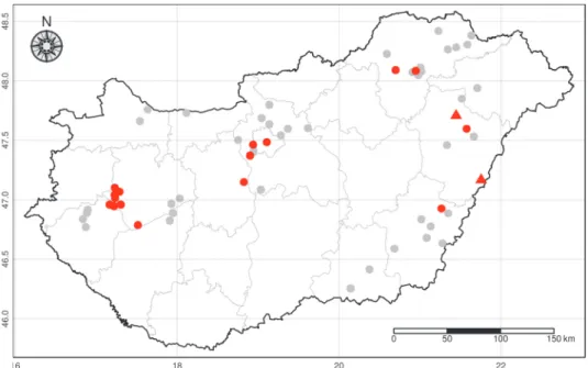
Fig. 1. Negative sampling sites (grey dots), sites of Aelurostrongylus abstrusus (red dots) and cats dually infected with Ae. abstrusus and Oslerus rostratus (red triangles)
Statistical evaluation
The independence of infection from age group, sex, and keeping mode was analysed by Fisher’s exact test. We used propensity score-based pairing to form sample pairs (Dinya and Solymosi, 2016). A limit value of P < 0.05 was used to evaluate the results. All analyses were performed in R environment (R Core Team, 2019).
Results
No E. aerophilus eggs occurred in the samples.
There were Ae. abstrusus L1 in the faeces of 60 cats (19.8%; 95% CI: 15.71–
24.65) living in 17 settlements and one kept in Budapest (Table 1 and Fig. 1). The LPG values showed significant differences, with an average number of 190.2 ±
304.9. Oslerus rostratus L1 were also found in the faecal samples of three cats infected with Ae. abstrusus. All infected cats belonged to the European short- haired breed, most of them (47/60, 78.33%; 95% CI: 66.38–86.87) lived out- doors, and 11 (18.33%; 95% CI: 10.56–29.92) stayed both indoors and outdoors.
Only two out of 74 animals kept indoors had lungworm larvae in their samples (2.7% 95% CI: 0.74–9.33). Significantly more animals which lived outdoors or both indoors and outdoors were infected than those kept exclusively indoors (OR: 12.15, 95% CI: 3.07–105.49, P < 0.001). The average age of the infected cats was 2.51 ± 1.26 years, the youngest and the oldest was 8 months and 6 years old, respectively. There was no significant difference (P = 0.5916) between the infection rate of cats younger than one year of age and 1- to 5-year-old cats. The animals under one year and between 1 and 5 years old had a higher risk of lung- worm infection than cats older than 5 years (OR: 7.85, 95% CI: 1.93–69.17, P <
0.001 and OR: 6.16, 95% CI: 1.24–60.20, P = 0.0157). The proportion of infect- ed males (34/178, 19.1%, 95% CI: 14.0–25.5) and females (26/125, 20.8%, 95%
CI: 14.61–28.73) did not differ significantly. A significantly higher proportion of non-neutered cats were infected with lungworms (OR: 2.98, 95%CI: 1.49–6.32, P < 0.001). When propensity score-based matching was applied, in order to have a balanced population (n = 120) for free range and neutering, the effect of young age remained (OR: 3.83, 95% CI: 1.09–17.22, P = 0.034). Nearly one-third of the infected animals (19/60, 31.67%, 95% CI: 21.31–44.23) showed respiratory signs (e.g. frequent sneezing, running nose) but their number did not differ significant- ly (P > 0.05) from those of infected cats not showing clinical signs.
Discussion
The results of this study are in line with previous findings showing that Ae. abstrusus infection of domestic cats is fairly prevalent in Hungary (Kávai, 1977; Dobos-Kovács, 1981; Takács and Takács, 2002; Capári et al., 2013). The occurrence of aelurostrongylosis in new regions can be considered a new finding.
In this survey the prevalence of this parasitosis was 19.8%. In the previous pathological or faecal examinations a lower rate of infections was detected: Ae.
abstrusus worms were found only in 2 out of 50 cats (4%) (Kávai, 1977) and in one out of 57 stray cats (1.7%) (Takács and Takács, 2002) at autopsy. In a para- sitological survey of 235 domestic cats carried out in the western part of Hunga- ry, the prevalence of aelurostrongylosis was 14.5% (Capári et al., 2013). In the present study, more than half of the infected animals (n = 35/60, 58.3%) lived in that region. Further studies could answer the question whether the endemic oc- currence of this parasitosis in that region is related to environmental and weather conditions more favourable for the intermediate hosts, which are also assumed in other parasitic infections (Patz et al., 2000; Morgan et al., 2009). Lungworm in-
fections of cats mostly caused by Ae. abstrusus have been found in several Euro- pean countries where different prevalence values were reported, e.g. Italy: 8.1–
17.8% (Traversa et al., 2008; Di Cesare et al., 2015; Giannelli et al., 2015, 2017), Portugal: 6.25–17.4% (Payo-Puente et al., 2008; Waap et al., 2014; Giannelli et al., 2017), Spain: 1.7–5% (Miró et al., 2004; Giannelli et al., 2017); Denmark:
8.86–13.6% (Olsen et al., 2015; Hansen et al., 2017). By examining more faecal samples collected during 2–3 consecutive days, it may have been possible to find a higher prevalence of lungworm infection than obtained in our study, as the in- termittent discharge of larvae was observed in chronically infected cats (Ribeiro and Lima, 2001; Payo-Puente et al., 2008). Recent European studies have report- ed that at least one of 10 cats is exposed to lungworm infections, the incidence of this parasitosis being more common on the continent than previously thought. In this context, it was noted that lungworm infection of cats is not regarded as a growing threat because there are no previous data to be used for comparison (Giannelli et al., 2017). The authors assumed that the lower occurrence of infect- ed cats found in the western part of Europe can be explained by the more fre- quent application of anthelmintic drugs.
Table 2
Morphometric features of first-stage larvae of Aelurostrongylus abstrusus, Troglostrongylus brevior, T. subcrenatus and Oslerus rostratus
Species Length (μm) Head Tail
Aelurostrongylus abstrusus 360–410 rounded, terminal
oral opening kinked (S shaped), distinct knob-like or small finger-like
projections Troglostrongylus brevior 300–360 pointed, subterminal
oral opening
gradually tapered to the extremity, deep dorsal and
a shallower ventral incisure Troglostrongylus subcrenatus 270–300 pointed, subterminal
oral opening
gradually tapered to the extremity, deep dorsal and
a shallower ventral incisure Oslerus rostratus 330–410 central oral opening,
surrounded by a cuticular ring with
dorsal and ventral prominences
with a constriction anterior to the end
and tip with a kinked appearance
This is a first record for Hungary that in the present study O. rostratus was also found in three cats (in the form of co-infection with Ae. abstrusus). The 30- to 40-mm-long adults of O. rostratus were mostly found in the bronchial submu- cosa and peribronchial tissues of wild cats and lynx (Brianti et al., 2014a). Since the first description of this nematode species (Gerichter, 1949), it has also been found in domestic cats of some European countries, which may have been in- fected accidentally (Juste et al., 1992; Millán and Casanova, 2009; Brianti et al., 2014a; Varcasia et al., 2015; Giannelli et al., 2017). Further studies could reveal how three cats kept in two settlements in the eastern part of the country became infected. One of the possible reasons for the rare occurrence of O. rostratus in domestic cats is that the living space of wild cats is narrowing, and their number is constantly decreasing in the country (Biró et al., 2009). Except for two infect- ed cats the third one lived outdoors or both indoors and outdoors. A similar result has been reported by others, suggesting that the living conditions have a decisive influence on the infection of animals, since the consumption of naturally infected intermediate or paratenic hosts are necessary for lungworm infections (Genchi et al., 2014; Giannelli et al., 2017). The two cats that were living indoors had prob- ably become infected before they were switched to indoor keeping.
The average age of infected cats (2.51 ± 1.26 years) indicates that lung- worm infection is more common among younger cats. Examining the infection rate of 303 animals divided into three age groups, there was a significantly lower risk of infection among animals above 5 years of age due to the acquired immun- ity (Pennisi et al., 2015). In other studies, there was no difference between the in- fection rates of the different age groups (Genchi et al., 2014; Tamponi et al., 2014). Some authors have reported that there is a higher risk of infection among young animals (Traversa et al., 2010; Barutzki and Schaper, 2013; Di Cesare et al., 2013). According to other authors, lungworms are more common among adult cats (Mircean et al., 2010; Knaus et al., 2014). We agree with those re- searchers who say that cats can be infected with lungworms at any age if they can reach the infective intermediate or paratenic hosts in the environment (Beug- net et al., 2014; Giannelli et al., 2015).
No relationship was found between lungworm infection and the sex of an- imals, as the parasitosis occurred in almost the same proportion in males and fe- males, as opposed to other findings (Traversa et al., 2008; Tamponi et al., 2014).
We agree with Traversa et al. (2008) and Genchi et al. (2014) who reported that the sex of cats did not affect their infection rate. Non-neutered cats had a signifi- cantly higher proportion of lungworm infections, which is probably due to a de- crease in their activity after the spaying operation.
Two-thirds of the infected cats were free of clinical signs, and only 19 showed respiratory signs. Italian researchers reported that more than 50% of an- imals infected with Ae. abstrusus had clinical signs (Traversa et al., 2008; Gen- chi et al., 2014; Di Cesare et al., 2015). The question arises whether or not there
is a correlation between LPG values and the occurrence of clinical signs. Contra- dictory data have been reported so far. There were scientists who did not find that LPG values were significantly higher in cats with clinical signs (Traversa and Di Cesare, 2016). Others have reported that the development of clinical signs results from the massive egg production of worms and the lesions produced in the lungs mainly by L1. Therefore, higher levels of LPG are found in such indi- viduals (Naylor et al., 1984; Gerdin et al., 2011; Genchi et al., 2014).
The knowledge regarding lungworm infection of domestic cats living in Hungary has been expanded with the results of the current studies. However, the following questions have arisen, which need to be answered by further studies:
What is the frequency of the parasitosis caused by O. rostratus in domestic cats?
Do Troglostrongylus spp. occur in the country? What are the most common in- termediate hosts of the nationwide distributed Ae. abstrusus? Can the geograph- ical distribution of cat lungworms be influenced by the climate change in the country?
Acknowledgements
We wish to thank the veterinarians involved in this study for their kind collabora- tion and all pet owners who consented to their pets’ involvement. This research was sup- ported partly by the 17896-4/2018/FEKUTSTRAT grant of the Hungarian Ministry of Human Capacities.
References
Anderson, R. C. (2000): The superfamily Metastrongyloidea. In: Anderson, R. C. (ed.) Nematode Parasites of Vertebrates. Their Development and Transmission. CABI, Wallingford, UK.
pp. 129–229.
Barutzki, D. and Schaper, R. (2013): Occurrence and regional distribution of Aelurostrongylus ab- strusus in cats in Germany. Parasitol. Res. 112, 855–861.
Beugnet, F., Bourdeau, P., Chalvet-Monfray, K., Cozma, V., Farkas, R., Guillot, J., Halos, L., Joa- chim, A., Losson, B., Miró, G., Otranto, D., Renaud, M. and Rinaldi, L. (2014): Parasites of domestic owned cats in Europe: co-infestations and risk factors. Parasit. Vectors 7, 291.
Biró, Zs., Szemethy, L. and Heltai, M. (2009): The wild cat: tribulations of an endangered predator 1. Current status and sources of risk [in Hungarian]. Vadászkutya [Hunting Dog] 3, 50–52.
Bowman, D. D. (2000): Respiratory system parasites of the dog and cat. Part I. Nasal mucosa and sinuses, and respiratory parenchyma. In: Bowman, D. D. (ed.) Companion and Exotic An- imal Parasitology. International Veterinary Information Service (www.ivis.org).
Brianti, E., Gaglio, G., Giannetto, S., Annoscia, G., Latrofa, M. S., Dantas-Torres, F., Traversa, D.
and Otranto, D. (2012): Troglostrongylus brevior and Troglostrongylus subcrenatus (Strongylida: Crenosomatidae) as agents of broncho-pulmonary infestation in domestic cats. Parasit. Vectors 5, 178.
Brianti, E., Gaglio, G., Napoli, E., Falsone, L., Giannelli, A., Annoscia, G., Varcasia, A., Giannet- to, S., Mazzullo, G. and Otranto, D. (2014a): Feline lungworm Oslerus rostratus (Stron- gylida: Filaridae) in Italy: first case report and histopathological findings. Parasitol. Res.
113, 3853–3857.
Brianti, E., Giannetto, S., Dantas-Torres, F. and Otranto, D. (2014b): Lungworms of the genus Troglostrongylus (Strongylida: Crenosomatidae): Neglected parasites for domestic cats.
Review. Vet. Parasitol. 202, 104–112.
Capári, B., Hamel, D., Visser, M., Winter, R., Pfister, K. and Rehbein, S. (2013): Parasitic infec- tions of domestic cats, Felis catus, in western Hungary. Vet. Parasitol. 192, 33–42.
Daikou, A., Di Cesare, A., Barros, L. A., Morelli, S., Halos, L., Beugnet, F. and Traversa, D.
(2015): Occurrence of Aelurostrongylus abstrusus and Troglostrongylus brevior in domes- tic cats in Greece. Parasit. Vectors 8, 590.
Di Cesare, A., Frangipane di Regalbono, A., Tessarin, C., Seghetti, M., Iorio, R., Simonato, G. and Traversa, D. (2014): Mixed infection by Aelurostrongylus abstrusus and Troglostrongylus brevior in kittens from the same litter in Italy. Parasitol. Res. 113, 613–618.
Di Cesare, A., Veronesi, F., Grillotti, E., Manzocchi, S., Perrucci, S., Beraldo, P., Cazzin, S., De Liberato, C., Barros, L. A., Simonato, G. and Traversa, D. (2015): Respiratory nematodes in cat populations of Italy. Parasitol. Res. 114, 4463–4469.
Di Cesare, A., Crisi, P. E., Di Giulio, E., Veronesi, F., Frangipane di Regalbono, A., Talone, T. and Traversa, D. (2013): Larval development of the feline lungworm Aelurostrongylus ab- strusus in Helixaspersa. Parasitol. Res. 112, 3101–3108.
Dinya, E. and Solymosi, N. (2016): Biometrics in Clinics 2. Solving Tasks in R Environment [in Hungarian]. Medicina, Budapest, ISBN 978 963 226 574 2.
Dobos-Kovács, M. (1981): On the pathological alterations of lungworm infestation of cats caused by Aelurostrongylus abstrusus [in Hungarian, with English abstract]. Magy. Allatorvosok 36, 552–556.
Genchi, M., Ferrari, N., Fonti, P., De Francesco, I., Piazza, C. and Viglietti, A. (2014): Relation be- tween Aelurostrongylus abstrusus larvae excretion, respiratory and radiographic signs in naturally infected cats. Vet. Parasitol. 206, 182–187.
Gerdin, J. A., Slater, M. R., Makolinski, K. V., Looney, A. L., Appel, L. D., Martin, N. M. and McDonough, S. P. (2011): Post-mortem findings in 54 cases of anesthetic-associated death in cats from two spay-neuter programs in New York State. J. Feline Med. Surg. 13, 959–966.
Gerichter, C. B. (1949): Studies on the nematodes parasitic in the lungs of Felidae in Palestine.
Parasitol. 39, 251–262.
Giannelli, A., Brianti, E., Varcasia, A., Colella, V., Tamponi, C., Di Paola, G., Knaus, M., Halos, L., Beugnet, F. and Otranto, D. (2015): Efficacy of Broadline® spot-on against Aeluro- strongylus abstrusus and Troglostrongylus brevior lungworms in naturally infected cats from Italy. Vet. Parasitol. 209, 273–277.
Giannelli, A., Capelli, G., Joachim, A., Hinney, B., Losson, B., Kirkova, Z., René-Martellet, M., Papadopoulos, E., Farkas, R., Napoli, E., Brianti, E., Tamponi, C., Varcasia, A., Alho, A.
M., Madeira De Carvalho, L., Cardoso, L., Maia, C., Mircean, V., Mihalca, A. D., Miró, G., Schnyder, M., Cantacessi, C., Colella, V., Cavalera, M. A., Latrofa, M., S., Annoscia, G., Knaus, M., Halos, L., Beugnet, F. and Otranto, D. (2017): Lungworms and gastrointes- tinal parasites of domestic cats: a European perspective. Int. J. Parasitol. 47, 517–528.
Hansen, A. P., Skarbye, L. K., Vinther, L. M., Willesen, J. L., Pipper, C. B., Olsen, C. S. and Mejer, H. (2017): Occurrence and clinical significance of Aelurostrongylus abstrusus and another endoparasites in Danish cats. Vet. Parasitol. 234, 31–39.
Jefferies, R., Vrhovec, M. G., Wallner, N. and Catalan, D. R. (2010): Aelurostrongylus abstrusus and Troglostrongylus sp. (Nematoda: Metastrongyloidea) infections in cats inhabiting Ibi- za, Spain. Vet. Parasitol. 173, 344–348.
Juste, R. A., Garcia, A. L. and Mencía, L. (1992): Mixed infestation of a domestic cat by Aeluro- strongylus abstrusus and Oslerus rostratus. Angew. Parasitol. 33, 56–60.
Kávai, A. (1977): To the knowledge of helminth parasites of cats in Hungary [in Hungarian]. Para- sit. Hung. 10, 91–93.
Knaus, M., Rapti, D., Shukullari, E., Kusi, I., Postoli, R., Xhaxhiu, D., Silaghi, C., Hamel, D., Visser, M., Winter, R. and Rehbein, S. (2014): Characterisation of ecto- and endoparasites in domestic cats from Tirana, Albania. Parasitol. Res. 113, 3361–3371.
Millán, J. and Casanova, J. C. (2009): High prevalence of helminth parasites in feral cats in Major- ca Island (Spain). Parasit. Res. 106, 183–188.
Mircean, V., Titilincu, A. and Vasile, C. (2010): Prevalence of endoparasites in household cat (Fe- lis catus) populations from Transylvania (Romania) and association with risk factors. Vet.
Parasitol. 171, 163–166.
Miró, G., Montoya, A., Jiménez, S., Frisuelos, C., Mateo, M. and Fuentes, I. (2004): Prevalence of antibodies to Toxoplasma gondii and intestinal parasites in stray, farm and household cats in Spain. Vet. Parasitol. 126, 249–255.
Morgan, E. R., Jefferies, R., Krajewski, M., Ward, P. and Shaw, S. E. (2009): Canine pulmonary an- giostrongylosis: The influence of climate on parasite distribution. Parasitol. Int. 58, 406–410.
Naylor, J. R., Hamilton, J. M. and Weatherley, A. J. (1984): Changes in the ultra-structure of feline pulmonary arteries following infection with the lungworm Aelurostrongylus abstrusus. Br.
Vet. J. 140, 181–190.
Olsen, C. S., Willesen, J. L., Pipper, C. B. and Mejer, H. (2015): Occurrence of Aelurostrongylus abstrusus (Railliet, 1898) in Danish cats: A modified lung digestion method for isolating adult worms. Vet. Parasitol. 210, 32–39.
Patz, J. A., Graczyk, T. K., Geller, N. and Vittor, A. Y. (2000): Effects of environmental change on emerging parasitic diseases. Int. J. Parasitol. 30, 1395–1405.
Payo-Puente, P., Botelho-Dinis, M., Urueña, A. M. C., Payo-Puente, M., Gonzalo-Orden, J. M. and Rojo-Vazquez, F. A. (2008): Prevalence study of the lungworm Aelurostrongylus ab- strusus in stray cats of Portugal. J. Feline Med. Surg. 10, 242–246.
Pennisi, M. G., Hartmann, K., Addie, D. D., Boucraut-Baralon, C., Egberink, H., Frymus, T., Gruffydd-Jones, T., Horzinek, M. C., Hosie, M. J., Lloret, A., Lutz, H., Marsilio, F., Rad- ford, A. D., Thiry, E., Truyen, U. and Möstl, K. (2015): Lungworm disease in cats. ABCD guidelines on prevention and management. J. Feline Med. Surg. 17, 626–636.
Philbey, A. W., Krause, S. and Jefferies, R. (2014): Verminous pneumonia and enteritis due to hy- perinfection with Aelurostrongylus abstrusus in a kitten. J. Comp. Path. 150, 357–360.
R Core Team (2019): A language and environment for statistical computing. R Foundation for Sta- tistical Computing, Vienna, Austria. URL https://www.R-project.org/.
Ribeiro, V. M. and Lima, W. S. (2001): Larval production of cats infected and re-infected with Ae- lurostrongylus abstrusus (Nematoda: Protostrongylidae). Rev. Méd. Vét. 152, 815–820.
Takács, A. and Takács, P. (2002): Data on worm infestation of domestic cats (Felis catus) in Hun- garian hunting areas [in Hungarian, with English abstract]. Magy Allatorvosok 124, 26–30.
Tamponi, C., Varcasia, A., Brianti, E., Pipia, A. P., Frau, V., Pinna Parpaglia, M. L., Sanna, G., Garippa, G., Otranto, D. and Scala, A. (2014): New insights on metastrongyloid lung- worms infecting cats of Sardinia, Italy. Vet. Parasitol. 203, 222–226.
Traversa, D. and Di Cesare, A. (2013): Feline lungworms: what a dilemma. Trends Parasitol. 29, 423–430.
Traversa, D. and Di Cesare, A. (2016): Diagnosis and management of lungworm infections in cats.
Cornerstones, dilemmas and new avenues. Review. J. Feline Med. Surg. 18, 7–20.
Traversa, D., Di Cesare, A. and Conboy, G. (2010): Canine and feline cardiopulmonary parasitic nematodes in Europe: emerging and underestimated. Parasit. Vectors 23, 62.
Traversa, D., Di Cesare, A., Milillo, P., Iorio, R. and Otranto, D. (2009): Infection by Eucoleus aerophilus in dogs and cats: Is another extra-intestinal parasitic nematode of pets emerging in Italy? Res. Vet. Sci. 87, 270–272.
Traversa, D., Lia, R. P., Iorio, R., Boari, A., Paradies, P., Capelli, G., Avolio, S. and Otranto, D.
(2008): Diagnosis and risk factors of Aelurostrongylus abstrusus (Nematoda, Strongylida) infection in cats from Italy. Vet. Parasit. 153, 182–186.
Traversa, D., Romanucci, M., Di Cesare, A., Malatesta, D., Cassini, R., Iorio, R., Seghetti, M. and Salda, L. D. (2014): Gross and histopathological changes associated with Aelurostrongylus abstrusus and Troglostrongylus brevior in a kitten. Vet. Parasitol. 201, 158–162.
Varcasia, A., Brianti, E., Tamponi, C., Pipia, A. P., Cabras, P. A., Mereu, M., Dantas-Torres, F., Scala, A. and Otranto, D. (2015): Simultaneous infection by four feline lungworm species and implications for the diagnosis. Parasitol. Res. 114, 317–321.
Waap, H., Gomes, J. and Nunes, T. (2014): Parasite communities in stray cat populations from Lisbon, Portugal. J. Helminthol. 88, 389–395.
This is an open-access article distributed under the terms of the Creative Commons Attribution 4.0 International License (https://creativecommons.org/licenses/by/4.0/), which permits unrestricted use, distribution, and repro- duction in any medium, provided the original author and source are credited, a link to the CC License is provid- ed, and changes – if any – are indicated. (SID_1)