HIPPOCAMPAL NECROSIS AND SCLEROSIS IN CATS:
A RETROSPECTIVE STUDY OF 35 CASES
Andrea KLANG1*, Sandra HÖGLER1, Nora NEDOROST1, Christiane WEISSENBACHER-LANG1, Ákos PÁKOZDY2, Bethan LANG3 and Herbert WEISSENBÖCK1
1Institute of Pathology and Forensic Veterinary Medicine, Department of Pathobiology and 2Clinic of Internal Medicine and Infectious Diseases, Clinical Department of Small Animals and Horses, University of Veterinary Medicine, Veterinärplatz 1, 1210 Vienna, Austria; 3Department of Clinical Neurology, Weatherall Institute of Molecular Medicine,
John Radcliffe Hospital, Oxford, England (Received 9 January 2018; accepted 12 February 2018)
Hippocampal necrosis and hippocampal sclerosis in cats is a neuropatho- logical entity which is a major concern in feline epilepsy. The aim of our study was to identify associated pathologic brain lesions possibly serving as aetiological triggers in this condition. Therefore, the formalin-fixed and paraffin wax- embedded brain tissue of 35 cats diagnosed with hippocampal necrosis or sclero- sis was examined retrospectively. In 26 cats inflammatory infiltrates could be found in the hippocampus or adjacent brain regions. Fifteen out of these animals demonstrated mild to moderate infiltrations by lymphocytes and complement deposition in the hippocampus similar to human limbic encephalitis, seven showed unspecific, predominantly non-suppurative inflammation, and two demonstrated suppurative inflammation of the hippocampus or adjacent brain re- gions. Additionally, one cat was diagnosed with central nervous manifestation of feline infectious peritonitis virus and another one with cerebral Toxoplasma gondii infection. Intracranial neoplasia was present in five cases altogether. Three of them comprised meningioma which was present additionally to lesions resem- bling limbic encephalitis in two cases, and a dentate gyrus alteration in one case.
The other two tumour-associated cases comprised oligodendroglioma. Structural alterations of the dentate gyrus together with hippocampal sclerosis were encoun- tered in three cases in total. Besides the case associated with a meningioma, one case demonstrated lesions resembling limbic encephalitis. A vascular infarct in the temporal lobe was encountered in one cat. In four cases no lesions other than hippocampal necrosis or sclerosis were found. The involvement of feline immu- nodeficiency virus infections, which may be able to produce hippocampal lesions, was not encountered in the cats examined.
Key words: Epilepsy, hippocampal sclerosis, hippocampal necrosis, feline diseases, brain
Research on feline epilepsy is now on the rise, pushed by increased com- pliance of patient owners, technical improvements, recommendations on system- atic sampling and processing of brain samples (Matiasek et al., 2015), and the possibility to serve as a natural animal model in certain instances, e.g. in limbic encephalitis (Klang et al., 2013). Presumably based on this above-mentioned progress, in recent years it has become clear that hippocampal necrosis and scle- rosis are common conditions in epileptic cats, which are commonly associated with characteristic clinical signs including acute complex partial cluster seizures with orofacial motor signs, secondary generalised seizures, mydriasis, motionless staring, salivation, and behavioural changes such as aggression (Pakozdy et al., 2011). In cats, numerous aetiologies have been demonstrated or suggested to in- duce hippocampal lesions including primarily autoantibody-mediated encephali- tis (Pakozdy et al., 2013), and infectious inflammatory disorders such as toxo- plasmosis and feline infectious peritonitis (Wagner et al., 2014). However, neo- plasia, vascular abnormalities, seizures, ischaemic episodes, metabolic, toxic or degenerative conditions and dentate gyrus structural alteration have also been de- scribed (Fatzer et al., 2000; Vanhaesebrouck et al., 2012; Gelberg, 2013; Mari- oni-Henry et al., 2012; Wagner et al., 2014; Fors et al., 2015; Klang et al., 2015).
Our aim was to give an overview on conditions associated with hippo- campal necrosis and sclerosis, and to examine whether feline immunodeficiency virus (FIV) infections, which are reported to cause similar lesions, have a role in the pathogenesis of hippocampal lesions.
Materials and methods
Thirty-five cases with hippocampal necrosis (n = 13) and hippocampal sclerosis (n = 22), examined between 2003 and 2016, were included in this study.
The study population comprised 33 European shorthair cats, one Longhair cat and one Chartreux cat with a gender distribution of 20 females and 15 males, aged between 0.25 and 16.5 years. Three cats were submitted from an external veterinarian. All others underwent clinical neurological examination. Clinical history revealed that, except from one cat which demonstrated head tilt and cir- cling, all animals showed focal or generalised seizures or status epilepticus. The duration time of neurological signs ranged from one day to 3.3 years. Two cats died spontaneously, all others were euthanised due to resistance to therapy or se- vere clinical course. In all cases postmortem examination was performed and brains were initially fixed in 10% neutral buffered formalin, sectioned trans- versely at the level of the basal ganglia, diencephalon, cerebellum, hippocampus, mesencephalon and medulla oblongata. Subsequently the brain was embedded in paraffin wax, sectioned and stained with haematoxylin and eosin. Depending on the characteristic histological lesions, selected cases were subjected to further
immunohistochemical staining, e.g. for Toxoplasma gondii and feline infectious peritonitis (FIP) virus. Immunohistochemical examinations were performed on an autostainer (Lab Vision AS 360). Antigen retrieval was conducted by heating in citrate buffer at pH 6. Primary antibodies (rabbit polyclonal anti-Toxoplasma gondii, dilution: 1:1000, Neomarkers, Thermo Fisher Scientific; mouse mono- clonal anti-feline infectious peritonitis virus, dilution: 1:10,000, Ingenasa) and horseradish peroxidase polymer (undiluted Lab Vision, Thermo Fisher Scientific) were applied and visualised with DAB, Lab Vision, Thermo Fisher Scientific).
Fifteen cases of this study were part of a previous study comparing feline cases of hippocampal necrosis with human voltage-gated potassium channel complex (VGKC)-associated encephalitis (Klang et al., 2014).
Nucleic acid extraction was performed in all cats from 10 formalin-fixed, paraffin-embedded (FFPE) brain sections at 10 µm slice thickness from the level of the hippocampus. Additionally, serum samples of three cats (cases 24–26) were available for FIV RT-PCR. RNA was extracted with the QIAamp® Viral RNA Mini Kit (Qiagen, Vienna, Austria) and eluted in a 60-µl volume of buffer.
Primers targeting the FIV envelope glycoprotein gene to 100% (primer forward:
5´-TAACHTTTGCAATGAGAAGT-3´, primer reverse: 5´- AGGTCATCTACC TTCATDGT-3´, PCR product size: 271 bp) were designed after extensive ho- mology studies using the Sci Ed Central software package (Scientific & Educa- tional Software, Cary, NC, USA) on all available GenBank sequences. Subse- quently, these primers were submitted to Basic Local Alignment Search Tool (BLAST; http://blast.ncbi.nlm.nih.gov/Blast.cgi) to search against GenBank se- quences and to exclude unintentional cross-reactivity with other organisms. The RT-PCR of the RNA samples was carried out using the Qiagen® OneStep RT- PCR kit (Qiagen, Vienna, Austria) according to the manufacturer’s instructions.
As positive control, no FFPE sample but only a FIV-infected cat kidney cell line from cell culture was available. These cells were fixed in formalin and embedded in paraffin wax in order to create the same preparation conditions for the positive control and the samples. The PCR reaction was started with a first reverse tran- scriptase step at 50 °C for 30 min, followed by an initial PCR activation step at 95 °C for 15 min, 40 cycles of heat denaturation at 94 °C for 30 s, primer anneal- ing at 60 °C for 30 s and DNA elongation at 72 °C for 1 min. Finally, a last DNA elongation step was carried out at 72 °C for 10 min. The reaction master mixture consisted of 5 µl QIAGEN OneStep RT-PCR Buffer (5×), 0.6 µM of each Pri- mer, 1 µl dNTP mix, 1 µl QIAGEN One Step RT-PCR Enzyme Mix, 1 µl tem- plate RNA and distilled water up to a volume of 25 µl. Each PCR product was analysed by gel electrophoresis using a 2% Tris acetate–EDTA–agarose gel.
Subsequently, the agarose gel was stained with ethidium bromide and bands were visualised using the BioSense gel imaging system software (GenXpress, Wiener Neudorf, Austria).
Table 1
Signalment, disease duration, hippocampal lesions and further brain lesions Signalment Disease
duration
HC lesion HS/NH
Further brain lesions
No. Breed Sex Age (y) INF NEO Others
01 ESH mc 3 7 d HS
02 ESH fc 8 14 d HS
03 ESH fc 7 7 d HN
04 ESH fc 4.5 5 d HS
05 ESH m 0.25 1 d HN FIP
06 ESH wc 10.5 3 d HN TOX
07 ESH mc 14 1 d HN INF S
08 ESH fc 5 1 mo HN INF S
09 ELH mc 0.75 14 d HN INF
10 ESH mc 13 1 mo 5 d HS INF
11 ESH mc 0.75 ? HS INF
12 CHA fc 8 3 d HS INF
13 ESH f 3.5 7 d HS INF
14 ESH mc 1.5 23 d HS INF
15 ESH mc 6 4 mo HS INF
16 ESH fc 11 5 d HN LE?
17 ESH fc 2 8 d HN LE?
18 ESH fc 3 10 d HS LE?
19 ESH fc 1 5 d HN LE?
20 ESH mc 7 3 d HS LE?
21 ESH fc 11 ? HS LE?
22 ESH mc 2 2 y 10 mo HS LE?
23 ESH fc 11 5 d HS LE
24 ESH fc 2.5 1 mo HS LE
25 ESH fc 3.5 5 d HN LE
26 ESH fc 1.5 2 mo 18 d HS LE
27 ESH mc 6 1 mo 24 d HS LE
28 ESH fc 16.5 5 y HS DGSA
29 ESH fc 12 1 d HN OLIGO
30 ESH fc 11 5 d HS OLIGO
31 ESH mc 5 1 y 3 mo HN VI
32 ESH fc 14 3 mo HS LE? MEN
33 ESH mc 5 1 mo 19 d HN LE? MEN
34 ESH mc 3 6 mo 24 d HS LE DGSA
35 ESH mc 13.5 3 y 4 mo HS MEN DGSA
CHA: Chartreux cat; DGSA: dentate gyrus structural alteration; ELH: European Longhair cat;
ESH: European Shorthair cat; d: days; f: female; fc: female castrated; FIP: feline infectious perito- nitis; FIV: feline immunodeficiency virus; HC: hippocampus; HN: hippocampal necrosis; HS: hip- pocampal sclerosis; INF: inflammatory; INF S: inflammatory suppurative; LE: serologically prov- en limbic encephalitis; LE?: suspected limbic encephalitis; m: male; MEN: meningioma; mc: male castrated; mo: month; NEO: intracranial neoplasia; OLIGO: oligodendroglioma; TOX: toxoplas- mosis; VI: vascular infarct; y: year; *part of the comparative study on VGKC-associated encephali- tis; ? unknown
In the course of preliminary examinations serological testing for antibod- ies against FIV antigen was performed in six outdoor cats with concurrent fever of unknown origin. In five cats the FIV Witness® test (Synbiotics Corporation, USA) with a sensitivity of 93.8% and a specificity of 93.4%, and in one animal the FIV Snap®-Combo Plus test (IDEXX Laboratories Inc., USA) with a sensi- tivity of 93.5% and a specificity of 100% was applied.
Serological testing for autoantibodies against voltage-gated potassium channel (VGKC) was performed in seven cats and six of them were additionally tested for antibodies to the VGKC-complex associated leucine-rich, glioma- inactivated 1 (LGI1) protein. Antibody titres were determined by routine im- munoprecipitation with rabbit brain extracts as described and applied for the di- agnosis of limbic encephalitis in humans (Irani et al., 2011) and previously im- plemented in cats (Pakozdy et al., 2013).
Results
As lesions were overlapping in many cases, for better comprehension the consecutive case number is provided in parentheses and, corresponding to the number of additional brain pathologies (no, dual, or three combined pathologies), the cases are listed in different sections in Table 1 together with the signalment and disease duration of the study population.
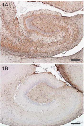
Cases with subtotal necrosis of pyramidal cells within the hippocampus were defined as hippocampal necrosis (n = 13). Cases with subtotal loss of py- ramidal cells together with gliosis were categorised as hippocampal sclerosis (n = 22). Figure 1 demonstrates strong immunohistochemical expression of glial fibrillary acidic protein (GFAP) (Fig. 1A) compared to a control hippocampus (Fig. 1B). Neurodegeneration including bilateral neuronal loss and eosinophilic necrosis could be found in different segments of the cornu ammonis to varying degrees. In some cases the subiculum and dentate gyrus were also affected.
Within the study population 31 cases (89%) showed other lesions in the brain in addition to hippocampal necrosis or sclerosis. Three different brain le- sions were present in four cats (no. 32–35).
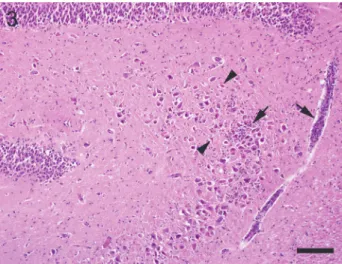
Inflammatory lesions, primarily localised within the hippocampus or adja- cent regions such as the piriform lobe, were present in altogether 26 cases (74%) with or without further lesions. Severe multifocal lymphoplasmohistiocytic en- cephalitis within the neocortex and hippocampus associated with glial nodules and Toxoplasma gondii organisms was encountered in one case (no. 6) associat- ed with hippocampal necrosis. Moderate pyogranulomatous meningitis with abundant fibrinous exudate and bilateral neuronal degeneration within the hippo- campus and the neocortex was associated with FIP virus (no. 5) (Fig. 2). In both cases the suspected pathogen was demonstrated by immunohistochemical exam-
ination. In two cases (no. 7 and 8) suppurative encephalitis was found. However, cultures were not performed and bacterial organisms could not be found during histological examination, and thus bacterial involvement can be presumed only.
Fig. 1. Immunohistochemical staining demonstrating strong gliosis in hippocampal sclerosis (A), compared to a control hippocampus (B). GFAP immunohistochemistry. Bar = 400 µm
Inflammatory infiltrates of unknown aetiology, including lymphocytic or lymphoplasmacytic meningitis, meningoencephalitis or encephalitis of varying degrees, restricted to the hippocampal region or with multifocal distribution, were present in seven cases (no. 9–15). In these cases no specific pathogen could be found during histological examination.
Fifteen cats (no. 16–27) were part of a previous comparative study (Klang et al., 2013) and demonstrated mild to moderate lymphocytic infiltrates and
complement deposition in the hippocampus and sometimes also in the adjacent leptomeninges, in the same way as in human limbic encephalitis.
Fig. 2. Intense neuronal necrosis in the cornu ammonis (arrowheads) and dentate gyrus (arrows) in a case of feline infectious peritonitis. Note dense, mixed, inflammatory infiltrates in adjacent lep-
tomeninges (asterisk). Haematoxylin and eosin (HE). Bar = 150 µm
Fig. 3. Neuronal degeneration and infiltration with lymphocytes (arrows) in the cornu ammonis (arrowheads) in limbic encephalitis. HE. Bar = 150 µm
Six (no. 23–27, no. 34) out of seven cases tested were categorised as lim- bic encephalitis as serological examination revealed VGKC-antibodies (Fig. 3).
Three of them revealed antibodies against VGKC-complex-associated LGI1 pro- tein.
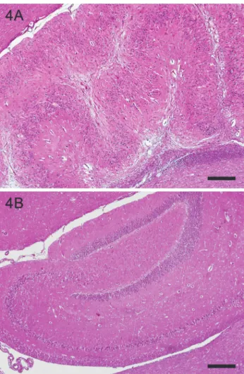
Fig. 4. Structural alteration of the dentate gyrus with abnormal folding and broadening together with hippocampal sclerosis (A) compared to a control hippocampus (B). HE. Bar = 400 µm
Nine cases (no. 16–22) with similar morphology and complement deposi- tion, in which serological examinations were not performed, were assigned as possible limbic encephalitis.
An intracranial neoplasia was present in five cases (14%). Two cases (no.
29 and 30) revealed an oligodendroglioma, which was localised in the right pi- riform lobe and the diencephalon, respectively. In three cases (no. 32, 33 and 35) a meningioma was present. In two of them the tumour was relatively small and inflammatory infiltrates resembling limbic encephalitis were concurrently pre- sent. In the third case (no. 35) an intraventricular meningioma was present in context with hippocampal sclerosis and a bilateral dentate gyrus structural altera- tion along the whole axis which appeared as abnormal folding in a festoon-like
pattern and broadening of the dentate gyrus (Fig. 4). Such alterations were en- countered in two further cases (no. 34 and 28) together with hippocampal sclero- sis, one of them together with VGKC autoantibodies (no. 34).
In one case (no. 31) (3%) a structural loss of the neocortex was found within the right ventral temporal lobe and adjacent hippocampus together with the proliferation of glial scar tissue and cavitation. This finding, interpreted as vascular change corresponding to the final stage of a cerebral infarct, was ac- companied by hippocampal sclerosis in the ipsilateral hippocampus and moder- ate hippocampal necrosis within the contralateral cornu ammonis.
Serological examination for FIV antibodies revealed negative results in all six cases (no. 1, 24, 26, 28, 32 and 33) performed. Viral RNA could not be de- tected by RT-PCR either in brain (n = 35) or in serum (n = 3) samples, whereas the control regularly revealed positive results.
Discussion
Our findings support the notion that there is a gradual transition from hip- pocampal necrosis to hippocampal sclerosis and the former precedes the latter.
Firstly, in some cases histological lesions are simultaneously evident in both hemispheres or are overlapping along the longitudinal axis. Secondly, it is obvi- ous that neuronal degeneration implies the depletion of neurons. Thirdly, cats with hippocampal sclerosis commonly show a long history of generalised sei- zures refractory to antiepileptic treatment, and status epilepticus. Finally, the ae- tiologies of both conditions are very similar. In addition, many cases described as hippocampal necrosis are rather matching the definition of hippocampal sclerosis than vice versa.
In the same way as in humans, a form of limbic encephalitis associated with hippocampal necrosis and sclerosis, characterised by the presence of high serum titres of autoantibodies against neuronal antigens such as LGI1, has been recently recognised in cats (Pakozdy et al., 2013; Klang et al., 2013; Pakozdy et al., 2014). The latter includes mild to moderate infiltrations of lymphocytes in the meninges, in perivascular cuffs or diffusely in the parenchyma, and varying degrees of acute neuronal degeneration and sclerosis within the hippocampus as- sociated with complement deposition (Klang et al., 2013). Serological tests for VGKC autoantibodies were performed in seven cases only, as this study was conducted retrospectively. Together with nine cases, which demonstrated histo- pathological findings such as complement activation and inflammatory infil- trates, together with clinical signs comparable to those of human limbic encepha- litis, we assume that altogether at least 46% of the examined cases may be asso- ciated with autoantibodies. This assumption correlates with limbic encephalitis in human temporal lobe epilepsy associated with hippocampal sclerosis, which ac-
Vascular events including hypertensive lesions, ischaemic forebrain in- farcts, parenchymal haemorrhage as well as middle cerebral artery thrombosis and vasculitis are also mentioned to be common in the literature regarding feline hippocampal sclerosis (Wagner et al., 2014). However, we encountered only a single case (3%) demonstrating lesions corresponding to a temporal lobe infarct.
Meningiomas represent the most common primary intracranial neoplasia in cats (De Lahunta and Glass, 2009). Other neoplasms such as oligodendrogli- oma are infrequently observed in the cat (Vandevelde et al., 2012) and there are just single reports describing the latter one associated with hippocampal necrosis in the piriform lobe (Vanhaesebrouck et al., 2012). While in one of our cats the tumour was localised in the piriform lobe, in the other case it extensively infil- trated the diencephalon.
Uni- and bilateral structural alterations of the dentate gyrus, similar to our findings in cats, were first described in dogs. One of these reports features a 4- year-old Pekingese dog with necrotising meningoencephalitis and neurological signs including seizures for six months (Cantile et al., 2001). The other one re- fers to a 5-year-old mixed-breed dog with granulomatous meningoencephalitis and refractory epilepsy for over 10 weeks (Klang et al., 2014). In both cases ad- ditional lesions resembling hippocampal sclerosis were histologically described.
At that point this alteration was interpreted as malformation. In the cat a dentate gyrus structural alteration was first published in 2015 (Klang et al., 2015), and this entity more likely represents rather a secondary seizure-induced process as- sociated with hippocampal sclerosis. First, the described broadening of the den- tate gyrus, the so-called granule cell dispersion, which is one feature of this al- teration, has recently been described in epileptic cats with hippocampal sclerosis (Wagner et al., 2014), and is also known in humans suffering from temporal lobe epilepsy (Lurton et al., 1998; Da Costa Neves et al., 2013). Secondly, all cats were adult or old at the onset of seizures, and structural alterations of the dentate gyrus were present together with hippocampal sclerosis.
FIV infection is a worldwide-occurring condition in domestic cats (Bendi- nelli et al., 1995). The literature on histopathological findings, especially in natu- rally infected cats, is limited. Lesions in experimental FIV infections include neuronal loss, perivascular mononuclear cell infiltrates, glial nodules and diffuse gliosis in the hilus and in the molecular layer of the dentate gyrus, midbrain, and thalamus (Dow et al., 1990; Mitchell et al., 1999; Maingat et al., 2009). Gliosis is described to be prominent in the hippocampus, especially at the border between the granule cell layer and the hilus of the dentate gyrus and in the molecular layer (Mitchell et al., 1999). Neuropathological findings of FIV-infected cats described in the literature share some features with feline hippocampal sclerosis. In the cat population examined FIV could not be demonstrated in FFPE brain or serum samples by PCR.
Summarising our results, hippocampal necrosis and sclerosis can be asso- ciated with inflammatory lesions other than limbic encephalitis such as FIP in- fections. In none of our cats could an underlying FIV infection be found. The ae- tiology remains to be defined in those cases which cannot be attributed to a spe- cific pathogen.
Acknowledgements
The authors would like to thank Ursula Glantschnigg-Eisl for providing serum samples and Angela Vincent for serological testing for VGKC auto-antibodies, Jan Bauer for characterisation of limbic encephalitis cases, Petra Kodajova for her skilled technical assistance, and the Institute of Virology from the University of Veterinary Medicine Vi- enna for performing the FIV test.
References
Bendinelli, M., Pistello, M., Lombardi, S., Poli, A., Garzelli, C., Metteucci, D., Ceccherini-Nelli, L., Malvaldi, G. and Tozzini, F. (1995): Feline immunodeficiency virus: an interesting model for AIDS studies and an important cat pathogen. Clin. Microbiol. Rev. 8, 87–112.
Bien, C. G., Urbach, H., Schramm, J., Soeder, B. M., Becker, A. J., Voltz, R., Vincent, A. and Elger, C. E. (2007): Limbic encephalitis as a precipitating event in adult-onset temporal lobe epilepsy. Neurology 69, 1236–1244.
Cantile, C., Chianini, F., Arispici, M. and Fatzer, R. (2001): Necrotizing meningoencephalitis as- sociated with cortical hippocampal hamartia in a Pekingese dog. Vet. Pathol. 38, 119–122.
Da Costa Neves, R. S., Jardim, A. P., Caboclo, L. O., Lancellotti, C., Marinho, T. F., Hamad, A. P., Marinho, M., Centeno, R., Cavalheiro, E. A., Scorza, C. A. and Targas Yacubian, E. M.
(2013): Granule cell dispersion is not a predictor of surgical outcome in temporal lobe epi- lepsy with mesial temporal sclerosis. Clin. Neuropathol. 32, 24–30.
De Lahunta, A. and Glass, E. (2009): Seizure disorders: narcolepsy. In: Veterinary Neuroanatomy and Clinical Neurology. Third edition. Saunders/Elsevier, St. Louis, MO. pp. 454–475.
Dow, S. W., Poss, M. L. and Hoover, E. A. (1990): Feline immunodeficiency virus: a neurotropic lentivirus. J. Acquir. Immune Defic. Syndr. 3, 658–668.
Fatzer, R., Gandini, G., Jaggy, A., Doherr, M. and Vandevelde, M. (2000): Necrosis of hippocam- pus and piriform lobe in 38 domestic cats with seizures: a retrospective study on clinical and pathologic findings. J. Vet. Intern. Med. 14, 100–104.
Fors, S., Van Meerveene, S., Jeserevics, J., Rakausas, M. and Cizinauskas, S. (2015): Feline hippo- campal and piriform lobe necrosis as a consequence of severe cluster seizure in two cats in Finland. Acta Vet. Scand. 57, 41.
Gelberg, H. B. (2013): Diagnostic exercise: sudden behaviour change in a cat. Vet. Pathol. 50, 1156–1157.
Irani, S. R., Michell, A. W., Lang, B., Pettingill, P., Waters, P., Johnson, M. R., Schott, J. M., Arm- strong, R. J., S. Zagami, A., Bleasel, A., Somerville, E. R., Smith, S. M. and Vincent, A.
(2011): Faciobrachial dystonic seizures precede Lgi1 antibody limbic encephalitis. Ann.
Neurol. 69, 892–900.
Klang, A., Leschnik, J., Schmidt, P. and Pakozdy, A. (2014): Bilateral hippocampal malformation and concurrent granulomatous meningoencephalitis in a dog with refractory epilepsy. J.
Comp. Pathol. 150, 424–428.
Klang, A., Schmidt, P., Kneissl, S., Bagó, Z., Vincent, A., Lang, B., Moloney, T., Bien, C. G., Ha-
loss in epileptic cats with anti-voltage-gated potassium channel complex antibodies. J.
Neuropathol. Exp. Neurol. 73, 403–413.
Klang, A., Thaller, D., Schmidt, P., Kovacs, G. G., Halasz, P. and Pakozdy, A. (2015): Bilateral dentate gyrus structural alterations in a cat associated with hippocampal sclerosis and in- traventricular meningioma. Vet. Pathol. 52, 1183–1186.
Lurton, D., El Bahh, B., Sundstrom, L. and Rougier, A. (1998): Granule cell dispersion is correlat- ed with early epileptic events in human temporal lobe epilepsy. J. Neurol. Sci. 154, 133–136.
Maingat, F., Vivithanaporn, P., Zhu, Y., Taylor, A., Baker, G., Pearson, K. and Power, C. (2009):
Neurobehavioral performance in feline immunodeficiency virus infection: integrated anal- ysis of viral burden, neuroinflammation, and neuronal injury in the cortex. J. Neurosci. 29, 8429–8437.
Marioni-Henry, K., Monteiro, R. and Behr, S. (2012): Complex partial orofacial seizures in Eng- lish cats. Vet. Rec. 170, 471.
Matiasek, K., Pumarola, I., Batlle, M., Rosati, M., Fernandez-Flores, F., Fischer, A., Wagner, E., Berendt, M., Bhatti, S. F. M., De Risio, L., Farquhar, R. G., Long, S., Munana, K., Patter- son, E. E., Pakozdy, A., Penderis, J., Platt, S., Podell, M., Potschka, H., Rusbridge, C., Stein, V. M., Tipold, A. and Volk, H. A. (2015): International veterinary epilepsy task force recommendations for systematic sampling and processing of brains from epileptic dogs and cats. BMC Vet. Res. 11, 216.
Mitchell, T. W., Buckmaster, P. S., Hoover, E. A., Whalen, L. R. and Dudek, F. E. (1999): Neuron loss and axon reorganization in the dentate gyrus of cats infected with the feline immuno- deficiency virus. J. Comp. Neurol. 411, 563–577.
Pakozdy, A., Gruber, A., Kneissl, S., Leschnik, M. and Halasz, P. (2011): Complex partial cluster seizures in cats with orofacial involvement. J. Feline Med. Surg. 13, 687–693.
Pakozdy, A., Halasz, P. and Klang, A. (2014): Epilepsy in cats: theory and practice. J. Vet. Intern.
Med. 28, 255–263.
Pakozdy, A., Halasz, P., Klang, A., Bauer, J., Leschnik, M., Tichy, A., Thalhammer, J. G., Lang, J.
G. and Vincent, A. (2013): Suspected limbic encephalitis and seizure in cats associated with voltage-gated potassium channel (VGKC) complex antibody. J. Vet. Intern. Med. 27, 212–214.
Vandevelde, M., Higgins, R. J. and Oevermann, A. (2012): Neoplasia. In: Veterinary Neuropathol- ogy: Essentials of Theory and Practice. 1st ed. John Wiley & Sons, Ltd., West Sussex, UK.
pp. 129–156.
Vanhaesebrouck, A. E., Posch, B., Baker, S., Plessas, I. N., Palmer, A. C. and Constantino-Casas, F. (2012): Temporal lobe epilepsy in a cat with a pyriform lobe oligodendroglioma and hippocampal necrosis. J. Feline Med. Surg. 14, 932–937.
Wagner, E., Rosati, M., Molin, J., Foitzik, U., Wahle, A. M., Fischer, A., Matiasek, L. A., Reese, S., Flegel, T. and Matiasek, K. (2014): Hippocampal sclerosis in feline epilepsy. Brain Pathol. 24, 607–619.