NEW AND NOTEWORTHY LICHEN-FORMING AND LICHENICOLOUS FUNGI 7
S. Y. Kondratyuk1#,2, L. Lőkös3, J. P. Halda4, E. Farkas5, D. K. Upreti6 A. Thell7, J.-J. Woo2, S.-O. Oh8 and J.-S. Hur2
1M. H. Kholodny Institute of Botany, Tereshchenkivska str. 2, 01004 Kiev, Ukraine;
#E-mail: ksya_net@ukr.net
2Korean Lichen Research Institute, Sunchon National University, Sunchon 540–742 Republic of Korea; E-mail: jshur1@sunchon.ac.kr
3Department of Botany, Hungarian Natural History Museum H-1431 Budapest, Pf. 137, Hungary; E-mail: lokos.laszlo@nhmus.hu
4Muzeum a Galerie Orlických hor, Jiráskova 2, 516 01 Rychnov nad Kněžnou, Czech Republic
5Institute of Ecology and Botany, Centre for Ecological Research Hungarian Academy of Sciences, H-2163 Vácrátót, Alkotmány u. 2–4, Hungary
6CSIR-National Botanical Research Institute
Rana Pratap Marg, Lucknow–226001 Uttar Pradesh, India; E-mail: upretidknbri@gmail.com
7Botanical Collections, Biological Museum, Lund University, Box 117, SE-221 00 Lund, Sweden;
E-mail: arne.thell@biol.lu.se
8Korea National Arboretum
Gwangneungsumogwon-ro 415, Pocheon-si, Gyeonggi-do 11186, Republic of Korea
(Received 27 March, 2017; Accepted 25 November, 2017)
Nineteen new to science species of lichen forming fungi, i.e.: Agonimia ascendens S. Y. Kondr., L. Lőkös et J.-S. Hur, A. sunchonensis S. Y. Kondr. et J.-S. Hur, A. yongsangensis S. Y. Kondr.
et J.-S. Hur, Biatora loekoesiana S. Y. Kondr. et J.-S. Hur, Caloplaca ivanpisutii S. Y. Kond., L.
Lőkös et J.-S. Hur, Candelariella makarevichiae S. Y. Kondr., L. Lőkös et J.-S. Hur, Huriella pohangensis S. Y. Kondr., L. Lőkös et J.-S. Hur, H. salyangiana S. Y. Kondr. et J.-S. Hur, Hyper- physcia oxneri S. Y. Kondr. et J.-S. Hur, Nectriopsis gangwondoensis S. Y. Kondr., L. Lőkös et J.-S. Hur, Porina ulleungdoensis S. Y. Kondr., L. Lőkös, J. Halda et J.-S. Hur, Psoroglaena gang- wondoensis S. Y. Kondr., L. Lőkös, J.-J. Woo et J.-S. Hur, Pyrenopsis cavernicola S. Y. Kondr., L.
Lőkös et J.-S. Hur, Rhizocarpon sunchonense S. Y. Kondr. et J.-S. Hur, Rufoplaca ulleungensis S.
Y. Kondr., L. Lőkös et J.-S. Hur, Sarcogyne ulleungdoensis S. Y. Kondr., L. Lőkös et J.-S. Hur, Skyttea bumyoungsungii S. Y. Kondr. et J.-S. Hur, Thelopsis gangwondoensis S. Y. Kondr., L.
Lőkös, J.-J. Woo et J.-S. Hur, Topelia loekoesiana S. Y. Kondr., J.-J. Woo et J.-S. Hur, all from South Korea, as well as Gallowayella awasthiana S. Y. Kondr. et D. K. Upreti from India and Franwilsia skottsbergii S. Y. Kondr., A. Thell, S.-O. Oh et J.-S. Hur from Chile are described, illustrated and compared with closely related taxa. A key to Agonimia species known from Eastern Asia is also included.
Lecanora helicopis is recorded for Korea for the first time, as well as a number of new to Jeju-do Island species (i.e.: Agonimia loekoesii, Biatora pseudosambuci, Buellia extremoorientalis, and Ivanpisutia oxneri) are recorded. Additional data on conidiomata and morphological characters of thallus and apothecia and illustrations as well as data on newly located iso- type specimens recently described from Canary Islands, Spain Fominiella tenerifensis are provided.
Two new combinations, i.e.: Phaeophyscia saxatilis (for Physcia saxatilis Kashiw.), and Xanthoparmelia umezuana (for Karoowia umezuana Moon K. H. et Kashiw.) are also proposed.
Key words: Chile, India, key, new species, phylogenetic analysis, revision, South America, South Korea, taxonomy
INTRODUCTION
Part of novelties found in Ulleung-do and Geumoh-do islands, as well as in a number of localities of Gangwon-do and Jeollanam-do Provinces of South Korea during field studies in 2016 was recently published (Kondratyuk et al.
2016b, c, d, 2017a, b). However, a number of still unnamed and unidentified specimens are also selected.
The aim of this communication to present legal descriptions of new set of taxa of the genera Agonimia, Caloplaca, Candelariella, Gallowayella, Hyperphyscia, Huriella, Porina, Psoroglaena, Rhizocarpon, Thelopsis, and Topelia, discovered in areas mentioned as well as to provide data on novelties and rare taxa found during revision as recent as previous collections kept in the KoLRI, KW-L, LWG and BP. Furthermore a number of taxa of the families Teloschistaceae, Candelariaceae, Physciaceae, Trapeliaceae appeared to be new from phyloge- netic analysis of families mentioned are also included and discussed.
MATERIAL AND METHODS
The specimens collected in 2016 in various areas of South Korea, as well as collections of previous years kept in the KoLRI and other herbaria (BP, KW-L, LE, LWG, VBI) included in comparative study were examined using standard microscopical techniques and hand-sectioned under a dissecting microscope (Nikon SMZ 645; Nikon, Tokyo, Japan). Anatomical descrip- tions were based on observations of these preparations under a microscope (Nikon Eclipse E200; Nikon, Tokyo, Japan, and Zeiss Scope, A1; Carl Zeiss, Oberkochen, Germany) with digital camera AxioCam ERc 5s. Section of apo- thecia were tested with water and with K and IKI (10% aqueous potassium iodide) for identification.
For identification chemical substances of critical taxa standard TLC and HPTLC methods with solvent C were carried out (Arup et al. 1993, Orange et al. 2001).
NEW SPECIES FOR SCIENCE
Agonimia ascendens S. Y. Kondr., L. Lőkös et J.-S. Hur, spec. nova
MycoBank no.: MB 824382.
Similar to Agonimia opuntiella, but differs in having microfruticose thallus consisting of mostly distant and scattered erect or ascending thalline lobes (not aggre- gated in compact thallus), in having usually longer and distinctly branched strip-like or finger-like lobes, and in having much longer spinulose hairs.
Type: Republic of Korea. Jeollanam-do, Suncheon-si, Samsan-dong, along river bank, on Metasequoia bark, growing together with Dirinaria appla- nata and Buellia aff. melaxanthella. Lat.: 34° 58’ 46.49” N; Long.: 127° 29’ 10.35”
E; Alt.: 23 m a.s.l. Coll.: Kondratyuk, S. Y. (163349), 02.10.2016 (KoLRI 041594 sub Agonimia – holotype).
Thallus very indistinct, microfruticose, consisting of rather scattered and distant small groups of ascending narrow thalline lobes, brown or brown- ish grey, with distinct whitish long hairs well contrasting darker thalline lobes (up to 1.5 mm diam., rarely aggregated to form a mat ca 1 cm diam. or more see after Harada et al. 2016). Thalline lobes to 0.6–0.8 mm long, and to (0.1–)0.12–0.18(–0.3) mm wide at the basis and somewhat slightly or distinctly widened and forming more or less rounded or spherical tips to 0.15–0.25(–0.3) mm, distinctly ascending or erect, with long, to 40–60(–80) µm long, (most- ly 0.04–0.08 mm long, colourless, or dark near the apices after Harada et al.
2016), white well contrasting to darker thalline lobes hairs, rather abundant especially on the tips of thalline lobes.
Thalline lobes in section 60–80 µm thick, lacking a distinct algal layer.
Upper cortex 5–20(–25) µm thick, euparaplectenchymatous, colourless or slightly brownish, lumina of the hyphae mostly isodiametric, roundish or po- lygonal, mostly 4–7 µm diam. (but 2–3 µm diam. near the apices of lobes), lacking distinct interhyphal spaces; walls of the hyphae 0.5–1 µm thick; papil- lae first ca 1 µm diam. and 2–3 µm long, later extended and broadened (ca 2.5 µm diam.) then adhering together throughout the length to form a spinulose hair. Medulla containing phycobiont cells, similar to upper cortex particularly in the upper parts, with hyphal cells becoming slightly separated (with inte- rhyphal spaces) below; lumina of the hyphae mostly roundish; phycobiont cells of trebouxioid algae, 5–7 µm diam., relatively dense in the upper parts of medulla, scattered below. Lower side of the thallus lacking cortex, rough, composed of roundish hyphal cells.
Upper surface of thalline lobes more or less slightly convex, with spinu- lose hairs, pale to somewhat dark brown or dirty brown, but greenish grey in shade, smooth, slightly glossy; underside usually flat to concave, generally paler than the dorsal surface, matt, lacking spinulose hairs, lacking rhizines (unfortunately we cannot confirm data of Harada et al. 2016 that it “soon be- coming granulose and finally granulose-sorediate”). Soredia-like formations to 60 µm diam., with the same long white hairs sometimes observed near or among thalline lobes, probably they are spherical tips of very short lobes, usu- ally very scarce and not forming abundant mass (as in Agonimia opuntiella).
Perithecia and pycnidia not seen.
Chemistry: not studied, all reactions negative.
Ecology: On bark of coniferous trees and among mosses over siliceous rock, where often associated with Hyperphyscia crocata, Phaeophyscia adiastola, as well as species of the genera Dirinaria, Lepraria and Endocarpon.
Etymology: Species name reflects characterictic ascending thalline lobes of this taxon.
Distribution: So far confirmed by specimens from scattered localities of South Korea (E Asia), while some records of A. opuntiella from China (Yun- nan, see Harada et al. 2016 and notes below) may belong to this taxon, too.
Taxonomic notes: From all Agonimia species A. ascendens differs in hav- ing microfruticose thallus consisting of very long strip-like thalline lobes and in having very long spinulose hairs. The closest species are the almost cosmo- politan A. opuntiella (Buschardt et Poelt) Vězda and A. cavernicola S. Y. Kondr., L. Lőkös et J.-S. Hur, which known so far only from E Asia.
Agonimia ascendens is similar to A. opuntiella, known also from Asia (mainland China (Yunnan), Taiwan, Korea), Australia, Europe, Africa, N and S Americas (see Harada et al. 2016), but differs in having microfruticose thallus consisting of mostly distant and scattered erect or ascending strip-like thalline lobes or small groups of thalline lobes (not aggregated in compact thallus vs.
minutely squamulose forming mat of densely aggregated and overlapping squamules to 1 cm across or more, with entire or lobed margin), in having usually longer and distinctly branched strip-like or finger-like lobes (vs. squa- mules 0.1–0.3 mm diam.), in having much longer spinulose hairs (mostly 40–
80 µm long vs. to 20 µm long), as well as in the lack of ellipsoid or subglobose blastidia forming abundant mass.
Agonimia ascendens is similar to A. cavernicola, in having somewhat fin- ger-like or isidia-like lobes with hyaline or whitish hairs, but differs in having thicker thalline lobes (0.2–0.3 mm diam. vs. 0.1–0.15 mm diam.), in having longer hairs (40–80 µm long vs. to 15 µm long), in having usually branched with somewhat to hemispherical thickened tips (vs. straight, without second- ary lobules and the same thickness throughout), and in different substrate (bark of mostly coniferous tress vs. rock).
Agonimia ascendens is similar to A. koreana Kashiw. et K. H. Moon, but differs in having distinctly ascending narrow lobes (vs. convex to 0.2–0.4 mm wide segments), in having cortical hairs (vs. cortical hairs absent), as well as in the lack of 0.8–1.2 mm thick paraplectenchymatous throughout thallus, and in the lack of papillate underside of lobes.
It is supposed that Harada et al. (2016) have investigated the same mate- rial, but they used the name Agonimia opuntiella for their specimens, while they emphasised about differences in the lack of blastidia in Eastern Asian material. Furthermore Harada et al. (2016) did not recognise important taxo- nomic characters as length of spinulose hairs (to 0.04–0.08 mm long, colour- less or dark near the apices, after Harada et al. 2016). See also illustrations of this material in cited paper (Harada et al. 2016).
Other specimens examined: Republic of Korea. Jeollanam-do, Suncheon-si, Samsan- dong, along river bank, on Metasequoia bark, growing together with Amandinea punctata and Phaeophyscia sp. Lat.: 34° 58’ 46.49” N; Long.: 127° 29’ 10.35” E; Alt.: 23 m a.s.l. Coll.:
Kondratyuk, S. Y. (163759), 30.10.2016 (KoLRI 042027 sub Amandinea punctata). – Jeolla- nam-do, Suncheon-si, Yongsang-dong, on bark of pine trees and on siliceous rocks in pine forest, on rock growing together with Rinodina sp. and Lepraria spp. Lat.: 34° 58’ 10.8” N;
Long.: 127° 30’ 10.1” E; Alt.: 189 m a.s.l. Coll.: Kondratyuk, S. Y. (163914), 4.12.2016 (KoLRI 042182 sub Rinodina), the same locality, growing together with Rinodina sp., Collema sp. and Phaeophyscia sp. (163916), (KoLRI 042184 sub Collema); the same locality, growing togeth- er with Lepraria sp. and Phaeophyscia sp. (163917), (KoLRI 042185 sub Lepraria); growing together with Physciella sp. (163918), (KoLRI 042186 sub Physciella). – China. Yunnan, en route from Kunming to Luquan, 5 km to Luquan, [25.3833° N, 102.4522° E], 2,150 m elev.
On limestone with mosses, 17.04.2014 Harada 28987 (CBM-FL–37590; KUN-L) [not seen].
Agonimia sunchonensis S. Y. Kondr. et J.-S. Hur, spec. nova (Fig. 1)
MycoBank no.: MB 824383.
Similar to Agonimia blumii, but differs in having granular thallus consisting of soredia sometimes aggregating in semi- or convex lax soredious aggregations of isidious to microcoralloid aggregations and in the lack of primary thalli areoles and not forming very thick soredious (Lepraria-like) thalline portions.
Type: Republic of Korea. Jeollanam-do, Suncheon-si, Yongsang-dong, along small stream, near water fall close to tourist point, on siliceous rocks, growing together with Endocarpon sp. Lat.: 34° 57’ 55.88” N; Long.: 127° 30’
03.98” E; Alt.: 110 m a.s.l. Coll.: Kondratyuk, S. Y. (163851), 20.11.2016 (KoLRI 042119 sub Agonimia – holotype).
Thallus from 0.5–1 mm across or larger aggregations to several cm across, fine granular or microgranular, sometimes to continuous crust in places with hardly distinct areoles to 0.2–0.3(–0.4) mm across, very irregular and better seen at the edges, consisting of soredia sometimes aggregating in semi- or convex lax soredious aggregations or ascending isidious to microcoralloid ag- gregations, with microlobules or isidia-like formations to 0.4–0.5 mm long and 0.1–0.15 mm wide; from dull green to grey, dark-lead-grey or tessellate dark grey and whitish grey to brownish grey or bluish lead-grey, in case of de- velopment of semiconvex aggregations and highly uplifted confluent soredi- ous mass sometimes upper portion/part of soredious mass is lighter coloured than base (light whitish grey in contrast to somewhat dark lead-grey); fine granular, from sorediate where soredia form rather uplifted hemispherical or spherical mass to 0.2–0.3(–0.5) mm across to somewhat confluent completely granular along several cm across (see holotype); initially small groups of sore- dia to (50–)70–150(–250) µm wide and almost the same high, very lax and highly uplifted, somewhat conical or hemispherical on substrate or thalli of other crustose lichens (see below ecology), rather scattered and distant, soon becoming aggregated in confluent mass. In section thallus to (100–)200–300 µm thick, no structural differences in upper and lower portion of thallus, and no hyphal structures, and no medulla tissue observed, only very lax aggrega- tions of soredia found.
Soredia (15–)17–22(–25) µm, regularly rounded or slightly elongated, hy- phal wall more or less regularly even, to 2(–3) µm thick, hyaline or sometimes greyish or light brownish; often aggregated in very irregular thalline portions to (30–)40–55 µm across (better seen in K), which difficult to call as conblastid- ia or consoredia as far they are very irregular and do not have certain shape, if they more than 30 µm across outer layer usually darker coloured, in K it is better seen that in some places the outermost cells have one side to 4–5 µm across somewhat darker making appearance of outer layer soredia aggrega- tions somewhat darker; sometimes isidioid-like coralloid formation with sep- arate fragments to (60–)80–100 µm wide observed, with very irregular thick- ness and in case when soredious mass seem to be more isidious and coralloid, in section the same aggregation of soredia observed, but darker outermost layers of hyphal walls of soredia in such sections present not regularly (prob- ably corresponding only exposed portions of branches/twigs of coralloid for- mations; coralloid formations are very crumble and easily broken in irregular fragments/portions (at any touch or sectioning/cutting).
Perithecia and pycnidia not seen.
Chemistry: No reactions observed, K– in thallus and in section, but hy- phal wall of soredia becoming somewhat lighter to hyaline in K, as well as that phenomenon that the larger aggregations consisting of small soredia bet- ter seen in K.
Ecology: Growing on bark of both deciduous (especially Salix) and conif- erous trees (Pinus spp., Metasequoia) in rather shaded and wet conditions of bark Salix alba at the edge of river, as in rather exposed conditions of road side tree, as well as in rather dry conditions of southeastern slopes of mixed forest and open areas on bark of Pinus trees.
Etymology: It is named after Suncheon city, Jeollanam-do Province of South Korea (E Asia), where the type collection was done.
Distribution: So far known from scattered localities in Jeollanam-do Province of South Korea (E Asia).
Fig. 1. Agonimia sunchonensis (holotype), general habit. Scale 0.5 mm. (Photos of S. Kondratyuk)
Taxonomic notes: We do not think that this material belong to Lepraria, while it is positioned in Agonimia rather preliminary now. However, this ma- terial differs from Lepraria species in having very spectacular coralloid forma- tions, as well as in the lack of any hyphal projections of hyphal cells, as well as in another colouration of thallus and in the lack of any chemical substances.
Of course the further, especially molecular study should solve this prob- lem. Unfortunately we have had rather poor collection in our disposition to use them for extracting DNA according to the current technology. We do hope that this step will be done by the next generation of Korean lichenologists.
From dark green crustose lichen Sculptolumina aff. japonica, with which it grows very often side by side, or above thallus of which it often was observed (see nos. 163942, 163943, 163944, 163945 in list of specimens examined below) it differs in distinctly soredious/isidious appearance being fine granular and in having grey or somewhat tessellate greyish-whitish colour (well contrast- ing to dark green thallus of Sculptolumina aff. japonica, as well as in having very lax and uplifted soredious mass in semi- or convex aggregations or in isidia-like coralloid hemispherical formations (and in the lack of distinct con- tinuous thallus) and being hitherto known only in sterile conditions.
It is difficult to make suggestion that it is sorediate or isidiate form of Sculptolumina aff. japonica because this soredious mass very often growing in- dependently of thallus of Sculptolumina or other lichens, as well as that colour of thalli are rather different and well contrasting each other.
After having somewhat tessellate grey or dark grey and whitish grey as well as very uplifted, semiconvex to convex soredious mass this species of Ago nimia is very similar to soredious mass of Phaeophyscia aff. adiastola. How- ever Agonimia differs in the lack of lobate portion of thallus, as well as in having much smaller soredious mass aggregations (to 0.2–0.3 mm across vs.
1–2 mm or to completely confluent upper surface in several cm across) and in having much smaller soredia (to 25 µm diam. vs. 25–40 µm diam. soredia and 40–55 µm across conblastidia).
It should be mentioned that soredious mass has somewhat water repel- ling property it is somewhat better to observe thallus details in K solution (than in water), because it was difficult to observe sections of thallus in water during our study.
Other specimens examined: Republic of Korea. Jeollanam-do, Suncheon-si, Maegok- dong, WSW of university area, on bark of trees in pine forest. Lat.: 34° 58’ 01.96” N; Long.:
127° 28’ 17.4” E; Alt.: 269 m a.s.l. Coll.: Kondratyuk, S. Y. (163942), 11.12.2016 (KoLRI 042210 sub Sculptolumina aff. japonica); the same locality, growing together with Buellia aff. mela- xanthella and Agonimia, (163943, 163944), (KoLRI 042211, KoLRI 042212 sub Sculptolumina aff. japonica); the same locality, growing together with Sculptolumina aff. japonica, Buellia aff. melaxanthella and Agonimia, (163945), (KoLRI 042213 sub Agonimia). – Jeollanam-do, Suncheon-si, Samsan-dong, along river bank, on Metasequoia?/Cryptomeria bark, growing
together with Endocarpon sp. Lat.: 34° 58’ 46.49” N; Long.: 127° 29’ 10.35” E; Alt.: 23 m a.s.l.
Coll.: Kondratyuk, S. Y. (163761), 30.10.2016 (KoLRI 042029 sub Agonimia); the same locali- ty, growing together with Agonimia sp. and green soredious crust, (163800, 163803), (KoLRI 042068, KoLRI 042071 sub Agonimia); the same locality, growing together with Agonimia sp. 163806 (KoLRI 042074 sub Agonimia). – Jeollanam-do, Suncheon-si, Songgwang-myeon, Jogyesan Mts, Sinpyeong-ri, near Songgwangsa Temple, along stream valley, on bark. Lat.:
35° 00’ 18.55” N; Long.: 127° 15’ 53.68” E; Alt.: 170 m a.s.l. Coll.: Kondratyuk, S. Y., Lőkös, L.
(163211, 163212, 163213), 24.09.2016 (KoLRI 041456, KoLRI 041457, KoLRI 041458 as Bacidia (green isidiate)).
Agonimia yongsangensis S. Y. Kondr. et J.-S. Hur, spec. nova (Fig. 2)
MycoBank no.: MB 824384.
Similar to Agonimia koreana in the lack of hairs on upper surface of thalline lobes but differs in having plane or concave (vs. more or less convex 0.2–0.4 mm wide, while the width of segment at the basis the same), and in having gradually becoming thinner and narrower towards the tips thalline lobes.
Type: Republic of Korea. Jeollanam-do, Suncheon-si, Yongsang-dong, Mt top behind artificial lake, on siliceous rocks in pine forest or in open areas, growing together with Endocarpon sp. and Lepraria sp. Lat.: 34° 58’ 22.11” N;
Long.: 127° 30’ 21.79” E; Alt.: 277 m a.s.l. Coll.: Kondratyuk, S. Y. (164036), 17.12.2016 (KoLRI 042304 – holotype); the same locality, growing together with Endocarpon sp. (164037), (KoLRI 042305 – isotype); the same locality, (164038), (KoLRI 042306 – isotype); the same locality, growing among mosses together with Lecania sp. (164035), (KoLRI 042303 sub Lecania – isotype).
Thallus consisting of microfruticose lobes, usually indistinct only some- times can be seen as fragments to 15–18(–25) mm across. Thalline lobes to 0.4–0.8 mm long, somewhat isidia-like, distinctly widened at the basis to 0.2–
0.3(–0.4) mm wide, and gradually becoming narrower towards the tips, often branched or dissected into narrower terminal portions to 40–50 µm across;
sometimes secondary lobules to 0.2–0.3(–0.4) mm long and (0.–06)0.08–0.15 (–0.2) mm wide distinctly developed, becoming gradually narrower towards the tips and dissected into smaller portions (resembling Candelaria-type of phyllidia or blastidia formations see Kondratyuk 1997); total width of lobe with all secondary lobules to 0.3–0.6(–0.8) mm wide; lobes horizontally ori- entated, dorsiventral, edges somewhat slightly folded downwards, and lobe seem to be slightly subconvex. Upper surface without hairs. No soredia or blastidia observed.
Perithecia and pycnidia not known.
Chemistry: not studied, all reactions negative.
Ecology: often growing among mosses as very close to moss mats, rather indistinct and ban be easily overlooked (recorded from bark of pine tree see below* and rock).
Etymology: It is named after the type locality, i.e. Yongsang-dong in southern part of South Korea (E Asia).
Distribution: So far known from scattered localities in southern part of South Korea (E Asia).
Taxonomic notes: Agonimia yongsangensis is similar to A. koreana in the lack of hairs on upper surface of thalline lobes but differs in having plane or concave (vs. more or less convex 0.2–0.4 mm wide, while the width of segment at the basis the same), and in having gradually becoming thinner and nar- rower towards the tips thalline lobes, as well as in having thalline lobes often dissected into narrower secondary lobules towards the tips.
* It is still heterogeneous as far recorded from bark of pine tree and on rock. The speci- men no. 164038 may resemble Phyllopsora loekoesii, but they have very large differences of measurements of thallus, thalline lobes and in character of dissection of thalline lobes in secondary lobules.
Fig. 2. Agonimia yongsangensis (holotype), general habit. Scale 1 mm (top left) and 0.5 mm (the others). (Photos of S. Kondratyuk)
In contrast to Agominia cavernicola Agonimia yongsangensis has much wider at the basis isidia-like lobes (0.2–0.3(–0.4) mm vs. 0.1–0.15 mm wide/
diam.), and in having distinctly becoming narrower towards the tips and of- ten branched or dissected into secondary lobules (vs. more or less cylindrical and scarcely branched), in having terminal portions dissected into smaller portions to 40–50 µm across.
When secondary thalline lobules of Agonimia yongsangensis are very small, to 0.1 mm diam. and somewhat isidia-like, they can be accepted as isidia-like formations of A. cavernicola, but A. yongsangensis differs in having rather larger, wider thalline lobes usually aggregated in wider thalline lobes, which are also only as part of more complex larger thallus, i.e.: small second- ary lobules of A. yongsangensis are only parts of much wider (to 0.2–0.3(–0.4) mm wide) thalline lobes, as well as A. yongsangensis differs in having shorter (to 0.3–0.5 mm long) and without hairs secondary thalline lobules.
When it is small, thallus has only to 0.4 mm long weakly branched thal- line lobes it can be accepted as young Cladonia thalli, but differs in having dull brownish green thallus and in having much smaller size.
Sometimes Agonimia yongsangensis growing side by side with Endocarpon sp., and can be similar to this lichen after having dull brownish green colour of thallus. However, A. yongsangensis differs in having usually smaller, especially narrower towards the tips lobes (vs. areoles 0.8–1 mm or more across), and in having ascending and somewhat hanging above substrate level lobes, while Endocarpon areoles are closely attached to the substrate by the whole surface.
KEY TO EASTERN ASIAN AGONIMIA SPECIES
1a Thallus of squamules up to 4 mm diam. or microfruticose of erect or as- cending lobules, with a minutely hairy surface 2 1b Thallus of smaller squamules up to 1 mm long or granular, surface at
most with minute papillae, rarely with hairs 3 2a Thallus microfruticose, mostly of distant and scattered erect or ascend- ing thalline lobes; spinulose hairs 40–80 µm long; ellipsoid blastidia very
rare A. ascendens
2b Thallus minutely squamulose forming mat of densely aggregated and overlapping squamules; spinulose hairs to 20 µm long; ellipsoid or sub- globose blastidia often forming abundant mass A. opuntiella
3a With perithecia 10
3b Without perithecia 4
4a Thallus without hairy surface 5 4b Thallus of distinctly ascending branched lobes or finger-like isidia, up- per surface with hairs from abundant to scarce 8 5a Thallus sorediate, continuous or isidiate areolate 7 5b Thallus squamulose or microfruticose, without soredia or isidia 6 6a Thallus squamulose, thalline lobes convex, a. 0.2–0.4 mm wide through-
out A. koreana
6b Thallus microfruticose, thalline lobes plane, gradually becoming nar- rower and thinner, as well as dissected into narrower secondary lobules
towards the tips A. yongsangensis
7a Thallus continuous with convex soredious mass A. loekoesii 7b Thallus areolate and isidious at first, then becoming sorediate and Lepra-
ria-like A. blumii
8a Thallus mostly finger-like isidia ca 0.1 mm diam., and to 0.5–1 mm long, primary plane areoles often soon disappearing, hairs short, seen only at
the largest magnification A. cavernicola
8b Thallus granular or with distinct thalline lobes 9 9a Thallus granular, usually of tessellate coloration, granules often forming
isidia-like uplifted aggregations A. sunchonensis 9b Thallus of separate, scattered ascending branched lobes; upper surface
with distinct hairs more than 40 µm long A. ascendens 10a Ascospores (25–)29–55 µm long; thallus finely verruculose or with
minute coralloid or finger-like squamules at most 0.24 × 0.1 mm; asci
8-spored 11
10b Ascospores (42–)57–120 µm long; thallus distinctly squamulose, squam- ules 0.05–1 × 0.05–0.3(–0.5) mm; asci (1–)2-spored or 8-spored 14 11a Perithecia rough, upper part with vertical cracks or furrows A. repleta 11b Perithecia smooth, not cracked in upper part 12 12a Perithecia 0.12–0.22 mm diam.; ascospores (25–)29–35(–44) µm long; usu-
ally on bark A. allobata
12b Perithecia 0.25–0.6 mm diam.; ascospores 31–55 µm long; on soil or on
mosses over soil and rock 13
13a Thallus usually with glossy, sterile black globules; thallus grey-green, cortical cells without pigment A. globulifera 13b Sterile black globules absent; thallus brown, cortical cells brown-pig-
mented A. gelatinosa
14a Asci (1–)2-spored; squamules sometimes elongate and terete, but often distinctly flattened, 0.1–1 × 0.1–0.3(–0.5) mm, green-fawn to brown
A. tristicula
14b Asci 8-spored; squamules often elongate, branched, terete or slightly flat- tened or globular, 0.05–0.25 × 0.05–0.12 mm, grey-green A. octospora
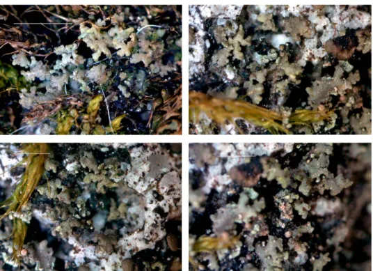
Biatora loekoesiana S. Y. Kondr. et J.-S. Hur, spec. nova (Fig. 3)
MycoBank no.: MB 824385.
Similar to Biatora pseudosambuci, but differs in having darker greenish thal- lus, somewhat smaller, from immersed in thallus to sessile, and much darker brown to blackish brown apothecia, more or less abundant pruina on apothecium disc, narrower true exciple in the uppermost lateral portion, lower hymenium, as well as 8-spored asci and wider ascospores.
Type: Republic of Korea. Jeollanam-do, Suncheon-si, Maegok-dong, WNW of university area, on bark of fruit trees in old fruit garden, growing together with Biatora pseudosambuci, Phaeophyscia and Lecanora spp. Lat.: 34°
58’ 18.16” N; Long.: 127° 28’ 03.8” E; Alt.: 203 m a.s.l. Coll.: Kondratyuk, S.
Y. (163874), 27.11.2016 (KoLRI 42142 sub Biatora loekoesiana – holotype, BP – isotype); the same locality, growing together with Buellia aff. pseudosubnexa, (163886), (KoLRI 42154 sub Buellia – isotype); the same locality, growing to- gether with Dimerella, (163884), (KoLRI 42152 sub Dimerella – isotype).
Thallus crustose, continuous, irregularly rounded [Note: Status of sore- diate material sometimes growing side by side with Biatora loekoesiana (see nos. 163996, 163994 and 163995 in KoLRI on Alnus) is still under revision.], greenish or greyish greenish. Thallus in section to 60–70 µm thick of densely packed algal cells, and cortical layer indistinct or to 10 µm thick, algal layer to 50(–70) µm thick, and medulla indistinct.
Apothecia 0.2–0.4(–0.45) mm diam., to 60(–90) µm thick in section, seem to be lecideine or biatorine, very small, dark brown, sometimes seem to be with abundant whitish pruina, while at highest magnification apothecia with- out pruina are observed too; in section apothecia distinctly biatorine, mostly
no algal cells in section, true exciple to 20–25(–30) µm thick in uppermost lateral portion and to 20–30(–40) µm wide in lower lateral and basal portions or basal portion becoming narrower in the centre to 10–15 µm thick or dis- appearing, more or less scleroplectenchymatous with matrix and hyphae lu- mina a. 1 µm diam.; hyaline to light brown in inner lateral and inner basal portions and dull yellow-brownish to medium brown in outermost layers;
hymenium to 50–55 µm high; epihymenium to 10–15 µm thick, dark greyish brown to blackish brown, or dull greyish brown; tips to 15 µm long becoming distinctly brownish, often curved above ascus tips, scarcely branched, tips to 2.5–3(–4) µm diam.; subhymenium 10–15(–30) µm thick, hyaline, sometimes with oil droplets to 2(–3) µm diam.; algal cells 7–13(–17) µm diam. below true exciple sometimes observed; asci 8-spored, sometimes seem to contain more of 8 ascospores owing to guttulae of ascospores (young ascospores usually bi- guttulate), but after careful checking ascospores simple and per 8 in ascus; as- cospores hyaline, simple sometimes seem to be 1-septate, ovoid, rather wide, to widely ellipsoid or hemispherical, distinctly guttulate, with one large to 5 µm diam. guttulae rarely 2–3-guttulate, (8–)9–13 × (5–)5.5–7(–8) µm [50 meas- urements].
Ecology: Material was collected from many localities but very often was overgrowing by Biatora pseudosambuci, and it was several times made wrong conclusion that material is conspecific with Biatora pseudosambuci. After sec- ondary revision of all collections of Biatora pseudosambuci it was shown that Bi- atora loekoesiana is presented among these collections, too. Several time it was observed that apothecia of Biatora loekoesiana and B. pseudosambuci can grow side by side. So suggestion that B. loekoesiana as earlier stage of development of Biatora pseusambuci colonies is made. And in nature Biatora pseudosambuci replaces thalli of B. loekoesiana very fast.
From our field experience we may make suggestion that Biatora loekoe- siana is very short living lichen, which very soon is replacing by Biatora pseu- dosambuci.
The latter species is probably especially successful in well-lit (well illu- minated) and rich on nitrates conditions. So Biatora loekoesiana can be rather rarely observed in conditions of road-side Prunus tree bark or in fruit gardens on bark of branches of fruit trees. In these conditions Biatora pseudosambuci can be mostly found. In contrast to this Biatora loekoesiana was rather abundant especially in shaded conditions of mixed mountain forest with small stream valleys at small waterfall on bark of Alnus trees.
Etymology: It is named after the well-known Hungarian lichenologist László Lőkös (1959–) (BP, Budapest, Hungary) in recognition of his contribu- tion, as well as with thanks for friendly support and help during joint work in South Korea.
Distribution: It is known hitherto from scattered localities in mainland and islands of South Korea (E Asia).
This material several times was especially collected in the vicinities of Sunchon city, in Ulleung-do Island, in Gangwon-do province as type collec- tions. But after revision unfortunately wrong conclusion that it is conspecific with Biatora pseudosambuci was done several times (see also above).
Taxonomic notes: Biatora loekoesiana is similar to B. pseudosambuci (S. Y.
Kondr., L. Lőkös et J.-S. Hur) S. Y. Kondr., L. Lőkös et J.-S. Hur, described
Fig. 3. Biatora loekoesiana (holotype), general habit. Scale 0.5 mm. (Photos of S. Kondratyuk)
recently from eastern Asia, with which often growing side by side, but differs in having darker greenish thallus (vs. grey or light grey), in having somewhat smaller, from immersed in thallus to sessile, and much darker brown to black- ish brown apothecia (vs. dark brown where own margin slightly lighter of disc, yellowish brown), in having more or less abundant pruina on apothe- cium disc, in having narrower true exciple in the uppermost lateral portion (20–25(–30) µm vs. 25–50(–60) µm wide), in having lower hymenium (50–55 µm vs. 70–80 µm high), as well as in having 8-spored asci (vs. (15–)16-spored) and in having wider ascospores ((8–)10–13 × (5–)6–7(–8) µm vs. (7–)8–11(–12)
× 4.5–5(–8) µm (Kondratyuk et al. 2016a, b).
Furthermore when B. pseudosambuci and B. loekoesiana growing side by side (see 163874 holotype and BP isotype), apothecia of both fungi can be on the same thallus, but thallus of B. pseudosambuci is much thinner, and apothe- cia are much larger and more shiny, with somewhat yellowish own margin of apothecia (like somewhat golden own margin as in apothecia of Lecidella mandshurica) (see Kondratyuk et al. 2015a).
Biatora loekoesiana cannot be simple juvenile stage of Biatora pseudosam- buci, as far these two taxa differs additionally to presence/absence pruina on apothecium disc, by measurements and structure of exciple (true exciple in Biatora pseudosambuci greyish or lightly brownish in outermost portion, where outermost portion to 15–20 thick of palisade plectenchyma with very thick 6–7 µm wide hyphae with lumina 1–1.2 µm diam.), in having 8 and 16-spored asci consequently, as well as in size of ascospores (see above).
Other specimens examined: Republic of Korea. Jeju-do, Jeju-si, Sangumbari Crater, around parking place at entrance, on bark of cherry trees, growing together with Buellia ex- tremorientalis and Scoliciosporum chlorococcum. Lat.: 33° 26’ 7.08” N; Long.: 126° 41’ 22.07” E;
Alt.: 408 m a.s.l. Coll.: Kondratyuk, S. Y., Kondratiuk, T. O., Yang, J.-H. (170153), 14.03.2017 (KoLRI 044277 sub Buellia extremorientalis). – Jeollanam-do, Suncheon-si, Yongsang-dong, on bark of trees in pine forest, growing together with Lecanora, Amandinea and Buellia. Lat.:
34° 58’ 10.8” N; Long.: 127° 30’ 10.1” E; Alt.: 189 m a.s.l. Coll.: Kondratyuk, S. Y. (163924), 4.12.2016 (KoLRI 42192 sub Biatora loekoesiana); the same locality, growing together with Biatora pseudosambuci, Sculptolumina aff. japonica, (163925), (KoLRI 42193). – Jeollanam-do, Suncheon-si, Yongsang-dong, along small stream, near water fall close to tourist point, on Alnus bark in mixed forest, growing together with Biatora pseudosambuci and Buellia aff. melanoxanthella. Lat.: 34° 57’ 55.88” N; Long.: 127° 30’ 03.98” E; Alt.: 110 m a.s.l. Coll.:
Kondratyuk, S. Y. (163861), 20.11.2016 (KoLRI 42214); the same locality, (163855, 163867), (KoLRI 42123, KoLRI 42135). – Jeollanam-do, Suncheon-si, Yongsang-dong, Mt top behind artificial lake, on Alnus bark, growing together with dull green punctiform soredious li- chen. Lat.: 34° 58’ 20.69” N; Long.: 127° 30’ 02.58” E; Alt.: 194 m a.s.l. Coll.: Kondratyuk, S.
Y. (163994, 163995, 163996), 17.12.2016 (KoLRI 44262, KoLRI 44263, KoLRI 42264).
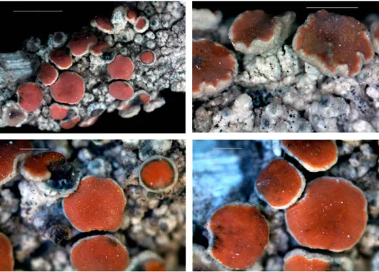
Caloplaca ivanpisutii S. Y. Kond., L. Lőkös et J.-S. Hur, spec. nova (Fig. 4)
MycoBank no.: MB 824386.
Similar to Caloplaca fuscoatroides, but differs in having thin continuous and grey cracked thallus, in having smaller apothecia, and in having dark brown apothe- cium disc, as well as in the lack of thick convex or semiconvex areoles, of warty forma- tions on thalline squamules, of cortical layer with cells containing brown pigment, of hyaline epinecral layer, and of orange-red own margin of apothecia.
Type: Republic of Korea: Mt Baekseokbong, Bukpyeong-myeong, Jeong- seon-gun, Gangwon-do, on rock. Lat.: 37° 28’ 44.34” N; Long.: 128° 39’ 45.60”
E; Alt.: ca 494 m a.s.l. Coll.: Joshi, Y., Wang, X. Y., Ryu, J. A., Hur, J. Y. (090453), 16.05.2009 (KoLRI 010157 – holotype, KW-L and BP – isotype).
Thallus to 1–5 cm across, or more larger, brownish grey or grey, from whitish grey to brownish grey, continuous, only slightly cracked, but not dis- tinctly areolate in most cases, in some thalli areoles to 0.4–0.8 mm can be ob- served; hypothallus not observed while black line to 0.1 mm wide between thalli of this species as well as other crustose lichens often observed.
Apothecia 0.3–0.8 mm diam., and 0.15 mm thick in section, rather numer- ous, abundant, dark brown to blackish brown or black, sometimes with bluish black tinge; at first completely immersed, than sessile, lecideine or biatorine to zeorine or rarely lecanorine; in biatorine own margin to 0.05 mm wide slightly arising level of apothecium, somewhat shiny in contrast to matt disc; thal- line margin (if observed) concolorous with thallus, from whitish grey to dark brown (and in this case concolorous with disc); only rarely distinctly zeorine, where true exciple slightly darker of disc, dark brown to blackish brown or medium brown to light brown in places as circle between thalline margin and disc observed; disc mostly flat; apothecia in section mainly zeorine and biato- rine; true exciple (30–)50–70 thick in uppermost lateral potion, in outermost portions to 15–20(–40) µm thick darkish/blackish or bluish blackish (and first impression that section of Lecidea/Lecidella type of apothecium) while inner portion hyaline; to 40–80 µm thick in lower lateral portion and to 50–80 µm thick in basal portion, palisade paraplectenchymatous with rounded cells, cell lumina to 4–5 µm diam.; thalline exciple often better developed at basis or if present in lateral portion to 60(–120) µm thick; hymenium 60–70 µm high;
epihymenium to 12–15 µm thick, dull blackish olive or brownish yellowish, epihymenium and uppermost cells of paraphyses bluish to dark aeruginose, N+ purple, paraphyses to 4–5 µm diam. in the uppermost portions [in water, and to 2–3.5 µm thick in K], becoming bluish-greenish or dark bluish green,
thick-walled; epihymenium K+ violetish tinge, while outermost layers of true exciple and aeruginose portions of epihymenium K– becoming slightly light- er but colour is the same; subhymenium 60–70 µm thick, hyaline, without oil;
asci with 4–8 bipolar and small size abortive ascospores, bipolar ascospores widely ellipsoid, distinctly attenuated towards the tips, often becoming or- ange with overmature, (11–)12–14(–16) × (4.8–)5.5–6.5 µm in water and 9–13 (–15) × (5.5–)6–7(–8) µm in K; ascospore septum wide, to (3–)4–5 µm wide in water and almost the same, (3.5–)4–5(–7) µm wide in K.
Conidiomata especially numerous at edge of thallus, in places of con- tact with other crust lichens, blackish or greenish dark brown, in section to 100–150 µm diam. with blackish or brownish cells at ostiole; conidia long ba- cilliform, (2.5–)3–5 × 0.8–1 µm.
Chemistry: True exciple N+ purple, thallus K+ yellow, KC–, C–, Pd– or slightly yellowish green.
Ecology: Growing on siliceous rock.
Etymology: It is named after the well-known Slovak lichenologist and our friend Ivan Pišút (1935–2017) (BRA, Bratislava, Slovakia), as well as after somewhat similarities with members of the genus Ivanpisutia (Ramalinaceae) when own margin of biatorine apothecia has lighter colour than apothecium disc.
Distribution: So far known from several scattered localities in South Ko- rea (E Asia).
Taxonomic notes: Caloplaca ivanpisutii is characterised by the following characters: grey to white-grey (as in Loekoesia austrocoreana) to brownish grey [as in some species of the genus Fuscidea], epihymenium and uppermost cells of paraphyses bluish to dark aeruginose, N+ purple, true exciple in outer por- tions darkish/blackish (and first impression after section of apothecium that section of Lecidea/Lecidella type apothecium), as well as narrow ascospores and wide ascospore septum.
Caloplaca ivanpisutii is similar to ‘Caloplaca’ fuscoatroides J. Steiner, saxi- colous species known from Europe (Italy, Greece and Ukraine) and Asia (Turkey), but differs in having thin continuous and grey cracked thallus (vs.
areolate to squamulose, thick, blackish brown to black), in having smaller apothecia (to 0.8 mm diam. vs. 0.8–1.5 mm diam.), in having dark brown apo- thecium disc (vs. rusty red to rusty brown), in the lack of thick convex or semiconvex areoles (vs. 0.4–0.7 mm thick and 0.7–1.8 mm diam.), in the lack of warty formations to 0.2–0.3 mm diam. on thalline squamules, in the lack of cortical layer with cells containing brown pigment, and in the lack of hyaline epinecral layer, as well as in the lack of orange-red own margin of apothecia, while ascospores are of the same range of variation (12–14 × 5.5–6.5 µm vs.
12–14(–16) × 5–7 µm), and the same septum ((3–)4–5 µm vs. 3–6 µm wide).
Status of ‘Caloplaca’ fuscoatroides is still not checked with molecular data, but it is mentioned that this species is similar to Blastenia crenularia (With.) Arup, Søchting et Frödén, but differs by darker and almost squamulose thal- lus, short ascospores and brownish tips of conidiomata.
Caloplaca ivanpisutii is similar to C. fuscoatra (Bayrh.) Zahlbr. (which sometimes considered to be synonym to C. aractina (Fr.) Häyrén), but differs in having thinner and light grey thallus (vs. rather thick to areolate, worthy, brownish black, greenish black, usually dark), in having wider range of vari-
Fig. 4. Caloplaca ivanpisutii (holotype), general habit. Scale 0.5 mm (top) and 1 mm (bottom).
(Photos of S. Kondratyuk)
ation of apothecia (vs. lecanorine or zeorine) apothecia, in having dark brown apothecium disc (vs. rusty red, dirty rusty red or rusty brick to black), in hav- ing true exciple N+ purple, while lack of matrix in true exciple, in having nar- rower ascospores (12–14 × 5.5–6.5 µm vs. (7.6–)10–15(–17) × 5–9 µm), and in having wider septum ((3–)4–5 µm vs. 2–4 µm wide).
After having whitish-greyish thallus and thalline margin, and dark brown to blackish apothecium disc Caloplaca ivanpisutii may be similar to Fus- cidea coreana S. Y. Kondr., L. Lőkös et J.-S. Hur, and to Gyalidea austrocoreana S. Y. Kondr., L. Lőkös et J.-S. Hur, but differs from both taxa in the lack of N+
reaction of epihymenium, as well as in having not constricted at the basis apo- thecia, in having Teloschistes-type of asci and bipolar ascospores.
Material of Lecanora spodoplacoides (= Lecanora viridirufa Ach. var. scoto- placoides Nyl.) as well as Placodium haematites var. ussuriense Tomin, both taxa described based on eastern Asian material should be compared in future with this taxon, too.
Caloplaca ivanpisutii may belong to the Loekoesia branch of the subfamily Caloplacoideae (see Kondratyuk et al. 2014, 2015b). However, freshly collected material of this taxon was not available for molecular study within this study.
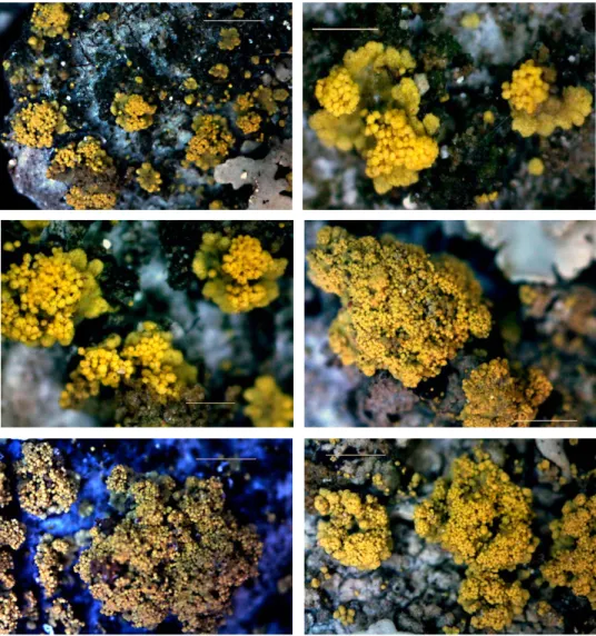
Candelariella makarevichiae S. Y. Kondr., L. Lőkös et J.-S. Hur, spec. nova (Fig. 5)
MycoBank no.: MB 824387.
Similar to Candelariella reflexa, but differs in having well-developed, distinct, minute areoles or squamules, and thalli usually consist of discrete, scattered areoles and do not form continuous leprose crust, in having mostly smaller thalline areoles/
squamules, in having well-developed soralia with uplifted soredious mass, in having smaller soredia.
Type: Republic of Korea. Gangwon-do, Sacheon-myeon, Gadunji-gill, small pine plantation along the seashore road (Halan-ro) at Sacheon Beach, on bark of Pinus densiflora and P. thunbergii, growing together with Agonimia sp. Lat.: 37° 49’ 43.21” N; Long.: 128° 52’ 41.50” E; Alt.: ca 5 m a.s.l. Coll.:
Kondratyuk, S. Y., Lőkös, L. (150828/A), 09.07.2015 (KoLRI 034061/A – holo- type); the same locality, growing together with Agonimia sp. and Amandinea sp. (150828/B, 150829), (KoLRI 034061/B, KoLRI 034062 – isotypes).
Thallus mostly macroscopically somewhat indistinct, dull yellow or dull citrine yellow spots, which consisting of very small scattered and distant small areoles, and often seem to leprose or consisting of confluent soredious mass, but at high magnification (×100 and more) consisting of small soredi-
ate areoles/squamules rarely aggregated in groups a. 2–3 mm across, while dispersed scattered minute areoles may cover spots to several cm across.
Thalline areoles 0.15–0.3(–0.5)[–0.8] mm diam./across, very distant each other, more or less rounded at first to irregular and somewhat semiconvex [on bark of deciduous trees sometimes to 1–1.5(–2) mm across in overmature thalli (see Taxonomic notes below)], from very thin, closely attached to the substrate and with very closely attached to the substrate peripheral portions, somewhat dissected in smaller portions to 0.04–0.06 mm wide, dull citrine; central por- tions usually with soredious mass more or less uplifted above substrate and thalline areoles level, brighter yellow; thalline areoles/squamules becoming somewhat thicker at the overmature or among thalli of other crustose lichens, when they do not have possibility to grow or occupy new surface, and mostly completely covered by soredia.
Soralia (0.1–)0.15–0.2(–0.3) mm diam. at first very well distinct as regu- larly rounded or slightly elongated, in the centre of thalline areole or close to the edge, but on the upper surface of areole, and with distinctly uplifted above thalline areole level, with somewhat semiconvex or convex soredious mass; however with time soralia becoming aggregated and indistinct while upper surface of thalline areoles/squamules becoming completely covered by soredious mass. Soredia regularly rounded and usually evenly sized, ca 20–30 µm diam. in uplifted hemispherical soredious mass in soralia or to 20–40 µm diam./across in case when upper surface of thalline areoles almost completely covered by soredious mass [spherical formations to 80(–100) µm across, prob- ably future soralia, especially along the edges of thalline areoles, can be ob- served on bark of deciduous trees sometimes (see Taxonomic notes below)].
Apothecia and conidiomata not observed.
Chemistry: Thallus contains calycin (HPLC).
Ecology: It is common on bark of pine trees (Pinus densiflora and P. thun- bergii), while was collected on broad leaved trees in well-lit conditions, on road side deciduous trees (see also below). Growing together with Kashiwadia orientalis, Dirinaria applanata, Biatora cf. pseudosambuci and species of the gen- era Agonimia, Lecanora, Lepraria.
Etymology: It is named after the well-known Ukrainian lichenologist Maria Florianivna Makarevich (1906–1989) who has provided important key to northern Eurasian Candelariella in the 1970s.
Distribution: It is so far known from South Korea (E Asia).
Taxonomic notes: Candelariella makarevichiae is similar to C. reflexa (Nyl.) Lettau, epiphytic lichen in nutrient enriched habitats such as the base of trees fouled by dogs, rough-barked trees around farm steads and village, on slop- ing trunks and boughs of large Salix and Sambucus, also as tiny thalli amongst other lichens on slightly nutrient-enriched broad-leaved trees in woodland;
shade and moderately pollution-tolerant, cosmopolitan taxon, as after ecol-
ogy (growing mainly on road side broad-leaved trees or in open conditions in woodlands) but differs in having well-developed distinct minute areoles or squamules (vs. granules or minute squamules), in having wide range of variation, but mostly smaller thalline areoles/squamules (0.2–0.3 mm or to 1–1.5(–2) mm across vs. to 1 mm diam.), in having well-developed soralia with uplifted soredious mass (vs. the margins of thalline squamules usually rapidly breaking down into fine-granular soredia), in having smaller soredia (20–30 µm diam. vs. 50–70 µm diam.), and usually consisting of discrete scat-
Fig. 5. Candelariella makarevichiae (holotype), general habit (top left) and enlarged thalline fragments with soralia. Scale 0.5 mm. (Photos of S. Kondratyuk)
tered areoles and do not forming continuous leprose crust, as well as in posi- tioning in separate from the C. reflexa subbranch in the phylogenetic tree after ITS nrDNA (Gilbert and James 2009).
Korean material of Candelariella makarevichiae can be keyed to Candelari- ella efflorescens R. C. Harris et W. R. Buck, known from mostly bark of broad- leaved trees of North America and Europe (Westberg 2004, 2007), but differs in having distinctly areolate to squamulose thallus, not being granular (vs.
from granular to areolate or minutely squamulose), in having plane areoles (vs. convex areoles to 0.4 mm wide), in having smaller soredia (20–30 µm diam. vs. 20–50 µm diam.), in having distinct rounded, somewhat uplifted soralia (vs. soredia on margin of the areoles and spreading inwards).
After having closely attached to substrate areoles and highly uplifted soredious mass material of Candelariella makarevichiae may resemble Mikhto- mia geumohdoensis, S. Y. Kondr., Liu D. et J.-S. Hur, but differs in having bright citrine, well distinct thallus, in having usually very distant scattered thalline areoles, and in having somewhat dissected peripheral zone of areoles (Kon- dratyuk et al. 2017a).
After ITS phylogeny Candelariella makarevichiae represented by three voucher specimens, i.e.: nos. 110993, 150828, 150829 forms separate clade which is positioning in rather distant position as from C. reflexa, as from C. efflorescens.
So far voucher specimens included in the phylogenetic analysis based on ITS nrDNA sequences were collected only on bark of pine trees. Material of Candelariella makarevichiae from bark of Zelkova serrata or cherry trees (Prunus avium) (see below) are included in this taxon with some hesitation. May be the latter belong to another taxon.
On bark of pine tree it is seen that thallus mainly consisting of scattered and distant, very small areoles, which may be accepted at first look as gran- ules or soredious portions.
Other specimens examined: Republic of Korea. Jeollanam-do, Jangheung-gun, Gwansan-eup, Okdang-ri, Cheongwansan Mts, near the entrance, on bark of Zelkova ser- rata, growing together with Kashiwadia orientalis, Dirinaria applanata, Biatora cf. pseudosam- buci and Lecanora sp. Lat.: 34° 32’ 54.91” N; Long.: 126° 55’ 59.88” E; Alt.: ca 91 m a.s.l.
Coll.: Kondratyuk, S. Y., Lőkös, L. (150412), 23.06.2015 (KoLRI 034007). – Jeollanam-do, Suncheon-si, Mageok, Sunchon National University territory, near the Humanitarian Fac- ulty, on bark of Pinus, growing together with Sculptolumina aff. japonica, Amandinea sp., Lecanora sp. and Lepraria sp. Lat.: 34° 58’ 10.8” N; Long.: 127° 28’ 36.7” E; Alt.: ca 180 m a.s.l.
Coll.: Kondratyuk, S. Y. (110993), 04.10.2015 (KoLRI 014068). – Jeollanam-do, Suncheon-si, along Jobi-gil road, on bark of Prunus avium. Lat.: 34° 58’ 34.68” N; Long.: 127° 28’ 16.78” E;
Alt.: 72 m a.s.l. Coll.: Kondratyuk, S. Y. (163430), 09.09.2016 (KoLRI 41675). – Jeollanam-do, Suncheon-si, Songgwang-myeon, Jogyesan Mts, Hugok-ri, along Hugogan-gil road in the settlement, on bark on Prunus avium, growing together with species of the genera Leprocau- lon and Sculptolumina. Lat.: 35° 00’ 47.17” N; Long.: 127° 12’ 34.08” E; Alt.: 135 m a.s.l. Coll.:
Kondratyuk, S. Y. (163458), 15.10.2016 (KoLRI 41703 sub Candelariella).
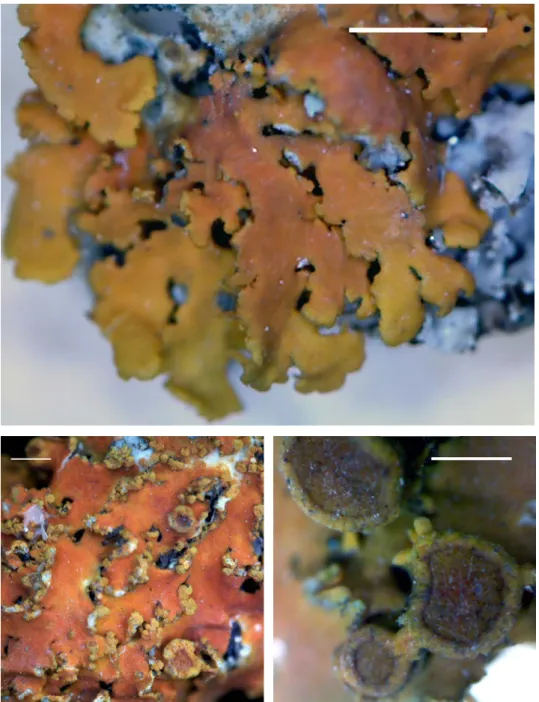
Franwilsia skottsbergii S. Y. Kondr., A. Thell, S.-O. Oh et J.-S. Hur, spec. nova (Fig. 6)
MycoBank nr. 824388.
Similar to Franwilsia renatae, but differs in having richly oil (Franwilsia-type plectenchyma) cortical layer of thallus and thalline margin of apothecium, in having mostly zeorine apothecia, in having much thicker thalline margin, in having Franwil- sia-type plectenchyma in true exciple, in having lower hymenium, in having shorter and wider ascospores, and in having black conidiomata, as well as in its distribution.
Type: Chile: Caleta Tototal Baja, on bark of tree [on small twigs], grow- ing together with Xanthoria ascendens, Caloplaca sp., Chrysotrix sp., and other crustose lichens; Franwilsia skottssbergii and Caloplaca sp. both damaged by lichenicolous fungus Endococcus aff. parietinarius. Lat.: 28° 17’ 15.6” S; Long.:
71° 10’ 37.6” W; Alt.: ca 203 m a.s.l. Coll.: Oh, S.-O., Hur. J.-S. (CL 130516), 14.11.2013 (KoLRI 020652 – holotype; for DNA SK H69 (=J69)).
Thallus 2–3 cm across, crustose, grey or whitish grey, continuous to some- what cracked, with portions 0.5–1 mm across, from rather thin to very thick and irregularly uneven to verruculose in the centre, often covered by highly uplifted apothecia with dull brownish orange discs as well as by blackish co- nidiomata. Hypothallus not observed. Thallus to 200–300 µm thick in section.
Cortical layer of thallus to 30–50 µm thick, with numerous oil agglomera- tions (not dissolved in K becoming somewhat yellowish or brownish and bet- ter seen in K, irregular, 3–7 µm across; oil of the Franwilsia-type), somewhat greyish, in places somewhat greyish or dirty in the upper portion owing to numerous small oil droplets; in lower portion more or less hyaline (lighter) with more or less scarce oil agglomerations; in some places seem to consist of two distinctly different portions: the upper to (7–)10–15 µm thick of epinecral layer or of hyphae parallely orientated to surface and without oil conglomer- ates), and the lower portion to 50–70 µm thick, somewhat brownish or greyish owing to numerous oil agglomerations (3–)5–7 µm diam./across; algal zone to 80–160(–180) µm thick.
Apothecia 0.7–1.5(–2) mm diam., and to 0.4–0.45 mm thick in section, ze- orine or lecanorine, mostly highly uplifted above thalline level and distinctly constricted at the basis, own margin/true exciple very thin, to 40–50 µm thick, from light yellowish or yellowish orange (lighter of disc) to bluish or bluish black (similar as in Franwilsia bastowii) to concolorous with disc, dull orange;
thalline margin entire or crenulate to developed only a side or at basis, grey- ish with whitish short line-like pseudocyphellae, somewhat eroded surface, sometimes with blackish conidiomata, often overgrowing by young smaller size apothecia; disc dull orange or dull brownish orange, sometimes dead,
colourless or greyish/concolorous with thalline margin or blackish in places and apothecia seem to be similar to Lecanora- or Rinodina-type; in section zeo- rine, where thalline exciple to 170 µm thick, with distinct cortical layer to 50 (–80) µm thick, distinctly Franwilsia-type, inspersed with oil agglomerations to 6–10 µm across; cortical layer of thalline exciple to 70(–100) µm thick (espe- cially on underside of apothecia, or in ‘conus’-like formations to 100(–120) µm thick, with numerous irregular oil agglomerations (oil of the Franwilsia-type);
true exciple to (30–)40–100(–120) µm thick in the uppermost lateral portion, to 20–40(–60) µm thick in lower lateral portion and especially not distinct in the basal portion where completely covered by oil agglomerations, probably to 40–50(–70) µm thick; true exciple with numerous oil agglomerations (oil of the Franwilsia-type) in both lateral and basal portion; algal zone continuous or of clusters to (50–)60–80(–100) µm diam. across, without oil; algal cells to (7–)12–18 µm diam./across, algae probably of the genus Asterochloris, richly inspersed with yellow drops of oil inside of algal cells (seen also in K); hyme- nium very thin, to (60–)70–80 µm high; epihymenium to 10–15 µm thick, dark brownish orange; subhymenium 150–230 µm thick, somewhat dirty, greyish or brownish, of the Franwilsia-type, with irregular oil agglomerations to 7–10
Fig. 6. Franwilsia skottsbergii (holotype), general habit (top left) and enlarged apothecia and thallus. Scale 2 mm (top left) and 1 mm (the others). (Photos of S. Kondratyuk)
µm diam./across in water (in K to 5–12 across or droplets to 7 µm diam.); para- physes slightly swollen towards the tips to 3.5 µm diam., poorly branched in the upper portion; asci 8-spored, often with ascospores of variegated size within the same ascus; ascospores elongated, slightly widened at the septum, and more or less attenuated to both ends; 9–13 × (4.5–)5–6 µm in water (25 measurements) and (8–)9–13(–14) × (4–)5–6(–7.5) µm in K (35 measurements);
ascospore septum to (2–)3–4 µm wide in water and (2–)3–4 µm wide in K.
Conidiomata to 0.1–0.2(–0.3) mm diam., blackish, often very abundant and making variegated black grey aspect of thallus; in section with blackish- bluish outermost cells of the upper half of exciple (in lower half usually hya- line), to 220–250(–280) µm diam. mostly regularly rounded; conidia narrowly bacilliform, 2.7–4 × 0.7–0.9(–1.2) µm.
Chemistry: Epihymenium and uppermost portion of lateral true exciple K+ purple with time becoming crimson, ascospore content becoming purple, too. Cortical layer of thallus, and thalline cortex of apothecium K–.
Ecology: Growing on bark of trees and twigs.
Etymology: This species is named after a Swedish botanist and explorer of Antarctica Carl Johan Fredrík Skottsberg (1880–1963).
Distribution: So far known from type collection in Chile, South America.
Taxonomic notes: This species is characterised by richly inspersed by oil subhymenium (with numerous irregular oil agglomerations, hereafter of Franwilsia-type (see also illustrations in Kondratyuk et al. 2009a, b), richly in- spersed true exciple (in both lateral and basal portions), as well as cortical layer of thallus and thalline exciple.
Franwilsia skottsbergii is similar to Franwilsia renatae (Wirth et S. Y. Kon- dr.) S. Y. Kondr., Kärnefelt, A. Thell, Elix, J. Kim, A. S. Kondratiuk et J.-S.
Hur in having greyish thallus and large zeorine apothecia with grey thalline margin and whitish pseudocyphellae, as well as in having richly oil exciple, and subhymenium, but differs in having richly oil cortical layer of thallus and thalline margin of apothecium, in having mostly zeorine apothecia (vs.
lecanorine), in having much thicker thalline margin (in section to 170 µm vs.
60–10 µm thick), in having thicker and Franwilsia-type plectenchyma in corti- cal layer (vs. 20–30 µm thick and palisade), in having Franwilsia-type plecten- chyma in true exciple (vs. leptodermatous paraplectenchymatous), in having lower hymenium (70–80 µm vs. 80–100 µm high), in having somewhat shorter and wider ascospores (9–13 × 5–6 µm vs. (10–)11–14(–15.5) × (3.5–)4–5 µm), as well as in its distribution (vs. African taxon), and in having black conidiomata (Wirth and Kondratyuk 2010).
Franwilsia skottsbergii is similar to F. bastowii (S. Y. Kondr. et Kärnefelt) S.
Y. Kondr., Kärnefelt, A. Thell, Elix, J. Kim, A. S. Kondratiuk et J.-S. Hur in hav- ing crustose thallus and zeorine apothecia often with bluish black own mar-
gin/true exciple, but differs in having rather thick grey or dull grey-whitish thallus (vs. whitish from endolithic to thick), in the lack of bluish or brownish black Rinodina-type apothecia, in having larger apothecia, in having much thicker and often more crenulate thalline margin, in having whitish pseudo- cyphellae especially well seen on thalline exciple, in having Franwilsia-type plectenchyma in cortical layer of thalline margin (vs. palisade), and in true exciple (vs. leptodermatous paraplectenchyma), in having narrower paraphy- ses tips (vs. 3–6(–7) µm) and poorly ramified (vs. richly ramified with 5–7(–10) long secondary branches, in having shorter and narrower ascospores (11–13
× 5–6 µm vs. (8–)10–15(–16) × (4.5–)5.5–7(–8) µm in water and much wider (10–)11–16(–18) × (5.5–)6–9(–11) µm in K), in having wider ascospore septum (3–4 µm vs. 2–3 µm wide), as well as in another substrate (vs. epilithic) and distribution (vs. Australian taxon), and in having black conidiomata (Kon- dratyuk et al. 2009a).
Franwilsia skottsbergii differs also from another Australian epilithic lichen F. kilcundensis (S. Y. Kondr. et Kärnefelt) S. Y. Kondr., Kärnefelt, A. Thell, Elix, J. Kim, A. S. Kondratiuk et J.-S. Hur in having shorter ascospores (9–13 µm vs. 13–18(–21) µm long), and in having black conidiomata (Kondratyuk et al.
2009b).
Unfortunately data on conidiomata of all species of the genus Franwilsia are hitherto missing.
Lichenicolous fungus Endococcus aff. parietinarius (Linds.) Clauzade et Cl.
Roux was identified preliminary because it differs in having longer and some- what narrower ascospores ((10–)13–14 × (3–)4–4.5(–5) µm vs. 10.5–12(–14) × (3.5–)4–5.5 µm). After ascospore measurements this material is closer to E.
ramalinarius (Linds.) D. Hawksw., but differs in having different host (mem- ber of the Teloschistaceae vs. Ramalinaceae) and in having semi-immersed ascomata (vs. immersed).
After ITS1/ITS2 nrDNA Franwilsia skottsbergii is positioned in outermost out position to the Franwilsia branch. However, we do hope that the further molecular data (i.e. on the 28S nrLSU and 12S mtSSU) will confirm closer po- sition of this taxon to the Franwilsia clade.
Gallowayella awasthiana S. Y. Kondr. et D. K. Upreti, spec. nova
(Figs 7–9)MycoBank nr. 824389.
Similar to Gallowayella sogdiana but differs in having smaller thallus, much longer thalline lobes, wider and mostly horizontally orientated thalline lobes, very long and easily distinct rhizines, smaller soredia, larger apothecia, much narrower ascospores, and narrower ascospore septum.
Type: India, Uttaranchal, Chamoli distr., Badrinath, Mana village, on twigs, growing together with Candelaria cf. concolor, Physcia, Phaeophyscia, Caloplaca aff. cerina aggr., Lecidella spp. Coll.: Shukla, V., Joshi, Y., 21.06.2005 (LWG 05–005210/B – holotype; voucher for DNA SK E15), (LWG 05–005210–
isotype; voucher for DNA SK E16).
Thallus 0.8–1(–3) mm across, but may form larger aggregations, foliose, deep yellow to reddish orange, often central portion dark reddish, or dark reddish orange to dark brownish reddish orange, deep brick orange or dark brownish yellow, while terminal portions of lobes much brighter yellowish, with punctiform to fissure-like yellowish-whitish or whitish yellow soralia along the thalline lobe edges well contrasting to dark upper surface of thallus.
Lobes to 2–4 mm long and (0.3–)0.5–0.7(–1) mm wide, more or less the same width throughout, neither becoming narrower, or becoming sometimes dis- tinctly widened towards the tips, scarce to richly branched; secondary lobules to 2–2.5(–3) mm long and 0.4–0.5 mm wide, mainly strip-like; often terminal and marginal portions of thalline lobes and lobules dissected into narrower portions 0.2–0.3(–0.5) mm wide and 0.4–0.7(–1) mm long; total width of the single lobe with all dissected portions and secondary lobules to 1.5 mm wide.
Underside white, usually with well distinct, very numerous (0.5–)0.8–1(–1.5) mm long Oxneria-type rhizines (see illustrations in Kondratyuk and Poelt 1997), yellow or white, especially well seen in overlapping portions of thallus.
Soralia punctiform at first (0.17–)0.2–0.3 mm long along the thalline lobe edge and gaped to 0.1–0.15(–0.2) mm wide (or to 0.2(–0.3) mm across), mostly marginal, of ‘micro-huculica’-type (i.e. with uplifted portions of upper cortex, somewhat micro-helmet, similar to Oxneria huculica, but smaller in size) (see illustrations in Kondratyuk et al. 2010), slightly brighter of thallus, yellowish white owing to naked medulla (similarly as in Oxneria ullophyllodes subsp.
subsorediosa (Räs.) ad int.), later in overmature thalli becoming somewhat larger to 0.3–0.6(–0.7) mm long (along the lobe edge) and to 0.15–0.2(–0.3) mm wide, usually very scarce and commonly empty, without soredia (seem to be washed or eaten). Soredia very small, to 20 µm diam., usually hardly seen, sometimes becoming isidious to 25–30 µm diam., and forming characteristic chains to 0.2(–0.3) mm long along the edge of lobes and the same wide; often seen among the rhizines, which are very close to lobe edges on underside, too.
Apothecia (0.5–)0.7–2(–2.5) mm diam., 0.28 mm thick in section, lecanor- ine, often with rhizines on thalline margin (what is characteristic for members of the genera Gallowayella and Oxneria (see illustrations in Kondratyuk 2004, Kondratyuk and Poelt 1997)), plane to somewhat undulating, constricted at basis, scattered to crowded, disc dark brownish yellow to brown, thalline margin to 0.1 mm wide mostly entire, rarely with soredious mass or rhizines;
in section zeorine, thalline exciple to 110–120 µm with well distinct to 30–35
Fig. 7. Gallowayella awasthiana (holotype), general habit (top) and enlarged fragments with apothecia (bottom). Scale 1 mm (top) and 0.5 mm (bottom). (Photos of S. Kondratyuk)