Di-(p-chlorophenyl)trichlorethane (DDT)
Herbert Keller
D D T is usually determined by a biological method, e.g. in the Drosophila assay. However, the accu
racy of this method is rather low. The quantitative determination of very small amounts of the in
secticide by a halogen determination with a colorimetric, spectrographic or polarographic method, is either relatively unspecific or requires a large expenditure on apparatus.
Very small amounts of D D T can be measured very accurately with carbonic anhydrase ( C A H ) by the inhibitory effect on the e n z y m e
1
) (see p. 7). The sensitivity which is attained can otherwise be obtained only by biological methods. Carbonic anhydrase is inhibited by D D T at concentrations at which other inhibitors (with the exception of sulphonamides) are inactive. A s the sample to be examined usually does not contain any sulphonamides, a special extraction and purification of the insecticide can be omitted.
Principle
Carbonic anhydrase catalyses the hydration of carbon dioxide to carbonic a c i d
2
) (7) C 0
2
+ H2
0 ~ = ± H2
C 03
H+ + H C O 3 -The equilibrium lies in favour o f carbon dioxide. By comparison with reaction (2), reaction (1) is a slow reaction. However, the equilibrium is attained relatively quickly even without a catalyst, so that a certain amount of expenditure on apparatus for recording enzyme activity is unavoidable.
The D D T inhibition of C A H is not competitive. The binding sites for the insecticide include not only the catalytically active "active centre", but also unspecific sites on the enzyme molecule. In addition, binding of D D T by contaminating material must be considered. Therefore for each enzyme preparation a new standard curve must be prepared.
Apparaturs for Measurements
A modified
3
) form of the apparatus designed by Maetz
4
^ has proved especially suitable for the deter
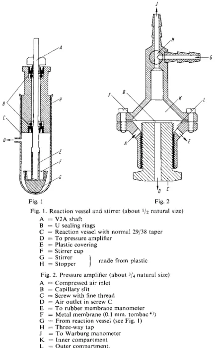
mination of the activity of C A H . It consists of the following (Fig. 1): The spindle of an electric motor is extended by a V2 A shaft at the end of which is a stirrer which has been widened to form a cup
like vessel to hold the substrate. The stirrer is just above the b o t t o m of the reaction vessel, which is covered with the enzyme-buffer solution. There is only a narrow clearance between the blades of the stirrer and the walls of the reaction vessel. On switching on the motor, both the enzyme-buffer solution and the contents of the stirrer cup are thrown onto the walls of the vessel. In this way an optimal gas exchange is guaranteed. The plastic stopper of the reaction vessel serves as a bearing for the stirrer shaft. The joints are sealed by U sealing rings. Parts of the stirrer shaft which might c o m e into contact with the reaction mixture are covered with plastic. The stirrer cup is made from the same material. Therefore contact between the reaction mixture and metal surfaces is impossible.
The CO2 liberated by dehydration can be measured in the ordinary Warburg manometer. However, it is better to record the pressure produced in the reaction automatically. A simple amplifier based on the principle of a reciprocally-acting pressure-reducing valve is suitable (Fig. 2): Air under constant pressure flows in through A and must pass through the capillary slit B before it can flow out again through the aperture D in the screw C. The pressure in the outer compartment can be measured with
1) H. Keller, Naturwissenschaften 39, 109 [1952].
2) Review: H. Gibian, Angew. Chem. 66, 249 [1954].
3) H. Keller, W. Miiller-Beisenhirtz and H. D. Ohlenbusch, Hoppe-Seylers Z. physiol. Chem. 316, 172 [1959].
4) / . Maet7, Bull. Soc. Chim. biol. 32, 830 [1950].
a rubber membrane manometer over the opening E. The movements of this rubber membrane rotate a mirror, which in its turn deflects the beam of a slit-lamp. The capillary slit B is formed by the screw
Fig. 2 Reaction vessel and stirrer (about V2 natural size)
= V 2 A shaft
= U sealing rings
= Reaction vessel with normal 29/38 taper
= T o pressure amplifier
= Plastic covering
= Stirrer cup
= Stirrer
= Stopper made from plastic Fig. 2. Pressure amplifier (about
3
U natural size) A = Compressed air inlet
= Capillary slit
= Screw with fine thread
= Air outlet in screw C
= T o rubber membrane manometer
= Metal membrane (0.1 mm. tombac*))
= From reaction vessel (see Fig. 1)
= Three-way tap
= T o Warburg manometer
= Inner compartment
= Outer compartment.
C and the metal membrane F. By pressure changes in the reaction vessel the slit B is made narrower or wider by F. A n increase in pressure in the inner compartment K leads to a narrowing of the slit,
*) Copper-base zinc alloy.
the air flow is impeded and the pressure in the outer compartment L increases. The screw C has a fine thread so that the width of the slit B can be regulated. The narrower the slit the greater is the sensitivity with respect to movements of the membrane F, so by adjustment of the screw and the flow of compressed air an amplification of the order of 1:100 can be obtained. The amplified pressure changes in the inner compartment are registered by the rubber membrane, mirror and slit-lamp beam.
The light beam activates a recorder (B. Lange, Berlin), consisting of a photocell, which continually follows the light beam and records its excursions on a synchronized drum. The running speed is 1 cm./sec.
If, with especially fast reaction rates, the photocell can no longer follow the light beam, the recordings can be made with a kymograph o n photographic paper.
5
Fig. 3. Recording Apparatus 1 = Warburg manometer
2 = Three-way tap 3 = Pressure amplifier
4 = Rubber membrane manometer 5 = Motor (2000 r.p.m.)
6 = Stopper
7 = Reaction vessel 8 = Ice bath 9 = Mirror (rotary) 10 = Slit-lamp 11 = Recorder
To standardize the apparatus the reaction vessel and the pressure amplifier can be connected by means of a three-way tap to a Warburg manometer (Fig. 3). During equilibration a connection between the closed reaction vessel and the outside atmosphere can be made by means o f the tap and the Warburg manometer. Measurements of activity and standardization are carried out at a constant temperature. The simplest method is to place the reaction vessel in an ice bath (Fig. 3).
The parts o f the apparatus are connected by PVC tubing (internal diameter 3 mm.).
Before using the apparatus for the determination of C A H activity two constants must be measured:
a) Amplification factor (f). With the three-way tap open to all positions, but with the tap of the Warburg manometer closed, successive pressures of 2, 4, 6, 8 and 10 cm. water are exerted on the amplifier head by means of the Warburg manometer. The recorder moves from the zero position to new positions which are marked.
The quotient
Pressure (cm./water) _ Manometer reading _ Deflection Recorder reading
is the amplification factor (f). This must be determined for each new series of measurements (e.g. daily).
b) Apparatus constant (k). So as to be able to give the activity of the enzyme in the usual Mitchell units
6
) it is necessary to k n o w the final pressure (apparatus constant k). With constant volumes and concentration of the reaction mixture, and constant temperature this value depends essen tially on the volume of the apparatus. The Warburg manometer is filled with mercury, the stirrer cup with substrate (solution I) and the reaction vessel with carbonic anhydrase solution (III).
Then the resulting pressure is measured by the usual Warburg technique with the three-way tap open to all positions. The enzyme catalysed reaction is allowed to proceed until the pressure no longer increases. (To protect the rubber membrane manometer it is recommended that the ampli
fier head is set to a lower sensitivity by loosening the screw C). Multiplication of the Hg reading in cm. by 13.595 gives the pressure in cm. water. It should be 40 — 50 cm. water. The rather difficult determination of the apparatus volume according to the m e t h o d of Damjanovic and Walen^ can be omitted.
Reagents
1. Sodium hydrogen carbonate, NaHC03, A. R.
2. Potassium hydroxide, KOH, 1 N, A. R.
3. Disodium hydrogen phosphate, Na 2 HP04-2 H2O, A. R.
4. Potassium dihydrogen phosphate, KH2PO4, A. R.
5. Dimethylformamide*), b. p. 153°C 6. Carbonic anhydrase, CAH
from bovine erythrocytes, see p. 632.
7. DDT**>
recrystallized from ethanol-ether, m. p. 108.5°C.
Purity of the e n z y m e preparation
The C A H preparation used should have a specific activity of at least 15 units/mg. according to Mitchell*\ corresponding to about 2 0 0 0 units/mg. according to Meldrum
7
K Lower activity considerably reduces the sensitivity of the method.
Preparation of Solutions (for ca. 80 determinations)
All solutions should be prepared with fresh, doubly distilled water.
I. Substrate:
Dissolve 1.5627 g. N a H C 0 3 in about 50 ml. doubly distilled water, add 3.8 ml. 1 N KOH and dilute to 100 ml.
II. Phosphate buffer (0.25 M; pH 6.8):
Dissolve 22.25 g. N a 2 H P 0 4 - 2 H 2 0 + 17.01 g. K H 2 P 0 4 in doubly distilled water and dilute to 1000 ml.
III. Carbonic anhydrase, CAH (0.1—1 u.g. protein/ml. buffer II):
Dissolve 10 mg. enzyme powder in 100 ml. doubly distilled water and dilute 1 ml. of this solution to 100 ml. with solution II. Dilute 1 ml. of this dilution to 10 ml. with solution II. With less active CAH preparations the last dilution should be smaller. It
*) e.g. Merck, N o . 3043.
**) e.g. from Fa. Geigy, Basle, Switzerland.
5
) Z. M. Damjanovic and R. J. Walen, Biochim. biophysica Acta 15, 586 [1954].
o) C. A. Mitchell, U. C. Pozzani and R. W. Fessenden, J. biol. Chemistry 160, 283 [1945].
7) N. U. Meldrum and F. J. W. Roughton, J. Physiology 80, 113 [1933].
is necessary to determine in preliminary experiments the concentration of enzyme which gives the highest, measurable reaction rate.
IV. DDT Standard:
Dissolve 10 mg. DDT in 10 ml. dimethylformamide, dilute 1 ml. of this solution to 10 ml. with dimethylformamide, mix and repeat the dilution a further three times. This gives solutions containing 1000, 100, 10, 1 and 0.1 ug. DDT/ml.
Stability of the s o l u t i o n s
Solution I should be stored, well stoppered, in an ice bath and should not be kept for longer than 6 hours. Solutions of carbonic anhydrase (stock solution III containing 100 (xg./ml.) keep for at least 2 weeks at 0 to 4 ° C without significant loss of activity.
Procedure
Experimental material
The samples should be dry. Tissue or biological fluids should be lyophilized or dried
in vacuoto constant weight. As DDT is quite stable no special precautions are necessary.
Add sufficient dimethylformamide to the powdered material so that a suspension is obtained that can be filtered. Stir the suspension for 15 min. at room temperature and filter through a hardened filter paper or centrifuge. Treat 0.5 ml. of the filtrate like the DDT standard solution (see under "Standaid curve").
Standard curve
To each 0.5 ml. DDT standard solution (IV) pipette 9.5 ml. enzyme solution (III) and incu
bate for 15 min. at room temperature. Then treat 1 ml. of each incubation mixture as de
scribed under "Assay". Calculate the enzyme activity of the five incubation mixtures inhi
bited with DDT (see below). Plot the values against the amounts of DDT used.
A s s a y
The following values must be measured:
(a) the rate of the non-catalysed reaction
(b) the rate of the reaction on addition of enzyme (from the difference (b) — (a) the enzyme activity is obtained)
(c) the rate of the reaction on addition of the enzyme inhibited with DDT (from the difference (c) — (a) the reduction in enzyme activity is obtained)
Therefore for each DDT determination three measurements are required, which only differ in the contents of the reaction vessel.
Pipette 1 ml. substrate solution (I) into the stirrer cup each time and for measurement (a) add 1 ml. phosphate buffer (II) for measurement (b) add 1 ml. CAH solution (III)
for measurement (c) add 1 ml. CAH solution (III) + sample
to the reaction vessel. Introduce the stirrer cup with the stopper into the reaction vessel and
equilibrate with taps 1 and 2 open (Fig. 3) for 15 min. Connect the amplifier head with the
reaction vessel by means of tap 2 and start the motor; at the same time set the recorder
drum in motion. The tracings of the recorder start from the moment of switching on. They
are stopped a few seconds later by opening tap 2 and switching off the motor.
Calculations
A straight line is drawn through the mean of the oscillatory record. The slope t g a of the line (refer to p. 8) is a measure of the reaction rate, tg a is multiplied by the amplification factor (f) (refer to p. 628). The corrected values for the measurements (a) and (b) are subtracted [(b) — (a)]. This gives a value for the enzyme catalysed reaction. Division by the apparatus constant (k) (refer to p. 629) gives the reaction rate v
0
of the enzyme preparation in Mitchell units6
*. T h e rate of the D D T inhi
bited reaction obtained in a similar manner from measurement (c) is Vj. The inhibitor coefficient h») is
V o - V i
h = , v
0
and the inhibition
I = . h
1 - h
After completing the measurements (a) to (c) determine the inhibition I, read off from the standard curve the corresponding amount of D D T and multiply by the dilution factor resulting from the preliminary treatment of the sample.
Example
Wheat flour (4.0 g.) was suspended in 5 ml. dimethylformamide and extracted by stirring for 10 min.
at room temperature. After centrifuging at high speed, 0.5 ml. supernatant was diluted with 9.5 ml.
enzyme solution, incubated for 15 min. and 1 ml. was taken for the assay.
Measurements (a): tgoq = 0.264; (b) tgoe
2
= 2.20; (c) t g a3
= 1.58. The amplification factor ( 0 and apparatus constant (k) were 0.634 and 40.6 respectively. Therefore it follows that:( t g a
2
- t g a i ) X f (2.20 - 0.264) X 0.634v
0 0
= - • = = 0.0302k 40.6 (tg a
3
- tg ai) x f (1.58 - 0.264) X 0.634Vi
== = = 0.02055k 40.6 0.0302 - 0.02055
h =
0 ^ 3 0 2 ^ ° '
32
I =
__M2__
1.0 - 0.32 = 0.47
Reading from the standard curve gave 2.0 fj.g. D D T / m l . dimethylformamide. Therefore there were 20 (j.g. D D T in 5 ml. dimethylformamide or 4.0 g. flour, i.e. 1 g. flour contained 5.0 \Lg. D D T .
Sources of Error
Errors in the standard or sample determinations can be due to only two reasons:
a) The enzyme preparation is impure. D u e to unspecific adsorption of the insecticide no measurable inhibition is observed in the low concentration range (below 10 [ig.).
b) The apparatus is leaking. It occasionally happens that tiny leaks occurring at the connections of the apparatus cause a very slight decrease in pressure. This error is easily detected since the non- catalysed reaction gives unreproducible curves which are too flat. By applying pressure by means 8) H. Netter: Theoretische Biochemie. Springer Verlag, Heidelberg 1959, p. 583 et seq.
of the Warburg manometer it is possible with the three-way tap to check whether the leak occurs in the area o f the reaction vessel or in the area o f the amplifier head. Naturally, a prerequisite for this procedure is that the three-way tap should be a perfect fit.
Specificity
In addition to D D T , the samples frequently contain the y-isomer of hexachlorocyclohexane (Lindane).
This contact insecticide does not inhibit at the concentrations of D D T which occur in practice.
Appendix
Isolation of carbonic a n h y d r a s e * ) (according to Keilin and M a n n
9
> ; modified)
I. Starting material: Bovine erythrocytes (preferably a citrate-containing concentrate, such as can be obtained from most of the large slaughter houses). 1000 ml. concentrate are equivalent to 3 500—4000 ml. blood. Wash 10000 ml. concentrate with the same volume of N a citrate saline (50 g. N a citrate + 80.0 g. N a C l to 10000 ml.). (CEPA centrifuge with a "serum sepa
rating cylinder"). Wash a further twice with the same volume of N a C l solution (90.0 g. N a C l / 10000 ml.) and add an equal volume of water to the serum-free suspension. A l l o w to stand for 30 min. at room temperature. With vigorous stirring, add 0.5 volumes of ethanol followed im
mediately by 0.5 volumes (per volume of the serum-free suspension) of chloroform. Continue stirring until the mixture solidifies to a paste. A l l o w to stand for two hours at 2 to 4 ° C then centrifuge. Decant the turbid, red supernatant and discard the precipitate.
II. Dialyse the supernatant in thin (2 cm. diameter) dialysis tubing for 24 hr. against running water.
III. Completely saturate the yellow, slightly turbid solution with ammonium sulphate at 2 ° C (plunger technique). Collect the precipitate by centrifuging in a cold room.
IV. Dissolve the precipitate in the smallest amount of distilled water and dialyse for 24 hr. against running water.
V. Saturate the dialysate with a m m o n i u m sulphate to 45 % saturation and centrifuge in refrigerated centrifuge (Spinco) for 60 min. at 4 0 0 0 0 g. Collect the supernatant and saturate with a m m o n i u m sulphate. Centrifuge again in a refrigerated centrifuge for 60 min. at 4 0 0 0 0 g. Discard the supernatant, dissolve the precipitate in a very small amount of distilled water and dialyse for 24 hr. in the cold against several changes o f distilled water.
VI. A d d 0.2 volumes of C
Y
-alumina-gel to the clear, colourless dialysate. Allow to stand for 30 min.in the cold and then centrifuge in a high speed centrifuge. Dialyse the clear supernatant for 24 hr. in the cold against distilled water.
VII. Lyophilize the dialysate. 10000 ml. b l o o d concentrate gives 2.5 to 3 g. highly purified enzyme.
1 mg. dry weight should contain 15 to 16.5 Mitchell u n i t s
6 )
. For the determination of activity, see p. 630.
*) A purer enzyme (chromatography on DEAE-cellulose and zone electrophoresis) has been de
scribed by S. Linkskog, Biochim. biophysica Acta 39, 218 [I960]. — The preparation (isolated according t o
9 )
) marketed by Mann Research Laboratories Inc., N e w York, U S A , is suitable.
9) D. Keilin and T. Mann, Biochem. J. 34, 1163 [1940].