Acta Microbiologica et Immunologica Hungarica
DOI:10.1556/030.66.2019.027
© 2019 Akadémiai Kiad´o, Budapest
ORIGINAL ARTICLE
* Corresponding author:
Dr. Tugce Unalan
Faculty of Medicine, Department of Medical Microbiology, Hacettepe University, Sihhiye, 06100, Ankara, Turkey
Phone: +90 0312 305 1560;
Fax: +90 0312 310 0580 E-mail:t.unalan91@gmail.com
An unusual case of peritoneal
dialysis-associated bacterial peritonitis caused by Weeksella virosa
TUGCE UNALAN
1* , ALPER KARAGOZ
2,
CIHANGUL BAYHAN
3, YASEMIN OZSUREKCI
3and GULSEN HAZIROLAN
11Faculty of Medicine, Department of Medical Microbiology, Hacettepe University, Ankara, Turkey
2Faculty of Molecular Biology and Genetics, Department of Microbiology, Usak University, Usak, Turkey
3Faculty of Medicine, Department of Pediatric Infectious Diseases, Hacettepe University, Ankara, Turkey
Received: May 27, 2019•Accepted: July 12, 2019
ABSTRACT
Weeksella virosais an atypical Gram-negative bacterium that does not grow on MacConkey agar. In this report, we present a 4-year-old female patient with Addison’s disease and end-stage renal failure secondary to focal sclerosing glomerulosclerosis. Continuous ambulatory peritoneal dialysis had been performed, and 3 months later, the patient developed fever, diarrhea, and vomiting. Peritonealfluid culture and dialysisfluid culture were positive forW. virosa. It was identified with Phoenix (BD, USA) and confirmed via 16S rRNA sequencing. It cannot be identified by Maldi Biotyper (Bruker). The isolate was found to be resistant to cephalosporins, ciprofloxacin, and amikacin by gradient test. Intraperitoneal cefepime was initiated but since antimicrobial susceptibility testing revealed cephalosporin resistance, therapy was changed to intraperitoneal meropenem. Following the removal of peritoneal dialysis catheter, fever, abdominal distention, and vomiting were resolved. Piperacillin, aztreonam, and carba- penems can be used for empirical therapy. Antimicrobial susceptibility testing should be performed to guide the choice of treatment. Removal of peritoneal dialysis catheter is an important step of management of this infection. To our knowledge, this is thefirst report ofW. virosa in a pediatric patient andfirst report from Turkey.
KEYWORDS
peritoneal dialysis catheter, peritonitis,Weeksella virosa, catheter-related infection
INTRODUCTION
Weeksella virosais a Gram-negative bacteriumfirst described in 1970 by Pickett and Manclark [1]. This organism grows on blood agar and chocolate agar after 48 h of incubation at 22, 35, and 42 °C, aerobic atmosphere, and forms cream-colored mucoid colonies with a non- diffusible yellowish pigment. It cannot be cultivated on MacConkey agar, which is accepted as a definitive property. The organism is catalase, oxidase, and indole positive. It is generally isolated from genitourinary tract and considered to be associated with sexual transmission.
This organism is frequently found in females and has a high correlation with comorbidities, such as renal diseases, obesity, liver diseases, and diabetes mellitus [2].
CASE PRESENTATION
A 4-year-old female patient with Addison’s disease and end-stage renal failure was admitted to the emergency department with diarrhea, vomiting, and fever. Four months before current 68 (2021) 1
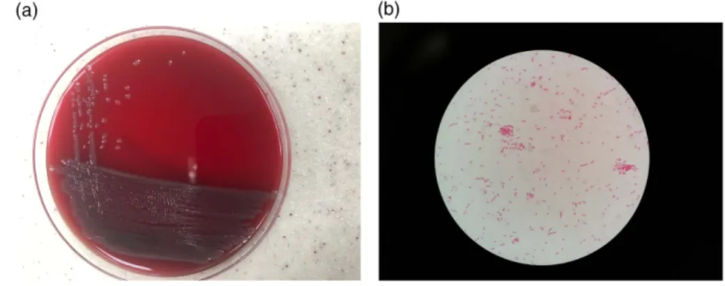
, 62 64–
admission, she had been diagnosed with nephrotic syndrome and chronic renal failure secondary to focal sclerosing glo- merulosclerosis and hospitalized for intractable hyperten- sion. Continuous ambulatory peritoneal dialysis (PD) had been begun to be performed 3 months ago. Upon her admission to the emergency department, there were no specific findings on physical examination and PD catheter exit-site was clean without any discharge. Intravenous ampicillin–sulbactam was started. Leucocyte or microorgan- ism on peritoneal fluid microscopic examination was not detected. On the second day of hospitalization, peritoneal fluid became cloudy and both peritonealfluid and dialysis fluid cultures yielded oxidase positive, catalase-positive Gram-negative bacterium that could not be identified by the method used routinely in our hospital, Bruker Maldi Biotyper (BD, USA) (Figure 1a and 1b). However, it was identified asW. virosaby Phoenix (BD, USA). The result was confirmed with polymerase chain reaction-based amplifica- tion of 16S rRNA.W. virosaNBRC 16016 (AB681031.1) was used as a positive control. DNA was isolated from 0.5 to 1 g of cell paste using MasterPure DNA purification kit (Qiagen, Hilden, Germany) following the manufacturer’s recommen- dations, with a modification of st/DL for cell lysis as de- scribed by Wu et al. [3]. The amplification of the 1,465-bp location of the 16S rRNA gene was performed using B162 forward primer (5′-CGCTCGTTGCGGGACTTAACCCAA- CATCTC-3′) and BR16SR reverse primer (5′-GAGAGTTT- GATCGTGGCTCAGATTGAACGC-3′) [4]. The cycles are initial denaturation at 95 °C for 3 min, 35 cycles of 95 °C for 20 s, annealing at 51 °C for 30 s, extension 72 °C for 1 min, andfinal extension 72 °C for 5 min. The amplified products were run and viewed in a 1.5% agarose gel (Sigma, St. Louis, MO, USA). Sequencing was performed using BigDye Terminator V3.1 Cycle Sequencing Kit (Applied Biosystems, MA, USA). DNA sequences of the purified products were identified using ABI Prism 3700 Genetic Analyzer (Applied Biosystems). The isolates were identified comparing the DNA reference isolates with data stored in the GenBank using the Basic Local Alignment Search Tool (BLAST version 2.0;
http://www.ncbi.nlm.nih.gov/BLAST) program. A phyloge- netic tree analysis was created using ClustalW MegAlign [5].
The antimicrobial susceptibility testing was performed and minimum inhibitory concentrations (MICs) were assessed by gradient test (bioMerieux, France). The results were interpreted using CLSI guideline [6]. The isolate was found to be resistant to cephalosporins, ciprofloxacin, and amikacin. The following susceptibility report and MICs (mg/L) were obtained: imipenem, 0.5, S; meropenem, 0.19, S;
cefepime, 48, R; ceftazidime,>256, R; cefotaxime,>256, R;
piperacillin, 16, S; ciprofloxacin, 1.5, I; and amikacin, 24, I.
Intraperitoneal (IP) cefepime was started, but after 6 days, therapy was changed to IP meropenem because cefepime resistance was detected in W. virosa. Intermittent fever, abdominal pain, and distention were persisted; the repeated blood culture yielded methicillin-resistant Staphylococcus haemolyticus, and intravenous teicoplanin was added to the therapy on the 14th day of hospitalization. Three days after this attempt, hematochezia was added to previousfindings without any abnormality in coagulation parameters or com- plete blood count and intravenous ampicillin–sulbactam therapy were changed to intravenous meropenem. Removal of PD catheter was planned but due to the patient’s renal comorbidities, it could only be removed after the 30thday of IP meropenem. Hemodialysis catheter was inserted. Fever, abdominal pain, vomiting, and abdominal distention complaints of the patient were resolved permanently after catheter removal. Parenteral antibiotic therapies were stopped on the 14thday of treatment.
W. virosais a rare pathogenic bacterium associated with diverse clinical presentations. There are nine different cases reported in the literature. Faber et al. [7] reported a 33-year- old female with spontaneous bacterial peritonitis and end-stage renal failure with PD and successful treatment with imipenem/cilastatin. Boixeda et al. [8] also reported spontaneous bacterial peritonitis in a 55-year-old male patient with hepatitis C infection and cirrhosis and had been successfully treated with cefoxitin. A urinary tract infection caused by W. virosa was outlined by Meharwal et al. [9].
Figure 1.(a) The macroscopic appearance ofW. virosaon blood agar after 24-h incubation. (b) The Gram-stained slide ofW. virosa
Acta Microbiologica et Immunologica Hungarica68 (2021) 1, 62–64 63
Manogaran et al. [10] described pneumonia and sepsis in a 53-year-old female patient where cefepime/vancomycin treatment had been failed, resulting in patient’s death.
Slenker et al. [11] reported four patients in which labial wound infection treated with incision and drainage in a 44-year-old female, amnionitis treated with ampicillin and gentamicin in 25-year-old female, urinary tract infection treated with trimethoprim/sulfamethoxazole in 26-year-old female, and sepsis in 31-year-old female where the treatment with aztreonam and tobramycin had failed. Finally, Toescu et al. [12] reported a 49-year-old female with craniotomy wound infection and ventriculitis, which were successfully eliminated with ceftriaxone and amoxicillin administration.
CONCLUSIONS
Our case was a 4-year-old girl who had renal comorbidities and PD-related bacterial peritonitis, which was successfully treated with meropenem and catheter removal. To our knowledge, this is the first report of a pediatric patient- infected with this bacterium andfirst report from Turkey.
Due to the small number of reports in the literature, the antimicrobial susceptibility profile of this organism has not been fully described. There are no species-specific antimicro- bial susceptibility testing forW. virosa, yet it has been studied using the “other non-Enterobacteriaceae” Gram-negative bacilli table of CLSI guidelines. The organism is reported to be susceptible to piperacillin, monobactams, cephalosporins, fluoroquinolones, and carbapenems. Aminoglycosides, nali- dixic acid, and nitrofurantoin should be avoided due to the reported resistancein vitro[10]. Our isolate was found to be resistant to cephalosporins. Although cephalosporin resistance was not described previously forW. virosa, high rates of this resistance were reported for non-enteric Gram-negative bacilli.
In a recent study, ceftriaxone resistance was reported as 49.2%
forAlcaligenesspp., 37.2% forBurkholderia cepacia, 42.4% for Chryseobacterium spp., 91.7% forStenotrophomonas malto- philia, 46.6% forPseudomonasfluorescens/putida, and 77.8%
forOchrobactrum anthropi[13].
Even thoughW. virosais a very rare organism, it should be considered when an atypical growth of oxidase and catalase positive Gram-negative bacterium is detected in blood, peri- toneal, or urinary cultures. W. virosa does not grow on MacConkey agar, which is a distinctive property of this bacterium. Empirical therapy with piperacillin, aztreonam, or carbapenems is recommended. However, ciprofloxacin and aminoglycosides should not be used empirically unless anti- microbial susceptibility testing was performed. In addition, the possibility of cephalosporin resistance should be kept in mind.
Acknowledgements:The authors declare nofinancial support regarding this publication.
Conflict of Interest:The authors declare no conflict of interest regarding this publication.
REFERENCES
1. Pickett, M. J., Manclark, C. R.: Nonfermentative bacilli associ- ated with man. I. Nomenclature. Am J Clin Pathol54, 155–163 (1970).
2. Holmes, B., Steigerwal, A. G., Weaver, R. E., Brenner, D. J.:
Weeksella virosagen. nov, sp. nov. (formerly group IIF), found in human clinical specimens. Syst Appl Microbiol8, 185–190 (1986).
3. Wu, D., Hugenholtz, P., Mavromatis, K., Pukall, R., Dalin, E., Ivanova, N. N., Kunin, V., Goodwin, L., Wu, M., Tindall, B. J., Hooper, S. D., Pati, A., Lykidis, A., Spring, S., Anderson, I. J., D’haeseleer, P., Zemla, A., Singer, M., Lapidus, A., Nolan, M., Copeland, A., Han, C., Chen, F., Cheng, J. F., Lucas, S., Kerfeld, C., Lang, E., Gronow, S., Chain, P., Bruce, D., Rubin, E. M., Kyrpides, N. C., Klenk, H. P., Eisen, J. A.: A phylogeny-driven genomic encyclopaedia of Bacteria and Archaea. Nature462, 1056–1060 (2009).
4. Lang, E., Teshima, H., Lucas, S., Lapidus, A., Hammon, N., Deshpande, S., Nolan, M., Cheng, J. F., Pitluck, S., Liolios, K., Pagani, I., Mikhailova, N., Ivanova, N., Mavromatis, K., Pati, A., Tapia, R., Han, C., Goodwin, L., Chen, A., Palaniappan, K., Land, M., Hauser, L., Chang, Y. J., Jeffries, C. D., Brambilla, E. M., Kopitz, M., Rohde, M., Göker, M., Tindall, B. J., Detter, J. C., Woyke, T., Bristow, J., Eisen, J. A., Markowitz, V., Hugenholtz, P., Klenk, H. P., Kyrpides, N. C.: Complete genome sequence ofWeeksella virosatype strain (9751). Stand Genomic Sci4, 81–90 (2011).
5. https://www.ncbi.nlm.nih.gov/genbank/.
6. Clinical and Laboratory Standards Institute: Performance Stan- dards for Antimicrobial Susceptibility Testing, 27th Edition.
CLSI supplement M100. Clinical and Laboratory Standards Institute, Wayne, PA, 2017.
7. Faber, M. D., del Busto, R., Cruz, C., Mezger, E.: Response of Weeksella virosa peritonitis to imipenem/cilastin. Adv Perit Dial7, 133–134 (1991).
8. Boixeda, D., de Luis, D. A., Meseguer, M. A., Aller, R., Martin de Argila, C., Lopez Sanroman, A.: A case of spontaneous peritonitis caused by Weeksella virosa. Eur J Gastroenterol Hepatol10, 897–898 (1998).
9. Meharwal, S. K., Taneja, N., Sharma, S. K., Sharma, M.:
Complicated nosocomial UTI caused by nonfermenters. Indian J Urol18, 123–128 (2002).
10. Manoragan, M., Marnejon, T., Sarac, E.: Pneumonia and sepsis due toWeeksella virosain an immunocompromised patient.
Infect Dis Clin Pract12, 286–287 (2004).
11. Slenker, A. K., Hess, B. D., Jungkind, D. L., De Simone, J. A.:
Fatal case of Weeksella virosa sepsis. J Clin Microbiol 50, 4166–4167 (2012).
12. Toescu, S. M., Lacey, S., Low, H. L.: First report of postopera- tive intracranial Weeksella virosa infection. Acta Neurochir 159, 2235–2238 (2017).
13. Sader, H. S., Jones, R. N.: Antimicrobial susceptibility of uncommonly isolated non-enteric Gram-negative bacilli. Int J Antimicrob Agents25, 95–109 (2005).
Acta Microbiologica et Immunologica Hungarica– 68 (2021) 1,
64 62–64