(Romania), Department of Applied Physics – Aalto University (Finland), Physics Department – Institute for Energy Technology (Norway), Helmholtz Zentrum Geesthacht – Centre for Materials and Coastal Research (Germany), National Institute for R&D of Isotopic and Molecular Technologies, Cluj-Napoca (Romania) and Department of Food Engineering – University of Szeged (Hungary).
High concentration aqueous magnetic fluids: structure, colloidal stability, magnetic and flow properties
Magnetic fluids, with the unique coexistence of magnetic and liquid properties, exhibit fascinating phenomena such as magnetic field induced pattern formation (shown in the bottom corners of the cover image). A wide range of advanced techniques were used to study two types of water based magnetic fluids with electro-steric (oleic acid, blue) and electrostatic (citric acid, green) nanoparticle stabilization, showing both similarities and important differences in their microscopic and macroscopic properties.
See K. D. Knudsen, L. Vékás et al., Soft Matter, 2018, 14, 6648.
rsc.li/soft-matter-journal
Cite this:Soft Matter,2018, 14, 6648
High concentration aqueous magnetic fluids:
structure, colloidal stability, magnetic and flow properties†
Corina Vasilescu, abM. Latikka, cK. D. Knudsen, *deV. M. Garamus, f V. Socoliuc, b Rodica Turcu, gEtelka Tomba´cz, hDaniela Susan-Resiga, bi R. H. A. Ras cjand L. Ve´ka´s *b
This paper is an in-depth analysis devoted to two basic types of water based magnetic fluids (MFs), containing magnetite nanoparticles with electrostatic and with electro-steric stabilization, both obtained by chemical coprecipitation synthesis under atmospheric conditions. The two sets of magnetic fluid samples, one with citric acid (MF/CA) and the other with oleic acid (MF/OA) coated magnetic nanoparticles, respectively, achieved saturation magnetization values of 78.20 kA m1 for the electrostatically and 48.73 kA m1 for the electro-sterically stabilized aqueous ferrofluids which are among the highest reported to date. A comprehensive comparative analysis combining electron microscopy, X-ray photoelectron spectroscopy, attenuated total reflectance Fourier transform infrared spectroscopy, vibrating sample magnetometry, small-angle X-ray and neutron scattering, dynamic light scattering and magneto-rheometry revealed similarities and essential differences on the microscopic and macroscopic level between the two kinds of water-based ferrofluids. While the saturation magnetization values are quite different, the hydrodynamic volume fractions of the highest concentration MF/CA and MF/OA samples are practically the same, due to the significantly different thicknesses of the particles’ coating layers. The results of volume fraction dependent structure analyses over a large concentration range by small-angle X-ray and neutron scattering, correlated with magneto-rheological investigations for the electrostatically stabilized MFs, demonstrate formation of short chains of magnetic nanoparticles which are relatively stable against coagulation with increasing concentration, while for MFs with electro-steric stabilization, magnetic field and shear rate dependent loosely bound structures are observed. These particle structures in MF/OA samples manifest themselves already at low volume fraction values, which can be attributed mainly to magnetic interactions of larger size particles, besides non-magnetic interactions mediated by excess surfactant.
aDepartment of Applied Chemistry and Organic and Natural Compounds Engineering, Faculty of Industrial Chemistry and Environmental Engineering, Politehnica University Timisoara, Carol Telbisz 6, 300001 Timis-oara, Romania
bLaboratory of Magnetic Fluids, Center for Fundamental and Advanced Technical Research (CFATR), Romanian Academy, Mihai Viteazul Str. 24, 300223 Timisoara, Romania. E-mail: vekas.ladislau@gmail.com
cDepartment of Applied Physics, School of Science, Aalto University, Puumiehenkuja 2, 02150 Espoo, Finland
dPhysics Department, Institute for Energy Technology (IFE), 2027 Kjeller, Norway. E-mail: kenneth.knudsen@ife.no
eDepartment of Physics, Norwegian University of Science and Technology (NTNU), 7495 Trondheim, Norway
fHelmholtz-Zentrum Geesthacht, Centre for Materials and Coastal Research, Max-Planck-Str. 1, 21502 Geesthacht, Germany
gNational Institute for Research and Development of Isotopic and Molecular Technologies (INCDTIM), Donat Str. 67-103, 400293 Cluj-Napoca, Romania
hDepartment of Food Engineering, Faculty of Engineering, University of Szeged, Moszkvai krt. 5-7, H-6725 Szeged, Hungary
iFaculty of Physics, West University of Timisoara, V. Parvan Bd. 4, 300223 Timisoara, Romania
jDepartment of Bioproducts and Biosystems, School of Chemical Engineering, Aalto University, Kemistintie 1, 02150 Espoo, Finland
†Electronic supplementary information (ESI) available: Fig. S1. ATR-FTIR spectra measured from dried solutions of the electrostatically stabilized magnetic fluid, uncoated iron oxide nanoparticles and citric acid. The pH of the MF/CA sample wasB7 and that of the reference samples was adjusted toB7. Fig. S2. ATR-FTIR spectra measured from dried solutions of the electro-sterically stabilized magnetic fluid, uncoated iron oxide nanoparticles and oleic acid. The pH of the MF/OA sample was B9 and that of the reference samples was adjusted toB9. See DOI: 10.1039/c7sm02417g
Received 8th December 2017, Accepted 30th June 2018 DOI: 10.1039/c7sm02417g
rsc.li/soft-matter-journal
PAPER
Published on 03 July 2018. Downloaded by UNIVERSITY OF SZEGED on 8/4/2021 9:15:53 AM.
View Article Online
View Journal | View Issue
1. Introduction
To ensure long-term colloidal stability of magnetic fluids (MFs), the overall particle interaction potential should provide an energy barrier in order to keep particles apart and homogeneously distributed in the volume of the carrier liquid.1 Attractive van der Waals and magnetic forces are ubiquitous and therefore must be balanced by Coulombic, steric or other interactions to control the colloidal stability of dispersed nanoparticle systems, even in intense and strongly non-uniform magnetic fields.2–5 High saturation magnetization ferrofluids should have the magnetic particle volume fraction as large as possible and, at the same time, avoid aggregate formation. These requirements are hard to satisfy, especially in the case of water based magnetic fluids. Long-term colloidal stability of concentrated magnetic fluids is more challenging for aqueous than for organic carriers. The increase of the physical volume fraction necessary to attain high saturation magnetization involves a corresponding increase of the hydrodynamic volume fraction, however to different extents depending on the stabilization mechanism, electrostatic or electro-steric.6 The advantage of electrostatically stabilized fluids is the reduction of the total suspended material at constant magnetic volume fraction compared with a surfactant stabilized fluid,4due to the much greater thickness of the steric stabilizing layer.
The surface charge has the main role in electrostatic stabili- zation of aqueous magnetic fluids,7consequently the pH and ionic strength of the dispersion medium strongly influence the stability of the ferrofluid.3,8 Surface ions are produced from surface groups through acid–base reactions (surface iron atoms are bridged by OH, an amphoteric group) or by complexing agents (ligands) of some surface atoms (e.g.citrate ions bound to iron atoms).9The adsorption of complexing ligands leads to a shift of the IEP value (the isoelectric point, i.e., the pH at which a colloidal particle carries no net electrical charge and so loses its electrostatic stabilization); using citrate ligands the coagulation range is shifted to pHo4. The colloidal stability is ensured by a balance between magnetic, van der Waals, hard core repulsion and screened electrostatic interactions of particles,10–12 as well as the osmotic pressure and ionic strength controlling the state of interaction of the dispersion. Exploring a very wide range of particle volume fraction values, 1–30%,13–15a critical volume fraction was determined expressing the freezing of the rotational dynamics of magnetic nanoparticles. The critical volume fraction strongly depends on the size of nanoparticles and the ionic strength of the dispersion.
Engineering applications, such as sink-float separators, stimulated the synthesis of relatively concentrated aqueous ferrofluids (saturation magnetization up to 38 kA m1) with surfactant double layer steric stabilization, a procedure intro- duced by Shimoiizaka and coworkers,16using sodium dodecyl- benzene sulfonate, poly(oxyethylene)nonylphenylethers, and di(2-ethylhexyl) adipate, which adsorb as a second layer on the chemisorbed oleic acid first layer. Increased dilution stability of water based ferrofluids was achieved by steric stabilization with lauric acid as the particle surface coating agent.17Mostly motivated
by biomedical and biotechnology applications,18–21the electro- steric stabilization of iron–oxide nanoparticles in an aqueous medium is widely applied, using various kinds of stabilizing bilayers, to produce stable water based magnetic fluids.22–27In the case of the fatty acids normally used as surfactants for forming double layers, the first layer is chemically bound on the particle surface (–COOH binds to the FeROH site), while the second layer forms via hydrophobic interaction of alkyl chains. This thick coating can effectively hinder the aggregation of magnetite particles due to the combined steric and electrostatic stabilization.28,29This procedure requires an excess of free surfactant in solution, which leads to the appear- ance of micelles that can affect negatively the stabilization of magnetic particles,30,31 as it was evidenced earlier in the case of ferrofluids with organic carriers.32 Unlike the charge stabilization of aqueous magnetic fluids, the stabilization in the above case is predominantly determined by steric factors due to the increase in the average distance between particles and the decrease in the dipole–dipole interaction energy that limits cluster formation.33The usually low iron oxide volume fraction (well below 10%) limits the saturation magnetization of water based magnetic fluids to approx. 100–200 G (8–16 kA m1), mainly because of the particle aggregation tendency at higher concentrations5and especially in biorelevant aqueous media.34 Progress has been related to use of triblock polymers synthesized with controlled concentrations of carboxylic acid binding groups in central polyurethane segments and poly(ethylene oxide) end blocks, which were successfully used as hydrophilic steric stabilizers for Fe3O4 particles with a mean size of 8.8 nm to obtain water based magnetite ferrofluids;35the highest concen- tration of dispersed surface coated magnetite nanoparticles attained 45 wt%. By stabilizing 7 nm mean size g-Fe2O3
nanoparticles with a very thin (approx. 1 nm) layer of a short, water soluble diblock copolymer of acrylic acid and acrylamide, high concentration (up to 65 wt%) colloidally stable magnetic fluids resulted36with 2 M NaCl solution as the carrier leading to a saturation magnetization of approx. 42.6 kA m1.
Colloidal stability issues, involving nanoparticle size and magnetic moment, dipolar interactions, excess surfactant and agglomerate formation, have strongly influenced the highest values of saturation magnetization achieved. Compared to magnetic fluids with organic carriers, in aqueous magnetic fluids significantly stronger interparticle interactions result in larger and more compact clusters with higher fractal dimensions.37Small-angle scattering techniques using neutron beams (SANS) or X-rays (SAXS) are sensitive to the onset and development of structuring in magnetic fluids and offer reliable information on colloidal stability5,38–41 up to the highest values of particle volume fraction. Rheological and magnetorheological investigations in the case of water based magnetic fluids refer mostly to bio- ferrofluids42,43 or to biopolymer based magnetoresponsive particle suspensions.44While the magnetic nanoparticle content of these magnetoresponsive colloidal systems is limited to 1–2%, the magnetic interaction parameter is well above 1 for multi-core magnetic particles explaining the observed high magnetoviscous effect.43The applications in biotechnology and biomedicine of
Published on 03 July 2018. Downloaded by UNIVERSITY OF SZEGED on 8/4/2021 9:15:53 AM.
magnetic nanoparticle dispersions stabilized by electrostatic repulsion or surfactant bilayers are governed by their sensitivity to conditions such as pH and ionic strength and/or protein adsorption in the biological environment. This is especially important when these particles are to be administered into a living organism and the colloidal dispersions therefore need to be very stable at both neutral pH and high ionic strength.
Considering the good colloidal behaviour of citric acid (CA) stabilized ferrofluids at different pH and electrolyte composi- tions, as well as their non-toxicity, CA coating of magnetic nanoparticles (MNPs) could be envisaged as a good strategy to increase the stability of MNPs in environmental applications,45as well as in preparation of high magnetization nanocomposites to be used as contrast agents and drug targeting vehicles46,47which involves the encapsulation of initially individually stabilized iron oxide nanoparticles up to high volume fractions. Oleic acid (OA) covered magnetite nanoparticles as-prepared or encapsulated in magnetoresponsive nanocomposites48 can represent optimal candidates to be used in magnetic bio-separation,24,49environ- mental sensing, imaging and remediation50 and as contrast agents for imaging the brain, since OA bilayer coated MNPs bearing a protein corona more enriched in lipoproteins and albumin than complement and immunoglobulin proteins might better escape the immune system.51OA bilayer structures were applied also to facilitate the transfer of iron oxide nanoparticles (IONPs), obtained by thermal decomposition of iron carboxylate salts at high temperature, from hexane to water.50Beside biotech- nology and biomedical applications, the aqueous ferrofluids with iron oxide nanoparticles characterized by high magnetization at saturation and long-term colloidal stability are of particular interest for magneto-gravimetric separators16,17,52–55and require large-scale cost-effective manufacturing, favouring ferrofluid synthesis by chemical coprecipitation.56
In this paper two types of highly concentrated water based magnetic fluids, one with electrostatic and the other with electro-steric stabilization, were prepared and investigated. In both cases the magnetite nanoparticles were synthesized by chemical coprecipitation, but the samples are significantly different concerning the particle hydrodynamic volume fraction and the highest saturation magnetization achieved, depending on the stabilization procedure applied. Manifold advanced techniques, such as TEM, DLS, SAXS, SANS, XPS, ATR-FTIR, VSM and magneto- rheometry, were applied to probe and evaluate the differences and similarities concerning the structure and behaviour of the two kinds of water based magnetic fluid samples. To our best knowledge these are among the highest magnetization water based magnetic fluids synthesized and comparatively analysed.
2. Materials and methods
2.1. Materials
The materials used to obtain the citrate sample were: FeCl36H2O, Z99%; FeCl24H2O, Z99.0%; NH4OH, 28.0–30.0%; citric acid monohydrate,Z99.5%; and acetone,Z99.8%. All primary materials were purchased from Sigma-Aldrich and used as received.
The materials used in the case of the oleic acid sample were:
FeCl3and FeSO4, purchased from Merck; and oleic acid (technical grade, 90%), purchased from Sigma-Aldrich. Ammonium hydroxide NH4OH 25% was purchased from a local manufacturer.
2.2. Synthesis of water based magnetic fluids with electrostatic stabilization
A successful synthesis route for magnetite nanoparticles with electrostatic stabilization in a water carrier, only slightly different from Massart’s procedure,7was described in ref. 57–59.
Iron oxide nanoparticles were synthesized by coprecipitation of an aqueous mixture of FeCl3and FeCl2salts and stabilized with citric acid near pH 7.2,5743.3 g of FeCl3and 15.9 g of FeCl2were dissolved in 1440 g of Milli-Q water. 160 mL of NH4OH was added to precipitate the particles in ambient conditions under vigorous stirring, with the pH reaching up to 11. 50.4 g of citric acid monohydrate was added after 5 minutes of stirring to stabilize the nanoparticles. Stirring was continued for an additional 5 minutes after which the supernatant was removed using magnetic decantation. 200 mL of Milli-Q water and 8.4 g of citric acid monohydrate were added and the suspension was stirred for an additional 5 minutes. The suspension was washed several times with acetone and Milli-Q water using magnetic decantation, and after that excess water was evaporated at room temperature to achieve a nanoparticle concentration of 20 vol%.
A series of samples from 0.5 vol% to 20 vol% were prepared by diluting the concentrated ferrofluid with Milli-Q water.
2.3. Synthesis of water based magnetic fluids with electro- steric stabilization
Iron oxide nanoparticles were synthesized by chemical coprecipi- tation from FeCl3and FeSO4salt solutions by addition of a base and subsequently coated with oleic acid at 801C according to the basic procedure outlined in ref. 28. In a typical synthesis, 60 grams of FeSO4 was dissolved in 200 mL distilled water and mixed with 130 mL of a FeCl3 solution of appropriate concentration. The Fe3+/Fe2+ ratio used was adjusted to 1.7 since the preparation was done in atmospheric conditions in the presence of oxygen. This mixture was heated to 801C under vigorous mechanical stirring and precipitated with the use of a concentrated ammonia solution, with the pH value of the reaction medium approaching 11. Immediately after the formation of a black precipitate, 25 mL of oleic acid was added and the stirring was continued for 15 minutes.
The obtained precipitate was washed several times with distilled water to remove residual salts and then dispersed in water with the addition of ammonia at pH around 8.5.
After repeated purification steps the above laboratory scale synthesis procedure leads to approx. 50 g OA coated hydrophilic magnetite NPs. Dilution series of the two ferrofluids were prepared (Tables 1 and 2) since their concentration dependent properties are of importance. The solid matter contents of ferrofluids range from 0.5% to 20% volume fraction for the electrostatically stabilized particles (MF/CA) and from 0.5% to 14% for the electro- sterically stabilized ones (MF/OA). In both cases the pH varies with dilution, but not by more than approx. 1.2 units. The citrated
Published on 03 July 2018. Downloaded by UNIVERSITY OF SZEGED on 8/4/2021 9:15:53 AM.
samples have a pH between 6.4 and 7.1, while the oleic acid samples present pH values between 8.0 and 9.2. Small quantities of the most concentrated samples of both types of water based magnetic fluids were placed on NdFeB permanent magnets to illustrate their strong magnetic response (Fig. 1).
It is worth mentioning that it is possible to synthesize both types of nanoparticles under the same conditions by using the so-called post-coating method where chemically adsorbing compounds like CA or OA are adsorbed on purified naked iron oxide NPs obtained in a single batch, as we have done in several cases before.34,51,60,61The drawback of this synthesis approach is that only low concentration samples can be prepared, while our goal in this work was to prepare and characterize highly concentrated magnetic fluids.
2.4. Characterization methods
The solid (physical), hydrodynamic and magnetic size distributions were studied using transmission electron microscopy (TEM),
dynamic light scattering (DLS) and magnetogranulometry, respec- tively. Different characterization techniques, such as X-ray photo- electron spectroscopy (XPS), zeta potential (NanoZS), Dynamic Light Scattering (DLS), Small-Angle X-ray Scattering (SAXS), Small-Angle Neutron Scattering (SANS) and magnetorheometry were used to evaluate particle surface properties, surfactant adsorption, colloidal stability at different pH and particle volume fraction values, as well as cluster formation and flow properties.
2.4.1. TEM.Transmission electron micrographs of the nano- particles were recorded with FEI Tecnai 12 Bio Twin and STEM Hitachi HD-2700 transmission electron microscopes. The TEM images were used to determine the statistics of the diameters of the iron oxide region of the nanoparticles using ImageJ.62
2.4.2. Dynamic light scattering.The mean hydrodynamic diameter of the iron oxide particles was determined at 25 0.11C by dynamic light scattering (DLS), using the Nano ZS device from Malvern (UK), operating in backscattering mode at an angle of 1731. The concentration of the dispersions was set to give an optimal intensity ofB105 counts per second. The diluted samples were homogenized in an ultrasonic bath for 10 seconds prior to the measurements, after which 50 seconds relaxation was allowed. First the size, then the zeta potential, was measured in a disposable zeta cell (DTS 1070). The Smoluchowski equation was applied to convert electrophoretic mobility to an electrokinetic potential value. The accuracy of the zeta potential measurements is5 mV. Cumulant analysis was used to evaluate correlation functions and to calculate Z-average hydrodynamic sizes. In the case of unstable, coagulating systems only the given kinetic stage can be compared, since the measurable hydrodynamic size increases in time.
2.4.3. Vibrating sample magnetometry. Magnetogranulome- try.The full magnetization curves, including the initial suscepti- bility and saturation magnetization of aqueous ferrofluids, were determined using a vibrating sample magnetometer, VSM 880 – ADE Technologies, USA, at room temperature, in the field range 0 kA m1to 950 kA m1.
The temperature dependence of the magnetization at low field (0.1 T) under zero-field cooling (ZFC) and field-cooling (FC) conditions in the range 4–300 K was determined using a Cryogenic vibrating sample magnetometer.
The magnetization data were used for magnetogranulometry analysis63which consists of determination of the nanoparticle concentration and magnetic diameter distribution from non- linear regression of the experimental data with the magnetiza- tion modelM(H) for dense ferrofluids developed by Ivanov and coworkers:64
MðHÞ ¼MLðHeÞ
¼n ð1
Dm¼0
m Dð mÞ f Dð mÞ L m0m Dð mÞHeðHÞ kT
dDm
HeðHÞ ¼HþMLðHÞ
3 1þ 1
48dMLðHÞ dH
f Dð mÞ ¼ 1 Dms ffiffiffiffiffiffi
p2pe
lnðDm=D0Þ
ð Þ2
2s2
(1) Table 1 Water based citrated ferrofluid samples with electrostatic stabilization
Sample code
Stabilizing ligand (monolayer)
Solid volume fraction [%]
Density [g cm3]
MF/CA1 Citric acid 0.50 1.019
MF/CA2 Citric acid 2.27 1.093
MF/CA3 Citric acid 4.05 1.167
MF/CA4 Citric acid 5.82 1.241
MF/CA5 Citric acid 7.59 1.315
MF/CA6 Citric acid 9.36 1.389
MF/CA7 Citric acid 11.14 1.463
MF/CA8 Citric acid 12.91 1.537
MF/CA9 Citric acid 14.68 1.611
MF/CA10 Citric acid 16.45 1.685
MF/CA11 Citric acid 18.23 1.759
MF/CA12 Citric acid 19.99 1.833
Table 2 Water based surfacted ferrofluid samples with electro-steric stabilization
Sample code
Surfactant (double layer)
Solid volume fraction [%]
Density [g cm3]
MF/OA1 Oleic acid 0.50 1.021
MF/OA2 Oleic acid 1.00 1.042
MF/OA3 Oleic acid 2.00 1.084
MF/OA4 Oleic acid 4.00 1.168
MF/OA5 Oleic acid 6.00 1.252
MF/OA6 Oleic acid 8.00 1.337
MF/OA7 Oleic acid 10.00 1.180
MF/OA8 Oleic acid 12.00 1.504
MF/OA9 Oleic acid 14.00 1.588
Fig. 1 Normal field instability ‘‘spikes’’ of the water based magnetic fluid samples on a permanent magnet (NdFeB): (a) with electrostatic stabili- zation; and (b) similar with electro-steric stabilization.
Published on 03 July 2018. Downloaded by UNIVERSITY OF SZEGED on 8/4/2021 9:15:53 AM.
whereHe(H) is the effective field,Dmis the magnetic diameter, nis the nanoparticle concentration,m(Dm) =msp(Dm)3/6 is the magnetic moment of the particle, Lis the Langevin function L(x) = coth(x)1/x,m0is the magnetic permeability of vacuum, ms is the saturation magnetization of magnetite, k is Boltz- mann’s constant and T is the temperature. f(D) is the log- normal probability distribution function of the magnetic dia- meter with a D0 median and a s standard deviation of the diameter natural logarithm. The nonlinear regression of the magnetization curve with the model in eqn (1) provides numer- ical values forn,D0ands.
2.4.4. Surface spectroscopy
XPS.The chemical composition (atomic concentrations) and the chemical state of the atoms at the surface of the magnetic nanoparticle the dried ferrofluid sample Fe3O4/OA9 or Fe3O4/ CA12 were determined by X-ray Photoelectron Spectroscopy (XPS). The spectra were recorded using a SPECS spectrometer equipped with a dual-anode X-ray source Al/Mg, a PHOIBOS 150 2D CCD hemispherical energy analyzer and a multi-channeltron detector with a vacuum maintained at 1109Torr. The AlKaX-ray source (1486.6 eV) operated at 200 W was used for the XPS investigations. The XPS survey spectra were recorded at 30 eV pass energy and 0.5 eV per step. The high-resolution spectra for individual elements (Fe, C, O) were recorded by accumulating 10 scans at 30 eV pass energy and 0.1 eV per step. The samples were dried on indium foil to allow the XPS measurements. The sample surface was cleaned by argon ion bombardment (300 V).
Data analysis and curve fitting was performed using CasaXPS software with Gaussian–Lorentzian product functions.
ATR-FTIR. The magnetic fluids were also characterized using attenuated total reflectance Fourier transform infrared spectroscopy (ATR-FTIR). The spectra were recorded with a Unicam Mattson 3000 spectrometer and a PIKE Technologies GladiATR ATR accessory, using 4 cm1resolution and 32 scans per measurement. Measure- ments were performed by letting a droplet of sample solution dry on the diamond of the ATR accessory and measuring the spectrum of a dry sample in the wavenumber range 400–4000 cm1in absorbance mode. The background spectrum was recorded before measuring the samples and subtracted from the sample spectra.
2.4.5. SAXS.Small-angle X-ray scattering data were collected at the P12 BioSAXS beamline (PETRA 3, EMBL/DESY, Hamburg, Germany). A beam of size 0.1 0.2 mm2 and photon energy 18.44 keV was applied. Calibration of theq-range was done using the diffraction pattern of silver behenate. The sample–detector distance was 4 m and the range for the scattering vector was 0.005–0.65 Å1. Data were normalized to the intensity of the transmitted beam. 20 mL of solutions of samples and buffer (H2O) were put in glass capillaries of 1 mm diameter and placed on a Linkam Heating stage HFSX 350 (Surrey, UK) for temperature control (T= 200.11C). A single measurement (1 second) consisted of accumulation of 20 frames, each of 0.05 seconds. Possible radiation damage effects were checked by comparison with the reference (typically the first exposure) and automatically integrated and subtracted with a standard acquisition program.65
2.4.6. SANS.The small-angle neutron scattering experiments were carried out at the SANS installation at the JEEP-II reactor at Kjeller, Norway. The wavelength was set with a velocity selector (Dornier), using a resolution (Dl/l) of 10%. The beam divergence was set by an input collimator (18.4 or 8.0 mm diameter) located 2.3 m from the sample, together with a circular 7 mm aperture located close to the sample which defined the beam cross section.
The detector was a 59 cm active diameter, 3He-filled RISØ type, mounted on rails inside the evacuated detector chamber. The sample–detector distance was varied between 1.0 and 3.4 m, and the wavelengths used were 5.1 and 10.2 Å. The resultingq-range for the experiment was 0.006–0.3 Å1. The solutions were put in 1 mm Starna quartz cuvettes. The cells were placed onto a copper-base for good thermal contact and mounted onto the sample stage in the sample chamber. Standard reductions of the scattering data, including transmission corrections, were conducted by incorporating data collected from the empty cell, and the blocked-beam background, according to the formula given in eqn (2):
IScor¼ IS MS IBG
MBG
TS TEC
IEC MEC IBG
MBG
(2) Here IS is the measured scattered intensity for the sample inside the quartz cell,IBGis the intensity of the blocked-beam background, andIECis the intensity of the empty quartz cell.TS andTECare the transmission values (o1) of the sample and of the empty cell, respectively. All the measurements were normalized to the beam monitor counts (Mi) to compensate for possible variations in the incoming beam flux. Finally, all data were transformed to an absolute scale (coherent differential cross section (dS/dO)), making use of the intensity value registered in open beam measurements (no sample or cell), with a calibrated attenuator (Cd-mask with holes) in the beam,66before averaging radially to produce anI(q)vs. qpattern.
2.4.7. Modelling of SAXS & SANS data. To account for possible deviations from spherical entities, the scattered inten- sityI(q) was modelled as coming from a population of ellipsoids of rotation or cylindrical particles.I(q) can be described in a decoupling approximation (no correlation between the size/
orientation and position of particles) by the following equation:67 I(q) =I(0)P(q)S0(q) +B (3) where
P(q) =h|F(q)|2i (4)
S0(q) = 1 +b(q)[S(q)1] (5) b(q) = |hF(q)i|2/h|F(q)|2i (6) The inner bracketsh iin eqn (4) and (6) represent an average weighted by the distribution of particle sizes and/or orienta- tions, I(0) is the scattering at zero angle (proportional to the concentration of particles, contrast, and particle volume),P(q) is the form factor,F(q) is the amplitude of the form factor,S(q) is the structure factor, andS0(q) is the effective structure factor modified by the anisotropy and polydispersity of particles.
Published on 03 July 2018. Downloaded by UNIVERSITY OF SZEGED on 8/4/2021 9:15:53 AM.
In the case of a core shell ellipsoid of rotation of semiaxesa, aandb,F(q) is expressed as:
FðqÞ ¼3ðrcorersolventÞVcore
sinðqRÞ qRcosðqRÞ ðqRÞ3 þ3ðrshellrsolventÞVshell
sinðqðRþTÞÞ qðRþTÞcosðqðRþTÞÞ qðRþTÞ
ð Þ3
(7)
whereR= [b2sin2a+a2cos2a]1/2andais the angle between the axis of the ellipsoidaand the scattering vectorq, andTis the thickness of the shell. A log normal distribution ofbwas used in the analysis.
In the case of core–shell cylinders, these expressions are slightly modified:
FðqÞ ¼2ðrcorersolventÞVcoresinðqL=2 cosbÞ2J1ðqRsinbÞ qL=2 cosb
ð ÞðqRsinbÞ þ2ðrshellrsolventÞVshell
sinðqðLþTÞ=2 cosbÞ2J1ðqðRþTÞsinbÞ qðLþTÞ=2 cosb
ð ÞðqðRþTÞsinbÞ
(8)
whereLis the length of the cylinder core,Ris the radius of the cylinder core,J1is the first-order Bessel function, andbis the angle between theq-vector and the axis of the cylinder.
Two kinds of structure factors were used in the analysis:
(i) the excluded volume interaction calculated with the Percus–
Yevick approximation for the closure relation,68which requires as input parameters the hard sphere volume fraction and the effective hard sphere radius. The detailed expression for the function can be found in ref. 69. (ii) The screened Coulomb potential in the rescaled mean spherical approximation (RMSA) in the Penfold and Hayter form.70,71TheS(q) model requires as input parameters the temperature, the dielectric constant of the medium, the added salt concentration, and the volume fraction.
2.4.8. Magnetorheology. Rotational rheometer with a MR cell.
The measurements of the flow properties of ferrofluids were performed using a PHYSICA MCR 300 (Anton Paar, Germany) equipped with a magnetorheological cell (MRD 170/1T-SN80730989).
The MR cell has parallel plates of 20 mm diameter, with the gap being fixed at h = 0.2 mm. All the measurements were done at 201C. The magnetic flux density was determined using a Hall probe located as described in ref. 72 and 73 to ensure on-line measurement of the magnetic induction in the MR cell gap.
3. Results and discussion
3.1. Particle morphology and sizes: transmission electron microscopy
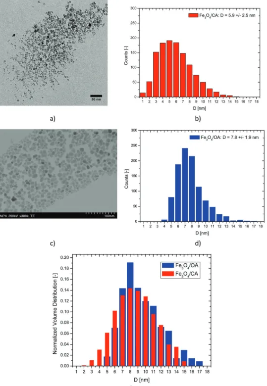
In Fig. 2 the TEM images and nanoparticle diameter histograms for the MF/CA and MF/OA samples are presented. The nano- particles in both ferrofluids show irregular shapes which is
typical for chemical coprecipitation synthesis. Table 3 presents the mean, standard deviation and skewness of the nanoparticle diameter, calculated based on measured sizes of more than 1000 particles for each ferrofluid sample. All distributions show positive skewness, in agreement with the log-normal distribution calculated from first principles by Cogoni and coworkers.74,75The MF/CA ferrofluid has particles of smaller average size than the MF/OA ferrofluid due to larger popula- tions of small diameter nanoparticles (1–4 nm). Fig. 2e shows the normalized volume distributions of magnetic nanoparticles in the MF/CA and MF/OA samples. Large nanoparticles are in excess in the MF/OA ferrofluid, while small nanoparticles are in excess in the MF/CA ferrofluid. This makes the MF/OA ferro- fluid more susceptible to magnetically driven structuring.
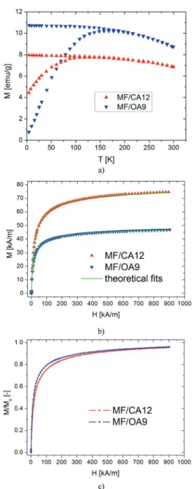
3.2. Magnetization curves and magnetogranulometry We considered the highest concentrations of both the electro- statically and the electro-sterically stabilized ferrofluid samples, i.e.MF/CA12 and MF/OA9 (cf.Tables 1 and 2) to investigate in detail the magnetization properties of the two aqueous colloidal systems.
The temperature dependence of the magnetization of the ferrofluid samples in ZFC and FC regimes (Fig. 3a) show a typical superparamagnetic behaviour. The ZFC curves exhibit a broad peak, indicating a transition from the magnetically blocked state at low temperatures to a superparamagnetic state at high temperatures. The blocking temperatures for magnetic nanoparticles depend on the size, size distribution, surface state and interparticle interactions,76the broad peak reflecting the relatively wide size range of magnetic nanoparticles typical for synthesis by chemical coprecipitation. From a qualitative examination of Fig. 3a one can conclude that the maximum of the ZFC curve, which could be assigned to the blocking temperature, is higher for the oleic acid ferrofluid (MF/OA) than for the citrate ferrofluid (MF/CA). This result is in agree- ment with the nanoparticles’ physical size and magnetic size determined from TEM and magnetogranulometry, respectively.
The broadness of the ZFC curve indicates the existence of dipolar interactions between nanoparticles in both types of ferrofluid.
The magnetization curves of the citric acid and oleic acid stabilized water based magnetic fluid samples are pre- sented in Fig. 3b. The initial susceptibility of the samples was 3.07 for MF/CA12 and 3.68 for MF/OA9 (Table 4). The saturation magnetization of the samples was determined using Chantrell’s method,77i.e.from the linear fit ofM(1/H) data at high fields. The saturation magnetizations are 78.20 kA m1for the MF/CA12 sample and 48.73 kA m1for the MF/OA9 sample (Table 4). The non-dimensional magnetization curves,i.e.the magnetization scaled to the saturation magnetization, reveal that citrate coated nanoparticles have a smaller magnetic diameter than the oleic acid coated ones. This is confirmed by the values obtained from magnetogranulometry in Table 4, which shows the results of the fitting. The magnetic volume fraction Fm (see eqn (12) below), mean magnetic diameter hDmi = D0es2/2 and magnetic diameter standard deviation
Published on 03 July 2018. Downloaded by UNIVERSITY OF SZEGED on 8/4/2021 9:15:53 AM.
dm¼D0es2=2 ffiffiffiffiffiffiffiffiffiffiffiffiffiffiffi es21 p
were calculated. The magnetic diameter of the nanoparticles is found to be 5.82.3 nm for the Fe3O4/CA sample and 6.72.7 nm for the Fe3O4/OA sample. The magnetic diameter is slightly smaller than the physical diameter, 5.9 2.5 nm and 7.8 1.9 nm, respectively, due to the nonmagnetic layer at the surface of the nanoparticles.78
Fig. 2 TEM images and nanoparticle diameter histograms for: (a and b) Fe3O4/CA and (c and d) Fe3O4/OA samples; and (e) normalized volume distributions.
Table 3 Nanoparticle diameter (physical size) statistics calculated from TEM images for MF/CA and MF/OA stabilized ferrofluid samples
Sample
Number of
particles Mean [nm] St. dev. [nm] Skewness []
Fe3O4/CA 1215 5.9 2.5 0.6
Fe3O4/OA 1014 7.8 1.9 1.0
Published on 03 July 2018. Downloaded by UNIVERSITY OF SZEGED on 8/4/2021 9:15:53 AM.
Thus, TEM, magnetogranulometry and ZFC/FC magnetization curves all show that the citrate stabilized nanoparticles have smaller sizes/volumes than the OA stabilized ones.
The magnetic interactions in MF/CA12 and MF/OA9 samples can be compared using the ratio g of the magnetic dipole–
dipole energy overkT:12 g¼m0m2
r3kT¼m0ms2v
kT F (9)
wherer%is the average distance between nanoparticles’ centers, m=ms*vis the magnetic moment,v=pd3/6 is the volume and F = v/%r3 is the solid volume fraction in the cubic lattice approximation. Eqn (9), rigorously valid for monodisperse magnetic nanoparticles, formally shows thatg*=g/Fis inde- pendent of the sample volume fraction and depends only on the nanoparticle diameter. On the other hand,gcan be directly computed from the measured initial magnetic susceptibilityw0 of the ferrofluid,12using an appropriate theoretical model for magnetization. The comparison ofg*values gives an indication of the susceptibility for magnetically driven structuring in different ferrofluid samples.
For the purpose of our investigation, because the magnetization model in eqn (1) is expressed in terms of the nanoparticle magnetic diameter,gwill be expressed as a function of the magnetic volume fraction. Given the nanoparticle size polydispersity in MF/CA and MF/OA ferrofluids, dg and dFm need to be defined for particles with diameters in the range (Dm,Dm+ dDm):
dg¼m0ms2vm
kT dFm; dFm¼vdn; dn¼nf Dð mÞ dDm; (10) where n is the nanoparticle concentration and f(Dm) is the magnetic diameter probability distribution function (see eqn (1)).
Thus, g can be expressed as an average over the diameter probability distribution function:
g¼p2m0ms2
36kT n ð
Dm6f Dð mÞ dDm (11) gis proportional to the 6-th moment of the magnetic diameter distribution. Using the general formula of the log-normal distribution momentshDkmi=Dk0exp(k2s2/2) and the expression of the volume fraction:
Fm¼ ð
dFm¼np 6 ð
Dm3f Dð mÞ dDm¼np 6Dm3
; (12)
one gets:
g¼pm0ms2 6kT Dm3
e9s2Fm: (13) Like in the monodisperse case,g*=g/Fmis independent of the sample volume fraction and depends only on nanoparticle diameter statistics, here assumed to be log-normal. In the Fig. 3 Magnetic behaviour of the non-diluted MF/CA and MF/OA ferro-
fluids: (a) ZFC/FC; (b) comparative experimental and fitted room tempera- ture magnetization curves, and (c) non-dimensional magnetization curves (corrected taking into account the demagnetizing field). The labels MF/CA12 and MF/OA9 correspond to the codes given in Tables 1 and 2.
Table 4 Saturation magnetization and nanoparticle diameter (magnetic size) statistics calculated from magnetization curves for CA and OA stabilized ferrofluid samples
Sample w0[] MS[kA m1]
M(1/H) lin.
fitR2 n[1022part per m3] D0[nm] s[] FitR2 Fm[] hDmi[nm] dm[nm]
MF/CA12 3.07 78.20 0.996 86.72 5.35 0.39 0.9999 0.14 5.8 2.3
MF/OA9 3.68 48.73 0.991 39.91 6.23 0.39 0.9997 0.10 6.7 2.7
Published on 03 July 2018. Downloaded by UNIVERSITY OF SZEGED on 8/4/2021 9:15:53 AM.
polydisperse case however, besides the average volume, g*
also depends on the standard deviation s. Because the log- normal skewness skw¼es2þ2 ffiffiffiffiffiffiffiffiffiffiffiffiffiffiffi
es21
p
is a monotonically increasing function of s and vice versa, g* accounts for the higher influence of large particles on the magnetic interactions taking place in the structure of ferrofluids.
By inserting eqn (13) in the low field approximation of eqn (1) (which is readily obtained using the Langevin function approximationL(x)Dx/3 for smallx) one obtains the following equation relatinggto the initial susceptibility of the ferrofluid:
w0¼g 3þg2
27þ g3
3888: (14)
Using the samples’ initial susceptibilities (Table 4) and mag- netic volume fractions determined with eqn (12) (Table 4),g*
was calculated to be 40.6 for MF/CA12 and 63.4 for MF/OA9.
One can notice thatg*is much larger for MF/OA9, which shows higher susceptibility for magnetically driven structuring in MF/OA9 than in MF/CA12.
Using eqn (12) for the magnetic volume fraction, the thick- nessrnmof the nonmagnetic layer can be calculated from the physicalFand magneticFmvolume fractions. If one assumes that the physical and magnetic diameter log-normal distribu- tions have the same s, but the mode D0p of the physical diameter is 2*rnm larger than the modeD0mof the magnetic diameter (D0p=D0m+ 2*rnm), the thickness of the nonmagnetic layer is 0.35 nm for MF/CA12 and 0.37 nm for MF/OA9. This result, bearing in mind that the nanoparticles in both ferro- fluids were synthesized by means of co-precipitation, shows that the thickness of the nonmagnetic layer is not influenced by the nature of the adsorbed molecules at the surface of the nanoparticles,i.e.citric acid and oleic acid, respectively. More- over, taking into consideration the dependence on the thick- ness of the surfactant layer rs of the maximum magnetic dipole–dipole interaction parameter per particle:
l¼pm0ms2
144kT Dm6 Dmþ2rnmþ2rs
ð Þ3 (15)
it follows that the magnetic interparticle correlations increase with decreasing surfactant layer thickness.
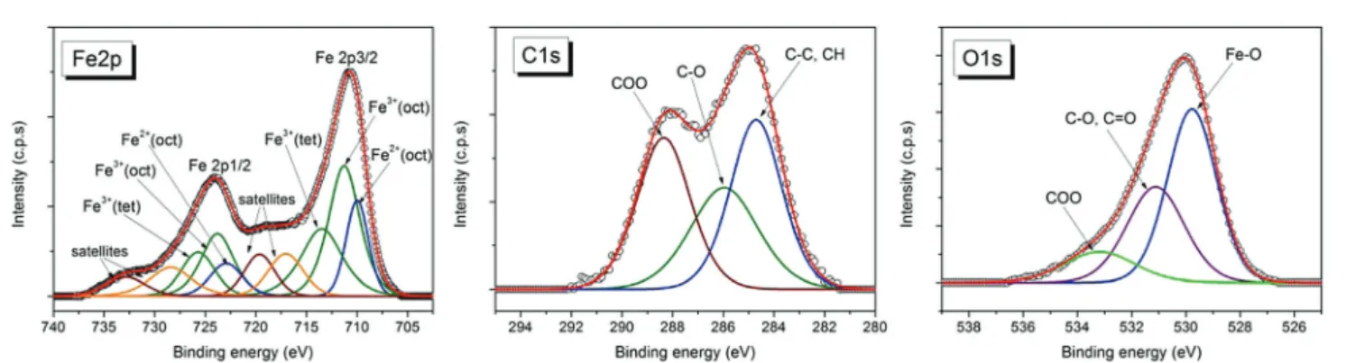
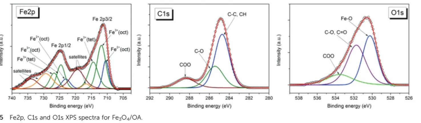
3.3. Particle surface properties: XPS and ATR-FTIR analyses Fig. 4 and 5 show high resolution XPS spectra of Fe2p, C1s and O1s for dried samples of the magnetic nanofluids Fe3O4/CA and Fe3O4/OA, respectively. Applying a similar analysis as in ref. 79, the deconvolution of these spectra shows the contributions from the peaks assigned to specific groups of the surfactants oleic acid or citric acid, and from magnetite.
The Fe2p spectrum contains the doublet Fe2p3/2 and Fe2p1/
2. The best fit for the Fe2p spectrum was obtained with the components corresponding to Fe2+octahedral, Fe3+octahedral, and Fe3+tetrahedral, respectively, and the satellites, in agree- ment with the reported data.80,81
For the Fe3+/Fe2+atomic concentration ratio calculated from the Fe3+and Fe2+peak areas we obtained values which are close to that expected for magnetite: Fe3+/Fe2+= 2.2 for Fe3O4/OA and Fe3+/Fe2+ = 2.08 for Fe3O4/CA. For each sample the fitting parameters including the peaks positions, FWHM, and calcu- lated atomic concentrations for the Fe2p components are given in Table 5. The best fit of C1s spectra for the nanofluid samples was obtained with 3 components (Fig. 4 and 5): the most intense component located at the binding energy 284.7 eV corresponds to C–C, and C–H groups; the component located around 286 eV corresponds to C–O attributed to the mono- dentate bond of carboxylate from oleic acid (OA) or citric acid (CA), respectively; and the higher binding energy component located at 288.4 eV corresponds to the bidentate bond of carboxylate from the surfactant80and to the carboxylate from the free surfactant molecules in the nanofluid samples. The intensity of the band located at 288.4 eV is higher for the Fe3O4/ CA sample in comparison with Fe3O4/OA due to the higher concentration of carboxyl groups.
For both nanofluid samples the oxygen spectra exhibit 3 components (Fig. 4 and 5) assigned to Fe–O from magnetite (530 eV), C–O/CQO (531.7 eV for Fe3O4/OA and 531.1 eV for Fe3O4/CA) which corresponds to monodentate carboxylate oxygen atoms, and O–CQO (533 eV) corresponding to oxygen atoms in the bidentate bond at the magnetite surface and to the carboxylate from the free surfactant molecules in the nanofluid samples.
The XPS analysis confirms that the facile coprecipitation process under atmospheric conditions applied in this work provides preponderantly magnetite nanoparticles,i.e.without
Fig. 4 Fe2p, C1s and O1s XPS spectra for Fe3O4/CA.
Published on 03 July 2018. Downloaded by UNIVERSITY OF SZEGED on 8/4/2021 9:15:53 AM.
the oxygen-free reaction medium frequently considered during synthesis, such as Ar24or N2,82to ensure formation of magne- tite instead of other iron oxides.
FTIR spectra for the electrostatically stabilized magnetic fluid MF/CA (FTIR absorbances: 1583, 1390, and 530 cm1), uncoated iron oxide nanoparticles (FTIR absorbances: 3113, 3011, 2805, 1757, 1394, and 570 cm1) and citric acid (FTIR absorbances: 1566, and 1389 cm1) are presented in Fig. S1 (ESI†). The pHs of the uncoated NP and citric acid solutions were adjusted to B7 (the pH of the MF/CA) with HCl and NaOH, respectively. The citric acid spectrum shows the asym- metric (1566 cm1) and the symmetric (1389 cm1) stretching bands of the carboxylate group. For MF/CA the asymmetric peak of adsorbed citric acid is moved to a higher wavenumber (1583 cm1) indicating the formation of direct metal-carboxylate complexes.79
Similar spectra for electro-sterically stabilized magnetic fluid MF/OA (FTIR absorbances: 2918, 2849, 1631, 1536, 1403, and 559 cm1), uncoated iron oxide nanoparticles (FTIR absor- bances: 3113, 3002, 2802, 1754, 1390, and 552 cm1) and oleic acid (FTIR absorbances: 2922, 2851, and 1561 cm1) are pre- sented in Fig. S2 (ESI). The pHs of the uncoated NP and oleic acid solutions were adjusted toB9 (the pH of the MF/OA) with HCl and NaOH, respectively.
The Na-oleate spectrum shows the aliphatic R0–CH2–R00 stretching bands at 2922 and 2851 cm1, which are also present in the MF/OA spectrum at the same positions. The carboxylate R–COOstretching of oleic acid (1561 cm1) moved to a lower
wavenumber (1536 cm1) in MF/OA, while the symmetric one remains almost unchanged (1403 cm1). In the MF/OA spectrum, the difference between the asymmetric and sym- metric COO– peaks is 133 cm1, which reveals bidentate type coordination of R–COO on the iron oxide surface.83,84 The 1631 cm1 peak in the MF/OA spectrum is assumed to come from a small amount of residual water in the dried sample.
Both types of magnetic fluids and uncoated NPs show a single peak at 530–570 cm1, which is consistent with Fe3O4 (a peak near 570 cm1). Furthermore, the spectra are missing peaks at 470 cm1, which suggests that the iron oxide in the samples is not a-Fe2O3. Similarly, the absence of additional peaks between wavenumbers 600 and 700 cm1suggests that the crystal form is notg-Fe2O3.85
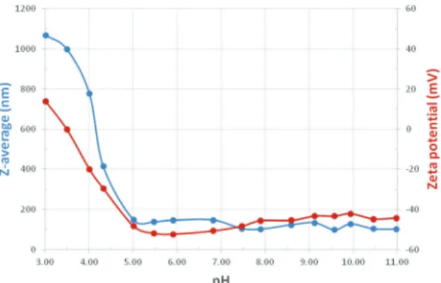
3.4. Colloidal stability: dynamic light scattering
The hydrodynamic diameter (Z-average) and the zeta potential for diluted samples of both types were measured as a function of pH in the range 3 to 11. The oleate and citrate stabilized magnetic fluid samples were stable almost over the whole pH range studied here except the most acidic pHs below 5 and 6, respectively.
Over the stable pH range, we obtained hydrodynamic dia- meters between 52 and 110 nm for the citric acid covered particles with a PDI of 0.26–0.46, and between 98 and 147 nm for the oleic acid covered particles with a PDI of 0.12–0.23. The citrate covered particles thus have smaller Z-average particle sizes than the oleic acid covered nanoparticles. The most acidic Fig. 5 Fe2p, C1s and O1s XPS spectra for Fe3O4/OA.
Table 5 Fitting parameters, including peaks positions, FWHM and calculated atomic concentrations from peak areas of Fe2p XPS spectra for the dried samples Fe3O4/OA and Fe3O4/CA
Peak name
Position (eV) FWHM (eV) Atomic conc. (%) Position (eV) FWHM (eV) Atomic conc. (%)
Fe3O4/OA Fe3O4/CA
Fe2+2p3/2 (octahedral) 710.5 2.2 6.44 710 2.5 8.499
Fe3+2p3/2 (octahedral) 711.8 3.2 14.535 711.2 3.6 17.122
Fe3+2p3/2 (tetrahedral) 714 3.6 10.343 713.5 4.7 11.717
Fe2+2p1/2 (octahedral) 723 3.2 6.234 723 3.7 8.228
Fe3+2p1/2 (octahedral) 724.6 2.9 14.072 724 3.8 16.575
Fe3+2p1/2 (tetrahedral) 726.7 3.2 10.012 725.7 3.8 11.341
Fe2+2p3/2 satellite 716.6 2.6 1.344 717 4 5.913
Fe3+2p3/2 satellite 719.4 5.1 10.527 719.6 3.5 5.083
Fe2+2p1/2 satellite 729.3 5.3 16.736 728.4 4.8 9.725
Fe3+2p1/2 satellite 733.9 5 9.756 732.8 4.5 5.798
Published on 03 July 2018. Downloaded by UNIVERSITY OF SZEGED on 8/4/2021 9:15:53 AM.
samples were unstable and started to coagulate right after sonication. The corresponding sizes,B1000 nm andB1400 nm, measured for oleate and citrate stabilized samples (Fig. 6 and 7), respectively, are thus only characteristic of the given kinetic stage. These samples settle down over a time of a few hours.
It should be noted that the diameters calculated from DLS include the effect of a hydration layer around the particles. The thickness of this layer can be relatively large, with the result that the DLS sizes will generally be higher than those obtained from the SAXS and SANS analysis (presented later).
Zeta potentials measured for the citrate and oleate stabilized samples show the characteristic pH-dependence due to the different dissociation behaviors of the acidic groups on the coating molecules.
Both samples are seen to be stable over a wide pH range;
however, the MF/CA sample loses colloidal stability below pH 6, while the MF/OA sample below pH 5, because of the difference in the charge state of these coated nanoparticles due to different dissociability (the citric acid has: pKa1= 3.13, pKa2= 4.76, and pKa3= 6.40, while the oleic acid has pKa= 5.02).
The charging behaviour of OA double layer stabilized MNPs can be understood considering its pKavalue. At pHB 5, the zeta potential is still high (50 mV) for the oleic acid sample, and the particles are charged, since about the half of oleic acid molecules in the 2nd layer are dissociated. However, the potential is significantly decreased below this limit. The system then loses its stability, the particles start to aggregate, and the average particle size increases abruptly. The citric acid is bound to the MNPviaone of its –COOH groups that connects to RFe–OH sites on the surface.61 Dissociation of surface complexed citric acid differs from that of molecules dissolved in water (see pKavalues above), so charging of citrated MNPs is supressed below pHB6, where the absolute value of the zeta potential drops just below 25 mV. We measured positive values of the zeta potential (5–15 mV) at and below pH 4 for the MF/CA samples (B5–10 mV in Fig. 6) and below pH 3.5 for the MF/OA sample (B15 mV in Fig. 7). These were not high, but systematic and above the reproducibility level (5 mV) of this method.
Under acidic conditions, positive zeta potential values are characterisitic of naked MNPs.34The results here thus indicate incomplete coverage of MNPs, as has been shown for citrated
MNPs containing less than B1 mmol COOH/g magnetite (i.e.,B0.33 mmol CA was added to 1 g MNP)61and for oleate coated MNPs with an added OA amount lower than 1.5 mmol g1.29On the other hand, incomplete coverage of the present citrated sample is not very likely, since CA was added in high excess to the magnetite during synthesis as explained in point 2.2. For the MF/OA sample, the existing free surfactant in solution is conditioning the formation of a physisorbed secondary OA layer which gives the hydrophilic character of dispersed magnetite NPs. The resulting degree of coverage is dependent on the added amount of OA during synthesis (given in point 2.3).
3.5. Structure
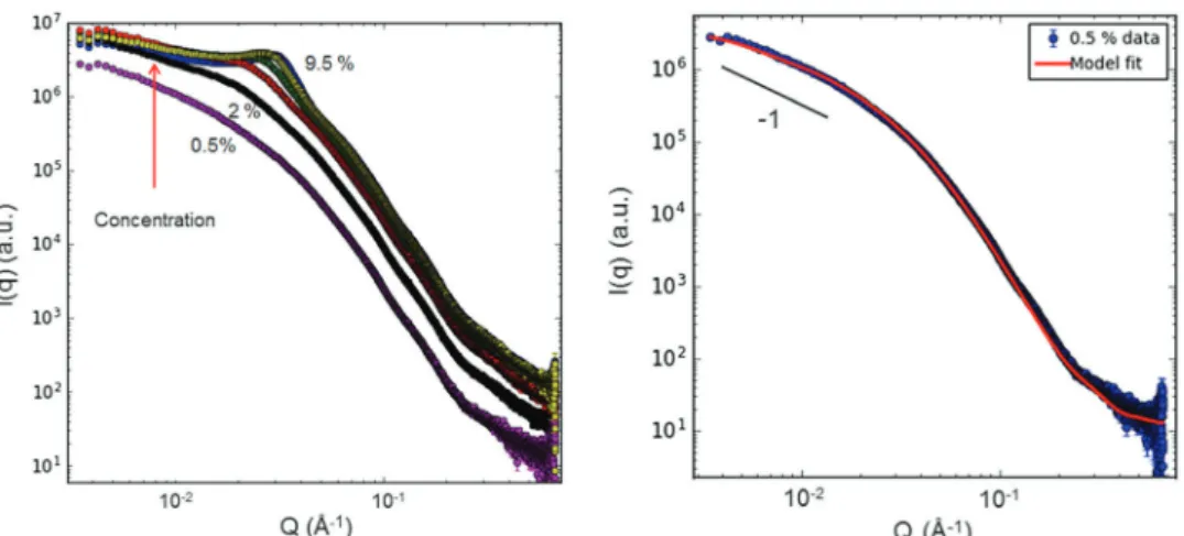
3.5.1. Electrostatically stabilized magnetic fluids analyzed by SAXS and SANS.SAXS and SANS data obtained for citric stabilized magnetic nanoparticles with the volume concentration of MFs varying from 0.5 to 20% are shown in Fig. 8. The scattering intensities have been normalized to the concentration of MNPs, which gives the possibility for direct comparison of size and interaction among MNPs (or their aggregates), depending on the concentration.
The SAXS and SANS data both show a continuous decrease of the normalized scattering intensity in the low-qregion with increasing MNP concentration, suggesting that the MNPs show dominating repulsive interaction and do not aggregate into larger complexes. This concentration dependence is thus an indication that the magnetic fluids are stable in the studied concentration range. The shape of curves for SAXS and SANS is quite similar, indicating only minor differences in the X-ray and neutron scattering length density profiles for these particles in H2O. This is corroborated by the calculated scattering length densities (Table 6), and that there is only a very small contribu- tion of magnetic scattering for SANS in these conditions. The log–log slope at high q-values is found to be close to 4, showing that the particle surface is smooth at short length scales for all concentrations studied.
Based on this initial inspection of the scattering data, one can proceed with model fitting. We first tried to describe the scattering curves using a model with individual polydisperse particles (log-normal distribution) as had been observed by Fig. 6 pH dependence of theZ-average particle diameter (blue) and the
zeta potential (red) for the citrated (MF/CA) sample.
Fig. 7 pH dependence of theZ-average particle diameter (blue) and the zeta potential (red) for the oleic acid double layer stabilized (MF/OA) sample.
Published on 03 July 2018. Downloaded by UNIVERSITY OF SZEGED on 8/4/2021 9:15:53 AM.
TEM (Fig. 2). However, it was not possible to obtain reasonable fits in this way, even when including an interaction potential.
On the other hand, a model of prolate ellipsoids of rotation with polydispersity in the small axis, including screened Coulomb interaction did produce satisfactory fits, see Fig. 9.
In the modelling we minimized the number of free fitting parameters, fixing values that are known from other methods or calculations (cf.SLD-values in Table 6). The shape (log-normal) and width of the distribution of the small axis were fixed to the values obtained for the MNPs from TEM. The thickness of the citric acid layer was fixed to 4 Å, which was the converging value obtained from the fit of SANS data at the lowest concentration (0.5%) where the particle interactions can be neglected. The parameters resulting from the fitting are thus the small axis, the
axial ratio, and the magnitude of the surface electrical charge; see fitted values for selected concentrations in Table 7. One should note that since the SANS data are normalized to an absolute scale (cm1), it is possible to verify if the total scattered intensity corresponds to what is expected from a given volume fraction of the magnetic particles. For all concentrations measured, the data fitted well to a model with a volume fraction fixed to the nominal value known from the preparation protocol, demonstrating that if any large agglomerates (outside the accessible range for SANS) exist in the sample, the amount of these must be very small.
The results from SAXS and SANS thus point towards the presence of short MNP aggregates in the form of ellipsoids of revolution, consisting of a moderate number (3–6) of MNPs and carrying a relatively low electrical charge. The aggregates become shorter with increasing concentration, and at the same time the interaction between aggregates becomes stronger.
A plot of the particle axial ratio as well as the charge based on the SAXS data is shown in Fig. 10.
We can also get information about the interactions in the system,i.e.the structure factor, by dividing all higher concentration data with one of the lowest concentrations where the Fig. 8 SAXS and SANS intensities normalized to the concentration of MNPs (Fe3O4/CA) with varying concentration. The SANS data have been background-subtracted for the H2O contribution.
Table 6 Scattering length densities (SLD) for the different components calculated using the SLD calculator in the SasView program86
Fe3O4 H2O Citrate Oleic acid
SAXS [Å2]106 40.3 9.42 14.7 8.5
SANS [Å2]106 6.93 0.56 3.3 0.078
Fig. 9 SAXS (left) and SANS data (right) of Fe3O4/CA magnetic fluids with fits (continuous lines) using a core–shell ellipsoidal model and a screened Coulomb interaction potential. Only selected concentrations are shown for the SANS data in order to better appreciate the difference between the curves with increasing concentration.
Published on 03 July 2018. Downloaded by UNIVERSITY OF SZEGED on 8/4/2021 9:15:53 AM.
![Table 2 Water based surfacted ferrofluid samples with electro-steric stabilization Sample code Surfactant (double layer) Solid volumefraction [%] Density[g cm 3 ]](https://thumb-eu.123doks.com/thumbv2/9dokorg/1339678.108716/5.892.63.438.92.304/surfacted-ferrofluid-samples-electro-stabilization-surfactant-volumefraction-density.webp)