Journal of Molecular and Cellular Cardiology 155 (2021) 10–20
Available online 22 February 2021
0022-2828/© 2021 Elsevier Ltd. All rights reserved.
Mapping genetic changes in the cAMP-signaling cascade in human atria
Anne Garnier
a, Nadja I. Bork
b,c, Eric Jacquet
d, Svante Zipfel
e, Christian Mu ˜ noz-Guijosa
f, Istvan Baczk ´ o
g, Hermann Reichenspurner
e, Patrick Donzeau-Gouge
h, Lars S. Maier
i, Dobromir Dobrev
j, Evaldas Girdauskas
c,e, Viacheslav O. Nikolaev
b,c,
Rodolphe Fischmeister
a,*,1, Cristina E. Molina
a,b,c,1aUniversit´e Paris-Saclay, Inserm, UMR-S 1180, Chˆatenay-Malabry, France
bInstitute of Experimental Cardiovascular Research, University Medical Center Hamburg-Eppendorf, Germany
cGerman Center for Cardiovascular Research (DZHK), partner site Hamburg/Kiel/Lübeck, Germany
dUniversit´e Paris-Saclay, Institut de Chimie des Substances Naturelles, CNRS UPR 2301, Gif-sur-Yvette, France
eDept. of Cardiovascular Surgery, University Heart Center Hamburg, Germany
fCardiac Surgery Department, Hospital de la Santa Creu i Sant Pau, Spain
gDept. Pharmacology and Pharmacotherapy, Univ. of Szeged, Hungary
hInstitut Hospitalier Jacques Cartier, Massy, France
iDept. Internal Medicine II, University Heart Center, University Hospital Regensburg, Germany
jInstitute of Pharmacology, West-German Heart and Vascular Center, Faculty of Medicine, University Duisburg-, Essen, Germany
A R T I C L E I N F O Keywords:
Atrial fibrillation Heart failure Right atria Left atria
cAMP-signaling cascade β-Adrenergic pathway
A B S T R A C T
Aim: To obtain a quantitative expression profile of the main genes involved in the cAMP-signaling cascade in human control atria and in different cardiac pathologies.
Methods and results: Expression of 48 target genes playing a relevant role in the cAMP-signaling cascade was assessed by RT-qPCR. 113 samples were obtained from right atrial appendages (RAA) of patients in sinus rhythm (SR) with or without atrium dilation, paroxysmal atrial fibrillation (AF), persistent AF or heart failure (HF); and left atrial appendages (LAA) from patients in SR or with AF. Our results show that right and left atrial appendages in donor hearts or from SR patients have similar expression values except for AC7 and PDE2A. Despite the enormous chamber-dependent variability in the gene-expression changes between pathologies, several distin- guishable patterns could be identified. PDE8A, PI3Kγ and EPAC2 were upregulated in AF. Different phospho- diesterase (PDE) families showed specific pathology-dependent changes.
Conclusion: By comparing mRNA-expression patterns of the cAMP-signaling cascade related genes in right and left atrial appendages of human hearts and across different pathologies, we show that 1) gene expression is not significantly affected by cardioplegic solution content, 2) it is appropriate to use SR atrial samples as controls, and 3) many genes in the cAMP-signaling cascade are affected in AF and HF but only few of them appear to be chamber (right or left) specific.
Topic: Genetic changes in human diseased atria.
Translational perspective: The cyclic AMP signaling pathway is important for atrial function. However, expression patterns of the genes involved in the atria of healthy and diseased hearts are still unclear. We give here a general overview of how different pathologies affect the expression of key genes in the cAMP signaling pathway in human right and left atria appendages. Our study may help identifying new genes of interest as potential therapeutic targets or clinical biomarkers for these pathologies and could serve as a guide in future gene therapy studies.
* Corresponding author at: Universit´e Paris-Saclay, Inserm, UMR-S 1180, 92296 Chˆatenay-Malabry Cedex, France.
E-mail address: rodolphe.fischmeister@inserm.fr (R. Fischmeister).
1 These authors contributed equally.
Contents lists available at ScienceDirect
Journal of Molecular and Cellular Cardiology
journal homepage: www.elsevier.com/locate/yjmcc
https://doi.org/10.1016/j.yjmcc.2021.02.006
Received 5 January 2021; Received in revised form 4 February 2021; Accepted 11 February 2021
1. Introduction
The β-adrenergic receptor (β-AR)/cyclic adenosine monophosphate (cAMP) pathway is a major pathway involved in the sympathetic regulation of heart function. It regulates cardiac contractility, relaxation and heart rate. β-AR activation by noradrenaline or adrenaline triggers a sequence of signaling events which starts by G-protein activation, stimulation of different adenylyl cyclase (AC) isoforms which synthetize cAMP leading to protein-kinase-A (PKA) mediated phosphorylation of several key proteins involved in excitation-contraction (EC) coupling, such as L-type Ca2+channels (LTCC), phospholamban (PLB), ryanodine receptors type-2 (RyR2) and troponin-I (TnI) [1]. A-kinase anchoring proteins (AKAPs) bind PKA [2] and phosphatases (PP2 and PP1), which act as signaling scaffolds to promote phosphorylation of target proteins.
cAMP has also PKA-independent targets in the heart, such as channels for cyclic nucleotides transport across the membranes (MRP) [3–5] or the small G-protein exchange-factor Epac (Exchange Protein directly Activated by cAMP) [6] involved in hypertrophic growth [7,8], which in turn activates calcium/calmodulin-dependent kinase type-IIδ (CaMKIIδ). The levels of cAMP and thus the degree of PKA and Epac activation are finely regulated by cyclic nucleotide phosphodiesterases (PDEs). There are 5 families (PDE1-4 and PDE8) of cardiac PDEs degrading cAMP [9]. Other proteins also play an important role in EC coupling by modulating β-AR desensitization (β-arrestins) or activating phosphorylation after an intracellular calcium increase (CaMKIIδ) (Fig. 5).
One important question is whether differences in neurohumoral regulation between right and left heart chambers are limited to elec- trophysiological and phenotypic features [10–12] or extend to genetics, and how it can differentially affect left and right atria. During the last years, many publications have highlighted the importance to study ge- netic differences between cardiac cavities. Phillips and colleagues [13]
reported no difference in the expression level of more than 600 proteins between the RV and the LV from pig and rabbit including contractile/
structural proteins and oxidative phosphorylation components. No plasma membrane voltage-gated ion channel was analyzed, but a few major Ca2+handling proteins (SERCA2, RyR2) and signal transduction components (Ca2+/CaMKIIδ, PKA type-I and type-II) showed no varia- tion in their expression level [13]. A recent single-cell resolution anal- ysis also showed similar nuclear transcriptional patterns when comparing right versus left atria from dead donors non-failing patients [14]. However, only limited information about β-AR/cAMP pathway genes and their expression in human healthy and diseased atria is available.
Several electrophysiological studies have shown the importance of the β-AR/cAMP pathway in the development and maintenance of many cardiovascular diseases such as heart failure (HF) and atrial fibrillation (AF). However, the relationship between electrophysiological changes in cellular signaling and changes in β-AR/cAMP pathway regulatory genes has mainly been investigated for individual targets in specific diseases and heart regions.
Unbiased genetic studies in the human heart considering both interchamber differences and pathological re-modeling are extremely important to characterize new potential therapeutic targets before bringing them into deeper mechanistic and translational studies as well as to better understand adaptation or maladaptation during disease.
Recently, gene therapy technology has begun to be tested to treat ven- tricular pathologies. However, expression of the tested genes and their disease driven changes have not always been studied in the human tis- sues, or only in a very small number of samples with variable reference genes, leading to contradictory results. Here, we aimed to study gene expression changes that occur in the human atrium with various pa- thologies at right and left chambers in order to gain a general under- standing of the remodeling of the β-AR/cAMP pathway promoted by different pathologies. We studied the expression of 48 target mRNAs in 113 appendage tissue samples from control donor patients, patients
being in sinus rhythm (SR) with or without atrial dilation, having different forms of AF, or suffering from HF. Our data reveal that many genes involved in the β-AR/cAMP pathway are affected in AF and HF but only few changes are right or left atrial specific.
2. Methods
2.1. Human atrial-tissue samples
A total of 113 human atrial appendage-tissue samples were collected from a total of 97 patients undergoing open-heart surgery either at the Institut Hospitalier Jacques Cartier, Massy, France, at the Cardiac Sur- gery Department, Hospital de la Santa Creu i Sant Pau/Universitat Aut`onoma de Barcelona, Spain, at the Cardiology and Pneumology Department, Georg-August-Universitat G¨ ottingen, Germany, at the ¨ Cardiac Surgery Department, University Hospital Essen, Germany, at the University Medical Center Hamburg-Eppendorf, Germany, or at the Department of Pharmacology and Pharmacotherapy, University of Szeged, Hungary. 81 specimens of right (RAA) and left (LAA) atrial appendage tissues were obtained from 80 patients in sinus rhythm with (SRd) or without (SRnd) atrial dilation, or with paroxysmal atrial fibrillation (pAF) or long-standing persistent atrial fibrillation (cAF), subjected to atrial cannulation for extracorporeal circulation. 32 right and left atrial appendage tissues were collected from 8 control donors (Ctl) and 9 end-stage heart failure dilated atria (HF) from explanted human hearts, respectively, at the time of the transplantation. All sam- ples were frozen in liquid nitrogen and stored at − 80 ◦C. Adipose tissue was removed before the cryopreservation of the sample. The study was conducted in accordance with the Declaration of Helsinki principles, and approved by the Ethical Committees of all included institutions.
Informed consent was obtained from each patient. Details regarding the clinical characteristics of the patients and their medication are shown in Tables 1 and 2.
2.2. RNA isolation and cDNA synthesis
76 snap-frozen tissue samples were weighed and placed in pre- cooled tubes containing TRIzol® reagent (Invitrogen, Life Technolo- gies, France) and rapidly subjected to automated grinding in a Bertin Precellys 24 (Bertin Technologies, France). Total RNA extraction was carried out using standard procedure according to the manufacturer’s instructions. RNA concentration and purity were evaluated by optical density (Biophotometer, Eppendorf, BioServ, France) and the integrity of the RNA samples were analyzed on a Bioanalyzer 2100 with the RNA6000 Nano Labchip Kit (Agilent Technologies, Santa Clara, CA, USA). The RNA-integrity number (RIN) was calculated by the instru- ment software. First strand cDNA synthesis was performed from 1-μg of total RNA with random primers and MultiScribeTM Reverse Transcrip- tase according to the provided protocol (Applied Biosystems, Life Technologies, France). To minimize intergroup variations, samples of each experimental group were processed simultaneously.
Total RNA content, 260 nm/280 nm optical-density (OD) ratio and RNA-integrity number (RIN) values were obtained from right and left atrial appendages without or with pathological conditions. RIN values were ranged between 5.5 and 8.5, with a mean value of 6.5 ±0.3 for RAA SRnd, 7.0 ±0.2 for RAA SRd, 6.4 ±0.3 for RAA pAF, 6.6 ±0.1 for RAA cAF, 7.5 ±0.6 for LAA SR, 6.5 ±0.3 for LAA AF, 6.9 ±0.4 for RAA Ctl, 7.0 ±0.4 for LAA Ctl, 7.0 ±0.5 for RAA HF and 7.1 ±0.3 for LAA HF. There were no differences in RNA content between the groups. The quality control parameters for human total RNA samples were similar regardless of human tissue-sample collection, with no difference be- tween the groups.
2.3. Real-time qPCR and quantification
Real-time PCR assays were performed using TaqMan® 384-well
microfluidic card technology from Applied Biosystems (TaqMan® Array Card or TAC, Life Technologies, France) and the TaqMan® Human Endogenous Control Panel. These TAC were designed to study the expression of 48 target genes: ABCC4 (MRP4), ABCC5 (MRP5), ADCY2 (AC2), ADCY4 (AC4), ADCY5 (AC5), ADCY6 (AC6), ADCY7 (AC7), ADCY9 (AC9), AKAP5, AKAP6, AKAP7, AKAP9, AKAP13, ARRB1 (Arrestin b1), ARRB2 (Arrestin b2), AURKAIP1 (AKIP), CAMK2D (CaM- KIID), NPPA (ANP), NPPB (BNP), PDE1A, PDE1C, PDE2A, PDE3A, PDE3B, PDE4A, PDE4B, PDE4D, PDE8A, PIK3CG (PI3Kγ), PKIA, PKIB, PPP1CA (PP1A), PPP1R1A (IPP1), PPP1R2 (IPP2), PPP2CA (PP2CA), PRKACA (PKACA), PRKACB (PKACB), PRKAR1A (RIA), PRKAR2A (RIIA), PRKAR2B (RIIB), RAPGEF3 (EPAC1), RAPGEF4 (EPAC2)), ADRB1 (β1-AR), ADRB2 (β2-AR), GNAI1 (Gi1), GNAI2 (Gi2), GNAI3 (Gi3), GNAS (Gs) (Supplemental Table 1) relative to 5 reference genes (POLR2A, YWHAZ, GAPDH, IPO8, PPIA) recently validated for a study using right and left cardiac cavities from different human tissue cohorts [15]. Each PCR reaction was performed on 4-ng of cDNA in a volume of 1-μl. The thermal cycling conditions for PCR amplification on TAC were 10-min at 94.5 ◦C, followed by 40 cycles of 30-s at 97 ◦C and 1 min at 59.7 ◦C, on an ABI-Prism 7900HT Sequence Detection Instrument (Applied Biosystems, Life Technologies, France). Each TaqMan® assay was previously validated by Applied Biosystems and the efficiency of amplification was certified to be superior to 90% by the supplier. For each group, an average Ct value was calculated. The determination of the relative gene-expression ratio was achieved using the ΔΔCt method and normalized by the geometric mean of a set of stable housekeeping genes.
2.4. Western blot analysis
37 snap frozen human atrial appendage-tissue samples were ho- mogenized in RIPA lysis buffer (NaCl 150 mM, Triton 1%, SDS 0.1%, SOD 0.5%, Tris 50 mM, Protease- and Phosphatase-Inhibitor Cocktail (Roche)) using a homogenisator (MICCRA D-1). After three homogeni- zation steps for 20 s each, always followed by cooling of the samples in liquid nitrogen, samples were incubated on ice for 1-h. After centrifu- gation (12,000 rpm, 5-min, 4 ◦C) supernatants were stored at − 20 ◦C until usage. Protein quantification was performed using BCA Protein Assay (Pierce BCA Protein Assay Kit, Thermo Scientific, #23227) and
50-μg of total protein were loaded on 8% SDS gels for SDS poly- acrylamide gel electrophoresis. Then, proteins were transferred to nitrocellulose membrane (Amersham, #106000 02) using a tank blot system. For immunoblot analysis, the following antibodies were used:
PDE2A (Fabgennix 101AP, rabbit polynclonal antibody, dilution 1:750 in 3% milk, sample cooking 70 ◦C 10-min), PDE8A (Santa Cruz Biotechnology sc-17,232, goat polynclonal antibody, dilution 1:500 in 3% BSA, sample cooking 70 ◦C 10-min), EPAC2 (Cell signaling #4156, mouse monoclonal antibody, dilution 1:250 in 3% BSA, sample cooking 55 ◦C 30-min), PIK3γ (kindly provided by Dr. Emilio Hirsch, mouse polyclonal antibody, dilution 1:100 in 3% BSA, 70 ◦C 10-min), GAPDH (HyTest #5G4 6C5, mouse monoclonal antibody, dilution 1:160,000 in 5% milk). For quantification, band densitometry analysis was done using ImageJ software.
2.5. Statistical analysis
Results are expressed as mean ±SEM. Statistical significance was evaluated using a Student’s t-test and ANOVA followed by Tukey’s test was used for comparison of multiple effects. A difference was considered statistically significant when p <0.05.
3. Results
Using RT-qPCR, we simultaneously evaluated the expression of 49 target genes and normalized it to the appropriate reference genes [15].
In total, 113 human atrial appendage-tissue samples (Tables 1 and 2) from right and left atria controls or with distinct cardiovascular diseases were analyzed. Western Blot was used to confirm protein changes for specific genes in 37 samples.
3.1. Validation of the control groups
Working with human cardiac samples entails intrinsic subject het- erogeneity. For instance, SR atrial appendage samples obtained from patients undergoing cardiac surgery due to cardiovascular diseases, which do not affect the atrium or the heart rhythm, were snap-frozen immediately after resection from the patient. In contrast, Ctl atrial appendage samples derived from donor hearts not suitable for Table 1
Clinical characteristics of patients/samples used for gene expression experiments.
Patients (n) SRnd SRd pAF cAF Ctl HF n =9
RAA, n =8 RAA, n =8 LAA, n =5 RAA, n =8 RAA, n =8 LAA, n =7 RAA-LAA, n =8 RAA, n =8 LAA, n =8
Female gender 3 (38%) 4 (50%) 2 (40%) 2 (25%) 3 (38%) 2 (29%) 3 (38%) 3 (38%) 3 (38%)
Age (years) 69.5 ±2.9# 69.8 ±3.1# 64.4 ±3.3# 76.0 ±4.0# 74.3 ±2.3# 71.6 ±3.8# 44 ±4.7 63.0 ±3.4# 61.9 ±3.7#
Smoking 2 (25%) 2 (25%) 1 (20%) 1 (13%) 3 (38%) 2 (29%) N/A N/A N/A
Hypertension 5 (62%) 4 (50%) 3 (60%) 4 (50%) 5 (62%) 4 (57%) 4 (50%) 8 (100%) 8 (100%)
CAD 5 (62%) 3 (37%) 1 (20%) 2 (25%) 2 (25%) 1 (14%) 0 (0%) 0 (0%) 0 (0%)
AVD/MVD 1 (13%) 4 (50%) 3 (60%) 3 (38%) 4 (50%) 4 (57%) 0 (0%) 0 (0%) 0 (0%)
CAD +AVD/MVD 2 (25%) 1 (13%) 1 (20%) 3 (38%) 2 (25%) 2 (29%) 0 (0%) 0 (0%) 0 (0%)
Transplantation 0 (0%) 0 (0%) 0 (0%) 0 (0%) 0 (0%) 0 (0%) 8 (100%) 8 (100%) 8 (100%)
LAD (mm) 39.2 ±1.4 44.3 ±0.9* 46.8 ±0.6* 43.8 ±2.7 46.0 ±3.9* 53.0 ±6.7* N/A N/A N/A LVEF (%) 61.4 ±2.3 62.7 ±2.0 49.4 ±0.4 57.9 ±2.6 62.8 ±2.6 64.0 ±5.2 N/A 25.5 ±1.5* 29.5 ±5.5*
Digitalis 0 (0%) 0 (0%) 0 (0%) 1 (13%) 0 (0%) 1 (14%) N/A N/A N/A
ACE inhibitors 3 (38%) 4 (50%) 4 (80%) 3 (38%) 4 (50%) 5 (71%) 4 (50%) N/A N/A
AT1 blockers 0 (0%) 0 (0%) 0 (0%) 0 (0%) 0 (0%) 0 (0%) N/A N/A N/A
Beta-blockers 5 (63%) 4 (50%) 5 (100%) 4 (50%) 5 (63%) 5 (71%) N/A N/A N/A
Ca2+-antagonists 0 (0%) 2 (25%) 0 (0%) 1 (13%) 3 (38%) 2 (29%) 0 (0%) N/A N/A
Diuretics 1 (13%) 2 (25%) 3 (60%) 3 (38%) 5 (63%) 4 (57%) 4 (50%) N/A N/A
Nitrates 0 (0%) 0 (0%) 0 (0%) 0 (0%) 0 (0%) 0 (0%) 0 (0%) N/A N/A
Statins 5 (63%) 2 (25%) 4 (50%) 2 (25%) 1 (13%) 2 (29%) 0 (0%) N/A N/A
Amiodaron 0 (0%) 0 (0%) 0 (0%) 6 (75%) 2 (25%) 2 (29%) 0 (0%) N/A N/A
Values are presented as mean ±SEM or number of patients (%). SRnd: Sinus Rhythm without atrium dilation; SRd: Sinus Rhythm with atrium dilation; pAF:
Paroxysmal Atrial Fibrillation; cAF: Chronic Atrial Fibrillation; HF: Heart Failure; Ctl: Control donors; ACE: angiotensin-converting enzyme; AT: angiotensin receptor;
AVD/MVD: aortic/mitral valve disease; CAD: coronary artery disease; LAD, left atrial diameter; LVEF, left ventricular ejection fraction; N/A: not available. * indicates p <0.05 vs. SRnd, # indicates p <0.05 vs. Ctl. CAD, AVD/MVD, and CAD +AVD/MVD reflect the indications for cardiac surgery (bypass surgery, valve surgery or both).
transplantation due to logistical reasons were usually perfused with, and stored in, a cardioplegic solution, histidine-tryptophan-ketoglutarat Custodiol® solution. We first tested the possibility that differences might exist between the cardioplegia treated or not treated samples under such conditions. We found comparable expression values between SR and Ctl atrial groups (Supplemental Table 2). These results suggest that 1) cAMP-signaling cascade related genes are not affected by the content of this cardioplegic solution, and that 2) it is appropriate to use diseased (SR) samples as potential controls.
3.2. Chamber-specific gene expression patterns in atria of the control donors human hearts
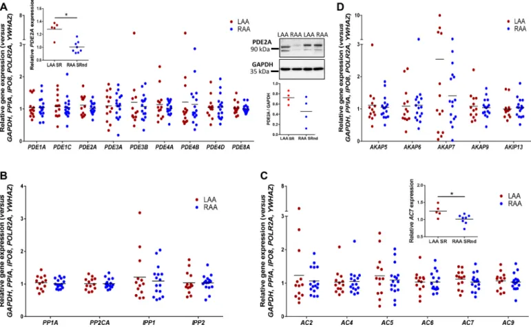
All tested genes were expressed in the right and the left atrial ap- pendages. Furthermore, most of the genes showed similar gene expres- sion values in the two chambers in the Ctl and SR samples (Fig. 1). The only significant differences were observed in AC7 and PDE2A, which tended to a higher expression in the left atrium when comparing right and left atrial appendages from SR patients. Western blotting experi- ments for PDE2A confirmed this tendency in protein expression.
3.3. Gene expression changes in human atrial fibrillation
Upregulation of the β-AR/cAMP pathway and increased phosphor- ylation of key proteins in EC coupling are the hallmarks of AF.
Accordingly, many genes involved into the β-AR/cAMP pathway are affected by AF (Fig. 2). Previous studies described gene expression changes in AF on PP1 [16] and BNP [17,18] and no change on IPP1, IPP2 [19], PP2CA [16,19] and CaMKIID [20]. By using a validated set of reference genes [15], we found a downregulation of AC9, AKAP9, AKIP, PDE3A, PDE4B, IPP2 and PKACA when comparing right and left atrial appendages in SR versus AF (Fig. 2A). AKAP5 and Epac1 also showed a tendency to diminution (Supplemental Table 2). PDE8A, PI3Kγ and EPAC2 were upregulated (Fig. 2A), with Arrestin b2 and PDE1A showing a clear trend augmentation in AF (Supplemental Table 2). We confirmed the upregulation of PDE8A, PI3Kγ and EPAC2 at the protein level using immunoblots (Fig. 2B). In contrast, MRP4, MRP5, AC2, AC4, AC5, AC6, AC7, AKAP6, AKAP7, AKAP13, Arrestin b1, BNP, CaMKIID, PDE1C, PDE2A, PDE3B, PDE4A, PDE4D, PKIA, PKIB, PP1A, PP2CA, IPP1, PKACB, RIA, RIIA, RIIB, β1-AR, β2-AR, Gi1, Gi2, Gi3 and Gs showed similar gene- expression patterns in all groups.
Interestingly, some genes appeared to be regulated in a chamber- specific manner. While BNP, PDE8A, PP2CA and EPAC2 expression increased in the RAA with pAF or cAF (Fig. 2C), PI3Kγ expression increased and MRP5 and PKIA expression decreased only in the LAA with AF (Fig. 2D).
3.4. Atrial heart failure-related gene-expression patterns
Although HF is extensively studied in the ventricle, less is known about whether and how HF affects the atria. Fig. 3A shows that HF strongly affects atrial cAMP-pathway gene expression. MRP4, MRP5, Arrestin b2, PDE4B, PDE4D and PKIB are downregulated, while AC2, AC5, AC6, AC9, AKAP9, AKIP, PDE1C, PDE3B, PKIA, PKIB, PP2CA, PKACA, PKACB, RIA, RIIB, EPAC1 and CaMKIID are upregulated when comparing right and left atrial appendages together in Ctl versus HF.
Although MRP4, PKACB and PDE1C expression tends to change in both atrial appendages in HF compared to Ctl, MRP4 downregulation and PKACB upregulation seem to be more specific to the RAA (Fig. 3B), and PDE1C is upregulated particularly in the LAA (Fig. 3C).
3.5. Gene-expression changes due to β-blocker treatment
The above results suggest that genetic remodeling of cAMP pathway in AF and HF patients might contribute to the altered sympathetic regulation of atrial function observed in these patients. To explore this further, we examined whether treatment of patients with β-blockers had any impact on their gene expression level. For this, samples from all groups of patients (see Table 1) were separated according to the absence or presence of AF, the absence or presence of atrial dilation and the absence or presence of β-blockers in their medication. Only few alter- ations in gene expression were found to be associated with β-blocker treatments (Fig. 4). Importantly, treatment of AF-patients with β-blockers did not seem to promote a reverse remodeling.
4. Discussion
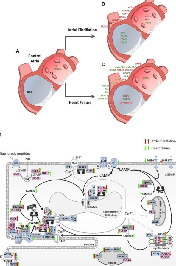
The present study is to our knowledge the first to compare mRNA- expression pattern of cAMP-cascade related genes in right and left atrial appendages of control human heart tissues and in hearts with different pathologies. The results are summarized in Fig. 5.
HF and AF are intrinsically associated with increased endogenous neurohormonal activation, which induces several changes in the heart and vascular system to maintain cardiovascular homeostasis. At the same time, chronic neurohormonal activation is known to be one of the most important mechanisms underlying pathological remodeling and promoting changes in gene expression of several signaling pathways.
Table 2
Clinical characteristics of patients/samples used for protein expression experiments.
Patients (n) SRnd SR pAF cAF
RAA,
n =8 LAA, n
=6 RAA, n
=8 LAA, n
=3 RAA, n
=6 LAA, n
=6 Female gender 1
(38%) 1 (38%) 1
(25%) 3
(100%) 0 (0%) 1 (29%) Age (years) 64.6 ±
4.2 60.8 ± 8.1 65.3 ±
4.8 61.7 ± 3.3 71.5 ±
3.1 69.8 ± Smoking 3 1.8
(37%) 1 (17%) 3
(38%) 1 (33%) 3
(50%) 2 (33%) Hypertension 5
(62%) 2 (33%) 5
(62%) 2 (67%) 5
(83%) 5 (83%)
CAD 5
(62%) 0 (0%) 2 (25%) 1
(33%) 2 (33%) 2
(33%)
AVD/MVD 2
(25%) 6 (100%) 5
(62%) 2 (67%) 3
(50%) 3 (50%) CAD +AVD/
MVD 1
(13%) 0 (0%) 1
(13%) 0 (0%) 1 (17%) 1
(17%) Transplantation 0 (0%) 0 (0%) 0 (0%) 0 (0%) 0 (0%) 0 (0%) LAD (mm) 39.6 ±
1.9 44.2 ± 3.3 49.7 ±
4.5* 52.5 ± 17.5 47.0 ±
4.5* 52.8 ± LVEF (%) 55.9 ± 6.2
2.9 48.3 ± 4.7 53.5 ±
2.4 60.3 ± 0.3 60.3 ±
0.4 54.5 ± Digitalis 0 (0%) 0 (0%) 0 (0%) 0 (0%) 0 (0%) 4.1 0 (0%) ACE inhibitors 6
(75%) 4 (67%) 3
(38%) 2 (67%) 4
(67%) 5 (83%) AT1 blockers 0 (0%) 0 (0%) 1
(12%) 0 (0%) 1
(17%) 0 (0%) Beta-blockers 4
(50%) 4 (67%) 7
(87%) 3 (100%) 5
(83%) 5 (83%) Ca2+-
antagonists 1
(12%) 0 (0%) 0 (0%) 0 (0%) 1 (17%) 3
(50%) Diuretics 0 (0%) 1
(17%) 1
(12%) 0 (0%) 2 (33%) 4
(67%)
Nitrates 1
(12%) 0 (0%) 0 (0%) 2
(67%) 0 (0%) 0 (0%)
Statins 3
(37%) 2 (33%) 1
(12%) 0 (0%) 3 (50%) 5
(83%) Amiodaron 0 (0%) 0 (0%) 2
(25%) 0 (0%) 0 (0%) 0 (0%) Values are presented as mean ±SEM or number of patients (%). SRnd: Sinus Rhythm without atrium dilation; SRd: Sinus Rhythm with atrium dilation; pAF:
Paroxysmal Atrial Fibrillation; cAF: Chronic Atrial Fibrillation; HF: Heart Fail- ure; Ctl: Control donors; ACE: angiotensin-converting enzyme; AT: angiotensin receptor; AVD/MVD: aortic/mitral valve disease; CAD: coronary artery disease;
LAD, left atrial diameter; LVEF, left ventricular ejection fraction; N/A: not available. * indicates p <0.05 vs. SRnd. CAD, AVD/MVD, and CAD +AVD/MVD reflect the indications for cardiac surgery (bypass surgery, valve surgery or both).
While changes occurring at the level of β-AR [21–23] have been extensively studied, only a few studies focussed on gene expression changes in the cAMP pathway.
In contrast to previous work in ventricular samples describing gene changes due to exposure to cardioplegic solutions [24], we found similar expression levels between SR patients and donor Ctl individuals. This suggests that gene expression is less sensitive to cardioplegic solution in the atria than in the ventricles. However, many studies do not specify the kind of cardioplegic solution used. One major limitation of studies using human is the selection of appropriate controls. Atrial appendage tissue from patients being in sinus rhythm at time of tissue collection is frequently used as control group for studies dealing with AF or HF [25,26]. Our results suggest that atrial appendage samples from SR patients match those obtained from donor Ctl individuals, validating the use of atrial samples from SR patients as putative controls. This is important considering the very limited availability of atria from healthy individuals. Also, the absence of differences between non dilated and dilated SR in our set of genes made possible to combine these two groups as controls versus different pathologies. However, this may not neces- sarily apply to other genes outside the cAMP-pathway.
We found that the right and left atrial appendages in control hearts had similar gene expression values for all studied genes with the exception of AC7 and PDE2A, which showed particularly higher abun- dance in left atria. However, β1-AR and β2-AR expression and density were previously found to be unchanged in AF [21,27] but, opposite to our results, higher in left versus right atrium [22]. Although similar gene expression profile between RAA and LAA was already described [14], it
is important to note that PDE4, AKAP7, IPP1, AC2 and AC5 showed highly-variable expression patterns. Even if human samples are difficult to obtain, using low number of samples, especially in studies involving highly variable genes, could lead to erroneous conclusions.
Despite the enormous variability in gene-expression profiles among specific pathologies, several distinguishable patterns could be identified.
Expression of AC4, AC7, AKAP5, AKAP6, AKAP7, AKAP13, Arrestin b1, PDE1A, PDE2A, PDE4A, PP1A, IPP1, RIIA, β1-AR, β2-AR, Gi1, Gi2, Gi3 and Gs remain unaltered during pathological remodeling in both atrial appendages. MRP5, PDE4B and PP2CA by contrast have similar expression changes in both pathologies. Expression of AC9, AKAP9, AKIP, PKIA and PKACA also change in all pathologies but in opposite direction: their expression increases in HF but is reduced in AF. The different PDE isoforms appeared regulated in a pathology-specific manner. For instance, PDE3A and PDE8A expression was modified in AF, but PDE1C, PDE3B and PDE4D expression was modified in HF.
PDE8A expression increased in AF together with EPAC2 and PI3Kγ but not CaMKIID. On the other hand, EPAC1 and CaMKIID gene expression was upregulated in HF, together with AC2, AC6, AC5, RIA, RIIB and PKACB while MRP4, Arrestin b2 and PKIB gene expression was downregulated.
We already reported reduced PDE4 and total PDE (IBMX-inhibited PDEs) activity in AF [25]. Here we confirm that PDE4B is downregulated in AF at the gene-expression level. Furthermore, PDE3A, one of the major isoforms in controlling cAMP-hydrolytic activity in human atria, is also downregulated in AF. Of note, the IBMX-insensitive PDE8A was upregulated in AF, particularly in the RAA.
Fig. 1.Plot showing an overview of the results on the gene expression values for the different phosphodiesterases (PDEs, A), phosphatases (PPs, B) adenylyl cyclases (ACs, C) and A-kinases-anchoring proteins (AKAPs, D) within the non-cardiac disease atrial cavities (LAA: dilated and non-dilated left atrial appendages, n =13; RAA:
dilated and non-dilated right atrial appendages, n =16). Separated small graphs in A and C panels show the only significant differences in gene expression between the samples from right and left non-cardiac disease atrial appendages (LAA SR: left atria, n =5; RAA SRnd: non-dilated right atria, n =8) as well as its corresponding protein expression (LAA SR n =4, RAA SRnd n =4). Right chambers values are indicated in blue, left chambers values are indicated in red. * indicates p ≤0.05 versus LAA in tissue from patients in sinus rhythm without atrial dilation (SRnd). (For interpretation of the references to colour in this figure legend, the reader is referred to the web version of this article.)
No change in PDE1A as well as an increase in PDE1C was observed in HF. Although PDE2 was previously found to be upregulated in the ventricles of HF patients [28], we found it stable in both atrial ap- pendages and all pathologies. The results about expression levels of phosphatases are inconsistent. PP1A mRNA levels were previously found to be unchanged in AF [19], in agreement with our results, but also downregulated in another study [16]. Both IPP1 and IPP2 were found to be unchanged in AF [19], in agreement with our results. However, PP2CA levels were reported to be unaltered in AF [16], while we found an increased expression in cAF versus SRd. Similar discrepancies exist for PP1A [29,30], which we found unaltered. Several factors can be responsible for such large variability among studies: intrinsic genetic differences among patients, underlying pathology or state of the pa- thology, medical treatments, age or sex differences. Also, various HF models and underlying aetiologies display different mRNA expression profiles [31]. Variability may also be caused by the use of a single and/
or inappropriate reference gene which could change the conclusions [15]. One example is MRP5. The multidrug resistant protein 4 (MRP4) and 5 (MRP5) act as cyclic nucleotides efflux pump regulating cytosolic cAMP and cGMP levels, respectively. MRP5 gene expression was pre- viously found to be downregulated in atria compared to ventricle and
upregulated in ischemic cardiomyopathy [32]. Unfortunately, this study used β-actin as housekeeping gene, which was found to be one of the most variable genes in human heart samples [15]. Our results based on the use of a validated set of 5 reference genes [15] show reduced MRP4 and MRP5 gene expression levels in HF. By contrast, only MRP5 expression was reduced in AF, yet only in the left atrial appendage.
Although some reports have evidenced the functional role of MRP4 in ventricular myocytes function [33], to date, no heart disease or atrial myocyte dysfunction has been linked to altered MRP4 gene expression.
EPAC shows a distinctive expression pattern, with very high levels of EPAC1 in HF and a tendency for downregulation in AF, while EPAC2 shows a strongly increased expression. On the other hand, although some studies found an increased protein expression or activity of CaMKIIδ in cAF [26], we and others [34] found no change in its gene expression level or even a tendency for a decrease. By contrast, we found an increased expression of CaMKIID in the atrial appendages of patients with HF.
Of particular interest is the increased expression of PI3Kγ in AF.
PI3Kγ regulates PDE3 and PDE4 activity in mouse ventricle [35] and acts as a PKA-anchoring protein for PDE3B, enhancing its activity [36].
Indeed, PI3Kγ links β2-AR signaling to PDE4A, PDE4B and PDE3A, Fig. 2.Relative quantification of gene expression in atrial appendage samples from patients in sinus rhythm (SR) or with atrial fibrillation (AF). (A, C, D) Plot of the mean and individual expression values only for the genes whose expression levels were significantly different (p ≤0.05) between SR (n =13) and AF (n =15) in both right and left atrial appendage samples (AA); or between right atrial appendage (RAA) samples from SR patients without atrial dilation (SRnd, n =8), SR patients with atrial dilation (SRd, n =8), patients with paroxysmal AF (pAF, n =8) and patients with persistent-chronic AF (cAF, n =8) (C); or between left atrial appendage samples (LAA) from SRnd (n =5) and cAF (n =7) patients (D). The data are normalized to the validated set of reference genes. (B) Representative immunoblots of EPAC2 and PDE8A in right atrial samples and of PI3Kγ in left atrial samples from patients in SR (n =8, 6 and 11, respectively) and AF (n =6, 6 and 11, respectively).
The GAPDH levels were used as internal control. Values from SR patients are indicated in black, values from AF patients in red.* indicates p ≤0.05 versus SR. Panels B and C show the genes with chamber-specific differences. (For interpretation of the references to colour in this figure legend, the reader is referred to the web version of this article.)
limiting β2-AR–induced cAMP elevation and PKA-dependent phosphor- ylation of LTCC and PLB, preventing spontaneous arrhythmogenic Ca2+ releases in ventricles [35]. Our results demonstrating PI3Kγupregulation suggest a potential role of this molecule in the AF-related remodeling of LTCCs. On the other hand, PI3Kγ upregulation may be a compensatory mechanism in order to limit arrhythmogenic Ca2+abnormalities linked to AF, the reduction of PDE3A and PDE4B expression observed here or the downregulation of PKACA expression.
Unexpectedly, BNP but not ANP was upregulated in pAF samples from RAA. Previous studies already reported an increase of BNP levels in AF [18] or ANP levels in HF [37]. Importantly, and in line with other authors [24], we found a highly variable natriuretic peptides (NP) expression, especially in donor Ctl individuals, probably because some non-failing hearts might have some pathology that made them unsuit- able for transplantation and/or subjected to strong medication cocktails until organs extraction. On the other hand, it is well known that plasma levels of NP change quickly depending on the clinical conditions, in- terventions and treatments of the patient but the relationship between transcriptional and plasma level changes in NP expression and its different metabolites is not clear.
AKAP9 binds to potassium channels (KCNQ1) and was shown to be linked to long-QT syndrome [38] so it was expected to change its expression in pathological conditions. While AKAP9 expression increased in HF in both atrial appendages, it is decreased in cAF.
Although AKAP5 binds to LTCC [39], this anchoring protein seems to be unchanged in AF. AKAP13 was reported to be upregulated during hy- pertrophy [40] as well as in the transverse aortic constriction mouse model [41], but its expression was unchanged between our pathological groups or between right and left atrial appendages, as previously shown [42]. It was also shown that AKAP6 might induce myocyte hypertrophy by regulating calcineurin [43] although we found no change in its expression. Our results on AKAP6 are in agreement with the study of Zakhary and colleagues [44] pointing towards a decreased PKA-RII autophosphorylation as a reason for limiting AKAP/PKA-RII interac- tion and affecting PKA targets. But these authors also described decreased PKA-RII and PKA-RI protein levels in human samples from patients with dilated cardiomyopathy. No change in RIIA and the PKACA subunit, as well as increased RIA expression in HF was also previously reported [45]. We found PKIA, PKACA, RIA, RIIB and PKACB upregulation and PKIB downregulation in HF, and no change in gene expression for RIIA.
In agreement with previous studies [21,22,46–48], no expression changes were observed in β1-AR, β2-AR, Gs, Gi1, Gi2 and Gi3 genes between chambers or in pathologies (Supplemental Table 2). However, β1-AR gene expression showed a tendency to decrease in atrial appendage samples from HF patients compared to Ctl. Furthermore, separation of the patients according to the absence or presence of β-adrenergic receptor blocker treatment revealed very few alterations in Fig. 3. Relative quantification of gene expression in atrial appendage samples from control donor patients (Ctl) or with end-stage heart failure (HF). (A–C) Plot of the mean and individual expression values only for the genes whose expression levels were significantly different (p ≤0.05) between atrial appendage samples (AA) from Ctl (n =16) and HF (n =16) patients (A); between right atrial appendage (RAA) samples from Ctl (n =8) and HF (n =8) patients (B); or between left atrial appendage samples (LAA) from Ctl (n =8) and HF (n =8) patients (C). The data are normalized to the validated set of reference genes. Values from Ctl patients are indicated in black, values from HF patients are indicated in red. Panels B and C show the genes with chamber-specific differences. (For interpretation of the references to colour in this figure legend, the reader is referred to the web version of this article.)
the expression of genes linked to cAMP pathway and, by comparing the effects of β-blocker treatment and the effects of AF on gene expression, none of those changes seemed associated to a reverse remodeling.
5. Limitations
Despite the statistically significant differences in gene expression between specific groups, the number of tissue samples for some com- parisons were relatively limited (LAA-SR n =5). In addition, only a few right and left atrial appendage samples were paired. On the other hand, patients of the Ctl group were younger than the other groups of patients.
Thus, age could be a confounding factor on the interpretation of the data, especially when comparing Ctl and HF. Furthermore, some infor- mation regarding medication of the Ctl and HF groups is missing making it not possible to check if gene expression could change because of the treatment of the patients in these groups. No comorbidities promoted differences that could change the results of the study.
Due to better accessibility we were able to use tissue only from right and left atrial appendages. Despite evidence for a potential role of these regions for AF pathophysiology, the atria are highly heterogeneous and other regions (e.g. left posterior free wall or the pulmonary veins) could be considered more important in the evolution and maintenance of AF [49]; these issues should be considered when translating our results to other atrial regions and interpreting their potentials roles in promoting AF or HF. In addition, experiments were performed in human whole tissue samples. Thus, the results are based not only on cardiomyocyte mRNA content, but also include many other cell types like fibroblasts, endothelial and immune cells. A nuclear but not whole cell
transcriptional analysis from a recent single-cell resolution study revealed that ADCY4, ADCY7, ADCY9, AKAP9, ARRB1, ARRB2, PRKAR2B, RAPGEF3, RAPGEF4, PDE1A, PDE1C, PDE2A, PDE3B, PDE4B and PDE8A are also relatively highly expressed in endothelial, fibroblast, macrophages, pericytes and/or neuronal cells [14]. So, it is important to keep this in mind even if cardiomyocytes account for the majority of heart mass and are the more common cell type in our sam- ples without fat. Nevertheless, cell-type heterogeneity does not change the main conclusion of our results and the possible functional conse- quences of the detected differences.
6. Conclusions
In the present manuscript, we studied the remodeling of the cAMP- signaling cascade induced by different pathologies in human atrial ap- pendages. Our data indicate that cAMP-signaling cascade related genes of atrial SR samples are not affected by cardioplegic solution content, providing a potential alternative for control donor individuals and serving as appropriate controls. Our data also reveal that in the control hearts, only AC7 and PDE2A expression is different between atrial chambers, being higher in the left versus right atrial appendages.
Furthermore, many genes in the cAMP pathway are affected in AF and HF, but only few changes were also chamber-specific. Of particular in- terest is the increased expression of PI3Kγ, EPAC2 and PDE8A in AF. By comparing mRNA-expression patterns of the cAMP-signaling cascade related genes in RAA and LAA of human hearts and across different pathologies, our study may help identifying new genes of interest as potential therapeutic targets or clinical biomarkers for these Fig. 4. Relative gene expression changes in response to β-blocker treatment. (A–C) Plot of the mean and individual expression values only for the genes whose expression levels were significantly different (p ≤0.05) between patients with (bb) or without β-blockers treatment (no-bb): A in right atrial appendage samples (RAA) from patients with persistent atrial fibrillation (cAF); B in left atrial appendage samples (LAA) from patients with atrial fibrillation (AF); C in RA samples from patients in sinus rhythm without atrial dilation (SRnd); and D in RAA samples from patients in sinus rhythm with atrial dilation (SRd). Values from SR patients are indicated in black, values from AF patients in red.
Fig 5.Graphical summary of the differences in gene expression between control right and left atrial appendages (A) or the differences within each atrial chamber induced by atrial fibrillation (B) or heart failure (C). Genes that are downregulated are indicated in red, and those upregulated in green. (D) Schematic representation of the changes in gene expression during atrial fibrillation and heart failure in human atrial myocytes. Only the genes involved in cAMP-signaling cascade studied here are represented by their respective transcribed proteins, enzymes or receptors with respect to their role in regulating excitation-contraction coupling. Up and down arrows indicate up- or downregulation in atrial fibrillation (in red) or heart failure (in green). (For interpretation of the references to colour in this figure legend, the reader is referred to the web version of this article.)
pathologies.
Funding
This work was supported by a Postdoctoral Lefoulon Delalande grant from the Institut de France to CEM, by the Deutsche For- schungsgemeinschaft (grant number ES 569/2-1) and Marie Curie IEF grant (grant number PIEF-GA-2012-331241) to CEM, by the Deutsche Forschungsgemeinschaft (grant number Ma 1982/5-1) to LSM, by the Deutsche Forschungsgemeinschaft (grant number Do 769/4-1) to DD, by the Nemzeti Kutat´asi, Fejleszt´esi ´es Innov´aci´os Hivatal (grant number GINOP-2.3.2-15-2016-00040) to IB, by the National Institutes of Health (grant numbers R01-HL131517, R01-HL136389, and R01-HL089598) to DD, and by a grant from Agence Nationale de la Recherche (ANR-16- ECVD-0007-01) to RF. UMR-S1180 is a member of the Laboratory of Excellence in Research on Medication and Innovative Therapeutics supported by the Agence Nationale de la Recherche (ANR-10-LABX-33) under the program “Investissements d’Avenir” (ANR-11-IDEX-0003-01).
Declaration of competing interest None declared.
Acknowledgments
We would like to acknowledge Priscilla Ponien for her contribution to the RT-qPCR experiments. We also thank Claudine Delom´enie (tran- scriptomic platform of UMS IPSIT) for her expertise in molecular biology. A special acknowledgement for Ramona Nagel and Bettina Mausa for their contribution to the western blotting experiments.
Appendix A. Supplementary data
Supplementary data to this article can be found online at https://doi.
org/10.1016/j.yjmcc.2021.02.006.
References
[1] D.M. Bers, Cardiac excitation-contraction coupling, Nature 415 (2002) 198–205.
[2] K.L. Dodge-Kafka, L. Langeberg, J.D. Scott, Compartmentation of cyclic nucleotide signaling in the heart: the role of A-kinase anchoring proteins, Circ. Res. 98 (2006) 993–1001.
[3] M. Kool, M. de Haas, G.L. Scheffer, R.J. Scheper, M.J. van Eijk, J.A. Juijn, et al., Analysis of expression of cMOAT (MRP2), MRP3, MRP4, and MRP5, homologues of the multidrug resistance-associated protein gene (MRP1), in human cancer cell lines, Cancer Res. 57 (1997) 3537–3547.
[4] G. Jedlitschky, B. Burchell, D. Keppler, The multidrug resistance protein 5 functions as an ATP-dependent export pump for cyclic nucleotides, J. Biol. Chem.
275 (2000) 30069–30074.
[5] Z.S. Chen, K. Lee, G.D. Kruh, Transport of cyclic nucleotides and estradiol 17-beta- D-glucuronide by multidrug resistance protein 4. Resistance to 6-mercaptopurine and 6-thioguanine, J. Biol. Chem. 276 (2001) 33747–33754.
[6] J. de Rooij, F.J. Zwartkruis, M.H. Verheijen, R.H. Cool, S.M. Nijman,
A. Wittinghofer, et al., Epac is a Rap1 guanine-nucleotide-exchange factor directly activated by cyclic AMP, Nature 396 (1998) 474–477.
[7] E. Morel, A. Marcantoni, M. Gastineau, R. Birkedal, F. Rochais, A. Garnier, et al., cAMP-binding protein Epac induces cardiomyocyte hypertrophy, Circ. Res. 97 (2005) 1296–1304.
[8] M. Metrich, A. Lucas, M. Gastineau, J.L. Samuel, C. Heymes, E. Morel, et al., Epac mediates β-adrenergic receptor-induced cardiomyocyte hypertrophy, Circ. Res. 102 (2008) 959–965.
[9] D. Mika, J. Leroy, G. Vandecasteele, R. Fischmeister, PDEs create local domains of cAMP signaling, J. Mol. Cell. Cardiol. 52 (2012) 323–329.
[10] T. Watanabe, L.M. Delbridge, J.O. Bustamante, T.F. McDonald, Heterogeneity of the action potential in isolated rat ventricular myocytes and tissue, Circ. Res. 52 (1983) 280–290.
[11] P.G. Volders, K.R. Sipido, E. Carmeliet, R.L. Spatjens, H.J. Wellens, M.A. Vos, Repolarizing K+currents ITO1 and IKs are larger in right than left canine ventricular midmyocardium, Circulation 99 (1999) 206–210.
[12] C. Ramakers, M.A. Vos, P.A. Doevendans, M. Schoenmakers, Y.S. Wu, S. Scicchitano, et al., Coordinated down-regulation of KCNQ1 and KCNE1 expression contributes to reduction of IKs in canine hypertrophied hearts, Cardiovasc. Res. 57 (2003) 486–496.
[13] D. Phillips, A.M. Aponte, R. Covian, E. Neufeld, Z.X. Yu, R.S. Balaban, Homogenous protein programming in the mammalian left and right ventricle free walls, Physiol.
Genomics 43 (2011) 1198–1206.
[14] N.R. Tucker, M. Chaffin, S.J. Fleming, A.W. Hall, V.A. Parsons, K.C. Bedi Jr., et al., Transcriptional and cellular diversity of the human heart, Circulation 142 (2020) 466–482.
[15] C.E. Molina, E. Jacquet, P. Ponien, C. Munoz-Guijosa, I. Baczko, L.S. Maier, et al., Identification of optimal reference genes for transcriptomic analyses in normal and diseased human heart, Cardiovasc. Res. 114 (2018) 247–258.
[16] T. Christ, P. Boknik, S. Wohrl, E. Wettwer, E.M. Graf, R.F. Bosch, et al., L-type Ca2+
current downregulation in chronic human atrial fibrillation is associated with increased activity of protein phosphatases, Circulation 110 (2004) 2651–2657.
[17] Y. Zhou, Y. Wang, S. Qiao, Apelin: a potential marker of coronary artery stenosis and atherosclerotic plaque stability in ACS patients, Int. Heart J. 55 (2014) 204–212.
[18] T. Inohara, S. Kim, K. Pieper, R.G. Blanco, L.A. Allen, G.C. Fonarow, et al., B-type natriuretic peptide, disease progression and clinical outcomes in atrial fibrillation, Heart 105 (2018) 370–377.
[19] A. El-Armouche, P. Boknik, T. Eschenhagen, L. Carrier, M. Knaut, U. Ravens, et al., Molecular determinants of altered Ca2+handling in human chronic atrial fibrillation, Circulation 114 (2006) 670–680.
[20] Y. Qin, Z. Zhang, J. Chen, X. Ding, S. Tong, Z. Song, Ca2+disorder caused by rapid electrical field stimulation can be modulated by CaMKIIδ expression in primary rat atrial myocytes, Biochem. Biophys. Res. Commun. 409 (2011) 287–292.
[21] J.B. Grammer, X. Zeng, R.F. Bosch, V. Kuhlkamp, Atrial L-type Ca2+-channel, β-adrenoreceptor, and 5-hydroxytryptamine type 4 receptor mRNAs in human atrial fibrillation, Basic Res. Cardiol. 96 (2001) 82–90.
[22] M. Gonzalez de la Fuente, A. Barana, R. Gomez, I. Amoros, P. Dolz-Gaiton, S. Sacristan, et al., Chronic atrial fibrillation up-regulates β1-Adrenoceptors affecting repolarizing currents and action potential duration, Cardiovasc. Res. 97 (2013) 379–388.
[23] F. Lezoualc’h, K. Steplewski, L. Sartiani, A. Mugelli, R. Fischmeister, A. Bril, Quantitative mRNA analysis of serotonin 5-HT4 receptor isoforms, calcium handling proteins and ion channels in human atrial fibrillation, Biochem. Biophys.
Res. Commun. 357 (2007) 218–224.
[24] A.S. Barth, A. Kumordzie, C. Frangakis, K.B. Margulies, T.P. Cappola, G.
F. Tomaselli, Reciprocal transcriptional regulation of metabolic and signaling pathways correlates with disease severity in heart failure, Circ. Cardiovasc. Genet.
4 (2011) 475–483.
[25] C.E. Molina, J. Leroy, W. Richter, M. Xie, C. Scheitrum, I.O. Lee, et al., Cyclic adenosine monophosphate phosphodiesterase type 4 protects against atrial arrhythmias, J. Am. Coll. Cardiol. 59 (2012) 2182–2190.
[26] N. Voigt, N. Li, Q. Wang, W. Wang, A.W. Trafford, I. Abu-Taha, et al., Enhanced sarcoplasmic reticulum Ca2+leak and increased Na+-Ca2+exchanger function underlie delayed afterdepolarizations in patients with chronic atrial fibrillation, Circulation 125 (2012) 2059–2070.
[27] U. Schotten, J. Ausma, C. Stellbrink, I. Sabatschus, M. Vogel, D. Frechen, et al., Cellular mechanisms of depressed atrial contractility in patients with chronic atrial fibrillation, Circulation 103 (2001) 691–698.
[28] H. Mehel, J. Emons, C. Vettel, K. Wittkopper, D. Seppelt, M. Dewenter, et al., Phosphodiesterase-2 is up-regulated in human failing hearts and blunts β-adrenergic responses in cardiomyocytes, J. Am. Coll. Cardiol. 62 (2013) 1596–1606.
[29] P. Nicolaou, E.G. Kranias, Role of PP1 in the regulation of Ca cycling in cardiac physiology and pathophysiology, Front. Biosci. 14 (2009) 3571–3585.
[30] S. Meyer-Roxlau, S. Lammle, A. Opitz, S. Kunzel, J.P. Joos, S. Neef, et al., Differential regulation of protein phosphatase 1 (PP1) isoforms in human heart failure and atrial fibrillation, Basic Res. Cardiol. 112 (2017) 43.
[31] J.C. Dosch, M.F. Szwerc, J.C. Lin, J.A. Magovern, J.G. Edwards, Pressure overload induces heterologous expression of the atrial natriuretic factor (ANF) gene, IUBMB Life 52 (2001) 315–319.
[32] P. Dazert, K. Meissner, S. Vogelgesang, B. Heydrich, L. Eckel, M. Bohm, et al., Expression and localization of the multidrug resistance protein 5 (MRP5/ABCC5), a cellular export pump for cyclic nucleotides, in human heart, Am. J. Pathol. 163 (2003) 1567–1577.
[33] A. Carillion, S. Feldman, C. Jiang, F. Atassi, N. Na, N. Mougenot, et al., Overexpression of cyclic adenosine monophosphate effluent protein MRP4 induces an altered response to β-adrenergic stimulation in the senescent rat heart, Anesthesiology 122 (2015) 334–342.
[34] A. Purohit, A.G. Rokita, X. Guan, B. Chen, O.M. Koval, N. Voigt, et al., Oxidized Ca2 +/calmodulin-dependent protein kinase II triggers atrial fibrillation, Circulation 128 (2013) 1748–1757.
[35] A. Ghigo, A. Perino, H. Mehel, A. Zahradnikova Jr., F. Morello, J. Leroy, et al., Phosphoinositide 3-kinase γ protects against catecholamine-induced ventricular arrhythmia through protein kinase A-mediated regulation of distinct phosphodiesterases, Circulation 126 (2012) 2073–2083.
[36] A. Perino, A. Ghigo, E. Ferrero, F. Morello, G. Santulli, G.S. Baillie, et al., Integrating cardiac PIP3 and cAMP signaling through a PKA anchoring function of p110γ, Mol. Cell 42 (2011) 84–95.
[37] T. Ichiki, J.A. Schirger, B.K. Huntley, F.V. Brozovich, J.J. Maleszewski, S.
M. Sandberg, et al., Cardiac fibrosis in end-stage human heart failure and the cardiac natriuretic peptide guanylyl cyclase system: regulation and therapeutic implications, J. Mol. Cell. Cardiol. 75 (2014) 199–205.
[38] J. Heijman, R.L. Spatjens, S.R. Seyen, V. Lentink, H.J. Kuijpers, I.R. Boulet, et al., Dominant-negative control of cAMP-dependent IKs upregulation in human long-QT syndrome type 1, Circ. Res. 110 (2012) 211–219.
[39] O.G. Shcherbakova, C.M. Hurt, Y. Xiang, M.L. Dell’Acqua, Q. Zhang, R.W. Tsien, et al., Organization of β-adrenoceptor signaling compartments by sympathetic innervation of cardiac myocytes, J. Cell Biol. 176 (2007) 521–533.
[40] G.K. Carnegie, J. Soughayer, F.D. Smith, B.S. Pedroja, F. Zhang, D. Diviani, et al., AKAP-Lbc mobilizes a cardiac hypertrophy signaling pathway, Mol. Cell 32 (2008) 169–179.
[41] D.M. Taglieri, K.R. Johnson, B.T. Burmeister, M.M. Monasky, M.J. Spindler, J. DeSantiago, et al., The C-terminus of the long AKAP13 isoform (AKAP-Lbc) is critical for development of compensatory cardiac hypertrophy, J. Mol. Cell.
Cardiol. 66 (2014) 27–40.
[42] W.L. Tan, B.T. Lim, C.G. Anene-Nzelu, M. Ackers-Johnson, A. Dashi, K. See, et al., A landscape of circular RNA expression in the human heart, Cardiovasc. Res. 113 (2017) 298–309.
[43] J. Li, A. Negro, J. Lopez, A.L. Bauman, E. Henson, K. Dodge-Kafka, et al., The mAKAPβ scaffold regulates cardiac myocyte hypertrophy via recruitment of activated calcineurin, J. Mol. Cell. Cardiol. 48 (2010) 387–394.
[44] D.R. Zakhary, C.S. Moravec, M. Bond, Regulation of PKA binding to AKAPs in the heart: alterations in human heart failure, Circulation 101 (2000) 1459–1464.
[45] Y.S. Han, J. Arroyo, O. Ogut, Human heart failure is accompanied by altered protein kinase A subunit expression and post-translational state, Arch. Biochem.
Biophys. 538 (2013) 25–33.
[46] G.L. Aistrup, I. Cokic, J. Ng, D. Gordon, H. Koduri, S. Browne, et al., Targeted nonviral gene-based inhibition of Gαi/o-mediated vagal signaling in the posterior left atrium decreases vagal-induced atrial fibrillation, Heart Rhythm. 8 (2011) 1722–1729.
[47] U.H. Frey, H. Nuckel, D. Dobrev, I. Manthey, I.E. Sandalcioglu, A. Eisenhardt, et al., Quantification of G protein Gαs subunit splice variants in different human tissues and cells using pyrosequencing, Gene Expr. 12 (2005) 69–81.
[48] J.D. Kilts, T. Akazawa, H.E. El-Moalem, J.P. Mathew, M.F. Newman, M.M. Kwatra, Age increases expression and receptor-mediated activation of Gαi in human atria, J. Cardiovasc. Pharmacol. 42 (2003) 662–670.
[49] N. Li, T.A. Csepe, B.J. Hansen, L.V. Sul, A. Kalyanasundaram, S.O. Zakharkin, et al., Adenosine-induced atrial fibrillation: localized reentrant drivers in lateral right atria due to heterogeneous expression of adenosine A1 receptors and GIRK4 subunits in the human heart, Circulation 134 (2016) 486–498.