Research Article
Elevated Expression of AXL May Contribute to the Epithelial-to- Mesenchymal Transition in Inflammatory Bowel Disease Patients
Éva Boros ,1Zoltán Kellermayer ,2,3Péter Balogh ,2,3Gerda Strifler,4Andrea Vörös,5 Patrícia Sarlós,6Áron Vincze,6Csaba Varga,7and István Nagy 1,4
1Institute of Biochemistry, Biological Research Centre, Hungarian Academy of Sciences, Szeged, Hungary
2Department of Immunology and Biotechnology, University of Pécs, Pécs, Hungary
3Lymphoid Organogenesis Research Group, Szentágothai János Research Center, University of Pécs, Pécs, Hungary
4Seqomics Biotechnology Ltd., Mórahalom, Hungary
5ATGandCo Biotechnology Ltd., Mórahalom, Hungary
61st Department of Internal Medicine, University of Pécs, Pécs, Hungary
7Department of Physiology, Anatomy and Neuroscience, University of Szeged, Szeged, Hungary
Correspondence should be addressed to István Nagy; nagyi@baygen.hu
Received 28 February 2018; Revised 27 May 2018; Accepted 12 June 2018; Published 22 July 2018
Academic Editor: Julio Galvez
Copyright © 2018 Éva Boros et al. This is an open access article distributed under the Creative Commons Attribution License, which permits unrestricted use, distribution, and reproduction in any medium, provided the original work is properly cited.
Understanding the molecular mechanisms inducing and regulating epithelial-to-mesenchymal transition (EMT) upon chronic intestinal inflammation is critical for understanding the exact pathomechanism of inflammatory bowel disease (IBD). The aim of this study was to determine the expression profile of TAM family receptors in an inflamed colon. For this, we used a rat model of experimental colitis and also collected samples from colons of IBD patients. Samples were taken from both inflamed and uninflamed regions of the same colon; the total RNA was isolated, and the mRNA and microRNA expressions were monitored. We have determined that AXL is highly induced in active-inflamed colon, which is accompanied with reduced expression of AXL-regulating microRNAs. In addition, the expression of genes responsible for inducing or maintaining mesenchymal phenotype, such as SNAI1, ZEB2, VIM, MMP9, and HIF1α,were all significantly induced in the active-inflamed colon of IBD patients while the epithelial marker E-cadherin (CDH1) was downregulated. We also show that, in vitro, monocytic and colonic epithelial cells increase the expression of AXLin response to LPS or TNFα stimuli, respectively. In summary, we identified several interacting genes and microRNAs with mutually exclusive expression pattern in active-inflamed colon of IBD patients. Our results shed light onto a possibleAXL- and microRNA-mediated regulation influencing epithelial-to- mesenchymal transition in IBD.
1. Introduction
Inflammatory bowel disease (IBD) is a group of multifacto- rial disorders characterized by chronic inflammation along the digestive tract, and in combination with lifestyle, it is associated with genetic and environmental factors [1, 2].
IBD comprises two main types of intestinal inflammation:
Crohn’s disease (CD) and ulcerative colitis (UC). While CD may occur from the mouth to the anus, UC is limited to the colon [3]. Of note, an increasing incidence of IBD has been reported worldwide, especially in economically well-developed countries [4]. As a life-long disease, the main
symptoms of IBD—such as abdominal pain, diarrhea, and fatigue—are highly reducing the quality of life; in addition, prolonged inflammation increases the risk of colitis- associated colorectal cancer (CRC) [5]. Inflammatory condi- tions, among others, play roles at different steps of tumor development such as initiation, invasion, metastasis, and epithelial-to-mesenchymal transition (EMT) [6]. The aber- rantly functioning common signaling pathways and mole- cules are able to link immune response and cancer progression to each other [7]. In line with this, defects in immune tolerance in the gut against commensal microor- ganisms or insufficient negative immune regulation may
https://doi.org/10.1155/2018/3241406
cause robust inflammation leading to colitis-associated colorectal cancer [8].
TAM receptors are known, among others, as pleiotropic negative regulators of the immune system [9, 10]. This family of cell surface transmembrane receptors has three members:
TYRO3, AXL, and MERTK. Their transmembrane domain is connected to the extracellular domain comprising tandem repeats and to the cytoplasmic protein tyrosine kinase domain, which is responsible for signal transduction after ligand-activated receptor dimerization [11]. The two ligands that bind to and activate TAM receptors are growth arrest- specific gene 6 (GAS6) and protein S (PROS) which have distinct expression patterns in mammalian tissues [12].
Apart of inhibiting inflammation TAM receptor signaling has been shown to have important regulatory roles in vascu- lar smooth-muscle homeostasis, erythropoiesis, and cancer development [12]. In addition, TAM receptors are promot- ing inflammatory responses; as phagocytic receptors, they also have a role in the clearance of apoptotic cells and in stimulating the maturation of natural killer cells [9, 13].
Importantly, AXL impinges on cell motility and invasion compared to TYRO3 and MERTK [14, 15], while TYRO3 acts as an inhibitor of type 2 immunity during allergic reac- tions [16]. TAM receptors AXL and MERTK are also known as protooncogenes: they are highly expressed in numerous tumor cells [17]. Hence, the therapeutic targeting of AXL and MERTK kinases is considered an anticancer strategy:
small-molecule inhibitors as well as biologics are in preclini- cal development [18].
The increased expression of AXL has been reported in several cancer types, such as lung, breast, and colorectal as well as head and neck cancer [11]. Furthermore, the AXL expression level inversely correlates with survival; hence, it is considered as a strong predictor of poor clinical outcome [19]. AXL has been shown to act through several signaling pathways: activation of NF-κB and JAK/STAT leads to the regulation of immune response and suppression of inflam- mation [12]; in contrast, the activation of transcriptional factors SNAI1/2, ZEB2, or TWIST stimulates EMT [20, 21];
moreover, AXL mediates tumor invasion by the regulation of matrix metalloproteinase-9 (MMP9) expression [22].
During EMT, transcription factors induce the breakage of cell–cell connections through the suppression of epithelial marker E-cadherin (CDH1), while expression of mesenchy- mal markers vimentin (VIM) and MMP9 induces reorganiza- tion of the extracellular matrix giving rise to mesenchymal phenotype [23]. Inflamed tissues and tumor microenviron- ment are characterized by limited supplies of oxygen, which provokes the expression of hypoxia-inducible factor 1 alpha (HIF1α) [24]. As a consequence, HIF1αinduces a NOTCH signaling pathway, critical in the regulation of EMT-related transcriptional factors, and also enhances the expression of AXL [14, 25].
Dysregulated expression of miRNAs has been described in various cancer types where miRNAs can act as tumor suppressors or oncogenes [26]. Several miRNAs, such as miR-34a, miR-199a, and miR-92b, are known to posttran- scriptionally regulate the expression of AXL [14, 27–29].
For example, miR-34a regulates AXL expression in lung
as well as in head and neck cancer cells, where high tumorAXLmRNA expression associates with poor survival [27, 30]; miR-34a and miR-199a reduce the expression of AXL in non-small-cell lung cancer and colorectal cancer cell lines [28]; and miR-92b reduces the amount of AXL in fibroblasts [29, 31].
Defining molecular relationships between chronic inflammation and EMT is critical for understanding the progression of IBD and colitis-associated CRC. We have previously shown that, among others, EMT is activated due to the opposed expression profile of genes and their regulat- ing microRNAs at the site of inflammation in a rat model of experimental colitis [32]. Here, we show that TAM family receptor Axl is highly induced in experimental colitis, which is accompanied with reduced expression of Axl-regulating microRNAs. In addition, we have now tested the expression of the selected genes and microRNAs on samples derived from colons of IBD patients and have determined that the expression pattern of all tested molecules is the same as in the case of a rat model of experimental colitis.
2. Materials and Methods
2.1. In Vivo Rat Model and Sample Collection.Experimental colitis in rats was induced by 2,4,6-trinitrobenzene sulphonic acid (TNBS) as described previously [32]. Briefly, male Wistar rats were randomly divided into two groups: thefirst group served as control (vehicle-treated hence noncolitis- induced) and the second group was induced by TNBS (colitis-induced) based on the method described by Morris et al. [33]. 72 hours after the treatment, all animals were sacrificed, and distal colons were removed. In the case of the control group, samples were taken from random colon sections; samples from colitis-induced animals were taken from inflamed colon region as well as from nonadjacent uninflamed region. All samples were kept in a TRIzol reagent (Thermo Fisher) at−80°C.
2.2. Patients. Colonic biopsies were obtained from 10 consenting patients with IBD (6 females and 4 males; median age 39 years, 28–48 years) undergoing colonoscopy for diag- nostic purposes approved by the Hungarian Medical Research Council’s Committee of Scientific and Research Ethics (ETT TUKEB). Sample collection and classification were performed according to the disease status of patients, active/relapsing or inactive/remission phase. Furthermore, samples from relapsing patients were subdivided as unin- flamed or inflamed according to the status of the colon tissue.
2.3. Cell Culture Conditions and Stimulations. The human THP-1 monocytic cells and HT-29 colonic epithelial cells were maintained in a RPMI-1640 (Gibco) or DMEM (Gibco) medium, respectively, supplemented with 10% fetal bovine serum (FBS; Gibco) and 1% antibiotic/antimycotic solution containing 10,000 units/ml of penicillin, 10,000μg/ml of streptomycin, and 25μg/ml of amphotericin B (Gibco). Cells were cultured at 37°C in an atmosphere of 5% v/v CO2in air. In order to activate THP-1 cells, the culture medium was additionally supplemented with 5 ng/ml phorbol myristate
acetate (PMA; Sigma) for 48 h prior to the experiment. For TNFα or LPS treatment, cells were seeded on six-well cell culture plates (Sarstedt) and stimulated with 1μg/ml LPS (Sigma) or 10 ng/ml TNFα(R&D Systems) for the indicated time, followed by RNA extraction.
2.4. Extraction of Total RNA, Reverse Transcription, and Quantitative Real-Time PCR (QRT-PCR).Samples from rat colons were homogenized in a TRIzol reagent by an ULTRA-TURRAX T-18 (IKA) instrument as described previously [32]. 0.1 ml of chloroform (Sigma-Aldrich) was added to a 0.3 ml homogenized sample with vigorous vortex- ing. The samples were centrifuged at 13000 rpm for 10 minutes. The total RNA was then extracted from the upper aqueous phase. RNeasy Plus Mini Kit (Qiagen) was used to purify the total RNA from rat colon samples, as well as human THP-1 and HT-29 cells, according to the manufac- turer’s protocol. Human colonic biopsies were obtained by experienced gastroenterologists at the 1st Department of Internal Medicine, University of Pécs, in accordance with the guidelines set out by the Medical Research Council of Hungary. The total RNA from human biopsies was isolated using NucleoSpin RNA Kit (Macherey-Nagel) according to the manufacturer’s protocol. The quality and the quantity of the extracted RNAs were determined by TapeStation (Agilent) and Qubit Fluorometer (Thermo Fisher).
Reverse transcription and QRT-PCR were performed as described previously [32]. All measurements were performed in duplicate with at least two biological replicates. Specific exon spanning primer sets and TaqMan Gene Expression Assays (Thermo Fisher) are listed in Tables 1 and 2, respec- tively. The ratio of each mRNA relative to the 18S rRNA was calculated using the 2−ΔΔCTmethod. The specific miRNA assays (Thermo Fisher) are shown in Table 3. The ratio of each miRNA relative to the endogenous U6 or RNU48 snRNA for rat or human, respectively, was calculated using the 2−ΔΔCTmethod.
2.5. Tissue Sections and Immunofluorescent Labelling. Rat colons were embedded in Technovit 7100, and tissue sections of 7μm thickness were cut with Reichert Jung 1140 Autocut microtome for immunofluorescent staining. Tissue sections were then blocked for 20 minutes in PBS (Gibco) containing 5% fetal bovine serum (FBS; Gibco) and 0.1% TritonX (Sigma). Next, sections were incubated overnight with mouse anti-Axl (Santa Cruz) primary antibody (1 : 100 dilution) or
isotype-matched negative control antibody, washed three times with PBT (PBS containing TritonX) for 10 min each, incubated with FITC conjugated anti-mouse IgG (Sigma) secondary antibody (1 : 250 dilution) for 90 minutes, and washed three times with PBT (10 minutes each). Finally, samples were washed with PBS containing 4′,6-diamidino- 2-phenylindole (DAPI; 1 : 10000 dilution) and mounted in Citifluor mounting media (Citifluor Ltd.). Samples were analyzed using epifluorescent illumination of the Axiovision Z1 fluorescent microscope (Zeiss), and images recorded by Axiovision software.
2.6. Statistical Analysis and Data Representation. Statistical evaluations were performed using the IBM SPSS Statistics program for Windows. Graphs were plotted with GraphPad Table1: SYBR Green primer sets used in QPCR experiments.
Gene Forward (5′–3′) Reverse (5′–3′)
CDH1 AGCCTGTCGAAGCAGGATTG AGTCCTGGTCCTCTTCTCCG
MMP9 TCTATGGTCCTCGCCCTGAA GCACAGTAGTGGCCGTAGAA
VIM GGACCAGCTAACCAACGACA AAGGTCAAGACGTGCCAGAG
HIF1α GCCGCTGGAGACACAATCAT CGTTTCAGCGGTGGGTAATG
ZEB2 CCAAGGAGCAGGTAATCGCA ACGTTTCTTGCAGTTTGGGC
SNAI1 ACCCCAATCGGAAGCCTAAC GGACAGAGTCCCAGATGAGC
Snai1 CGGAAGCCCAACTATAGCGA AGAGTCCCAGATGAGGGTGG
Table2: TaqMan Gene Expression Assays.
Gene Assay number
Axl Rn01457771_m1
Tyro3 Rn00567281_m1
Mertk Rn00576094_m1
Pros1 Rn01527321_m1
Gas6 Rn00588984_m1
AXL Hs00242357_m1
MERTK Hs01031979_m1
TYRO3 Hs00170723_m1
PROS1 Hs00165590_m1
GAS6 Hs00181323_m1
18S RNA Hs99999901_s1
TNFα Hs00174128_m1
SOCS3 Hs01000485_g1
Table3: miRNA-specific TaqMan microRNA assays.
miRNA Assay number
RNU48 tm001006
U6 tm001973
miR-199a tm002304
miR-192 tm000491
miR-34a tm000426
Prism 6 software. Quantitative data are presented as the mean±SEM, and the significance of difference between sets of data was determined by one-way analysis of variance (ANOVA) following LSD post hoc test; apvalue of less than 0.05 was considered significant.
3. Results and Discussion
3.1. Expression Pattern of TAM Receptors and Their Ligands in Rat Experimental Colitis. Increasing evidence suggests the malfunction in the negative regulation of immune
Tyro3
0.0 0.5 1.0 1.5 2.0 5 2.5
Relative expression (fold change) Control Uninflamed Inflamed
⁎
(a)
Axl
Relative expression (fold change)
0 1 2 3 4
Control Uninflamed Inflamed
⁎
⁎
(b)
Relative expression (fold change)
Mertk
0.0 0.5 1.0 1.5 2.0
Control Uninflamed Inflamed
(c)
0.0 0.5 1.0 1.5 2.0 5 2.5
Relative expression (fold change) Gas6
Control Uninflamed Inflamed
(d)
Relative expression (fold change) Pros1
0 1 2 3 4
Control Uninflamed Inflamed
(e)
Relative expression (fold change) Snai1
0 1 2 3 4
Control Uninflamed Inflamed
⁎
⁎
(f)
Control (g)
Uninflamed (h)
Inflamed (i)
Figure1: Diverse expression patterns of TAM receptors and their ligands at the site of colon inflammation in rat experimental colitis. The relative gene expression of TAM receptorsTyro3(a),Axl(b), andMertk(c) and their ligandsGas6(d) andPros1(e) as well as the EMT- inducerSnai1(f) is shown from control (left columns,n= 2), uninflamed (middle columns,n= 6), and inflamed (right columns,n= 6) rat colon sections. (g–i) Protein expression of Axl on rat colon sections. Data on (a–f) are presented as the mean±SEM;∗p< 0 05. Scale bar for (g–i): 100μm; blue: DAPI; green: Axl protein.
TYRO3
0 2 4 6 8
Relative expression (fold change) Inactive Active-uninflamed Active-inflamed
(a)
AXL
0 2 4 6 8
Relative expression (fold change) Inactive Active-uninflamed Active-inflamed
⁎
⁎
(b)
MERTK
0 2 4 6 8
Relative expression (fold change) Inactive Active-uninflamed Active-inflamed
(c) GAS6
0 2 4 6 8
Relative expression (fold change) Inactive Active-uninflamed Active-inflamed
(d)
PROS1
Relative expression (fold change)
0 2 4 6 8
Inactive Active-uninflamed Active-inflamed
(e)
Relative expression (fold change)
SNAI1
Inactive Active-uninflamed Active-inflamed
0 20 40 60 80 100
⁎
⁎
(f)
Relative expression (fold change) Inactive Active-uninflamed Active-inflamed
ZEB2
0 2 4 6
8 ⁎
⁎
(g)
Relative expression (fold change) Inactive Active-uninflamed Active-inflamed
MMP9
5 0 10 15 20 200 400 600 800
(h)
Relative expression (fold change) Inactive Active-uninflamed Active-inflamed
0
CDH1
0.5 1.0 1.5
⁎
⁎
(i) Figure2: Continued.
response, thereby robust and prolonged inflammation leads to colitis-associated CRC [11, 12]. Since TAM receptors are involved in both negative regulation of the immune response and in tumorigenesis, wefirst sought to analyze their expres- sion level in rat experimental colitis. We have determined that the gene expression of Axl is significantly induced in inflamed regions of the colon as compared to both controls and uninflamed regions (Figure 1(b)). In contrast, we could not detect any significant change in gene expression of neither receptors Tyro3 and Mertk (Figures 1(a) and 1(c)) nor ligandsGas6andPros1(Figures 1(d) and 1(e)) as com- pared to controls. Next, we performed immunofluorescent labelling using anti-Axl mAb on tissue sections. In line with the increased gene expression ofAxl, we detected a marked increase of Axl protein in the lamina propria of inflamed regions (Figures 1(g)–1(i)). Given that AXL exerts a wide array of functions including immune regulation, clearance of dead cells, cell survival, proliferation, migration, and adhe- sion [34], it is difficult to conclude which process is affected by its increased expression in experimental colitis.
3.2. Association of Axl with EMT in Rat Experimental Colitis.
AXL is associated with mesenchymal phenotype [35] with increased expression in non-small-cell lung cancer cell lines [36]. The knockdown ofAXLin MDA-MB-231 breast cancer cell line reduced the expression of mesenchymal marker vimentin [37]. In contrast, AXL overexpression led to the elevated expression of EMT inducers, such as SNAI1, and repression of CDH1 in breast cancer stem cells [21]. We have previously reported the induction of EMT-related genes including VimandZeb2 in the inflamed rat colon samples accompanied with decreased expression of epithelial marker E-cadherin (Cdh1) [32]. Here, we show that in parallel with increased Axl expression (Figure 1(b)), Snai1 is also
upregulated in inflamed regions of rat colons (Figure 1(f)).
These data suggest that the increased expression of Axl in rat experimental colitis may trigger the expression of EMT inducers Zeb2 and Snai1, thereby linking increased Axl expression to EMT.
3.3. AXL and EMT Genes Show Altered Expression Pattern in IBD Patients.In relapsing disorders, such as IBD, active and inactive/remission intervals alternate. In the active phase, symptoms manifest and inflammationflames up with dam- aged and phenotypically intact regions sporadically following each other along the gut lumen [38]. We have shown that in the case of rat experimental colitis, several genes and miR- NAs show distinct expression patterns between uninflamed and inflamed regions of colitis-induced colons [32]. Thus, we have collected colonic biopsies from IBD patients simi- larly to our sampling model applied in the case of rat exper- imental colitis. Two samples from the colons of IBD patients in active phase were collected: one sample corresponds to the phenotypically uninflamed region while the other to the severely inflamed region, referred to as active-uninflamed and active-inflamed, respectively.
Next, we determined the expression of TAM receptors and their ligandsGAS6andPROS1as well as genes involved in EMT on these samples. In the case of TAM receptors and their ligands, only AXL was significantly induced in the active-inflamed colon biopsies compared to both the inactive and the active-uninflamed samples (Figures 2(a)–2(e)).
Recent reports have shown that AXL regulates EMT through the activation of SNAI1, ZEB2, and MMP9 in several human malignancies [20, 21]. In line with these data, all three genes were significantly induced in the active-inflamed regions of relapsing IBD patients (Figures 2(f)–2(h)). These data are in agreement with those observed in rat experimental colitis
Relative expression (fold change) Inactive Active - uninflamed Active - inflamed
VIM
0 5 10
15 ⁎
⁎
(j)
Relative expression (fold change) Inactive Active - uninflamed Active - inflamed
0
HIF1훼
2 4
6 ⁎
⁎
(k)
Relative expression (fold change) Inactive Active - uninflamed Active - inflamed
0
miR-192
1 2 3
⁎
⁎
(l)
Figure2: Distinct expression of genes and microRNA regulating epithelial-to-mesenchymal transition in IBD patients. The relative gene expression of TAM receptors TYRO3(a), AXL(b), andMERTK (c); their receptorsGAS6 (d) andPROS1 (e); and genes involved in epithelial-to-mesenchymal transitionSNAI1(f),ZEB2(g),MMP9(h),CDH1(i),VIM(j), andHIF1α(k) as well as microRNA miR-192 (l) are shown from inactive (left,n= 5), active-uninflamed (middle,n= 7), and active-inflamed (right,n= 5) colon samples of IBD patients.
Dots and lines represent individual values and the mean, respectively;∗p< 0 05.
(Figure 1(f) [32]). The main role of the increased ZEB2 in EMT is the suppression of E-cadherin coding CDH1 gene [39]. In line with this and our earlier data [32], the expression ofCDH1was significantly downregulated in active-inflamed regions (Figure 2(i)). Elevated MMP9 and vimentin are responsible for the formation of mesenchymal phenotype
and the maintenance of microenvironment by, among others, inducing HIF1α[23, 24]. We have determined that all these genes were upregulated in the active-inflamed regions of relapsing IBD patients (Figures 2(h), 2(j), and 2(k)) corroborating earlier data from rat experimental colitis [32]. Taken together, these data suggest that EMT takes place
miR-199a
Relative expression (fold change) Control Uninflamed Inflamed
0.0 0.5 1.0 1.5
⁎
⁎
(a)
Relative expression (fold change)
miR-199a
Inactive Active-uninflamed Active-inflamed
0.0 0.5 1.0 1.5
2.0
⁎
⁎
(b)
Relative expression (fold change) Control Uninflamed Inflamed
miR-34a
0.0 0.5 1.0 1.5
⁎
⁎
(c)
Relative expression (fold change) Inactive Active-uninflamed Active-inflamed
0.0 0.5 1.0 1.5
2.0 miR-34a
⁎
⁎
(d)
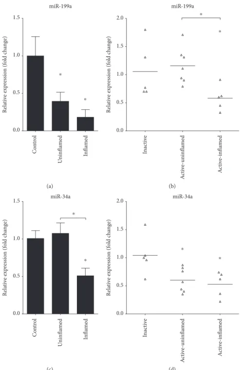
Figure3: Decreased expression of miRNAs regulating Axl in inflamed colon. The relative expression of miR-199a (a, b) and miR-34a (c, d) is shown from rat (a, c) and human (b, d) samples. Data on panels (a) and (c) represent control (left columns,n= 2), uninflamed (middle columns,n= 6), and inflamed (right columns,n= 6) rat colon sections and are presented as the mean±SEM; data on panels (b) and (d) represent inactive (left,n= 5), active-uninflamed (middle,n= 7), and active-inflamed (right,n= 5) colon samples of IBD patients where dots and lines represent individual values and the mean;∗p< 0 05.
in the active-inflamed regions of IBD patients in whichAXL may play a regulatory role.
3.4. Dysregulated MicroRNA Expression Drives EMT in IBD.
Altered expression of miRNAs is a hallmark of various can- cer types. MicroRNAs regulating genes involved in EMT, such as miR-192 and miR-375, are downregulated in the inflamed colon regions in rat experimental colitis [32] and also in active-inflamed colons of IBD patients (Figure 2(l)).
Importantly, microRNAs miR-199a and miR-34a, which posttranscriptionally regulate the expression ofAXL[27–29], are also significantly downregulated in inflamed rat colons (Figures 3(a) and 3(c)) as well as in active-inflamed regions of IBD patients (Figures 3(b) and 3(d)). As a consequence, the posttranscriptional regulation of AXL is dysregulated;
hence, its transcript (Figures 1(b) and 2(b)) and protein (Figure 1(i)) levels increase.
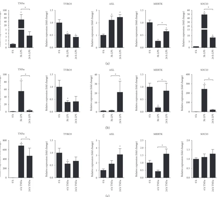
3.5. LPS Induces AXL Expression in THP-1 Monocytic Cell Line. TAM receptors exhibit different tissue expression patterns, and their promoters are regulated by distinct extra- cellular stimuli [40, 41]. Next, we have used inactivated and PMA-activated THP-1 cell line of monocytic origin to deter- mine if the LPS challenge alters the expression of TAM receptors. For this, we have stimulated the cells for 3 or 24 hours with LPS and havefirst determined the expression of TNFαas a positive control of induction. As expected, both inactivated and activated THP-1 cells responded with ele- vated expression of TNFα (Figures 4(a) and 4(b)). The expression of TAM receptors showed diverse expressions:
0 h 3h LPS 24 h LPS 0 h 3h LPS 24 h LPS 0 h 3h LPS 24 h LPS 0 h 3h LPS 24 h LPS 0 h 3h LPS 24 h LPS
0 1 2 3 4 5 20 40 60 80 100
*
0.0 0.5 1.0 1.5
*
*
0 1 2 3
0.0 0.5 1.0 1.5
Relative expression (fold change)
0 5 10 15 20 30 40 50 60
*
TNF훼 TYRO3 AXL MERTK SOCS3
Relative expression (fold change)
Relative expression (fold change)
Relative expression (fold change)
Relative expression (fold change)
⁎
⁎
⁎
⁎
⁎
⁎ ⁎
⁎
⁎
⁎
(a)
0 h 3h LPS 24 h LPS 0 h 3h LPS 24 h LPS 0 h 3h LPS 24 h LPS 0 h 3h LPS 24 h LPS 0 h 3h LPS 24 h LPS
0 20 40 60 80 100
*
0.0 0.5 1.0 1.5
0 10 20 30 40
0.0 0.5 1.0 1.5
Relative expression (fold change)
0 100 200 300 400
*
TNF훼 TYRO3 AXL MERTK SOCS3
Relative expression (fold change)
Relative expression (fold change)
Relative expression (fold change)
Relative expression (fold change) ⁎
⁎
⁎
⁎
⁎ ⁎
⁎
⁎
⁎
(b)
0 h 0 h 0 h 0 h 0 h
TNF훼
0 200 400 600 800
*
TYRO3
0.0 0.5 1.0 1.5
AXL
0 1 2 3 4 5
MERTK
0.0 0.5 1.0 1.5 2.0 2.5
SOCS3
Relative expression (fold change)
Relative expression (fold change)
Relative expression (fold change)
Relative expression (fold change)
Relative expression (fold change)
0.0 0.5 1.0 1.5 2.0
*
⁎ ⁎
⁎
⁎
⁎
24 h TNF훼
6 h TNF훼 24 h TNF훼
6 h TNF훼 24 h TNF훼
6 h TNF훼 24 h TNF훼
6 h TNF훼 24 h TNF훼
6 h TNF훼
(c)
Figure4: Distinct expression of TAM receptors in LPS triggered THP-1 and TNFαtriggered HT-29 cells. The relative gene expression of the proinflammatory moleculeTNFα, TAM receptors TYRO3, AXL, andMERTK, and the suppressor of cytokine signalling SOCS-3 in (a) inactivated LPS-stimulated THP-1, (b) PMA-activated LPS-stimulated THP-1, and (c) TNFα-stimulated HT-29 cells. Data are presented as the mean±SEM;n= 3;∗p< 0 05.
while LPS downregulated the expression of TYRO3 and MERTK, it significantly induced the expression of AXL (Figures 4(a) and 4(b)). Interestingly, while the expression ofAXLin inactivated THP-1 cells shows modest induction at both 3 and 24 hours post LPS stimuli (Figure 4(a)), induc- tion in activated THP-1 cells at 24 hours after LPS challenge is robust (Figure 4(b)). Since TAM receptors inhibit TLR- induced inflammation by the activation of, among others, SOCS3 anti-inflammatory molecule [13], we have next deter- mined its expression. The expression pattern of SOCS3 (Figures 4(a) and 4(b)) follows that ofTNFαthat is opposing expression pattern to that of AXL suggesting that even though increased AXL may act as a negative regulator of innate immunity upon LPS stimulation of THP-1 cells, the downstream effector molecule is probably not SOCS3.
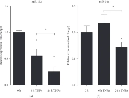
3.6. TNFαInduces Gene Expression of AXL in HT-29 Colonic Epithelial Cell Line.In order to determine if TAM receptors show altered expression in colonic epithelial cells, we have stimulated HT-29 colonic epithelial cells with TNFα for 6 or 24 hours and have againfirst monitored the expression of TNFα which is known to be self-induced. The robust expression of TNFα indicated successful induction of HT-29 cells (Figure 4(c)).AXLagain showed induced expres- sion at 24 hours postinduction (Figure 4(c)). Importantly, in contrast to THP-1 cells, the expression ofSOCS3remained unaltered (Figure 4(c)) suggesting that the function of inducedAXLin these cells is probably not the inhibition of inflammation, at least not through SOCS3. Finally, the expression of both miR-192 and miR-34a showed significant downregulation at 24 hours post-TNFαstimuli (Figures 5(a) and 5(b), resp.). These data are in complete agreement with
those obtained from both rat and human colon samples indi- cating that TNFα-stimulated HT-29 cells are mimicking in vivoobservations making them a perfectin vitrosystem for modelling the gene expression changes of colonic epithe- lial cells in IBD.
4. Conclusion
Our present study demonstrates that the epithelial-to- mesenchymal transition is triggered in IBD patients. These data are in agreement with our previous report showing the activation of EMT in rat experimental colitis [33]. In addition, we show that the increased expression of AXL is accompanied with the decreased expression of its microRNA regulators. These findings shed light onto a possible AXL- and microRNA-mediated regulation influencing epithelial- to-mesenchymal transition in IBD. In addition, by using in vitromonocytic and colonic epithelial cells, we show that upon stimulation with LPS or TNFα, respectively, the expres- sion ofAXLincreases in both cell types.
Rothlin et al. in a recent review have underlined an apparently contradicting role of TAM receptors in the con- text of IBD and CRC, asking if individual members of the TAM family have tumor-promoting or antitumor effects [16]. The present study provides preliminary evidence on the possible oncogenic role of AXL in IBD: the increased expression of AXL—possibly due to the downregulated expression of its microRNA regulator/s, such as miR-199a and miR-34a—along with malfunctioning of the negative regulation of inflammation points towardAXLindeed being an important player in bridging IBD and CRC. Since the therapeutic targeting of AXL inhibits tumor growth and
miR-192
Relative expression (foldchange)
0 h 24 h TNF
훼
0.0 0.5 1.0 1.5
6 h TNF
훼
⁎
⁎
⁎
(a)
miR-34a
Relative expression (fold change)
0.0 0.5 1.0 1.5
0 h 6 h TNF
훼
24 h TNF훼
⁎
⁎
(b)
Figure5: Decreased expression of miRNAs regulating Axl in TNFαtriggered HT-29 colonic epithelial cells. The relative gene expression of miR-199a (a) and miR-34a (b) is shown from TNFαtriggered HT-29 cells. Data are presented as the mean±SEM;n= 3;∗p< 0 05.
metastasis in ovarian cancer models [42], it is likely that AXL may become a potential therapeutic target in colitis- associated colorectal cancer.
Data Availability
The authors have not used high-throughput analyses; hence, the data are not deposited.
Conflicts of Interest
The authors have no competing interests to declare.
Authors’Contributions
Éva Boros and István Nagy conceived and supervised the project and designed the experiments. Zoltán Kellermayer, Péter Balogh, Patrícia Sarlós, and Áron Vincze obtained colonic biopsies. Csaba Varga performed the experiments with rats. Éva Boros, Gerda Strifler, and Andrea Vörös per- formed thein vitroexperiments with THP-1 and HT-29 cells.
Éva Boros performed all other experiments. Éva Boros and István Nagy analyzed and interpreted the data and wrote the paper.
Acknowledgments
The authors thank Dr. Zita Szalai for the help with animal experiments as well as Dr. JenőSolt, Dr. Anikó Szabó, and Dr. Benedek Tínusz for providing help in obtaining colonic biopsies. This work was funded, in part, by grant from the National Research, Development and Innovation Office (Grant no. GINOP-2.3.2-15-2016-00039). Éva Boros was funded by the European Union and the State of Hungary, cofinanced by the European Social Fund in the framework of “National Excellence Program” (Grant no. A2-ELMH- 12-0082). István Nagy was supported by the János Bolyai Research Scholarship of the Hungarian Academy of Sciences.
Zoltán Kellermayer was supported by the ÚNKP-17-4-I New National Excellence Program of the Ministry of Human Capacities and the postdoctoral research grant of the Faculty of Medicine, University of Pécs. Péter Balogh was supported by OTKA K108429.
References
[1] J. Z. Liu, S. van Sommeren, H. Huang et al., “Association analyses identify 38 susceptibility loci for inflammatory bowel disease and highlight shared genetic risk across populations,”
Nature Genetics, vol. 47, no. 9, pp. 979–986, 2015.
[2] D. Ellinghaus, E. Ellinghaus, R. P. Nair et al., “Combined analysis of genome-wide association studies for Crohn disease and psoriasis identifies seven shared susceptibility loci,” American Journal of Human Genetics, vol. 90, no. 4, pp. 636–647, 2012.
[3] D. K. Podolsky,“Inflammatory bowel disease,”New England Journal of Medicine, vol. 325, no. 13, pp. 928–937, 1991.
[4] J. Burisch and P. Munkholm,“The epidemiology of inflamma- tory bowel disease,”Scandinavian Journal of Gastroenterology, vol. 50, no. 8, pp. 942–951, 2015.
[5] J. K. Triantafillidis, G. Nasioulas, and P. A. Kosmidis,“Colo- rectal cancer and inflammatory bowel disease: epidemiology, risk factors, mechanisms of carcinogenesis and prevention strategies,” Anticancer Research, vol. 29, no. 7, pp. 2727– 2737, 2009.
[6] S. I. Grivennikov, F. R. Greten, and M. Karin, “Immunity, inflammation, and cancer,”Cell, vol. 140, no. 6, pp. 883–899, 2010.
[7] K. Taniguchi and M. Karin,“NF-κB, inflammation, immunity and cancer: coming of age,” Nature Reviews Immunology, vol. 18, no. 5, pp. 309–324, 2018.
[8] M. Saleh and G. Trinchieri,“Innate immune mechanisms of colitis and colitis-associated colorectal cancer,” Nature Reviews Immunology, vol. 11, no. 1, pp. 9–20, 2011.
[9] C. V. Rothlin, S. Ghosh, E. I. Zuniga, M. B. A. Oldstone, and G. Lemke,“TAM receptors are pleiotropic inhibitors of the innate immune response,” Cell, vol. 131, no. 6, pp. 1124– 1136, 2007.
[10] J. H. M. van der Meer, T. van der Poll, and C. van 't Veer,
“TAM receptors, Gas6, and protein S: roles in inflammation and hemostasis,”Blood, vol. 123, no. 16, pp. 2460–2469, 2014.
[11] D. K. Graham, D. DeRyckere, K. D. Davies, and H. S. Earp,
“The TAM family: phosphatidylserine sensing receptor tyro- sine kinases gone awry in cancer,” Nature Reviews Cancer, vol. 14, no. 12, pp. 769–785, 2014.
[12] G. Lemke and C. V. Rothlin, “Immunobiology of the TAM receptors,” Nature Reviews Immunology, vol. 8, no. 5, pp. 327–336, 2008.
[13] A. Zagórska, P. G. Través, E. D. Lew, I. Dransfield, and G. Lemke, “Diversification of TAM receptor tyrosine kinase function,”Nature Immunology, vol. 15, no. 10, pp. 920–928, 2014.
[14] E. B. Rankin, K. C. Fuh, L. Castellini et al.,“Direct regulation of GAS6/AXL signaling by HIF promotes renal metastasis through SRC and MET,”Proceedings of the National Academy of Sciences of the United States of America, vol. 111, no. 37, pp. 13373–13378, 2014.
[15] S. G. Kimani, S. Kumar, V. Davra et al., “Normalization of TAM post-receptor signaling reveals a cell invasive signature for Axl tyrosine kinase,”Cell Communication and Signaling:
CCS, vol. 14, no. 1, p. 19, 2016.
[16] P. Y. Chan, E. A. C. Silva, D. de Kouchkovsky et al.,“The TAM family receptor tyrosine kinase TYRO3 is a negative regulator of type 2 immunity,”Science, vol. 352, no. 6281, pp. 99–103, 2016.
[17] L. Bosurgi, J. H. Bernink, V. Delgado Cuevas et al.,“Paradox- ical role of the proto-oncogene Axl and Mer receptor tyrosine kinases in colon cancer,”Proceedings of the National Academy of Sciences of the United States of America, vol. 110, no. 32, pp. 13091–13096, 2013.
[18] A. Verma, S. L. Warner, H. Vankayalapati, D. J. Bearss, and S. Sharma,“Targeting Axl and Mer kinases in cancer,”Molec- ular Cancer Therapeutics, vol. 10, no. 10, pp. 1763–1773, 2011.
[19] C. Gjerdrum, C. Tiron, T. Hoiby et al., “Axl is an essential epithelial-to-mesenchymal transition-induced regulator of breast cancer metastasis and patient survival,”Proceedings of the National Academy of Sciences of the United States of Amer- ica, vol. 107, no. 3, pp. 1124–1129, 2010.
[20] C. M. Gay, K. Balaji, and L. A. Byers,“Giving AXL the axe:
targeting AXL in human malignancy,” British Journal of Cancer, vol. 116, no. 4, pp. 415–423, 2017.
[21] M. K. Asiedu, F. D. Beauchamp-Perez, J. N. Ingle, M. D.
Behrens, D. C. Radisky, and K. L. Knutson, “AXL induces epithelial-to-mesenchymal transition and regulates the func- tion of breast cancer stem cells,” Oncogene, vol. 33, no. 10, pp. 1316–1324, 2014.
[22] K. Y. Tai, Y. S. Shieh, C. S. Lee, S. G. Shiah, and C. W. Wu,“Axl promotes cell invasion by inducing MMP-9 activity through activation of NF-kappaB and Brg-1,” Oncogene, vol. 27, no. 29, pp. 4044–4055, 2008.
[23] S. Lamouille, J. Xu, and R. Derynck,“Molecular mechanisms of epithelial-mesenchymal transition,”Nature Reviews Molec- ular Cell Biology, vol. 15, no. 3, pp. 178–196, 2014.
[24] G. Peng and Y. Liu,“Hypoxia-inducible factors in cancer stem cells and inflammation,”Trends in Pharmacological Sciences, vol. 36, no. 6, pp. 374–383, 2015.
[25] Z. Wang, Y. Li, D. Kong, and F. H. Sarkar,“The role of Notch signaling pathway in epithelial-mesenchymal transition (EMT) during development and tumor aggressiveness,”Cur- rent Drug Targets, vol. 11, no. 6, pp. 745–751, 2010.
[26] Y. Peng and C. M. Croce,“The role of microRNAs in human cancer,” Signal Transduction and Targeted Therapy, vol. 1, no. 1, p. 15004, 2016.
[27] C. Y. Cho, J. S. Huang, S. G. Shiah et al.,“Negative feedback regulation of AXL by miR-34a modulates apoptosis in lung cancer cells,”RNA, vol. 22, no. 2, pp. 303–315, 2016.
[28] G. Mudduluru, P. Ceppi, R. Kumarswamy, G. V. Scagliotti, M. Papotti, and H. Allgayer,“Regulation of Axl receptor tyro- sine kinase expression by miR-34a and miR-199a/b in solid cancer,”Oncogene, vol. 30, no. 25, pp. 2888–2899, 2011.
[29] H. Y. Zhu, W. D. Bai, J. Li et al.,“Peroxisome proliferator- activated receptor-γ agonist troglitazone suppresses trans- forming growth factor-β1 signalling through miR-92b upregulation-inhibited Axl expression in human keloidfibro- blasts in vitro,”American Journal of Translational Research, vol. 8, no. 8, pp. 3460–3470, 2016.
[30] K. M. Giles, F. C. Kalinowski, P. A. Candy et al.,“Axl mediates acquired resistance of head and neck cancer cells to the epider- mal growth factor receptor inhibitor erlotinib,” Molecular Cancer Therapeutics, vol. 12, no. 11, pp. 2541–2558, 2013.
[31] A. Belfiore, M. Genua, and R. Malaguarnera, “PPAR-γ agonists and their effects on IGF-I receptor signaling: implica- tions for cancer,”PPAR Research, vol. 2009, Article ID 830501, 18 pages, 2009.
[32] E. Boros, M. Csatári, C. Varga, B. Bálint, and I. Nagy,“Specific gene- and microRNA-expression pattern contributes to the epithelial to mesenchymal transition in a rat model of experi- mental colitis,”Mediators of Inflammation, vol. 2017, Article ID 5257378, 9 pages, 2017.
[33] G. P. Morris, P. L. Beck, M. S. Herridge, W. T. Depew, M. R.
Szewczuk, and J. L. Wallace, “Hapten-induced model of chronic inflammation and ulceration in the rat colon,”Gastro- enterology, vol. 96, no. 2, pp. 795–803, 1989.
[34] S. Hafizi and B. Dahlback,“Signalling and functional diversity within the Axl subfamily of receptor tyrosine kinases,”Cyto- kine & Growth Factor Reviews, vol. 17, no. 4, pp. 295–304, 2006.
[35] C. Wilson, X. Ye, T. Pham et al.,“AXL inhibition sensitizes mesenchymal cancer cells to antimitotic drugs,” Cancer Research, vol. 74, no. 20, pp. 5878–5890, 2014.
[36] L. A. Byers, L. Diao, J. Wang et al., “An epithelial- mesenchymal transition gene signature predicts resistance to
EGFR and PI3K inhibitors and identifies Axl as a therapeutic target for overcoming EGFR inhibitor resistance,” Clinical Cancer Research, vol. 19, no. 1, pp. 279–290, 2013.
[37] A. Abu-Thuraia, R. Gauthier, R. Chidiac et al.,“Axl phosphor- ylates Elmo scaffold proteins to promote Rac activation and cell invasion,”Molecular and Cellular Biology, vol. 35, no. 1, pp. 76–87, 2014.
[38] B. Khor, A. Gardet, and R. J. Xavier,“Genetics and pathogen- esis of inflammatory bowel disease,”Nature, vol. 474, no. 7351, pp. 307–317, 2011.
[39] T. S. Wong, W. Gao, and J. Y. W. Chan,“Transcription regu- lation of E-cadherin by zincfinger E-box binding homeobox proteins in solid tumors,” BioMed Research International, vol. 2014, Article ID 921564, 10 pages, 2014.
[40] E. D. Lew, J. Oh, P. G. Burrola et al., “Differential TAM receptor-ligand-phospholipid interactions delimit differential TAM bioactivities,”eLife, vol. 3, 2014.
[41] W. I. Tsou, K. Q. N. Nguyen, D. A. Calarese et al.,“Receptor tyrosine kinases, TYRO3, AXL, and MER, demonstrate distinct patterns and complex regulation of ligand-induced activation,” The Journal of Biological Chemistry, vol. 289, no. 37, pp. 25750–25763, 2014.
[42] P. Kanlikilicer, B. Ozpolat, B. Aslan et al.,“Therapeutic target- ing of AXL receptor tyrosine kinase inhibits tumor growth and intraperitoneal metastasis in ovarian cancer models,”Molecu- lar Therapy - Nucleic Acids, vol. 9, pp. 251–262, 2017.
Stem Cells International
Hindawi
www.hindawi.com Volume 2018
Hindawi
www.hindawi.com Volume 2018
INFLAMMATION
Endocrinology
International Journal ofHindawi
www.hindawi.com Volume 2018
Hindawi
www.hindawi.com Volume 2018
Disease Markers
Hindawi
www.hindawi.com Volume 2018
BioMed
Research International
Oncology
Journal ofHindawi
www.hindawi.com Volume 2013
Hindawi
www.hindawi.com Volume 2018
Oxidative Medicine and Cellular Longevity
Hindawi
www.hindawi.com Volume 2018
PPAR Research
Hindawi Publishing Corporation
http://www.hindawi.com Volume 2013
Hindawi www.hindawi.com
The Scientific World Journal
Volume 2018
Immunology Research
Hindawi
www.hindawi.com Volume 2018
Journal of
Obesity
Journal ofHindawi
www.hindawi.com Volume 2018
Hindawi
www.hindawi.com Volume 2018
Computational and Mathematical Methods in Medicine
Hindawi
www.hindawi.com Volume 2018
Behavioural Neurology Ophthalmology
Journal ofHindawi
www.hindawi.com Volume 2018
Diabetes ResearchJournal of
Hindawi
www.hindawi.com Volume 2018
Hindawi
www.hindawi.com Volume 2018
Research and Treatment
AIDS
Hindawi
www.hindawi.com Volume 2018
Gastroenterology Research and Practice
Hindawi
www.hindawi.com Volume 2018
Parkinson’s Disease
Evidence-Based Complementary and Alternative Medicine
Volume 2018 Hindawi
www.hindawi.com