Gasotransmitters
Edited by Rui Wang
Laurentian University o f Sudbury, Canada Email: rui. wang@laurentian. ca
ÊT
ROYAL SOCIETY OF CHEMISTRYEditor-in-chief:
C. David Garner, University o f Nottingham, UK Series editors:
Stefano L. Ciurli, University of Bologna, Italy Julie Kovacs, University o f Washington, USA Emma Raven, University o f Leicester, UK Hongzhe Sun, University o f Hong Kong, China Anthony Wedd, University o f Melbourne, Australia Titles in the Series:
1: Mechanisms and Metal Involvement in Neurodegenerative Diseases 2: Binding, Transport and Storage of Metal Ions in Biological Cells 3: 2-Oxoglutarate-Dependent Oxygenases
4: Heme Peroxidases
5: Molybdenum and Tungsten Enzymes: Biochemistry
6: Molybdenum and Tungsten Enzymes: Bioinorganic Chemistry 7: Molybdenum and Tungsten Enzymes: Spectroscopic and Theoretical
Investigations
8: Metal Chelation in Medicine
9: Metalloenzymes in Denitrification: Applications and Environmental Impacts
10: The Biological Chemistry of Nickel 11: Gas Sensing in Cells
12. Gasotransmitters
How to obtain future titles on publication:
A standing order plan is available for this series. A standing order will bring delivery of each new volume immediately on publication.
For further information please contact:
Book Sales Department, Royal Society of Chemistry, Thomas Graham House, Science Park, Milton Road, Cambridge, CB4 OWF, UK
Telephone: +44 (0)1223 420066, Fax: +44 (0)1223 420247, Email: booksales(a)rsc.org
Visit our website at www.rsc.org/books
Print ISBN: 978-1-78262-924-5 PDF ISBN: 978-1-78801-300-0 EPUB ISBN: 978-1-78801-480-9 Print ISSN: 2045-547X Electronic ISSN: 2045-5488
A catalogue record for this book is available from the British Library
© The Royal Society of Chemistry 2018 A ll rights reserved
Apart from fa ir dealing fo r the purposes o f research fo r non-commercial purposes or for private study, criticism or review, as permitted under the Copyright, Designs and Patents Act 1988 and the Copyright and Related Rights Regulations 2003, this publication may not
be reproduced, stored or transmitted, in any form or by any means, without the prior permission in writing o f The Royal Society o f Chemistry or the copyright owner, or in the
case o f reproduction in accordance with the terms o f licences issued by the Copyright Licensing Agency in the UK, or in accordance with the terms o f the licences issued by the appropriate Reproduction Rights Organization outside the UK. Enquiries concerning reproduction outside the terms stated here should be sent to The Royal Society o f Chemistry at the address printed on this page.
Whilst this material has been produced with all due care, The Royal Society o f Chemistry cannot be held responsible or liable fo r its accuracy and completeness, nor fo r any consequences arising from any errors or the use o f the information contained in this publication. The publication o f advertisements does not constitute any endorsement by
The Royal Society o f Chemistry or Authors o f any products advertised. The views and opinions advanced by contributors do not necessarily reflect those o f The Royal Society o f Chemistry which shall not be liable fo r any resulting loss or damage arising as a result o f reliance upon this material.
The Royal Society of Chemistry is a charity, registered in England and Wales, Number 207890, and a company incorporated in England by Royal Charter (Registered No. RC000524), registered office: Burlington House, Piccadilly, London W1J 0BA, UK, Telephone: +44 (0) 207 4378 6556.
For further information see our web site at www.rsc.org
Printed in the United Kingdom by CPI Group (UK) Ltd, Croydon, CRO 4YY, UK
Preface
Gasotransmitters are real and exist eveiywhere in our body. Gaso- transmitters are unique and specifically regulate biological functions.
Gasotransmitters are novel signaling gas molecules we cannot live without.
Since its inception in 2002 (R. Wang, Two’s company, three’s a crowd—
Can H 2S be the third endogenous gaseous transmitter? FASEB /., 2002, 16,
1792-1798), the concept of ‘gasotransmitters’ has been widely accepted and applied in different life-science disciplines. The initial members of the gasotransmitter family included nitric oxide (NO), carbon monoxide (CO), and hydrogen sulfide (H2S). This concept has continuously evolved and been refined with the inclusion of new members, such as ammonia (NH3) (R. Wang, Gasotransmitters: Growing pains and joys, Trends Biochem. Sci., 2014, 39, 227-232*). The birth and growth of the gasotransmitter framework have deepened our understanding of cellular signaling processes and led to the discovery of new pathogenic mechanisms and therapeutic strategies for related diseases. Research on gasotransmitters has also gone beyond the boundaries of mammalian biology and medicine. The production and function of gasotransmitters in plant and bacteria, for example, have attacted the attention and stimulated the interest of numerous research teams and researchers worldwide.
Over the last 15 years, the gasotransmitter literature has exponentionally grown. Each year, about 15 000 papers are published on gasotransmitters.
A recent study revealed that, before 2004, H 2S biology-related publications were less than 100 per year. By 2015, the annual publications on H2S biology- related research had increased by about seven-fold (G. Yang and L. Wu, Trends in H2S biology and medicine research—A bibliometric analysis,
'Note: This seminar paper framing the concept and establishing the qualification standards for gasotransmitters has been reprinted as an appendix of this book.
Metallobiology Series No. 12 Gasotransmitters
Edited by Rui Wang
© The Royal Society of Chemistry 2018
Published by the Royal Society of Chemistry, www.rsc.org
CHAPTER 8
Production and Signaling o f Methane
M . BOROS*3 AND F. KEPPLERb
a University of Szeged, Institute of Surgical Research, Szokefalvi Nagy B. 6, 6720 Szeged, Hungary; b Heidelberg University, Institute of Earth Sciences, Im Neuenheimer Feld 234-236, Heidelberg 69120, Germany
*Email: boros.mihaly(a)med.u-szeged.hu
8.1 Introduction
Many gases are biologically active in vivo, but contrary to ‘classical’ pathways of signal transduction, the exact roles of gaseous compounds in the medi
ation of extra- or intracellular events are to date not completely understood.
These molecules are also at the forefront of medical research due to the anti
inflammatory effects of nitric oxide (NO), carbon monoxide (CO), and hydrogen sulfide (H2S). Methane (CH4) has a long evolutionary history on Earth, and it is also part of the gaseous environment that maintains the metabolism within the aerobic eukaryote cells but, in fact, the role of CH 4 in physiology is largely unmapped. Conventionally believed as physiologically inert, studies cited in this review suggest that it can modulate the pathways involved in oxidative and nitrosative stress responses and key events of inflammation.
This chapter is divided in two parts, the first one being devoted to the biogenesis of CH4 in eukaryotes and the interactions of CH 4 with other biological gases in vivo, while the second part deals with well-documented biological responses and their potential physiology- and pathology-related implications.
Metallobiology Series No. 12 Gasotransmitters
Edited by Rui Wang
© The Royal Society of Chemistry 2018
Published by the Royal Society of Chemistry, www.rsc.org
8.2 Physico-chemical Properties and Toxicity of C H 4
The bioactivity or toxicity of gas mediators NO, CO, and H 2S is related to their tendency to react with biologically important molecules. Despite this, CH4 is intrinsically non-toxic in vivo; rabbits can inhale a mixture of one volume of oxygen and four volumes of CH 4 for any length of time without showing any obvious side-effects. It is a simple asphyxiant, which means that tissue hypoxia might occur when an increasing concentration of CH 4 dis
places the inhaled air in a restricted area, and the concentration of oxygen is reduced to below approx. 16-18% in the internal milieu. With a density of 0.716 g lT 1 under standard conditions of temperature and pressure, CH4 is lighter than air; nevertheless, CH4 intoxication can occur in open fields as well.1 In such cases, respiratory arrest is not due to the chemical specificity of the gas, but to the decreased 0 2 content. C H 4 will displace oxygen down to 18% in air when present at about 14% (or 140 000 parts per million by vol
ume, ppmv). It should be mentioned that CH 4 is combustible and forms explosive mixtures with air at concentrations between 5% (lower explosive limit) and 15% (upper explosive limit) at room temperature. In the early days of colonoscopy, an accumulation of colonic gas and the use of electro- surgical devices sometimes led to intracolonic explosions.2 It is expected to cause unconsciousness due to central nervous system (CNS) depression when it reaches high concentrations (30% or so), well above the lower ex
plosive limit and level for asphyxiation.
The inhalation of normoxic air containing 2.5% CH 4 for 3 h or CH 4-rich saline (MRS) treatments for several days had no side-effects on the blood gas chemistry (pH, Pa02, PaC02), in unstressed animals.3,4 When the effect of exogenous CH 4 on the respiratory activity of the mitochondrial oxidative phosphorylation (OxPhos) system was investigated by high-resolution re
spirometry, the incubation of 2.2% CH4 did not affect the activity of OxPhos complexes of intact rat liver mitochondria.5 MRS treatment had no signifi
cant effect on the redox system of normal retinal tissue and did not affect the cytochrome c release or activation of caspase-9 and caspase-3 in rats.4 When 10 mL kg-1 MRS was applied intraperitoneally and micromolar C H 4 levels were detected in the circulation, the plasma glucose and hematocrit levels did not change. Data on human cardiovascular effects are sparse, but in a case report with a 45 min CH 4 exposure to liquid manure, the unconscious patient presented spontaneous breathing with an arterial pH value of 7.26 and made a full recovery later.6
8.3 Methanogenesis - Biotic and Abiotic Sources in the Environment
In the atmosphere, which currently contains over 1.8 ppmv, CH4 is an im
portant greenhouse gas. The atmospheric concentration of CH 4 has in
creased dramatically since pre-industrial times from about 715 parts per billion by volume (ppbv) to 1800 ppbv in the year 2010.7'8 The current global
budget of atmospheric CH4 is of the order of 550-650 Tg (million tonnes) of CH4 per annum.8 The global atmospheric CH 4 budget is determined by many natural and anthropogenic terrestrial and aquatic surface sources, balanced primarily by one major sink (hydroxyl radicals) in the atmosphere.
However, a large fraction of the emissions is mainly the result of environ
mental microbial processes, such as archaeal methanogenesis in wetlands, rice fields, and ruminant and termite digestive systems.
8.3.1 Abiotic Sources of C H 4 (Including Thermogenic Degradation of Organic Matter)
About a quarter of all CH 4 sources are associated with chemical processes, including emissions from mining and combustion of fossil fuels, and the burning of biomass or geological sources such as volcanoes and geothermal systems. The largest fraction of this chemically formed CH 4 comes from thermal degradation of organic matter and it is sometimes not classified as abiotic because the precursor substance was delivered from biological compounds.
Strictly speaking, abiotic (or abiogenic) CH 4, formed by chemical re
actions, does not directly include organic matter and it is produced in much smaller amounts on a global scale. These reactions occurs on Earth in sev
eral specific geological environments and they may be produced by either high-temperature magmatic processes in volcanic and geothermal areas, or via low-temperature (<100 °C) gas-water-rock reactions in continental set
tings. For example, these reactions might involve the hydrogenation of C 0 2, also known as the ‘Sabatier reaction’. For more details regarding the abiotic formation of CH4 on Earth, we refer the reader to a review by Etiope and Sherwood Lollar.9
8.3.2 Microbial Methanogenesis - Formation of C H 4 by Archaea
Usually, biogenic methanogenesis is regarded as a microbial process carried out by a unique class of prokaryotes (archaea). Methanogens do not use oxygen to respire and the terminal electron acceptor is carbon. The two best- described pathways involve the use of acetic acid and inorganic carbon di
oxide (C02) as terminal electron acceptors. Methanogenic archaea and CH 4 production are typically found in ruminants or termites or in the environ
ment in wetlands and landfills, and at those sites where organic matter is decomposing in the absence of oxygen or other oxidants, such as nitrate, sulfate, or ferric iron. In all methanogenic archaea, CH4 is formed from methyl-coenzyme M (CH3-S -CoM) by reduction, where the methyl coenzyme M reductase catalyzes the reaction between thioether methyl coenzyme M and thiol N-(7-mercaptoheptanoyl)threonine 3-O-phosphate to give CH4 and a mixed disulfide.10’11
In both rumen and the hindgut of insects (termites, cockroaches, scarab beetles), CH4 seems to be exclusively produced by hydrogenotrophic me- thanogens that utilize hydrogen gas (H2) and C 0 2 and their activity is dependent on the availability of H 2. CH 4 production from acetate plays no obvious role and acetoclastic methanogens are usually not detected in such environments.7 In the rumen, CH 4 production largely occurs in the gut lumen where it reduces H2 to concentrations that are not permissive for acetogenesis. In the insect hindgut, however, methanogenesis is restricted to the gut wall or other gut structures, or it is associated with gut flagellates, while in the gut lumen acetogenesis is often the main H2-consuming pro
cess. The microbiology and methanogenic processes in ruminants and ter
mites have been reviewed in detail.12“15
In contrast to ruminants, where all animals produce and emit substantial amounts of CH 4, only around one third of humans are considered to emit CH4 at measureable rates. CH 4 is produced in the human large intestine by microbial activity, with Methanobrevibacter smithii being the predominant archaeon that utilizes H 2 and C 0 2. Human CH 4 production will be further discussed later (Section 8.5).
8.3.3 Non-archaeal C H 4 Formation in Eukaryotes
Until recently, biological CH4 formation had been associated exclusively with anoxic environments and microbial activity (prokaryotes - archaea). How
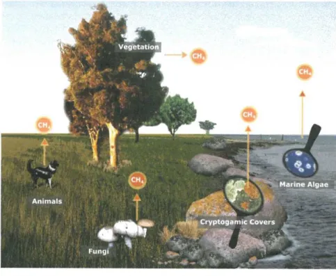
ever, recent studies have unambiguously confirmed direct (endogenous) CH 4 release from eukaryotes, including plants, fungi, lichens, marine algae, and animals, even in the absence of microbes and in the presence of oxygen (Figure 8.1).16-26 Thus, the recently found formation of C H 4 in eukaryotes is often termed ‘aerobic’ or ‘non-microbial’ CH 4 formation. However, in this chapter, we prefer to use the term ‘non-archaeal CH 4 formation’.
Various in vitro and in vivo experimental data have established the possi
bility of biotic, non-archaeal generation of CH 4 under various conditions.
Precursor compounds such as pectin, cellulose, lignin, ascorbic acid, leaf waxes, and methionine have been shown to deliver methyl groups for CH4 production.18,21,27-30 Furthermore, it was found that aerobic CH4 release may be stimulated in plants by increasing the temperature, physical injury, re
active oxygen species (ROS) and UV radiation, and inhibition of cytochrome c oxidase by sodium azide (NaN3) 30-33 Along these lines, significant in vivo CH4 release was demonstrated in a rodent model of chemical asphyxiation, after chronic inhibition of the activity of mitochondrial cytochrome c oxidase by NaN3.34 However, at this time, the biochemical reactions leading to CH4 production in plants and mammals and the potential enzymes involved have not been identified yet, even though some experimental data seem to indi
cate that CH 4 formation may be linked to redox regulation and be connected with hypoxic events.35 In this respect, it was hypothesized that hypoxia- induced CH 4 generation may be a necessary phenomenon of aerobic life, and perhaps a surviving evolutionary trait in eukaryote cells.1"
Figure 8.1 Conceptual scheme showing the recently identified novel sources of non- archaeal CH4 in the biosphere (designed by Thomas Klintzsch, Heidel
berg University, and reproduced with permission).
Future research should particularly seek to answer the question of whe
ther CH4 generation is a by-product of the chemical degradation of biomo
lecules, e.g. , induced by UV irradiation, increased temperatures, or hypoxia, or whether it also plays a more general physiological role (see below). It appears that, in plants, high rates of CH 4 generation may be linked to en
vironmental stress. Similar mechanisms might be active in animals, and probably humans as well, producing CH 4 when the organisms are under external or internal {e.g., inflammation) stress.
8.4 Potential Pathways of C H 4 Formation in Eukaryotes
While the biochemistry of methanogenesis in archaea (procaryotes) has been well described, pathways of aerobic CH 4 generation from eukaryotes including plants, mammals, fungi, and algae have yet to be elucidated and its precursor compounds identified.36 However, some studies are available proposing potential reaction schemes that may possibly occur in eukaryotes.17,21,33’37
It has been hypothesized that the hetero-bonded methyl groups of bio
molecules, such as the sulfur-containing amino acid methionine or the
ammonium salt choline, might be carbon precursors of CH 4 in living cells.
In this context, it was proposed that electrophilic methyl groups (EMGs) bound to positively-charged nitrogen moieties (such as the choline mol
ecule) may potentially act as electron acceptors, and that these reactions may entail the generation of CH 4 in animal cells.38 A continuous lack of the electron acceptor 0 2 will maintain an elevated mitochondrial NADH/NAD 1 ratio, causing the formation of a nucleophilic hydride ion that is transferred to the EMG (see Figure 8.2). Such an anomalous increase in reducing power also occurs in pathologies involving the interruption of electron flow down the mitochondrial electron transport chain (ETC). This may be supported by the observation that non-microbial CH4 generation from animal cells was observed when endothelial rat liver cells were exposed to site-specific in
hibitors of the ETC.25
It has been argued that similar mechanisms might occur in plant mi
tochondria and chloroplasts. The ETC in plant mitochondria consists of four multi-subunit complexes, respiratory complexes I-IV , assisted by ubiquinone and cytochrome c (see Figure 8.3). The mitochondrial elec
tron transport chain is a potential source of ROS such as superoxide and H 20 2, with complexes I and III being their main sites of production.
Complex IV receives electrons from reduced cytochrome c and transfers them to an oxygen molecule. The complex functionality can be inhibited by NaN3, which blocks the electron transport.39 Whiskerman et al. in
vestigated the possibility of non-microbial C H 4 formation in hetero- trophic plant cell cultures such as tobacco BY-2 (Nicotiana tabacum), grape vine (Vitis vinifera), and sugar beet (Beta vulgaris L.).33 They also examined the disturbance of mitochondrial functionality using ETC in
hibitors such as NaN3, rotenone, and salicylhydroxamic acid (SHAM).
Under non-stress conditions, the plant cell cultures produced trace amounts of C H 4, but these could be increased by one to two orders of magnitude when NaN3 was added to the cell cultures. The addition of other ETC inhibitors did not result in significant C H 4 formation, indi
cating that a site-specific disturbance of the ETC at complex IV causes C H 4 formation in plant cells.33
CH 3
R1 — N: + h-c h3
C H 3
Figure 8.2 The proposed reaction scheme for CH 4 formation in hypoxic animal cells. The nucleophilic hydride-ion (H_ ) is transferred to an electrophilic methyl group. This is followed by separation of the methyl group and the formation of CH 4.
Adapted with permission from F. Keppler, M. Boros, C. Frankenberg, J.
Lelieveld, A. McLeod, A. M. Pirttila, T. Rockmann and J. P. Schnitzler, Environ. Chem ., 2009, 6, 459 © The Authors.
Krebs cycle
Figure 8.3 Electron transport chain in the inner membrane of plant mitochondria (after Moller 2007, modified). CI-CIV stand for complexes I-IV, UQ - ubiquinone, Cyt c - cytochrome c. Inhibitors: rotenone, SHAM, and NaN3. Methane is formed when sodium azide, a compound known to disrupt the electron transport flow at cytochrome c oxidase (complex IV) in plant mitochondria, is added to cell cultures.
A. Wishkerman, S. Greiner, M. Ghyczy, M. Boros, T. Rausch, K. Lenhart and F. Keppler, Plant Cell Environ., 2011, 34, 457, John Wiley and Sons,
© 2010 Blackwell Publishing Ltd.
Furthermore, a reaction of ROS with methoxyl groups (OCH3) of pectic polysaccharides was suggested as a possible route to CH 4 formation in plants exposed to UV radiation.32 This study concluded that UV radiation evokes CH 4 production from pectic methyl groups by interacting with UV photosensitizers to generate hydroxyl radicals, but it was also suggested that other diverse processes might generate hydroxyl radicals and contribute to CH 4 emissions independently of UV irradiation.
Recently, Althoff et al. presented a novel chemical reaction that readily forms C H 4 from organosulfur compounds, such as methionine, di- methylsulfide (DMS), and dimethylsulfoxide (DMSO), under highly oxi
dative conditions, ambient atmospheric pressure and temperature.21 In the first phase of the reaction, methyl sulfides are oxidized to the cor
responding sulfoxides.21 Then, in the next phase, déméthylation of the sulfoxide via homolytic bond cleavage leads to methyl radical formation and finally to CH 4. In this reaction, the oxidant for both phases was pro
posed to be a ferryl species (see Figure 8.4). However, other oxidants (in
cluding ROS) are also conceivable. Because sulfoxidation of methyl sulfides is ubiquitous in the environment, they suggested that this novel chemical route might be involved in CH 4 formation in living aerobic or
ganisms. Thus, it could be envisaged that these thioethers and sulfoxides might be a direct precursor of the CH 4 formed from eukaryotes. The highest C H 4 formation rates might be expected from aerobic organisms, especially when oxygen availability is limited or the organisms are under hypoxia. This conclusion is in broad agreement with previous results, which demonstrated enhanced C H 4 formation in animal cells under re
duced oxygen content.25
■ = starting material
■ = products/intermediates
■ = postulated intermediate ABA = 2-Amino-butanoicacld ASC = Ascorbic acid DHASC = Dehydroascorbicacid FH * Ferrihydrite HCyA ■ Homocysteicacid MSA - Methanesulfonicacid MET = Methionine MSO = Methionine sulfoxide MSO2 = Methioninesulfone
COOH
Figure 8.4 Potential route for CH 4 formation using amino acid methionine as the methyl precursor. The carbon atom present in the CH„ molecule is highlighted in red.
Adapted with permission from F. Althoff, K. Benzing, P. Comba, C. McRoberts, D. R. Boyd, S. Greiner and F. Keppler, Nat. Commun., 2014, 5, 4205. © The Authors.
8.5 Human C H 4 Production - Archaeal and Non-archaeal Sources
In humans, several gaseous products are formed as a result of various eu
karyotic (human) and prokaryotic (bacterial) activities by enzymatic or non- enzymatic processes. Methanogenesis in humans has long been considered an exclusive attribute of methanogenic archaea, a group quite distinct from the usual bacteria and eukaryotes.40-42 These strictiy anaerobic inhabitants of the gastrointestinal tract produce CH4 from decomposing organic matter through hydrogenotrophic, methylotrophic, and acetotrophic classes of methanogenesis. From a breath analysis, approximately 30-60% of adults were classed as CH4 producers when production was defined as a >1 ppmv increase above the ambient air level.43 CH4 on breath testing is associated with higher levels of Methanobrevibacter smithii in stool and the proportion of M. smithii in stool also correlates well with the amount of breath CH4.44 Some conditions are thought to increase the CH4 production within the colon, such as excessive intracolonic anaerobiosis and elevated intracolonic pH.
In regard to CH 4 production, gender, age, and ethnic differences have been observed.42,45,46 Moreover wide day-to-day (inter-day) variations and reduced emission after physical exercise have also been reported.47’48 However, the fundamental factors influencing the number of methano- gens and the amount of CH 4 produced are still not known. Many studies have reported correlations between breath CH 4 levels and afflictions such as irritable bowel syndrome, large bowel cancer, and constipation.42,43,49-51 However, the findings of these studies remain controversial and the impact of endogenous microbial CH 4 generation still has not been determined.
The relationship between age and breath CH4 concentration is crucial when interpreting the results from studies investigating the correlation be
tween breath CH 4 levels and specific diseases such as diverticulosis and large bowel cancer.43,d2 Interestingly, two recent studies showed that, in a German population with an age range from 4 to 95 years, the percentage of breath CH4 producers greatly varied with age.46’53 When subjects were div
ided into age groups of 15 years, a significant increase in the percentage of breath CH 4 producers with age was observed. The incidence of many dis
eases increases with age and, if this is not taken into account, the concurrent increase observed in the percentage of breath CH 4 producers with age might easily lead to a misinterpretation of the data arising from supposed correl
ations.46 For more detailed information regarding the occurrence of CH4 in man, and its possible link to certain diseases, we refer the reader to reviews by de Lacy Costello.42’43
Quite significantly, a recent study using a stable carbon isotope and high precision concentration measurements provided evidence that the exhaled CH4 levels of all volunteers investigated (n = 112) in an age range from 1 to 80 years were above (on average ~118 ppbv) the inhaled CH 4 concen
tration.53 Based on their data, the authors hypothesized that next to mi
crobial sources in the gastrointestinal tracts there might be other, as yet unidentified, non-archeal processes involved in CH4 formation, which sup
ports the idea that humans might also produce CH 4 endogenously in cells.
In this sense, the physiological levels of CH4 in the human body have not yet been determined.
In the case of the gastrointestinal tract, various data suggest that the ex
cretion of CH4 in the breath of mammals may predominantly reflect intes
tinal archeal production, but a variable amount is possibly linked to a mitochondrial dysfunction. Significant CH 4 formation was detected in un
restrained rats treated with NaN3, which led to the selective and stable in
hibition of mitochondrial cytochrome c oxidase activity.54 The NaN3-induced global mitochondrial dysfunction was evidenced by hepatic ATP depletion, and a systemic inflammatory reaction. CH 4 exhaled from the airways, to
gether with the amounts discharged from the skin and body orifices, was quantified by means of whole-body photoacoustic spectroscopy. Through the determination of the amount of CH4 released from the animals at dif
ferent times, this study demonstrated that chronic NaN3 administration was accompanied by increasing emanation of endogenous CH 4 throughout the
entire duration of the experiments, irrespective of the concomitant anti
biotic treatment targeting the potential CH 4-producer gastrointestinal mi
crobial flora.26 To sum up, it has been shown that CH 4 excretion reflects the intestinal microbial fermentation along with an unknown and variable amount of generation induced from target cells, and if non-archaeal CH 4 is added to the constitutive low-level CH 4 steady-states or microbial pro
duction, this addition could occur at such a low rate that it is impossible to detect by any conventional technique.
Apart from the above considerations, the designation of subjects as
‘producers’ or ‘non-producers’ based on CH 4 breath testing only accounts for one way of escape, but not the large amount that is passed directly as flatus (between 50% and 80% of the total). Indeed, there is a release of
~150 pgC H 4cmT2 in 30 min through the skin in healthy individuals, cor
responding to a 313 fmol cm“ 2 min-1 discharge.55 It should be added that a recent study using highly sensitive open-circuit respiration chambers (which account for the total CH 4 emission) found that natural differences in CH 4 yields between individual sheep were not due to naturally differing densities of methanogenic archaea in the rumen, i.e., greater densities of methano- gens in high CH 4 animals and lower densities in low CH 4 animals, sug
gesting that other - genetic, epigenetic, or environmental - factors are present in the background.56
Another significant issue is that the large differences in breath gas an
alysis data and the rather wide day-to-day (interday) variations47 are pre
sumably not only due to variations in the personal background, bacterial strains, sampling, and analysis techniques, but have hemodynamic and microcirculatory causes as well. It should be borne in mind that in- traluminally generated CH 4 traverses the gastrointestinal mucosa and enters the splanchnic microcirculation freely. Thereafter, owing to its physico
chemical properties, CH 4 is transported by the circulating blood and, when reaching the lungs, it is partially released into the breath if the partial pressure is higher than that in the atmosphere. In this context, it may be presumed that exhaled CH 4 levels will vary in association with gastro
intestinal perfusion changes and, in this way, the breath CH 4 output may also be regarded as a marker for mesenteric microcirculatory alterations.
However, the correct characterization of this association warrants the dy
namic measurement of breath gases. Indeed, in a recent human study with CH4-producing volunteers, the on-line measured alveolar breath CH 4 level decreased dramatically (from 11.4 to 2.8 ppmv) during treadmill exercises, while the lung ventilation-perfusion ratio increased by a factor of 2-3. Based on mass balance equations and a three compartment model, the dynamics of CH4 profiles were described and it was found that the breath CH4 con
centration was affected not only by changes in the ventilation-perfusion ratios, but also by changes in the fractional large intestinal blood flow.48
A great deal of effort has been devoted toward the measurement of breath CH 4 levels in humans, but the clinical utility of CH 4 measurement methods is controversial.57 The intra- and intersubject variabilities are usually very
large, and the interpretation of the results is often difficult. CH 4 producers may stop excreting, non-producers may start to excrete CH 4, and occasion
ally the CH 4-producing status does not change after antibiotic treatments targeting the intestinal methanogenic flora.58-60 Indeed, it has recently been suggested that the clinical implications of breath CH 4 analyses should themselves undergo an in-depth revision.61 It should be mentioned that, in clinical laboratory practice, breath CH4 levels are usually determined by sampling of breath air in gastight bags, which are then analyzed by means of gas chromatography (GC) equipped with either flame ionization (FID), thermal conductivity, or mass spectrometry detectors. Thus, the sampling frequency of these traditional methods is rather limited. The major prob
lems are that discontinuous detection methods cannot accurately reflect the overall profile of in vivo CH4 generation and that, because CH4 distributes itself evenly across membrane barriers, the production is manifested not only in the exhaled air but also through other body surfaces.
Recent advances in analytical methods for high temporal resolution measurements of CH 4 might further improve our understanding of CH 4 formation in humans. Tuboly et al. applied a near-infrared laser technique- based photoacoustic spectroscopy (PS) system for in vivo studies, which had previously been validated for real-time whole-body and single breath meas
urements of CH4 emissions in human and animal studies.26,34 With this technique, a daily CH4 production profile can be determined and stress- caused changes or treatment effects may be accurately evaluated with re
producible results. In addition, Keppler et al. used high resolution optical spectroscopy (Cavity Ring Down Spectroscopy, CRDS) for the investigation of CH 4 in breath samples.53 This method is capable of precisely determining the concentration and the stable carbon isotope composition of CH 4 with high temporal resolution.
8.6 Intestinal Gases and the Influence of C H 4 on Gastrointestinal Motility
In a pioneering study, pulmonary CH4 excretion ranged from undetectable to 0.66 mLmin ', and 20% of the total CH 4 produced was excreted via the lungs.40 There are two major sources of gas in the gut; namely, swallowed air and gas produced locally during the fermentation of colonic contents or independently from the gut microbiome. A third component (COa) is gen
erated by an acid-base reaction in the duodenum, but this is rapidly diffused back into the circulation and exhaled. At rest, the intestines receive about 15% of the total blood flow, which is completely adapted to meet the transport and metabolic needs of gut tissue. In fact, the volume of intestinal gas is fairly stable because expeditious gas transit and evacuation prevent gas pooling; hence, gaseous distension of the bowel does not occur so long as absorption by blood or elimination as flatus keeps pace with pro
duction.62 According to previous studies, values for intra-intestinal gas
volumes range from 176 to 199 mL. More recently, CT image analyses gave similar results (median volumes were 155-220 mL) and the endogenous gas production or composition was not that different between patients with gas- related complaints and healthy subjects.63,64 Despite this, it is likely that the quantitative equilibrium of gas concentrations in the intestinal compart
ments is influenced by qualitative changes. While the breathing of NzO causes the expansion of air in the bowel lumen and additionally augments the accumulation of intestinal gases, it has been demonstrated in a canine study with CH4- or C 0 2-filled bowel segments that, when dogs breathed oxygen, the bowel gas volume decreased, while breathing N20 just increased the volume of the CH 4-containing segment. Breathing oxygen after 30 min of breathing N20 reduced the volume of the CH 4-containing segment toward control volumes.65 In brief, alterations in one or other gas components in the intestinal lumen can affect the other gases and the net result of these processes determines the final composition of the gaseous environment.
Gas molecules in this milieu are likely to have access to a variety of ion channels and the receptor components of the neuromuscular apparatus that are involved in gastrointestinal motility regulation and, in this respect, the association between CH4 production (again, defined as more than 1 ppm increase for breath testing above the atmospheric CH 4 level) and intestinal transit seems to be well established: CH4 production is usually associated with a constipation-related phenotype.66,67 It has been shown not just in patients, but also in healthy volunteers that the total colonic transit times are significantly more prolonged in CH 4-producers than in non-CH4 pro
ducers; those who present CH 4 in exhaled air during the lactulose H2 breath test have a delayed orocecal transit compared to those with no CH 4 pro
duction.68’69 Other reports also support the observation that diarrheal con
ditions such as inflammatory bowel disease (IBD) are negatively associated with CH 4 production and a link was suggested between the low gastro
intestinal transit time and the CH 4 production capacity [i.e., a higher abundance of methanogenic archaea) in IBS patients.67,70’71 It has also been shown that a constipation phenotype is associated with a higher abundance of methanogenic archaea. Based on these observational data, Gottlieb et al.
proposed a causative role for CH 4 in constipation-related gastrointestinal disorders (which supports the CH 4-first hypothesis). In this case, a feedback loop exists where CH 4 produces better survival conditions for methanogen archaea, and an increase in the gastrointestinal transit time promotes the growth of archaea until a steady state is reached.72 Nevertheless, there are still many inconsistencies in human clinical investigations, and we refer here to a recent review in which the authors summarize the key character
istics of CH 4-linked gastrointestinal motility changes in humans, especially those linked to microbial methanogenesis.67
Data are also available from in vitro models of intestinal peristalsis. There are obvious limitations of perfusion of gases to mimic the actual physio
logical setting of the small bowel or colonic environment, but with a con
stant concentration of 980-1010 ppmv of infused CH4 and constant pH in
the organ bath, regardless of the rate of flow, Pimentel et al. demonstrated that CH4 exposure significantly augmented the contractile force of the gui
nea pig ileal segments.73 Based on these data sets, it was proposed that CH 4 predisposes one to constipation because it slows the transit process and promotes segmental (non-propagating) contractions.73 These results are consistent with another series of studies involving dog ileal segments; here, CH 4 infusion at a rate that corresponded to an increase of 50 ppmv in ex
haled air induced a 59% slowing down of the intestinal transit, whereas room air had no effect. In another report, Liu et al using circular and lon
gitudinal muscle strips after pre-treatment with tetrodotoxin to distinguish between the direct action of CH 4 on smooth muscle cells and the indirect action mediated by intrinsic nerves and N-nitro-L-arginine methylester to inhibit nitrergic mechanisms, demonstrated that CH 4 significantly attenu
ated the spontaneous contractile amplitude of longitudinal muscle strips isolated from rat intestines.74 Thereafter, Jahng et al used a similar setup to detect velocity changes in peristaltic contractions using isolated guinea pig ileum and right and left colon segments gassed with a control (95% 0 2-5%
C 0 2), H2, or CH4, and again, the velocity of ileal peristaltic contraction de
creased while the amplitude of peristaltic contraction increased after CH4 infusion.75
Taken together, these findings strongly suggest that C H 4 might modu
late the peristaltic activity and signaling mechanisms of the enteric ner
vous system. Although there is no direct evidence that CH 4 influences gastrointestinal mediator levels, in key in vitro studies, CH 4 infusion augmented the intestinal contractions of isolated intestinal segments in both the orad and aborad directions relative to a stimulus. Freely diffusing through membranes, CH 4 could affect any of the neuromuscular elements participating in a reflex mechanism of the enteric nervous system without the involvement of the brain-gut axis. Indeed, there is some pertinent data in the literature that suggests C H 4 may influence the gastrointestinal membrane structures embedded in the lipid bilayer. In a recent study, Liu et a l demonstrated that adding a 3% CH 4 solution significantly increased the density of voltage-dependent potassium channels (IKV) (from 13.3 ± 1.0 pA/pF to 18.5 ± 1.4 pA/pF at ±60 mV) in an isolated single co
lonic smooth muscle cell system.74 Furthermore, Pimentel et a l showed that C H 4 production is associated with a lower postprandial serotonin (5-hydroxytryptamine, 5-HT) response in CH 4-producing IBS subjects with constipation.76 Serotonin is a potent stimulator of gut peristalsis, and 95%
of all serotonin secreted by enterochomaffin cells is found in the gastro
intestinal tract. It has been demonstrated that the baseline serotonin levels were not different between CH 4 producers and non-producers but, after a carbohydrate challenge, the serum serotonin concentration was signifi
cantly lower in the CH 4-producing IBS subjects compared to those who were H2-producers. Interestingly, an early study with isolated, perfused, and ventilated rat lung preparations demonstrated the potency of halo- genated methane to inhibit the uptake of 5-HT.77
8.7 Effects of CH 4 on the Metabolism
More attention is being paid to the role of gastrointestinal microflora in association with the energy metabolism of the human body. The physio
logical effects of endogenous CH 4 levels on extra-intestinal systems have not yet been evaluated, but lots of data suggest that CH 4 itself may play a specific role in the metabolism and energy homeostasis in humans. The energy balance is defined as the equilibrium between the ingested meal energy and the energy excreted as waste products, the energy being used for metabol
ism, and the energy incorporated into the body as growth, reproduction, and fat stores. Quite surprisingly, higher exhaled CH 4 levels are associated with greater body mass indexes (BMIs) among obese human subjects; and hu
mans with increased concentrations of exhaled CH4 exhibit increased levels of obesity compared to individuals with lower breath CH4 concen
trations.78,79 Elevated breath CH 4 in humans is associated with a higher increase in the absolute glucose levels when undergoing an oral glucose challenge than their non-CH4 producing counterparts, independent of the BMI.80 If CH4 per se is slowing the intestinal transit, this may increase the duration of the postprandial nutrient absorption (with a direct effect on the gastrointestinal motility), and the slowing of the transit process could result in higher levels of methanogenic microflora as well (the indirect effect on motility); hence, both of these effects could lead to increased weight gain and the development of obesity.78
The coexistence of H2-producing bacteria with H2-utilizing methanogenic archaea in obese individuals leads to the hypothesis that H2 transfer between bacterial and archaeal species is an important mechanism for increasing the energy uptake by the human large intestine in obese persons.81 Indeed, using real-time polymerase chain reaction, Zhang et al. detected significantly higher numbers of H2-utilizing methanogenic archaea in the gastrointestinal tract of obese population than in normal-weight individuals.81
An increased abundance of methanogen strains has been observed in the cecal flora of Ob/Ob mice as well. It has been suggested that methanogens affect the caloric harvest by increasing the capacity of polysaccharide
consuming bacteria to digest polyfructose-containing glycans, which leads to increased weight gain in mice.82 Studies of gnotobiotic normal mice colonized with the principal methanogenic archaeon in the human gut, Methanobrevibacter smithii, and/or B. thetaiotaomicron revealed that co
colonization not only increases the efficiency but also changes the specificity of bacterial polysaccharide fermentation, leading to a significant increase in adiposity compared to mice colonized with either organism alone.83
In a study, the colonization of the rat gut with M. smithii was not limited to the large bowel, but rather extended to the small bowel, including the ileum, jejunum, and duodenum. Therefore, it was suggested that obese human subjects may have increased numbers of methanogens in the small bowel, rather than in the colon, thus exerting slowing effects in the small bowel while preserving the colonic transit.84
Rats that had gained more weight had higher stool levels of M. smithii than rats that had gained less weight, and the extent of colonization of the bowel with M. smithii colonization also corresponded with weight gain in these rats, irrespective of the diet. Taken together, these findings support the opinion that the level and extent of colonization of the intestinal tract with M. smithii is predictive of the degree of weight gain in this animal model.
We need to ask whether these changes are causally linked to the presence of methanogenic microorganisms, and more precisely, to the presence of their product. The direct link between CH4 and energy metabolism needs further elucidation, but chronic oral administration of bromochloromethane (BCM), a compound that reduces the activity of the methanogen populations, in
duced an obese trend in Sprague Dawley rats. What is more, the expression of peroxisome proliferator-activated receptor gamma (PPAR-y), lipoprotein lipase, protein phosphatase 2A, and adiponectin genes was universally upre- gulated, and the expression of the fasting-induced adipose factor (Fiaf) gene, a target of PPAR-a, was downregulated.85 After termination of BCM treatment and followed either with or with no re-incubation with a faecal methanogen mixture, blood parameters and gene expression returned to the original levels only in rats with faecal methanogen populations. These results suggest a transient, direct effect of CH4 on energy homeostasis and imply that that influencing the CH4 production in either direction might influence the energy homeostasis. Moreover, PPAR-y, originally identified as a key regulator of lipid metabolism, inhibits the activation of the nuclear factor NF-kB, while the upregulation of the macrophage/Kupffer cell PPAR-y leads to the attenuation of the pro-inflammatory tumor necrosis factor (TNF-a) response and endo
thelial dysfunction.86-88
It should be mentioned that a shift in the energy balance may alter the inflammatory status as well and emerging evidence strongly supports an anti-inflammatory role for CH4.3 Also, it is worth noting that physical exer
cise, particularly endurance training, has been shown to produce substantial amounts of ROS in skeletal muscle from both mitochondrial and non- mitochondrial sources, which include xanthine oxido-reductase (XOR), NADPH oxidases, and phospholipase A2, among others. In a rat model of one-time exhaustive exercise, treadmill running induced a weight loss, a decrease in blood glucose levels, and an increase in blood lactate, creatine kinase (CK), and urea nitrogen (UN) concentrations (parameters of muscle injury and protein metabolism, respectively); in addition to structural muscle changes, the signs of inflammatory activation, including leukocyte accumulation (evidence via myeloperoxidase activity), increased plasma le
vels of interleukins (IL-1(3, IL-6, IL-10), and the TNF-a were present.89 In this model of intense endurance exercise where the main sources of energy were aerobic oxidation and glycolysis, exogenous CH4 administration (in- traperitoneal injection of MRS) prolonged the treadmill running time by 27 min, normalized the changes in blood lactate and glucose, reduced the elevations in CK and UN, and the parameters of exercise-induced pro-in
flammatory activation.89 These findings partly suggest that exogenous CH4
may improve the skeletal blood flow, which increases the oxygen supply and the percentage of aerobic oxidation (not only was the production of lactate in muscle reduced, but the metabolic clearance of blood lactate was also higher), and they partly indicate that there is an anti-inflammatory activity for MRS as well. Along these lines, it has recently been shown that the nu
clear factor-eiythroid2 p45-related factor 2 (Nrf2)/Kelch-like ECH-associated protein 1 (Keapl) pathway is one of the major cellular defence mechanisms that operates during acute exercise stress in the skeletal muscle and, more significantly, it has also been demonstrated that the anti-inflammatory ac
tivity of CH 4 is partially mediated by the activation of Nrf2 signaling.4,90
8.8 Interaction with Other Biological Gases:
CO , NO, and H 2S
Biological gases form complex extra- and intracellular pathways and gas mediators may regulate a great many processes in an antagonistic or syn
ergistic way. The gastrointestinal lumen also contains a range of potentially bioactive gas metabolites such as CO, H 2, NH3, and H 2S. The gas com
position of the stomach is actually quite similar to that of inhaled air, but the composition of the ileal and colonic gas environment is different. In the complex ecosystem of the gastrointestinal tract, methanogens are compelled to compete with other microorganisms such as sulfate-reducing bacteria for their common substrates in the colon; hence, CH4 is present in the intestinal atmosphere in variable amounts, in close symbiosis with other gas mol
ecules. The details and consequences of such in vivo relationships are nevertheless basically unknown, because detection of the in vivo dynamics and distribution of these gas molecules is technically limited. However, the reciprocal and synergistic relation among gasotransmitters with diverse ef
fects on basal cell functions has recently been experimentally demon
strated.91 It was shown that CO induces an elevation in H2S production and NO, one of the most important biological transmitters, and that it can interact with H2S, which may determine the final biological effects of both
. QO
gaseous transmitters.
H2 can also act as an electron donor for dissimilatory sulfate reduction.
The major end product of this process is sulfide, which is rapidly hydrolyzed to H2S, defined as a gasotransmitter. Sulfate-reducing bacteria can also get energy by oxidizing molecular hydrogen while reducing sulfate to H 2S.
Hydrogen may pass through the gut wall into the blood and be transported to the lungs, where large excretion rates have been found.93’94
The volume of H 2 in the bowel of healthy subjects varies from 0.06 to 29 mL and H2 production, which averages 0.24 mL min-1 in the fasting state, increased by seven-fold after intestinal instillation of lactose. Indeed, me
thanogens are unique in that their metabolism increases in the presence of gaseous products as they use molecular H 2 to reduce C 0 2 to CH 4.94,95 The conversion of H2 into CH 4 is a reaction associated with the reduction of
intestinal gas volume, because the reaction reduces five volumes of gas to one (C02 + 4H2-> CH4 + 2H20).96
To date, the interplay of CH4 with H2, NO, CO, or H 2S in mammals has not yet been investigated systematically. Yet, on the one hand, it has been shown that the inhibition of mitochondrial cytochrome c oxidase (complex IV), an important target of NO under hypoxia, leads to CH 4 generation.33,34 On the other hand, it was demonstrated that normoxic CH 4 ventilation decreases the tyrosine nitrosylation after ischemia-reperfusion (IR) injury, a process that involves NO and peroxynitrite formation.3 Notably, CH 4 can influence the ROS (mainly superoxide) production of activated immune cells or ROS- producing enzymatic and non-enzymatic pathways in parenchymal cells. In principle, the diffusion-limited reaction between superoxide and NO leads to the formation of peroxynitrite (ONOO) and nitrotyrosine (3-NT), the latter being widely recognized as a biochemical marker of the post-translational modification of proteins. The nitrotyrosine generation process involves a covalent modification, but it may also be a dynamic and reversible process.97 In our in vivo animal model, nitrotyrosine formation was significantly sup
pressed by increasing the CH 4 input prior to stress induction, which means the removal of peroxynitrite from the reaction. In a recent study, CH 4 also suppressed 3-NT production in the cortex and hippocampus, as compared to controls in a rat model of CO poisoning.98 This further suggests that the effect of an increased CH4 input on tissue nitrosative stress is linked to a process that depends on the tissue superoxide and/or NO generation.
Moreover, some of the effects exerted by CH4 in model systems of inflam
mation can be accounted for by the indirect modulation of functions of NO.
It is also likely that the two gases are able to modulate the effect of each other at membrane interfaces, where their concentration is at its peak.99 NO can directly inhibit mitochondrial functions via several pathways, and if the ef
fects of CH4 are NO-influenced or mediated, the NO-mediated inhibition could be reversed by CH 4-containing gas mixtures.
Somewhat clearer data are available on the links of CH 4 with other gas messengers in plant pathophysiology. The effects of CH4 supplementation to CO and NO biology were repeatedly observed in adventitious root formation, during the adaptation to abiotic stress and germination inhibition by NaCl or copper.100-104 The administration of CH4-rich water (MRW, ca. 0.021 g CH 4kg 1 water, i.e. , ca. 1 mM CH 4) increased the adventitious root for
mation partly through a pathway including heme oxygenase-1 (HO-l).100 Dose-response experiments demonstrated differences in the most effective doses of CH 4 among the species, but a 24-h pre-treatment with MRW not only induced the gene expression of target genes of adventitious root for
mation, but also the increased expression of CsH O l and protein levels of H O -l. Pre-treatment with zinc protoporphyrin, a specific inhibitor of HO-l, abolished both CH4-triggered root formation and the expression of genes involved in the process. O f note, by direct application of CO (the product of HO-l thought to mediate the above effects), the CH 4-mediated rooting could be partially restored. Moreover, the involvement of Ca2+ was demonstrated
in the effects of CH 4 as the removal of Ca2+ from the incubation medium by chelation abolished the beneficial effects of CH 4.
Interestingly, adventitious root formation is reported to be partly medi
ated by NO and H 2S, and, in a recent paper, it has been shown that NO is a downstream signaling molecule involved in the CH 4-induced adventitious root formation of cucumber plants.101,105’106 In these studies, the same concentration of CH 4 as a pre-treatment did indeed trigger increased NO generation through a mammalian NOS-like enzyme-dependent and a diamine-oxidase-dependent pathway. cPTIO and PTIO, specific NO scav
enger compounds, managed to abolish the effects of CH 4, much like the NOS inhibitor L-NAME and diamine oxidase inhibitor p-HEH. In contrast, the inhibition of nitrite reduction, which is an alternative source of NO, did not influence the root formation or NO levels. Furthermore, CH 4 treatment did indeed increase the protein S-nitrosylation, an important post-translational modification mediated by NO. Interestingly, similar effects were observed in a similar model with CO treatment and hydrogen gas treatment, where NO also played a downstream mediator role for CH 4. 107,108
In a salinity toxicity model with 100 mM NaCl-treated alfalfa seeds, sus
tained endogenous CH 4 production was detected during the germination process, in general agreement with previous reports of the research group of Keppler.16,102 MRW (30%) alleviated the NaCl toxicity and, interestingly, further increased the endogenous CH4 production, similar to the NaCl-in- duced stress. MRW re-established the altered ion homeostasis upon NaCl stress, especially by increasing the K+/Na+ ratio. MRW led to an increase of the antioxidative capacity of ascorbate peroxidase, superoxide dismutase (SOD), and guaiacol peroxidase isoforms, while the superoxide anion pro
duction and oxidative damage decreased. The expression of HO-1 was ele
vated during salt stress, and MRW pre-treatment further increased its expression. By blocking HO-1 with the zinc protopophyrin inhibitor, the beneficial effects of CH 4 were partly diminished, which demonstrates the contribution of HO-1 and CO to CH 4-induced protection, like that for ad
ventitious root formation. Hence, it was suggested that HO-1 might be a central enzyme of NO and CH 4-linked responses for salinity stress in plants.
A copper (Cu) overdose also induces the inhibition of seed germination and, as a redox active metal, elevated Cu levels directly increase ROS for
mation. Like other stress factors, Cu treatment did significantly increase the CH4 formation in alfalfa, and exogenous CH 4 was able to hinder Cu accu
mulation.103 Furthermore, MRW prevented membrane lipid peroxidation, as indicated by the reduced thiobarbituric acid reactive-substance levels. An assessment of plasma membrane integrity using Evans blue and Schiffs reagent also supported the above findings. Interestingly, CH 4 treatment in the absence of Cu also caused slightly increased staining, which may demonstrate the direct effects of CH 4 on the plasma membrane fluidity.109 Besides showing the beneficial effects on antioxidant enzyme systems, the authors were able to demonstrate pathologically increased proline accu
mulation under Cu stress, which was prevented using MRW. In conclusion,
exogenous CH 4 in plants is able to reduce oxidative stress and promotes the activation of antioxidant defence systems, much like in mammals.
8.9 Bioactivity of Exogenous C H 4
The oxidative or reductive by-products of cells can influence the physio
logical responses but, once released, CH 4 is widely regarded as biologically inactive. Although a historical paper reported an increased survival time in hemorrhaged rats after treatment with a CH 4-air mixture, such paradigms are deeply rooted and not easy to modify.110 Apart from studies on the biological role of endogenously generated CH 4, several studies have dem
onstrated explicit effects of exogenous CH4 in eukaryotic biological systems.
In the case of NO or H 2S, the administration of precursors may stimulate endogenous release or the enzymatic synthesis of the compounds can be induced.111’112 However, with CH4, direct delivery is a feasible option for increasing the in vivo concentration up to the theoretical upper limit [i.e., 5%). In our studies, gas inhalation was used with artificial air containing 21% 0 2 and variable amounts of CH 4. Increasing the CH 4 concentration up to 2.5% in normoxic artificial air did not influence the blood gas chemistry and hemodynamics in mechanically ventilated anesthetized dogs or in ro
dents. In this way, the inspired CH4 is quickly transported from the lungs to the organs by the circulating blood and attains levels of 2-3-fold or more over basal concentrations in tissues, which is sufficient to modulate the local ROS and RNS production {i.e., peroxynitrite and superoxide generation).109 Another possible way of administration is MRW in plants or MRS in mam
mals, using pure CH 4 dissolved in distilled water or a 0.9% saline solution under 0.4 MPa for 3 to 8 h to reach a supersaturation level. These solutions are stored under atmospheric pressure at 4 °C in y-radiation-sterilized alu
minium bags with no dead space, and freshly prepared to ensure that the CH4 concentration exceeds a minimum of 1.5 m m olL-1 before adminis
tration. Chen et al. reported that the average CH4 concentration one day after preparation was 1.6 m m olL-1, which remained relatively stable over four weeks, and that the reduction in CH4 concentration was <15%.113
The kinetics of CH 4 was studied after the inhalation of normoxic CH4-air mixtures and ip MRS treatment in rat models. A 15 min inhalation of a normoxic CH4-air mixture at a flow rate of 300 m Lm in-1 resulted in 6.6 ppm mg-1 tissue CH4 in ileal tissue (detected by the PS technique) and 1.5 ppm mL-1 CH 4 in the caval vein.109 Substantially elevated concentrations of CH4 were measured in spinal cords as early as 10 min after a 10 mL kg-1 ip MRS injection and the levels remained high after 12, 24, 48, and 72 h of treatment (117.3 ±14.1, 105.1 ±12.3, 93.5 ±11.6, 90.2 ±10.2 pmolg-1, re
spectively).4 Chen et al. detected the CH 4 concentration in myocardial tissue 10 min after a 10 mLkg-1 ip injection and the tissue CH 4 level was about six times higher than that in the control group, demonstrating that ip-injected CH4 reaches the target tissues.113
When CH4 is absorbed into the mesenteric veins from the peritoneal cavity or the intestinal lumen, it is transferred by circulation to the lungs. In the lungs, it may be exhaled and the remaining fraction of CH 4 will be further circulated (which does not interfere with the oxygen saturation of the blood) and may again diffuse into tissues with a CH 4 concentration gradient.
It should be added that, in a clinical study with non-pregnant gynaecological patients with closed-system mechanical ventilation, a progressive accumu
lation of CH 4 was found at the end of the ventilation (with a mean CH 4 concentration of 941 ppmv).114
The solubility of CH 4 in blood is rather low (with a blood/air partition coefficient of 0.066), so if there are no physical barriers to prevent its cellular entry, its concentration in all regions should be equal to the equilibrium concentration in the atmosphere or that within the lumen of the gastro
intestinal tract, if these are the sole or predominant sources of CH 4. If there is no additional exogenous administration, the CH 4 concentration will de
crease over time due to exhalation, falling to a low level.
Only a few historical and contradictory findings are available concerning the fate of CH 4 in non-archaeal biological systems. No detectable utiliza
tion of inhaled CH 4 was observed in healthy human volunteers, whereas 0.33% of intraarterially administered [14C]CH4 was converted to [14C]C02 in sheep. The significance of these observations is uncertain, but a recent study with a comprehensive data set demonstrated high levels of oxidation and organic fixation of 14C originating from [14C]CH4 in many organs, and especially the liver, in rats. It was proposed that interactions with free radical reactions might lead to a higher level of fixation and perhaps the oxidation of CH 4 in lipid environments, such as the mitochondrium membrane.115,116
Although the fate of extra- or intracellular C H 4 is an open question, there are many hydrophobic and hydrophilic interfaces in the cytoplasm and CH4 may enter the hydrophobic non-polar lipid tails of phospholipid biomembranes. This effect will be even stronger at high salt concen
trations, because the hydrophobic interactions are enhanced as a result of salting-out effects. This entry should be temporary, however, because without a new supply, C H 4 will enter the circulation and then be excreted through the lungs if its partial pressure is higher than that in the atmos
phere. In this round rotation, there is a close association with CH 4 em
anations, gastrointestinal perfusion, and the cardiac output. In a recent human study with CH 4-producing volunteers and dynamic online breath CH4 measurements, the alveolar breath CH 4 data revealed substantial changes under exercise and non-exercise conditions, and apart from an increased dilution of breath CH 4 within the lungs (due to a rise in the ventilation-perfusion ratio), exercise also altered the fractional perfusion of the intestine, which represents a production site of CH 4 in the body.
With this line of reasoning, it may be inferred that, under constant resting or workload conditions, the breath CH 4 concentration is affected by changes in the intestinal blood flow.48’117