L . E . ROTH
Committee on Cell Biology and Department of Biochemistry and Biophysics, Iowa State University, Ames, Iowa
Introduction
Filaments are integral components of several movement systems.
T h e intent of this report is to present an overview of those filament systems in which individual filaments are continuous throughout their functional length. For example, the individual filaments in cilia extend essentially continuously from base to tip. By contrast, the filament system of striated muscle is composed of filaments not continuous over the functional unit, the sarcomere.
As concepts are derived by consideration of the comparative morphol- ogy, biochemistry, and physiology of such filamentous systems, under- standing of other types of movements may be enhanced. Similarities and differences between the functions of these systems and the function of muscles may become clearer.
Structural Features of Filament Systems
The movements of chromosomes in mitosis are produced by a well- organized mitotic apparatus (MA). T h e following descriptive survey includes the four types of nuclear divisions—anastral, astral, intranu- clear, and amitotic—and other filament systems that are similar. My purpose is not to describe in ultimate detail; the reader will be directed to numerous publications for this information. The intent is to com- pare and contrast filament systems and to utilize information available about one to gain further understanding about the others.
The most detailed structural information available to date on mitosis comes from electron-microscopic studies of the anastral division process in the giant, multinucleate amebae (Roth and Daniels, 1962;
Daniels and Roth, 1963). The anastral MA has no centrioles nor does it converge to a pole and, therefore, has no asters (Fig. 1). In cross section, fibrils are observed that are irregular in size and shape (Fig.
2, F) and are composed of filaments that measure 15 ιημ in diameter and
1 Supported by a research grant C-5581 from the National Cancer Institute, United States Public Health Service.
527
528 L. E. ROTH
have a circular cross section with a dense cortex and light center (Fig.
3, F). Surrounding each filament is a randomly arranged grouping of fine material (Fig. 3, M). This appearance is found in all filaments ob-
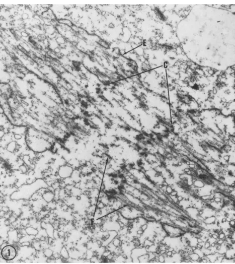
FIG. 1. Survey electron micrograph of the mitotic apparatus of the giant ameba, Pelomyxa carolinensis, about 5 min after the beginning of the anaphase. T h e chro- mosome plates (C) are about 4 μ apart, filaments (F) are present both in the inter- zone (continuous filaments) and poleward (continuous and chromosomal filaments), and fragments of the nuclear envelope (E) are within and exterior of the mitotic apparatus. Fixation in osmium tetroxide solution at pH 8.0 with 0.002 M cobalt added. Magnification: χ 8700. (Micrograph by R. A. Jenkins.)
served, regardless of whether they might be continuous or chromosomal filaments, located on the poleward or interzonal sides of the chromosome plate, or in the metaphase or anaphase.
Numerous ribosomelike particles are scattered between the filaments in a manner not readily suggesting a structural association of particle and filament (Fig. 3, R ) . Since it is first observed in the prometaphase
\vhen the envelope has only a few interruptions, this distribution is un- doubtedly the cause of the intense basophilia found in the mitotic apparatus of many different cells (e.g., Boss, 1955; Rustad, 1959).
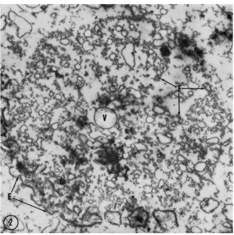
FIG. 2. Cross-sectional survey micrograph on the poleward side of an anaphase mitotic apparatus when the chromosome separation is about 9 μ. Filaments are arranged into irregular fibrils (F), fragments of the envelope (E) are leading the chromosomes (not in section) at the periphery of the MA, and small vesicles in- cluding a recently formed pinocytic vesicle (V) are contained within the MA. Fixa- tion in an osmium tetroxide solution at pH 8.0 with 0.002 M cobalt. Magnification:
χ 12,000. (Micrograph by R. A. Jenkins.)
By the telophase, the filaments have become detached from the chromosomes and can be observed in the cytoplasm some distance from the chromosomes that are now surrounded by a continuous nuclear
530 L. E. ROTH
Fie. 3. Detail of mitotic apparatus at the same stage and with the same fixation as Fig. 1. Filaments (F) both on the interzonal and poleward sides of the chromosome plates (C) have the same diameter and appearance and have fine material (M) on their surfaces. Ribosome-like particles (R) are scattered throughout the MA usually without indications of attachment to filaments. Magnification: χ 37,000. (Micrograph by R. A. Jenkins.)
envelope. The length of these bundles suggests that they are the con- tinuous filaments that become perhaps 65 μ long in these cells owing to a marked spindle elongation. At this time, the filaments are arranged in very close bundles and regular configurations, since the material sur- rounding filaments is no longer present. With the exception of this difference, telophase filaments show no observable difference in diameter or appearance from those in the anaphase and metaphase.
Mitosis of the astral type has been observed in several different cells.
The most careful and detailed observations are those made by Harris (1961, 1962) on sea urchin blastomeres; they show that many of the previously mentioned features are also present. The filament diameter and appearance are essentially identical, and ribosomelike particles are similarly distributed. T h e adhering material seen in anastral mitoses has not been so adequately observed, but is probably present though masked by the many other particles present. No observations of such cells in the telophase have shown filament bundles. Kane (1962) has also studied these cells by isolation with his method of controlled pH in hexandiol and by electron microscopy and has seen similar structures with the exception that the filament diameter is reported to be 21 πιμ rather than the 15-ιημ diameter reported by Harris and also found in the giant ameba MA. The possibility of his experimental procedure causing alteration of the filaments exists, and the hypothesis follows that such filaments may be amenable to experimental alteration.
Intranuclear mitosis takes place in the micronuclei of protozoan ciliates. Here the nuclear envelope remains continuous throughout division, and the entire process, which is otherwise essentially an anastral mitosis, takes place within the confines of the envelope. The MA in this case is composed of filaments of identical size and appearance to those found in astral and anastral mitosis. T h e major difference is the com- plete lack of ribosomal particles (Roth and Shigenaka, 1963). Movement of chromosomes is largely by spindle elongation.
Amitotic division takes place in the separation of ciliate macronuclei.
Filaments are contained within the macronuclear volume and are con- centrated either at the nuclear constriction (Roth, 1959; Roth and Minick, 1961) or extend in random directions (Roth and Shigenaka, 1963). However, another, more highly organized macronuclear appara- tus has been found in the oligotrichous, rumen ciliates (Roth and Shi- genaka, 1963); this filament system is external to and encloses both the macronucleus and micronucleus. Although present during the inter- phase, this system is more highly developed during division. Whether this filament system actually produces a major part of the force that
532 L. E. ROTH
causes division of the macro- and micronuclei is still not established al- though no adequate mechanism of macronuclear division has yet been suggested. This system has the appearance of an extranuclear division apparatus.
The demonstration of MA filaments by electron microscopy is pos- sible only by the choice of somewhat unusual conditions of osmium tetroxide fixation (Roth and Daniels, 1962; Roth and Jenkins, 1962).
Filaments of the MA will be preserved if divalent cations—calcium, magnesium, strontium, or cobalt at 0.002 M—are added to an osmium tetroxide fixation fluid in the pH range of 7.2 to 8.0 or without divalent cations if fixation is carried out at a slightly acid pH such as 6.0 (see also Harris, 1962). Such stabilizing conditions are, by contrast, not neces- sary for preservation of the filaments of cilia, infraciliature, or centrioles.
Functional lability is, in fact, a property of the MA as Östergren (1949) demonstrated so well in his observations of the mitotic process in Luzula where the chromosomes have diffuse kinetochores. Thus the evidence shows that the MA filaments are more labile than other filaments of similar morphology to which we now turn our attention.
The cytoplasmic infraciliature of many protozoa is composed of fila- ments. These filaments usually have a diameter of 15 to 21 ιτιμ and are found in hexagonally packed bundles extending from one group of cilia to another (Fig. 4, F); sheetlike groups are also found beneath the pellic- ular membranes particularly surrounding the orifice of the contractile vacuole (Fig. 5, F). In a few cases, large concentrations of these elements within a small volume of the cytoplasm are present so there is an inti- mate association of bundles from many different parts of the cell (Fig.
6); this region is probably the "motorium" described first by Sharp (1914) in this genus. The infraciliature is typically seen connected to filaments lying close to the nuclear envelope.
Flagellate protozoa have similar filaments in their cytoplasm. How- ever, the groupings are usually smaller with only a few filaments in sheetlike arrangements coursing beneath the membranes of the pellicle and associated in small numbers with the base of flagella (Roth, 1958).
Amebae typically lack such filaments in their cytoplasm. Rather, the only filaments demonstrated are those already described in the mitotic apparatus.
Cilia are now widely known to be composed of filaments. The shaft structure is rigidly standardized with nine peripheral filaments of essen- tially doublet cross sections and two central filaments of essentially circular cross sections. For our purposes it is important to pay particular attention to the structure of the ciliary tip. In the tips of protozoan cilia
(Fig. 7), the peripheral filaments have single, circular cross sections (Roth, 1957; Gibbons and Grimstone, 1960; Roth and Shigenaka, 1963).
The peripheral filaments do not all terminate at the same level so that cross sections at the tip frequently show diminished numbers. However, the important consideration is the structure of the tips of the peripheral filaments where circular cross sections appear similar to those of the infraciliature and the MA.
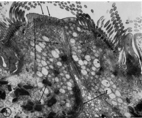
FIG. 4. Survey electron micrograph of a section through the anterior end of the oligotrichous protozoan Diplodinium ecaudatum. Filaments (F) form bundles extend- ing to groups of cilia, sheets that are located at the region of ingestion of food par- ticles and fluids, and a barrier wall that separates the digestion region from the re- mainder of the cytoplasm. Ingesta would move in the direction of the unlettered arrows through the esophageal tube into an enlarged volume of cytoplasm. T h e dense granules (S) are food-storage bodies that are localized in the anterior, peripheral portions of the cell. Magnification: χ 8300.
Another system of movement closely related to cilia is found in the stalk of several peritrich protozoa. When disturbed, these organisms re- tract quickly by coiling their stalks tightly; recovery to the extended condition usually takes several seconds so that contraction is not rapidly repeated. T h e fibrils contained within these stalks are extensions from
FIG. 5. T h e orifice of the contractile vacuole in Diplodinium ecaudatum showing the filament ( F ) system around it. Magnification: χ 48,000.
FIG. 6. Cross sections of filaments of the infraciliature in Diplodinium ecaudatum.
Individual filamentous bundles are nearly ubiquitous in the cytoplasm, but in this
534
typical basal bodies and, though they have marked striations, are similar to ciliary shafts (Randall, 1956; Rouiller et al., 1956).
The structural resemblance of metazoan centrioles to the triplet- filament complex of ciliary basal bodies adds another major link to this series of similarities. New impetus has been given to this long-established concept by the extremely close morphology of basal body (e.g., Gibbons and Grimstone, 1960) and centriole (Gall, 1961; Slautterback, 1963).
With mention of the centriole, we have again returned to considéra-
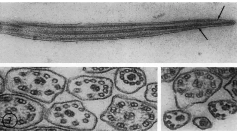
FIG. 7. T h e structure of the tips of cilia. T h e upper micrograph from Isotricha sp. is a longitudinal section in a plane including both central filaments which fuse and then end in a terminal enlargement. T h e peripheral filaments are still doublets at the left end of the upper micrograph but soon have circular cross sections and end before the terminal enlargement; peripheral filaments do not all end at the same level as indicated by the unlettered arrows. Magnification: χ 57,000. T h e lower micrographs are from Diplodinium ecaudatum and form a series, with a few excep- tions, of more distally located cross sections. Lower left, magnification: χ 137,000;
lower right, magnification: χ 118,000. (Lower micrographs by Y . Shigenaka.)
tion of the mitotic apparatus. In spite of similarities, a major difference between basal bodies and centrioles is evident since filaments of the astral MA do not usually contact the centriole; rather, a distance of separation exists with the filaments appearing to end at the periphery of the centrosome which is a clear homogeneous region.
The foregoing survey demonstrates that filaments from many cells and from several different cell structures are very similar in appearance,
region many bundles are closely packed immediately below the pellicle (unlettered arrow). This organism is the one in which Sharp (1914) originally described a "motor- ium." Magnification: χ 57,000. (Micrograph by Y . Shigenaka.)
536 L. E. ROTH
size, and arrangement and also suggests that chemical and functional similarities exist.
Evidence for chemical similarity is available from two areas. First, divalent cations have been implicated as integrally involved. Bishop (1962a) in his valuable detailed review paper lists numerous species in which motility of isolated sperm flagella is activated by the divalent cations of calcium and magnesium and inhibited by proper concentra- tions of compounds that bind cations by chelation. In another presenta- tion, Bishop (1962b) summarizes the evidence relating to glycerol-KCl- extracted sperm and suggests not only that calcium and magnesium are involved in inducing flagellar movement but that they also perhaps regulate this function according to their position and binding. Divalent cations in this regard probably regulate the ATPase activity of the fila- ment protein. Jahn (1962) has discussed similar effects on cilia particu- larly in regard to reversal of beat. Although the effect of divalent ions in this case is attributed to membrane binding, he has proposed that limitations caused by bond angles can restrict the ions capable of occu- pying binding sites and thus explain the biological specificity of certain ions. In regard to the MA, Mazia (1959) has described an effect of cal- cium on the isolated MA of sea urchin blastomeres where the fibrous appearance is increased by application of cations. As described earlier, stabilization of the MA for electron microscopy by such ions when
fixation at pH 8.0 is used is another example of divalent cation involve- ment with filament systems. Still another filament system in which calcium ions are vitally involved is that in the stalk of peritrich proto- zoa; the modified ciliary shaft filaments are described by Hoffmann- Berling (1958) to contract when traces of calcium are added and to relax either spontaneously or upon addition of the chelating agent, ethylene- diaminetetraacetate (EDTA).
Protein chemists state that one of the common features of the action of inorganic electrolytes on proteins is their binding to the charged groups of proteins (Putnam, 1953). More specifically, the alkali earth cations—calcium in particular—are thought to combine with the free dicarboxylic acid residues of proteins (Hober, 1945), thus suggesting the importance of aspartic and glutamic acids which are present in the protein of the MA in larger quantity than most other amino acids (Mazia, 1956). Binding at these sites is necessary for stabilization at pH 8.0 but not at pH 6.0; the latter pH is probably nearer and sufficiently close to the isoelectric point of MA protein where a lesser hydration of the protein exists (Höber, 1945), and stabilization in addition to osmium tetroxide fixation is not required. In muscle function, the importance of calcium and magnesium ions is clearly established (Weber, 1958). There-
fore, divalent cations are apparently necessary components of all motile systems.
Further chemical similarities are suggested by evidence pointing toward the presence of actomyosin-like proteins. Weber (1958) summarizes this subject well and concludes that marked similarities exist although major differences are found in regard to the involvement of adenosine triphosphate (ATP) and calcium ions. Later evidence suggests close similarities in the amino acid composition of protein from the MA (Mazia, 1956), from the flagella of Chlamydomonas (Jones and Lewin, 1960), and the cilia of Tetrahymena (Watson and Hopkins, 1962); high amounts of aspartic and glutamic acids are present in all three cases.
The ability of these proteins to act as nucleoside triphosphatases has been widely established (Weber, 1958), although varying degrees of specificity for this capability are found. For example, Mazia et al. (1961) have shown that MA protein can split the terminal phosphate from A T P but not from adenosine diphosphate (ADP), guanosine triphos- phate (GTP), cytosine triphosphate (CTP), or uridine triphosphate (UTP). Thus the proteins involved in the movement systems are quite similar though not identical.
SUMMARY
Filaments with the general appearance of a dense cortex and light center and having a diameter of 15 to 21 ηιμ are present in systems of several different types and in many different cells. Evidence pointing to their chemical similarity is sufficient to suggest that they are quite closely related in their composition and, therefore, that their functional capabilities are similar.
Formation of Filaments
How are such structural filaments formed? If the small amount of available data on the formation of these systems is compiled, a hypoth- esis of synthetic stages can be formulated. In regard to the MA, signif- icant studies by Went (1960) have shown that synthesis of filament protein has largely been completed before the prophase begins. How- ever, it is obvious by observation of prophase cells that no filament formation has yet taken place. Rather, filaments are formed at the time when the nuclear envelope is first interrupted in the typical astral and anastral divisions. At this time electron microscopy of giant amebae nuclei has shown filaments with a cross-sectional appearance not greatly different from that in later stages: circular cross sections are present with fine material along their surfaces (Roth and Daniels, 1962). Form- ing filaments in the prometaphase differ very little from functioning
538 L. E. ROTH
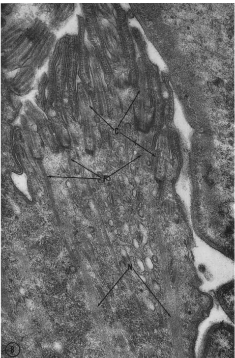
FIG. 8. Section through the tuft of forming cilia present in Diplodinium ecauda- tum during cytokinesis. The short shafts (less than 1 μ long) of the cilia (C) already
filaments in the anaphase to the extent of our present observations.
Thus, synthesis of MA protein takes place in the interphase whereas filament formation takes place later. Therefore, the two processes are separable events both in time and probably also in location.
Centriole formation has been studied in two recent significant studies. Mazia et al. (1960) have shown evidence that replication of the cell centers involves the formation of a pro-body of smaller size than the parent structure. Gall (1961), in a detailed electron-microscopic study of centriole formation in spermatogenesis, presented good struc- tural evidence that a procentriole is initially formed close to but not in contact with the centriole. These presentations together suggest that the procentriole is shorter but equal in diameter and filamentous structure to the parent centriole. Thus centriole formation is currently thought to be composed of at least two processes: formation of a short pro- centriole followed by an elongation of the filamentous structures to form the centriole.
Observations on formation of the protozoan infraciliature are mea- ger. Recent studies of the rumen protozoa (Roth and Shigenaka, 1963), however, have given some evidence that the formation of these filaments takes place by an aggregation and orientation in the cytoplasm from masses of otherwise unoriented material. In dividing Diplodinium, an oligotrichous ciliate with a highly developed infraciliary system, a tuft of cilia is formed when cytokinesis takes place. Organisms with a con- striction furrow are easily selected and allow detailed observation of the process of formation of the shafts of cilia and the infraciliary filaments.
The infraciliature in some of the cases studied in composed of filaments extending for a few microns from the basal bodies of the forming cilia but then fade into a region of oriented but nonfilamentous material (Fig. 8, N). An initial molecular synthesis apparently takes place fol- lowed by an aggregation into filaments which finally elongate to the length of the interphase infraciliature. The basal body of the cilium may influence formation of such filaments but direct elongation is un- likely since the number of infraciliary filaments per basal body may be fifty or more.
The formation of cilia is another case of filament formation that should be considered. The basal bodies, because of their extremely close structural similarity to the centriole, are probably formed like centrioles.
have filaments attached to their basal bodies. Near the basal bodies, filaments (F) are well defined whereas deeper in the cytoplasm aligned material (N) is present but not filaments. This phenomenon occurs in division stages in several portions of the cyto- plasm and probably represents the formation of infraciliary filaments. Magnification:
χ 36,000.
540 L. E. ROTH
A pro-body would be formed and elongate to a full basal body, move to and become oriented perpendicular to the plasma membrane, and then greatly elongate to form a shaft of somewhat different, fila- mentous composition than itself. Unfortunately little evidence is availa- ble in support of this hypothesis. Many questions arise: Can the basal body of a mature cilium give rise to such a probasal body; how do the triplet filaments in the base function in forming the doublets in the shaft; and how do the central filaments arise?
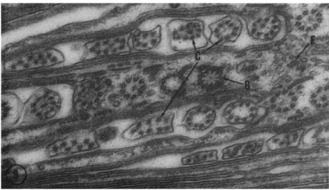
FIG. 9. A section perpendicular to that in Fig. 8 showing cross sections of the forming cilia. Notice that the short shafts (C) are composed of filaments with circular cross sections and are not yet arranged in a circle distally. T h e basal bodies (B) are not different from those of mature cilia and the infraciliary filaments (F) are well formed. Magnification: χ 57,000. (Micrograph by Y. Shigenaka.)
The first stages of ciliary shaft formation have been observed in studies of rumen protozoa (Bretschneider, 1962; Roth and Shigenaka, 1963). T h e first elements in the shaft of the very short forming cilium are about nine filaments of circular cross sections with material ran- domly adhering to their surfaces (Fig. 9, F). At this stage the filaments have not yet been oriented into a circular configuration but are ran- domly arranged within the short tapering part of the cilium thus far formed. T h e membrane of the cell has been extended so there is always an enclosure of these forming filaments in the cytoplasm of the cell.
The appearance of filaments with doublet cross sections, their arrange- ment into a circular configuration, and the addition of central filaments takes place at a later time by processes not known. Central filaments form without apparent connection with other filaments whereas periph-
eral filaments seem to form by direct contact with the basal triplets.
Thus, the formation of the shafts of cilia can probably be arranged into several parts: first, synthesis of the molecular components; second, simple molecular aggregation to form circular cross sections; third, aggregation of a modified type to form doublet filaments; and fourth, the arrange- ment of filaments into the nine-and-two complex.
SUMMARY
The formation of filament systems probably involves four events which may be partially separable in time and location. (1) Synthesis of protein molecules must take place. (2) Polymerization of these mole- cules and (3) cross-bonding of polymer chains result in the formation of filaments. Degrees of intimacy of filament formation with parent filaments have been observed; formation involving direct contact takes place but considerably greater independence must also be possible. (4) Grouping of filaments takes place, e.g., the rather crude grouping to form irregular fibrils in the MA. In cilia, however, modification of the filament-forming process from the first formation of circular filaments to doublet filaments takes place as an additional step followed by a very precise filament arrangement into the nine-and-two pattern. Therefore, four steps of the formation process are recognized: synthesis, polymeri- zation, polymer cross-bonding, and filament grouping.
Production of Movement by Filaments
Progress is being made in understanding the mechanism by which filaments produce movement in striated muscles. Neither the molecular conformation theory nor the molecular interaction (sliding) theory can be either confirmed or excluded at present. In regard to other cell move- ments, Weber (1958) lucidly summarizes biochemical and comparative physiological evidence to establish important apparent differences in the contraction of muscle and the mechanisms of other cell movements.
Structural differences also suggest such differences since the systems pre- viously described, in contrast to muscle, are characterized by filaments continuous throughout their functional length; in addition, neither two different filament types nor regular arrays are necessarily present.
Where rapid and repeated movements are to be produced as in cilia, configurational changes of proteins could account for rapid lengthening and shortening of segments of filaments. Semiconduction along the fila- ments may take place as postulated in recent papers (Eley, 1962; Green and Fleischer, 1962) from the volume honoring Albert Szent-Györgi who first proposed that such conduction is important in biological systems.
542 L. E. ROTH
Semiconduction can be suggested in biological systems whenever a close and regular linear arrangement of molecules occurs such as in filaments.
However, while configurational changes and semiconduction can ex- plain rapidly repeated movements, they seem inadequate to explain the anaphase movements of chromosomes.
The filament-related anaphase movement has two components:
shortening of the chromosome-to-pole distance and elongation of the pole-to-pole distance. Both are active processes. Elongation is so marked in the giant amebae that the final pole-to-pole distance is five times as long as it was in the metaphase; no change is observed in the length of chromosomal filaments in these cells (Short, 1946; Berkeley, 1948). Simi- larly, chromosome-to-pole shortening can be so marked that the kineto- chore-to-pole distance at the end of the anaphase is one-fifth the meta- phase distance (Hughes, 1952). Configurational changes of individual protein molecules cannot explain such great changes in length. Rather, removal of molecules from chromosomal filaments and insertion of new molecules into continuous filaments is likely.
A dynamic process seems to be involved. A dynamic polymerization and cross-bonding may take place whereby a filament has added mole- cules inserted to greatly increase its length or, conversely, molecules removed from it to shorten it. Such a mechanism would be easily ex- plained if the addition and subtraction of molecules took place at the polar ends of individual filaments, but the evidence is against this being the location involved. Rather, polarizing microscope studies as reported and summarized by Inoué (1953, 1959, 1963) are interpreted to show anaphase changes near centers of organization—centrioles, kinetochores, or phragmoplasts—but not at the ends of filaments. Thus contraction of chromosomal filaments may be considered as the removal of mole- cules at points along the chromosomal filament. Spindle elongation or the lengthening of continuous filaments can be the insertion of mole- cules into largely the interzonal portion of the continuous filaments which would, therefore, be increased in mass; some evidence for an increase of mass in the interzone has been given from two interference microscopic studies (Rustad, 1959; Longwell and Svihla, 1960). There- fore, the dynamic mechanism involved can best be explained as the insertion and removal of molecules from filaments not at their ends but within their lengths.
In many cells where some amount of both anaphase movements take place, the interzone must be suggested to favor polymerization whereas the region poleward from the chromosomes would favor depolymeriza- tion; the possibility exists, therefore, that molecules removed from chromosomal filaments may become incorporated later into continuous
filaments. Interestingly, movement by spindle elongation frequently fol- lows chromosome-to-pole movement in those cells where both move- ments are important. In many cells the two movements take place more simultaneously, but I am not aware of any cases where spindle elonga- tion precedes chromosome-to-pole movement.
SUMMARY
Filaments are dynamic structures. Just as membranes are regarded as being dynamic and capable of great surface enlargement in short periods of time, so, also, filaments may need to be similarly regarded as dynamic and capable of considerable elongation and shortening. It seems appropriate to suggest that, as a membrane may increase its sur- face area, similarly a filament may increase its length. T h e dynamic mechanism involved may account for filament formation and, further- more, may also produce the mitotic movements caused by filaments.
Movements without Filaments
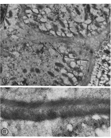
In the holotrichous protozoan Isotricha prostoma, the extranuclear division apparatus does not contain filaments of the type discussed.
Instead, the thick fibers or sheets present (Fig. 10, F) are composed of a fine material in parallel array (Fig. 11). Similar material has been re- ported to occur in Physarum (Wohlfarth-Bottermann, 1962) and also has been shown at this Symposium to be present in amebae at determined sites and times relating to movement (Wohlfarth-Bottermann, 1963). In ciliate protozoa, such as Stentor (Randall, 1957) and Diplodinium (Fig.
5), both filamentous bundles and nonfilamentous fibers are present in the pellicle in juxtaposition. Such nonfilamentous bundles could be in- volved in a similar mechanism of movement, differing only in the man- ner of molecular linkage.
Summary
Filament systems having structural and chemical similarities have been described and discussed in order to derive hypotheses of their for- mation and function. Formation is suggested to include four steps:
synthesis, polymerization, polymer cross-bonding, and filament grouping.
Some cell movements involving filaments can be explained by local changes in polymerized molecules. For example, in cilia configurational changes of the protein molecules or changes in cross-bonding of helically arranged polymers could explain shortening by a small percentage of its length.
Some other cell movements, though perhaps involving very similar molecules and arrangements, cannot be explained by such mechanisms.
544 L. E. ROTH
The functioning of the filaments of the MA is such an example since the changes in filament length are too great to be explained in this way.
Filaments of the MA and other filaments at the time of their forma-
FIG. 10. A survey electron micrograph of Isotricha prostoma including portions of the pellicle (P) and macronucleus (M). A system of fibrils (F) is present forming a layer slightly below the plasma membranes and another surrounding the nuclei; the layers are interconnected. Magnification: χ 11,000.
FIG. 11. Higher magnification of the fibril in Isotricha (Fig. 10). Each section of the pellicular layer shows two sublayers in which parallel, fine elements are found;
the orientation of elements in the sublayers is perpendicular. Magnification: χ 41,000. (Micrograph by J . Chakraborty.)
tion are similar. The terms "function" and "formation" in regard to the M A filaments may be synonymous. Slightly varying conditions may be found in cells to alter the equilibrium that controls the addition (to lengthen) or removal (to shorten) of molecules from filaments in meta- stable states. Filament function in the mitotic apparatus is thus con- sidered to be a dynamic process.
Cell movements must be explained at the molecular level. The func- tion of the mitotic apparatus can only begin to be explained as a dy- namic molecular phenomenon. T o understand ameboid movement, terms such as "contraction" must give way to terms that are chemically more precise; to understand all cell movements, we must apply the principles of the physical chemistry of proteins (Höber, 1945; Putnam, 1953;
Weber, 1958 are exemplary).
Varying bonding of similar molecules can produce the several levels of organization known in motile systems: fibrils composed of filaments, fibrils composed of finer elements in biréfringent array, isometric arrays, and free molecules constituting the precursor pool. Movement is con- trolled molecular bonding.
REFERENCES Berkeley, E. (1948). Biol. Bull. 9 4 , 169.
Bishop, D. (1962a). Physiol. Rev. 4 2 , 1.
Bishop, D. (1962b). In "Spermatozoan Motility" (D. W. Bishop, ed.), pp. 251-268. Am.
Assoc. Advan. Sei., Washington, D.C.
Boss, J . (1955). Exptl. Cell Res. 8 , 81.
Bretschneider, L. H. (1962). Koninkl. Ned. Akad. Wetenschap. Proc., Ser. C 6 5 , 423.
Daniels, E. W., and Roth, L. E. (1963). / . Cell Biol. In press.
Eley, D. D. (1962). In "Horizons in Biochemistry" (M. Kasha and B. Pullman, eds.), pp. 341-380. Academic Press, New York.
Gall, J . G. (1961). / . Biophys. Biochem. Cytol. 1 0 , 163.
Gibbons, I. R., and Grimstone, Α. V. (1960). / . Biophys. Biochem. Cytol. 7, 697.
Green, D. E., and Fleischer, S. (1962). In "Horizons in Biochemistry" (M. Kasha and B. Pullman, eds.), pp. 381-420. Academic Press, New York.
Harris, P. (1961). / . Biophys. Biochem. Cytol. 1 1 , 419.
Harris, P. (1962). / . Cell Biol. 1 4 , 475.
Höber, R. (1945). "Physical Chemistry of Cells and Tissues," pp. 293ff. Blakiston, Philadelphia, Pennsylvania.
Hoffmann-Berling, H. (1958). Biochim. Biophys. Acta 2 7 , 247.
Hughes, A. F. W. (1952). "The Mitotic Cycle," p. 128. Academic Press, New York.
Inoué, S. (1953). Chromosoma 5 , 487.
Inoué, S. (1959). In "Biophysical Science—A Study Program" (J. L. Oncley, ed.), pp.
402-408. Wiley, New York.
Inoué, S. (1963). In "Primitive Motile Systems in Cell Biology" (R. D. Allen and N.
Kamiya, eds.), pp. 549-598. Academic Press, New York.
Jahn, T. (1962). / . Cellular Comp. Physiol. 6 0 , 217.
546 L. E. ROTH
Jones, R. F., and Lewin, R. A. (1960). Exptl. Cell Res. 1 9 , 408.
Kane, R. E. (1962). J. Cell Biol. 1 5 , 279.
Longwell, A. C , and Svihla, G. (1960). Exptl. Cell Res. 2 0 , 294.
Mazia, D. (1956). Advan. Biol. Med. Phys. 4 , 95.
Mazia, D. (1959). In "Sulfur in Proteins" (R. Benesch et ed., eds.), p. 367. Academic Press, New York.
Mazia, D„ Harris, P. J . , and Bibring, T. (1960). / . Biophys. Biochem. Cytol. 7, 1.
Mazia, D., Chaffee, R. R., and Iverson, R. M. (1961). Proc. Natl. Acad. Sei. (U.S.) 4 7 , 788.
Östergren, G. (1949). Hereditas 3 5 , 445.
Putnam, F. W. (1953). In "The Proteins" (H. Neurath and K. Bailey, eds.), Vol. IB, pp. 807-892. Academic Press, New York.
Randall, J . T . (1956). Nature 1 7 8 , 9.
Randall, J . T. (1957). Symp. Soc. Exptl. Biol. 9, 185.
Roth, L. E. (1957). / . Biophys. Biochem. Cytol. 3 , 985.
Roth, L. E. (1958). / . Ultrastruct. Res. 1, 223.
Roth, L . E. (1959). Proc. Intern. Conf. Electron Microscopy, 4th, West Berlin, 1958, 2, 241-244.
Roth, L. E., and Daniels, E. W. (1962). / . Cell Biol. 1 2 , 57.
Roth, L. E., and Jenkins, R. A. (1962). Proc. Intern. Conf. Electron Microscopy, 5th, Philadelphia, 1962, 2 , NN-2.
Roth, L. E., and Minick, Ο. T. (1961). / . Protozool. 8 , 12.
Roth, L. E., and Shigenaka, Y. (1963). J. Cell. Biol. In press.
Rouiller, C , Faure-Fremiet, Ε., and Gauchery, M. (1956). Exptl. Cell Res. 1 1 , 527.
Rustad, R. C. (1959). Exptl. Cell Res. 1 6 , 575.
Sharp, R. (1914). Univ. Calif. (Berkeley) Puhl. Zool. 1 3 , 43.
Short, R. B. (1946). Biol. Bull. 9 0 , 8.
Slautterback, D. B. (1963). / . Cell. Biol. 1 8 , 367.
Watson, M. R., and Hopkins, J . M. (1962). Exptl. Cell Res. 2 8 , 280.
Weber, H. H. (1958). "The Motility of Muscle and Cells." Harvard Univ. Press, Cambridge, Massachusetts.
Went, H. A. (1960). Ann. Ν. Y. Acad. Sei. 9 0 , 422.
Wohlfarth-Bottermann, Κ. E. (1962). Protoplasma 5 4 , 514.
Wohlfarth-Bottermann, Κ. E. (1963). In "Primitive Motile Systems in Cell Biology"
(R. D. Allen and N. Kamiya, eds.), pp. 79-110. Academic Press, New York.
DISCUSSION
DR. INOUÉ: I am very pleased with Dr. Roth's conclusion that this film of mitotic apparatus represents a dynamic state of organization. I wonder what electron- microscopic evidence one can find to support this notion.
DR. ROTH: I think the information has to come mostly from other studies, but the electron-microscopic evidence which I think is most important is that in the giant ameba, material adheres to the exterior of filaments in prometaphase, metaphase, and anaphase. Those filaments that are in the interzone and, therefore, are presumed to be continuous filaments are the ones that we see in telophase and that pack more closely because material is now absent from their surfaces. This evidence suggests that material has now been incorporated into the filaments and that they have concluded their formation at the same time they have concluded their function.
DR. ALLEN: Are the continuous filaments thicker than the chromosomal filaments?
DR. ROTH: W e can see no difference in the diameter of continuous and chromo- somal filaments.
DR. TERU HAYASHI: T O a naive nonelectron microscopist, the films you showed of the mitotic apparatus, especially in those regions where they were not too closely packed, looked very much like the sort of thing one sees with sarcoplasmic reticulum.
DR. ROTH: YOU are suggesting perhaps they are not filaments, but rather endo- plasmic reticulum?
DR. T E R U HAYASHI: Yes.
DR. ROTH: I think they are not. You may, if you wish, follow the argument which many people do, that any membranous structure is part of the endoplasmic reticulum;
if you do, you may wish to call them endoplasmic reticulum. Cross-sectional views show uniform packing of uniform elements, more than is characteristic of the endo- plasmic reticulum.
DR. TERU HAYASHI: What are the dimensions of the separate units? You men- tioned diameter.
DR. ROTH: T h e filament diameter in the mitotic apparatus is 15 ηΐμ, except in Dr. Kane's isolated mitotic apparatus, where it is 20. T h e infraciliary filaments also seem to have 15-20 πΐμ diameters. Perhaps there is either 15 or 20 πΐμ, but we are not ready to say that yet.
DR. REBHUN: I would not stress the differences in the 15 vs. 20 πΐμ fibers, es- pecially if there are differences of embedding media and different types of fixation.
With respect to the endoplasmic reticulum, in Dr. Harris' work you do find the 15-ηΐμ fibers in the asters as well as chains of endoplasmic reticulum, and it looks certainly as if they are different systems.
The thing I would stress is that in your work no changes in diameter can be seen in the chromosomal filaments at any stage in mitosis.
DR. ROTH: T h a t is exactly right.
DR. REBHUN: This does not necessarily mean that there is no shortening of the filaments. They might be, for example, shortening by material "dissolving" from the ends. If so, you cannot tell whether there is active shortening which has nothing to do with the generation of force, or not. It would be very difficult to decide whether the shortening were actually coupled to the performance of work.
DR. ROTH: First of all, a very active process is involved in the spindle elongation of the giant ameba; that the filaments are performing some work seems to be some- what well established there, if one believes the work that Berkeley (1948) and Short (1946) report in very nice detail in their papers on giant amebae. Elongation, if im- peded on one end, results in a "barreling-out" of the spindle in the interzone, and they stress very strongly that the elongation must be an active process.
In relation to your first remark, I suggest only that 15 and 21 ηΐμ filaments may not be the same, or they may be. Such a difference in size is very close to the limit of error in magnification of microscopes, but, nevertheless, in the dividing Diplodin- ium, instead of having only one diameter, we seem to have a range of diameters. If a lot of measurements are made, they turn out to have a slightly bimodal distribution clustering around 15 and 21 πΐμ when sections are studied from the same or identically handled specimens.
DR. ZIMMERMAN: A comment I would like to make is that you must remember Dr. Kane's work on mitotic apparatus was done on isolated preparations. It is amaz- ing to me that he got any kind of evidence indicative of fibers, because all previous work on the isolated mitotic apparatus had given no such indications. T h e fact that Dr. Harris' work gave measurements very comparable to yours indicates in all proba- bility that the measurements are valid.
548 L. E. ROTH
DR. ROTH: Yes. I have very great respect for Dr. Kane's work. It is interesting because it may mean that these filaments can be altered. Perhaps they transform or can be transformed from one diameter to another. Perhaps by his experimental pro- cedure he transformed them in some way. With this in mind, we can look back at the suggestion of bimodal diameter distribution in Diplodinium and speculate that the filaments are formed at one diameter but function at another.
DR. ZIMMERMAN: It could be a difference in fixation.
DR. ROTH: The fixation in his case could be the altering factor. However, I am suggesting the organism may be able to alter the filaments; there may be two forms of filaments also, the smaller one being that one which is first formed.
DR. INOUÉ: Dr. Kane has been able to alter artificially the filaments into three different forms; one, a so-called tubular filament of the type you see; another, a solid-appearing filament; and, third, he can put these in solution and cause small droplets to form. I think it could be a simple ionic environment that introduces differences which do not result from the fixation process itself.
DR. ROTH: I think Dr. Inoué will emphasize in his paper the metastability or lability of the filaments of the mitotic apparatus. I would agree that this metasta- bility follows from the many studies that have gone before, particularly the one by Östergren on Luzula where the chromosomes may be pulled through filaments in a typical mitotic process. It is possible that the tubular filament does not correspond exactly to the in vivo functional unit of the mitotic apparatus. This may possess a metastability that we can alter by fixation procedures and perhaps which the cell alters during function.