Accepted Manuscript
Biological evaluation of microbial toxin degradation by microinjected zebrafish (Danio rerio) embryos
Zsolt Csenki, Edina Garai, Anita Risa, Mátyás Cserháti, Katalin Bakos, Dalma Márton, Zoltán Bokor, Balázs Kriszt, Béla Urbányi
PII: S0045-6535(19)30660-5
DOI: https://doi.org/10.1016/j.chemosphere.2019.04.014 Reference: CHEM 23522
To appear in: ECSN
Received Date: 4 February 2019 Revised Date: 1 April 2019 Accepted Date: 2 April 2019
Please cite this article as: Csenki, Z., Garai, E., Risa, A., Cserháti, Máá., Bakos, K., Márton, D., Bokor, Zoltá., Kriszt, Balá., Urbányi, Bé., Biological evaluation of microbial toxin degradation by microinjected zebrafish (Danio rerio) embryos, Chemosphere (2019), doi: https://doi.org/10.1016/
j.chemosphere.2019.04.014.
This is a PDF file of an unedited manuscript that has been accepted for publication. As a service to our customers we are providing this early version of the manuscript. The manuscript will undergo copyediting, typesetting, and review of the resulting proof before it is published in its final form. Please note that during the production process errors may be discovered which could affect the content, and all legal disclaimers that apply to the journal pertain.
M AN US CR IP T
AC CE PT ED
Biological evaluation of microbial toxin degradation by microinjected zebrafish (Danio 1
rerio) embryos 2
3
Zsolt Csenki 1†*, Edina Garai1†, Anita Risa2, Mátyás Cserháti2, Katalin Bakos1, Dalma 4
Márton2, Zoltán Bokor1, Balázs Kriszt2, Béla Urbányi1 5
6
1Department of Aquaculture, Institute of Aquaculture and Environmental Safety, Faculty of 7
Agricultural and Environmental Sciences, Szent István University, 1. Páter Károly St., H- 8
2100 Gödöllő, Hungary 9
2Department of Environmental Safety and Ecotoxicology, Institute of Aquaculture and 10
Environmental Safety, Faculty of Agricultural and Environmental Sciences, Szent István 11
University, 1. Páter Károly St., H-2100 Gödöllő, Hungary 12
† The authors contributed equally to this work 13
*Address correspondence to: Zsolt Csenki csenki.zsolt@mkk.szie.hu 14
15
Declarations of interest: none 16
17
M AN US CR IP T
AC CE PT ED
18
Abstract 19
The use of microinjection of newly fertilized zebrafish eggs as an appropriate tool for 20
qualifying the biodetoxification properties of toxin-degrading microbes was investigated.
21
Ochratoxin A (OTA), bacterial degradation products of OTA and bacterial metabolites of the 22
Cupriavidus basilensis ŐR16 strain were microinjected. Results showed that variations in the 23
injected droplet size, and thus treatment concentrations, stayed within ±20%, moreover 24
embryo mortality did not exceed 10% in controls, that is in accordance with the 25
recommendations of the OECD 236 guideline. The highest lethality was caused by OTA with 26
a significantly higher toxicity than that of bacterial metabolites or OTA degradation products.
27
However, toxicity of the latter two did not differ statistically from each other showing that the 28
observed mortality was due to the intrinsic toxicity of bacterial metabolites (and not OTA 29
degradation products), thus, the strain effectively degrades OTA to nontoxic products.
30
Sublethal symptoms also confirmed this finding.
31
Results confirmed that microinjection of zebrafish embryos could be a reliable tool for testing 32
the toxin-degrading properties of microbes. The method also allows comparisons among 33
microbial strains able to degrade the same toxin, helping the selection of effective and 34
environmentally safe microbial strains for the biodetoxification of mycotoxins in large scale.
35
Keywords: Cupriavidus basilensis, mycotoxin, ochratoxin, biodegradation, biodetoxification 36
37
M AN US CR IP T
AC CE PT ED
1. Introduction 38
Ochratoxin A (OTA) is a hazardous mycotoxin produced during the secondary metabolism of 39
filamentous fungi belonging to the genera Aspergillus and Penicillium (Bui-Klimke and Wu, 40
2015). OTA is a potent nephrotoxic mycotoxin that has several harmful effects in Vertebrates, 41
including fish, such as hepatotoxicity (Gagliano et al., 2006), teratogenicity (Haq et al., 2016;
42
O’Brien et al., 2005) and immunosuppression (Marin and Taranu, 2015). OTA has been 43
reported to play a role in the development of different types of tumors in Rodent models and 44
humans (Pfohl-Leszkowicz and Manderville, 2007). Chronic OTA exposure proved to be a 45
leading factor in mycotoxin-induced porcine nephropathy and Balkan endemic nephropathy 46
(BEN) in humans (Stoev and Denev, 2013; Vrabcheva et al., 2000).
47
The toxin is present in various agricultural products (e.g., fruits, cereals, meats, coffee beans, 48
spices) (Bui-Klimke and Wu, 2015) and survives many common food-processing procedures, 49
such as roasting, brewing and baking, thus, it can be found in bread (Scudamore et al., 2004), 50
juicy fruits (Fernández-Cruz et al., 2010), beer (Odhav and Naicker, 2002) and wine 51
(Otteneder and Majerus, 2000). Because of its potential health risks, many countries and 52
international organizations have introduced a limit value for the OTA content of cereals and 53
cereal products (ECR, 2006; FAO, 2003).
54
Global occurrence of mycotoxins in the food chain is a problem worldwide, so several 55
strategies have been developed to decrease mycotoxin levels in animal feeds and human food 56
e.g. prevention, physical and chemical methods and biodegradation (Binder, 2007; EFSA, 57
2010). Among these, toxin biodegradation by microorganisms or their enzymes is the most 58
promising approach which could be an important postharvest strategy to reduce or eliminate 59
mycotoxin contamination.
60
There is growing need for the selection of microbial strains for efficient mycotoxin 61
biodegradation in large scale use, which are able to eliminate the hazardous effects of a toxin 62
M AN US CR IP T
AC CE PT ED
and its breakdown products in addition to the degradation of their chemical structure 63
(Ferenczi et al., 2014; Sheikh-Zeinoddin and Khalesi, 2018; Vanhoutte et al., 2016).
64
Traditional analytical and immunological methods are sufficient to test biodegradation of the 65
parent compound, but they are unable to detect the toxic effects of potential degradation 66
products and bacterial metabolites. In addition, biodegradation does not always mean 67
biodetoxification. According to the statements described above and the scientific advice of 68
EFSA (EFSA, 2010), it is important to develop and use new in vivo toxicological approaches 69
for investigating biodegradation and detoxification efficiency directly.
70
Various microorganisms have been reported to be suitable for degrading and detoxifying 71
OTA, some of them are highly efficient (Abrunhosa et al., 2014; Hathout and Aly, 2014).
72
Two pathways may be involved in OTA microbiological degradation. The primary is the 73
hydrolytic cleavage of the amide bond in OTA, resulting in the production of phenylalanine 74
and ochratoxin α (OTα), which - in most cases – is the major degradation product. Since OTα 75
and phenylalanine are presumably non-toxic, this mechanism can be considered as a 76
detoxification pathway. The second is a hypothetical process where OTA is degraded via the 77
hydrolysis of the lactone ring (Karlovsky Petr, 1999). In this case, the final degradation 78
product is an opened lactones form of OTA, which has similar toxicity to the parent 79
compound (Li et al., 1997; Xiao et al., 1996).
80
In the present report, Cupriavidus basilensis (ŐR16 strain), the first Cupriavidus species with 81
proven OTA degradation potency has been selected. The strain ŐR16 can degrade almost 82
100% of OTA in solutions with concentrations below 20 mg/L in laboratory conditions 83
during 5 days of incubation, and the major metabolite of OTA is OTα. The degradation 84
efficiency of the strain was tested in mice, where neither the metabolites produced in a 85
modified LB medium, nor the degraded OTA residuals evoked pathological disorders, or 86
disturbed the expression of the examined genes (Ferenczi et al., 2014). Based on these 87
M AN US CR IP T
AC CE PT ED
phenomena, the strain ŐR16 seems to be suitable for developing new in vivo test methods for 88
Vertebrate models to examine and evaluate the detoxification ability of mycotoxin degrading 89
microorganisms.
90
Zebrafish embryo tests are widely used bioassays in toxicological and ecotoxicological 91
testing, and are often used to analyze organic-matter rich samples (e.g. waste water and 92
sediment samples) (Braunbeck et al., 2005; Nagel, 2002). Since these assays should be carried 93
out at temperatures above 25°C, many factors may interfere with toxicity evaluation, of which 94
low oxygen supply in the embryo test vessel is one of the most important (Küster and 95
Altenburger, 2008; Strecker et al., 2011). Deviations from oxygen saturation increase the 96
frequency of malformations or suspension of embryo development, and distinction between 97
effects of hypoxia and the toxicity of a sample is not always possible. For organic-matter rich 98
samples, the microinjection of fish embryos could be an alternative method to eliminate the 99
secondary effects of hypoxia.
100
Microinjection is a simple way to introduce substances into newly fertilized fish eggs. It has 101
previously been used for testing polar and nonpolar substances in many fish species (Colman 102
et al., 2004; Mizell and Romig, 1997; Walker et al., 1992). Effects on embryonic development 103
are visible shortly after microinjection, and even minor toxic effects can be distinguished 104
from background mortality and other sublethal symptoms. Although, microinjection of 105
substances into the yolk of zebrafish eggs is feasible, introduction of accurate volumes (e.g.
106
constant volumes) through a series of injections seems to be problematic so nominal and real 107
injected volumes may be different (Schubert et al., 2014).
108
The objective of this in vivo toxicological study was to investigate whether microinjection of 109
newly fertilized zebrafish eggs could be an appropriate tool for qualifying the 110
biodetoxification efficiency of toxin-degrading microbes. Therefore OTA, breakdown 111
products of OTA and bacterial metabolites of Cupriavidus basilensis ŐR16 strain were 112
M AN US CR IP T
AC CE PT ED
injected into zebrafish eggs at different volumes and mortality and sublethal effects were 113
compared. Additionally, we investigated the injected volume fluctuations during a series of 114
microinjections, to see if desired treatment concentrations are reached and to ensure that the 115
results are reliable.
116 117
M AN US CR IP T
AC CE PT ED
2. Material and methods 118
2.1. Animal protection 119
The Animal Protocol was approved under the Hungarian Animal Welfare Law (XIV-I- 120
001/2303-4/2012).
121 122
2.2. Zebrafish maintenance and egg collection 123
Laboratory-bred AB strain zebrafish were held in breeding groups of 30 females and 30 males 124
at the Department of Aquaculture, Szent István University, Hungary, in a Tecniplast ZebTEC 125
recirculation system (Tecniplast S.p.A., Italy) at 25.5°C ± 0.5°C, pH 7.0±0.2, conductivity 126
550±50 µS (system water) and light:dark period of 14 h:10 h. Fish were fed twice a day with 127
dry granulate food (Zebrafeed 400-600 µm, Sparos Lda., Portugal) supplemented with freshly 128
hatched live Artemia salina twice a week. Fish were placed in breeding tanks (Tecniplast 129
S.p.a.) late in the afternoon the day before the experiment and allowed to spawn by removing 130
the dividing walls next morning. Spawning of individual pairs was delayed through time to 131
allow a continuous supply of 1-cell embryos.
132
133
2.3. Bacterial strain cultivation and metabolite preparation 134
The bacterial Cupriavidus basilensis ŐR16 strain (stored at −80 °C) was thawed on ice, 135
streaked on Luria-Bertani (LB) agar plates (10 g tryptone, 5 g yeast extract, 9 g sodium- 136
chloride and 18 g bacteriological agar (Biolab Ltd., Hungary) in 1L (pH 7.0) ion-exchanged 137
water) and incubated at 28 °C for 72 hours. Then single colonies were inoculated into 50 mL 138
100% LB medium (10 g tryptone, 5 g yeast extract and 9 g sodium-chloride in 1L (pH 7.0) 139
ion-exchanged water) in 250 mL flasks and cultures were grown for 120 h at 28 °C, 170 rpm 140
in a shaking incubator (Sartorius Certomat BS-1, Germany). Liquid cultures were centrifuged 141
at 3220 g, 4 °C for 20 min (Eppendorf 5810R, Germany), the pellet was resuspended in 50 142
M AN US CR IP T
AC CE PT ED
mL 20% LB medium (100% LB medium diluted with ion-exchanged water), then was 143
centrifuged again at the same conditions. The procedure was repeated twice. After 144
resuspension, the optical density of the culture was measured at 600 nm (OD 600) (GENESIS 145
10S UV-VIS, Thermo Fischer Scientific) and adjusted to 0.6±0.05 to prepare bacterial 146
inoculum. 5 mL bacterial suspensions were inoculated into 45 mL sterile 20% LB medium in 147
triplicates and incubated on a laboratory shaker at 28 °C, 170 rpm for 120 h. Cultures were 148
then centrifuged at 3220 g, 4 °C, for 15 min. Supernatants were filtered through 0.2 µm 149
syringe filters (VWR International Ltd., Hungary) to gain bacteriologically sterile samples 150
containing bacterial metabolites only. Samples were stored at −20 °C until microinjection.
151
152
2.4. Ochratoxin A biodegradation and OTA concentration measurement 153
Bacterial inocula (5 mL) were prepared as above, and added to 45 mL 20% LB medium 154
containing OTA (7 mg/L final concentration). Similar inocula were prepared in parallel 155
without OTA to test the effects of bacterial metabolites. Uninoculated LB medium (20%) 156
contaminated by OTA (7 mg/L) was used as negative control. Both of the cultures and control 157
were incubated at 28 °C, 170 rpm for 120 h in triplicates. After the incubation, cultures were 158
centrifuged at 3220 g, 4 °C, for 20 min. Supernatants were filtered with 0.2 µm syringe filters, 159
and samples were stored at −20 °C until microinjection.
160
For the measurement of OTA concentration, high-performance liquid chromatography with 161
tandem mass spectrometry (HPLC-MS/MS) was applied. Prior to measuring toxin 162
concentration, 100 μL sample were mixed with 25 μL isotope-labelled internal standard (13C20- 163
OTA), the mixture was evaporated under nitrogen gas, thereafter it was reconsituted in 50-50 164
V/V% A-B eluent (A : water, 5mM ammonium-acetate, 0.1% acetic acid ; B : methanol, 165
5mM ammonium-acetate, 0.1% acetic acid). For the separation Agilent 1100 HPLC (Agilent 166
Technologies, USA) equipped with Agilent Zorbax C18 column (3.5µm, XDB-C18, 2.1 x 167
M AN US CR IP T
AC CE PT ED
50mm) was used. 10 μL prepared samples were injected into the mobile phase containing A- 168
B eluent. 400 µL/min flow rate and 40°C column temperature was set. 3200 QTRAP 169
LC/MS/MS system (Applied Biosystems, USA) in positive ion mode was used for the 170
determination of OTA concentration in samples. During the measurement, LOD was 2 µg/L 171
and LOQ was 6 µg/L.
172
2.5. Microinjection 173
A Narishige (Japan Model PN-31) micropipette puller (heater level: 89.1, magnet sub level:
174
15.7, magnet main level: 84.3) was used to pull microinjection pipette tips (injection needle) 175
(Narishige Japan G-1 borosilicate glass capillary, 1 mm o.d. x 0.6 mm i.d., 90 mm length).
176
Injection needles were backfilled with 20 µ L substance without air bubbles by a Microloader 177
pipette tip (Eppendorf, Germany).
178
The needle was placed in the microinjection manipulator (microINJECTOR MINJ-2, TriTech 179
Research Inc. Los Angeles, USA) connected to a nitrogen gas bottle. Injections were carried 180
out under a stereomicroscope at 15× magnification (Leica LED2500, Leica Microsystems 181
GmbH, Germany). Injection volumes were determined in immersion oil (Merck Ltd., 182
Hungary, An affiliate of Merck KGaA, Darmstadt, Germany) on the basis of droplet 183
diameters by a calibrated software (Leica M205 FA, Leica DFC 7000T camera, Leica 184
Application Suite 3.4.2.18368, Leica Microsystems GmbH, Germany). Injection volumes 185
were administered five times into the oil droplet until appropriate volume was achieved 186
(pressure or capillary orifice size change). According to the sphere volume formula 187
(V=1/6πd
3
), a sphere diameter of 50 µm corresponded to an injection volume of 0.22 nL, 100 188
µm to 0.52 nL, 150 µm to 1.77 nL, and 200 µm to 4.17 nL. Injection volume needed to be 189
measured and adjusted for each solution, concentration and control.
190
M AN US CR IP T
AC CE PT ED
One-cell stage zebrafish embryos were lined up against the side of a microscope slide placed 191
in a 10 cm diameter Petri dish. Excess water was removed with a plastic pipette. Treatment 192
groups of 20 eggs were injected in a minimum of three replicates per treatment. Following 193
microinjection, eggs were incubated in system water with methylene blue (2 mL 0.1%
194
methylene blue in 1 L system water) (25°C ± 2°C) in 10 cm diameter Petri dishes. After 2 195
hours, coagulated and/or non-fertilized eggs were discarded and developing embryos were 196
transferred in groups of twenty into 6 cm diameter Petri dishes. Embryos were then incubated 197
in system water at 26°C ± 1°C and a 14 h:10 h-light:dark period and checked for lethal and 198
sublethal effects under a microscope. System water was replaced in every 24 hours until 120 199
hpf (hours post-fertilization). Digital images of embryos (72 hpf) and larvae (120 hpf) in 200
lateral orientation were taken under a stereomicroscope at 30× magnification (Leica M205 201
FA, Leica DFC 7000T camera, Leica Application Suite 3.4.2.18368, Leica Microsystems 202
GmbH, Germany).
203
204
2.6. Determination of the variations in the injection volume 205
Zebrafish Ringer’s solution (ZFR) (116 mM sodium-chloride, 2.9 mM potassium-chloride, 206
1.8 mM calcium-chloride and 5 mM HEPES (pH 7.2) (Sigma-Aldrich, Hungary) in system 207
water, filtered with 0.2 µm syringe filters) was injected into the yolk of zebrafish eggs. Prior 208
to treatments, microinjection parameters (pressure and capillary orifice size) were set 209
according to the volumes calculated on the basis of injected droplet sizes in immersion oil.
210
When the desired volume was reached, five eggs were injected, and the diameter of five 211
droplets was measured again in immersion oil. This egg injection - droplet measurement cycle 212
was repeated five times to test the accuracy of injection.
213
214
M AN US CR IP T
AC CE PT ED
2.7. Effect of the highest used injection volume and the LB media on the viability of 215
embryos 216
The effect of the largest injection volume (4.2 nL) on egg viability was tested with Zebrafish 217
Ringer’s solution, the negative control of the experiments. The effect of the bacterial growth 218
medium and the effects of the solvent were tested following the injection of 4.2 nL of 20%
219
LB medium and 20% LB medium with acetone (250 µ L acetone in 50 mL 20% LB medium).
220
221
2.8. Determination of the initial OTA concentration of the reference curve 222
OTA (99.5% Fermentek, Israel) was dissolved in acetone (98.8% Sigma-Aldrich, Hungary) at 223
1000 mg/L concentration, of which 1; 7; 10 mg/L concentrations were prepared in 20% LB 224
medium. OTA contaminated medium was injected in 0.22 nL, 0.52 nL, 1.77 nL and 4.17 nL 225
volumes into the embryos to find the optimal concentration for the reference curve.
226 227
2.9. Examining the toxicity of samples derived from OTA degradation experiment 228
Samples containing ŐR16 metabolic products as well as OTA degradation products were 229
injected in 0.22 nL, 0.52 nL, 1.77 nL and 4.17 nL volumes into the zebrafish embryos.
230
231
2.10. Examination of injected embryos 232
Embryo mortality was determined at 72 and 120 hpf on the basis of egg coagulation, the lack 233
of somite formation and the lack of heart function. Sublethal effects were examined at 72 and 234
120 hpf, the endpoints were pericardial edema, yolk edema, tail deformation, craniofacial 235
deformation and disintegrated abnormal embryo shape. Abnormalities were recorded 236
separately, irrespective of the number of deformities per individual.
237 238
M AN US CR IP T
AC CE PT ED
2.11. Statistics 239
Results were analysed and graphs were plotted by GraphPad Prism 6.01 (GraphPad Software, 240
San Diego, USA). Data were checked for normality with Shapiro-Wilk normality test and 241
non-compliance with the requirements of parametric methods was established. Significant 242
differences were verified by Kruskal-Wallis analysis with Dunn's multiple comparisons test.
243
244
M AN US CR IP T
AC CE PT ED
3. Results and discussion 245
246
3.1 Examination of variations in the microinjection volume 247
In toxicology including ecotoxicology, the concentrations used should remain as stable as 248
possible to obtain reliable results. The microinjection method may cause volume fluctuations, 249
the rate of which depends on the injection time, the applied pressure, the diameter of the 250
needle tip and the viscosity of the cytoplasm of the injected cell (Minaschek G. et al., 1989;
251
Schubert et al., 2014). These volume variations cause concentration shifts, and so nominal 252
and real concentrations may differ from each other.
253
The best method for volume determination is the measurement of droplet diameters in the 254
yolk after each injection (Schubert et al., 2014). However, with diffuse substances – such as 255
those used in these experiments – this is not possible, therefore droplet size was measured in 256
immersion oil, prior to microinjection to the yolk. To examine alterations in the injection 257
volume during the microinjection procedure, a microinjection series was carried out with 258
zebrafish Ringer’s solution and the diameter of injected droplets were measured after the 259
injection of every 5 embryos. In general, no significant difference was observed between 260
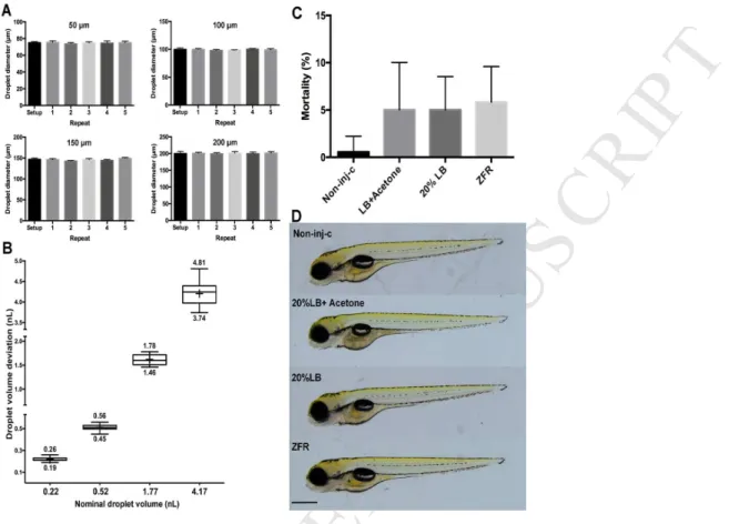
replicates compared to the desired diameter (Fig 1 A). Minimal and maximal droplet volumes 261
calculated from the measured diameters are shown in Fig 1 B. The largest decrease in volume 262
was detected in case of the 1.77 nL droplet size (17.51% (1.46 nL)), while the largest volume 263
increase was seen in case of the 0.22 nL droplet size (18.18% (0.26 nL)).
264
According to the OECD 236 guideline for the Fish Embryo Toxicity Test, nominal and real 265
concentrations should not differ from each other by more than ±20% (OECD236, 2013). In 266
this experiment deviations from the nominal volume stayed within this range for all volumes 267
tested, thus presumably our experiments would meet this basic requirement.
268
M AN US CR IP T
AC CE PT ED
Results show that with the above described experimental settings, the method is 269
dimensionally stable for all used droplet sizes, if the capillary is not clogged during injection.
270 271
3.2 Effect of the largest injection volume and the media on the viability of embryos 272
Prior to testing bacterial products, potential toxic effects of three basic media, the Zebrafish 273
Ringer’s solution, the LB medium, the medium supplemented with the solvent acetone (which 274
served as a bacterial propagation medium and carrier for OTA) and the largest used injection 275
volume (selected according to the work of Schubert and co-workers (2014)) was examined.
276
The injection volume is a critical factor in postinjection embryo survival, but potentially does 277
not cause egg trauma if the administered volume is bellow 10% of the total volume of the 278
yolk (Walker et al., 1992). For the same substance and same concentration, smaller injection 279
volumes cause less mortality and malformations in injected embryos (Zabel et al., 1995).
280
According to these, LB media were administered in the largest droplet volume too.
281
As the conditions did not have significant toxic effects, only results for 5 days of exposure are 282
shown in Figure 1 C. In the non-injected control no dead embryos were found, and the 283
average mortality rate was also very low in case of the LB medium (5%), the solvent 284
supplemented LB (5%) and the ZFR (5.83%) and there was no significant difference between 285
treatments. Malformations were not detected either in injected or non-injected (control) 286
embryos (Fig. 1 D).
287
The OECD guideline for fish embryo test allows a maximum of 10% lethality in the control 288
during an experiment (OECD236, 2013). This criterion was fulfilled in this study, since LB 289
media and ZFR caused lower lethality. Based on the mortality and morphology results, the 290
injection settings and droplet sizes used here seemed to be suitable for further work, the 291
examined conditions are not toxic to zebrafish embryos and so do not affect the outcome of 292
subsequent tests.
293
M AN US CR IP T
AC CE PT ED
294
3.3 The effect of metabolites produced by the Cupriavidus basilensis ŐR16 strain on the 295
survival of microinjected embryos 296
Bacterial metabolites produced during the primary metabolism of the strain (ŐR16) might 297
also have toxic effects on embryos, therefore the effect of the LB medium following 3 and 5 298
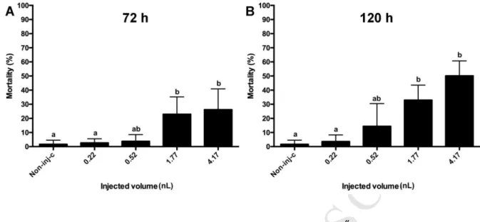
days of bacterial incubation was tested in 4 injection volumes (Fig. 2 A and B). The solution 299
decreased the survival rate of embryos at 72 and 120 hpf too and dose-response relationship 300
was found between injection volumes and lethality. After 72 hours of exposure, significant 301
increase was detected in mortality in the groups injected with 1.77 and 4.17 nL (p < 0.05) 302
compared to the control, and the group injected with the largest volume (4.17 nL) and the 303
group injected with 0.22 nL (p < 0.01). Mortality in the groups injected with the two largest 304
volumes was 22.92% (1.77 nL) and 26.15% (4.17 nL), with no significant difference between 305
the groups. Mortality increased in all injected groups after 120 hours of exposure, but 306
compared to the control, significant difference was only detected in the groups injected with 307
the two largest volumes where mortality was 32.92% (1.77 nL, p < 0.01) and 50.13% (4.17 308
nL, p < 0.01). Results clearly show that the strain ŐR16 produces toxic metabolites that – 309
following administration by microinjection – decrease the survival of zebrafish embryos.
310
Ferenczi et al. (2014) examined OTA biodegradation efficiency of the strain ŐR16 and the 311
toxicity of breakdown products derived from degradation in feeding experiments with mice.
312
Animals were exposed to ŐR16 metabolites via intragastric gavage once a day through 21 313
days. Toxic effects were examined via the expression of several marker genes and 314
histolopathological examination of the kidney and spleen. In mice, metabolic products of the 315
strain ŐR16 did not seem to be toxic compared to the control. According to the results 316
described above, zebrafish embryos seem to be more sensitive to the bacterial metabolites 317
than mice, however, difference may be due to different exposition pathways.
318
M AN US CR IP T
AC CE PT ED
319
3.4 Determination of the initial OTA concentration for further experiments 320
In order to determine the initial OTA concentration for further degradation experiments, OTA 321
was injected into the yolk of embryos in 1, 7 and 10 mg/L concentration, in different volumes.
322
All concentrations fell within the degradable concentration range of strain ŐR16. Mortality 323
was checked at 3 and 5 dpf and results were plotted on a dose response curve for mortality.
324
The graph of the potentially optimal initial concentration should serve as a reference for 325
further experiments even if toxicity is higher following degradation, so should meet the 326
following requirements: the maximum mortality should not exceed that of the bacterial 327
metabolic products and the curve should not reach its maximum early.
328
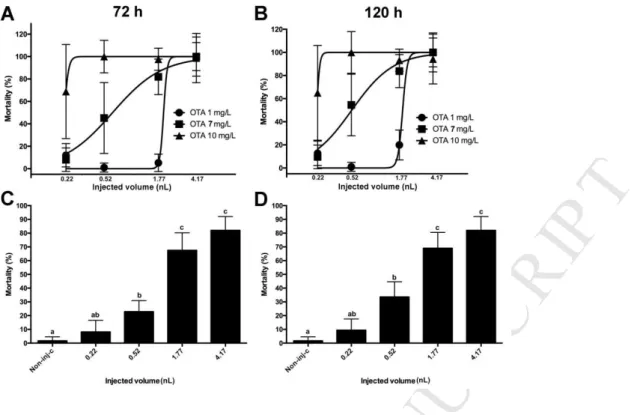
Mortality increased along with the injection volume in case of all three OTA concentrations, 329
and reached the maximum after 72 hours of exposure in all cases. Mortality did not change 330
significantly for 120 hours following exposure (Fig. 3 A and B).
331
The slope of the dose-response curve for 1 mg/L OTA was lower than the others and 332
mortality maximum was reached only with the largest injection volume following 72 and 120 333
hours of exposure. In case of lower injection volumes, mortality was below 10%.
334
Dose-response relationship was detected between injection volumes and mortality in case of 7 335
mg/L OTA as well. Mortality increased gradually with injection volumes at 72 and 120 hours 336
of exposure too, and the maximum (100%) was reached with the largest injection volume.
337
From 0.52 nL, significant difference (p < 0.05) was detected in mortality compared to the 338
control. Differences between mortality values of the groups injected with volumes ≤0.52 nL 339
compared to the 1.77 (p < 0.001) and 4.17 nL injection volumes were also significant (p <
340
0.05), however, no significant difference was found in case of the two largest volumes (Fig.
341
3C and D). Mortality reached its maximum (75%) early with 10 mg/L OTA with the lowest 342
injection volume (0.52 nL) and did not show to be higher with larger volumes.
343
M AN US CR IP T
AC CE PT ED
On the basis of our results, 7 mg/L was selected to be an initial concentration in further 344
experiments. The mortality curve of this concentration shown here served as reference for 345
subsequent tests.
346
The present study was the first to examine acute toxic effects of OTA following 347
microinjection, and high mortality was detected even after short exposures to low 348
concentrations. However, these results are difficult to compare to the results of classical tests 349
where embryos are exposed via waterborne exposure. It is still unclear how substances are 350
distributed in the yolk following injection but it is inhomogenous in most cases, so 351
presumably embryos are not exposed uniformly. Moreover, zebrafish embryos consume their 352
yolk sac completely to 165 ±12 hpf (Litvak and Jardine, 2003), thus, some of the substance 353
may remain unabsorbed during the exposition period presented here, however, with longer 354
exposure the experiment would fall under animal testing regulations. The microinjection 355
technique enables the administration of exact amounts, so theoretically it would be possible to 356
determine doses per bodyweight as seen in feeding experiments with vertebrates.
357 358
3.5 Toxicity of samples derived from ochratoxin degradation experiment 359
In order to clarify the toxicity of OTA-metabolites produced during microbial toxin 360
degradation with strain ŐR16, degradation products were microinjected in four concentrations 361
into zebrafish embryos. Mortality was examined on the 3rd and 5th day of exposure.
362
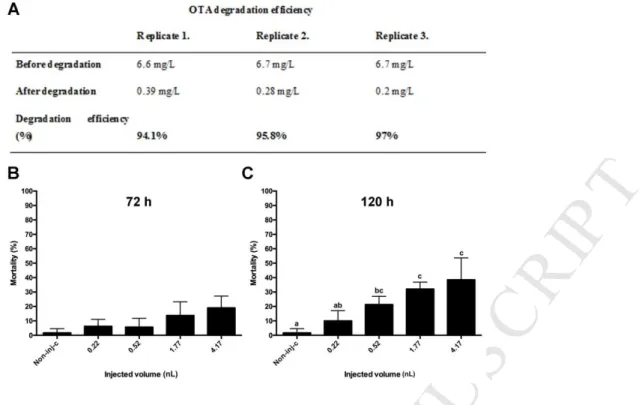
Mortality increased with the injected volume as seen previously. At 72 hours of exposure, 363
mortality in the non-injected control, and in the 0.22 nL and 0.52 nL injection volumes was 364
bellow 10%, and did not reach 30% even with the highest volumes. No significant difference 365
was observed between treated groups (Fig. 4 B). At 120 hours of exposure, dose-response 366
relationship was found between the injected volume and embryo mortality, as mortality 367
increased gradually along with the injection volume and reached 38.5% in the largest volume.
368
M AN US CR IP T
AC CE PT ED
Statistically significant decrease was detected in the number of survivals in the groups 369
injected with 0.52 nL, 1.77 nL and 4.17 nL compared to the non-injected control (p < 0.05), 370
and the two largest injection volumes compared to 0.22 nL (p < 0.05) (Fig. 4 C).
371
OTA degrading efficiency of strain ŐR16 was tested prior to exposure and it was found to be 372
95.6% (Fig. 4 A). OTA degradation of the strain ŐR16 is possibly mediated by a peptidase 373
enzyme. Ferenczi et al. (2014) showed that the major metabolite of OTA degraded by strain 374
ŐR16 is ochratoxin alpha (OTα). They found that OTA content in the supernatants decreased 375
gradually, OTα content increased in parallel during the 5-day incubation period and OTA was 376
completely degraded (94% decrease was measured by ELISA and 100% by HPLC), that is in 377
accordance with the results of the above described experiments. OTα is not potentially toxic, 378
according to the results of previous Vertebrate studies (Bruinink, 1998; Ferenczi et al., 2014).
379
Haq and co-workers (2016) tested the toxicity of OTα with ZETA test on zebrafish embryos 380
in concentrations ≤2.5 µM. In contrast to OTA, no significant difference was detected 381
between the mortality of embryos exposed to OTα and the untreated negative controls during 382
the 5 days exposure. On the basis of these, mortality in our experiments is probably due to 383
other metabolites of strain ŐR16.
384
Ferenczi et al. (2014) also studied OTA degradation products of strain ŐR16 in mouse 385
feeding experiments. Subchronic exposure did not cause mortality in mice and physiological 386
or gene expression alterations in the examined organs, compared to controls. However 387
degradation products were lethal to injected zebrafish embryos, so the zebrafish embryo is 388
probably a more sensitive model, than the mouse.
389 390
3.6 Comparison of mortality values of 7 mg/L OTA, and the bacterial and degradation 391
products of the strain ŐR16 392
M AN US CR IP T
AC CE PT ED
Mortality caused by 7 mg/L OTA, and the bacterial and degradation products of the strain 393
ŐR16 were plotted on joint graphs. In order to investigate the degradation characteristics of 394
the bacterial strain, mortality values of equal volumes were compared to each other (Fig. 5 A 395
and B).
396
Following 72 hours of incubation, mortality did not show significant difference between 397
groups injected with the smallest volumes. In case of larger volumes, there was no difference 398
between results of the bacterial metabolites and the degradation products of the strain, 399
however, mortality values of 7 mg/L OTA differed significantly from these (p < 0.05).
400
Highest mortality was caused by 7 mg/L OTA injected in 0.52 nL and above.
401
120 hours after microinjection, no significant difference was seen between the mortality 402
values of groups injected with 0.22 nL. In case of the groups injected with 0.52 nL, significant 403
difference was detected between 7 mg/L OTA and the metabolites of the strain ŐR16 (p <
404
0.05). Mortality values of the degradation products of strain ŐR16 did not differ nor from that 405
of the bacterial metabolites neither from the OTA solution. In larger volumes, only mortality 406
values of 7 mg/L OTA differed significantly from other groups (p < 0.001 - degradation 407
products, p < 0.01 – ŐR16 bacterial metabolites), however, bacterial metabolites of the strain 408
and degradation products of OTA did not show significant difference. The highest mortality 409
was detected in 7 mg/L OTA injected in 0.52 nL and above.
410
As no statistical difference was found between the mortality values of the bacterial and 411
degradation products, it can be concluded that OTA breakdown products are not toxic, and 412
mortality is probably caused by metabolites of the strain ŐR16. Results also show that 413
exposure via microinjection is a potential, functional, alternative way to test the detoxification 414
efficiency of toxin degrading microbes on zebrafish embryos in vivo. Mortality in itself may 415
provide a sufficient endpoint when testing the differences between the toxicity of the bacterial 416
metabolites of a strain and the degradation products of the toxin following microinjection, and 417
M AN US CR IP T
AC CE PT ED
toxicity of toxin degradation products can be predicted. There was no detectable difference 418
between the mortality curves of 3 and 5 days of exposure, so it seems that a 3 days exposure 419
period is sufficient for studying the degradation characteristics of bacterial strains.
420 421
3.7 Sublethal effects in injected embryos 422
Beyond mortality, sublethal endpoints were also analyzed in treated embryos following 72 423
and 120 hours of exposure. Generally, compared to the non-injected controls all treatments 424
with all injected volumes increased the frequency and severity of developmental deformities 425
(Fig. 6 A and B). Following 72 hours of exposure, the highest frequency of morphological 426
disorders was detected in the 7 mg/L OTA group, and in some replicates of treatments with 427
the highest volumes of this concentration, all surviving embryos showed abnormalities. A 428
statistically significant difference was only observed between the 1.77 nL OTA (7 mg/L), and 429
1.77 nL samples containing bacterial metabolites or degradation products (p < 0.01).
430
Following 120 hours of exposure, it was also evident that compared to other treatment groups 431
the ratio of deformed embryos was the highest in the groups treated with OTA from 0.52 nL 432
and above. Statistically significant differences were observed in ŐR16 bacterial metabolites 433
(p < 0.01) and breakdown products of OTA (p < 0.05) compared to 7 mg/L OTA, injected in 434
1.77 nL. Significant differences were also found between OTA 7 mg/L and metabolites of 435
strain ŐR16 (p < 0.01) or degradation products (p < 0.05), injected in 4.17nL. However no 436
significant difference was detected between the deformation frequencies in the groups 437
injected with the bacterial metabolites of the strain and OTA degradation products during the 438
whole exposure period with any injection volumes.
439
It can be concluded that notwithstanding the significant differences detected in morphology, 440
OTA degradation products seem to be nontoxic on the basis of deformation frequencies, 441
however the metabolites of the strain were proved to be toxic.
442
M AN US CR IP T
AC CE PT ED
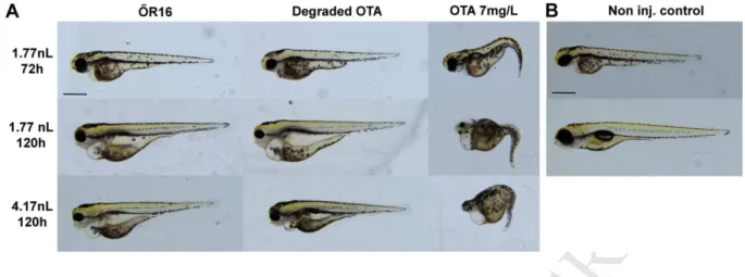
Figure 7. shows representative development dysfunctions in embryos from treatment groups 443
with statistically significant differences. Following 3 days of OTA injection (1.77 nL) 444
embryos displayed craniofacial deformities, small eyes, curvature of the body axis, yolk 445
deformities, reduced growth rates and edemas in some cases. Most of them have previously 446
been described in OTA treated zebrafish embryos (Haq et al., 2016), and teratogenic effect 447
was observed at sub-micromolar concentrations with an EC50 of 20 nM OTA.
448
Similarly to zebrafish OTA proved to be teratogenic in the amphibian Xenopus laevis model 449
(FETAX) too, causing mainly craniofacial deformities (O’Brien et al., 2005) like in the 450
experiments described above. These developmental abnormalities (craniofacial deformities) 451
were also detected in a wide range of Vertebrates, including rats (Brown and Purmalis, 1976), 452
mice (Arora, 1983), hamsters (Hood et al., 1976) and chicken (Wiger and Starrmer, 1990).
453
Decreased hatching rate described by Haq et al. (2016) was not seen in our experiments.
454
Embryos injected with the same volume (1.7 nL) of bacterial metabolites and OTA 455
degradation products displayed shorter body, yolk sac deformations, grey coloration in the 456
yolk, pericardial edema, small eyes and deformities of lower facial structures in embryos 457
following 3 days of injection. Curvation of the body as a common sign of OTA exposure has 458
not been detected.
459
Five days after microinjection, sympthoms got more pronounced in OTA treated embryos and 460
severe deformations appeared all through the body. Embryos injected with bacterial 461
metabolites of the strain ŐR16 and OTA degradation products displayed shorter body, yolk 462
sac deformations, pericardial edema, edema around the abdomen, small eyes, small and not 463
well defined olfactory region and deformities of lower facial structures on the 5th day of 464
exposure. As in 3 dpf exposed embryos, curvation of the body axis was not seen here either.
465
In contrast, OTα did not seem to be toxic in Vertebrates. Haq and co-workers (2016) 466
examined the effects of OTα (along with OTA) on zebrafish embryos and neither 467
M AN US CR IP T
AC CE PT ED
teratogenicity nor mortality differed significantly from that of the negative control embryos 468
during 5 days of exposure. Ferenczi et al. (2014) demonstrated apparent hydrolysis of OTA to 469
OTα, and consequent detoxification by using a bacterial species Cupriavidus basilensis, as 470
evidenced by comparative toxicological studies in a mouse model of nephrotoxicity.
471
In the present study, morphological examination showed that phenotype of OTA treated 472
embryos differed significantly from the morphology of embryos exposed to bacterial 473
metabolites or OTA degradation products in both experimental time points, however embryos 474
in the latter groups showed similar phenotypes. In conclusion, it seems that strain ŐR16 475
degrades OTA to nontoxic metabolites, the strain is able to degrade OTA even in 7 mg/L 476
concentration, and deformations resulted from the injection of OTA degradation products are 477
probably due to the metabolites of the bacteria. In addition, zebrafish exposed via 478
microinjection appeared to be more sensitive to the metabolites of strain ŐR16 than mice.
479
All injected solutions contained high levels of organic matter. No deformation implied 480
oxygen deprivation in morphological examinations of exposed zebrafish embryos (Küster and 481
Altenburger, 2008; Strecker et al., 2011). Results suggest that microinjection can be an 482
alternative way to test samples with high organic matter content.
483
High organic matter content of samples often causes hypoxia during zebrafish embryo tests, 484
and its effects (developmental disorders, suspension of embryo development) can hardly be 485
differentiated from those of the sample itself (Küster and Altenburger, 2008; Strecker et al., 486
2011). With microinjection hypoxic effect of such samples can be avoided and results can 487
easily be evaluated.
488 489
M AN US CR IP T
AC CE PT ED
490
4. Conclusions 491
Microinjection is a simple way to introduce organic matter-rich test substances into newly 492
fertilized fish eggs and helps to eliminate hypoxia that cause a wide range of secondary 493
effects. If the method is well optimized, injection volume variations can be kept within ±20%, 494
according to the OECD 236 test guideline’s recommendations and so result reliability can be 495
ensured.
496
Results clearly showed that investigation of zebrafish embryos microinjected with toxin 497
solutions, metabolites of bacterial strains and OTA degradation products could provide an 498
alternative way for studying the toxin detoxification-properties of microbial strains. The 499
zebrafish embryo – thanks to their sensitivity – proved to be a good model for the studies.
500
Toxicity differences between substances may be detected even after 3 days of exposure on the 501
basis of mortality, that can be completed and further refined by the evaluation of sublethal 502
data.
503
Microinjection enables the selection of microbial strains that are able to degrade the toxin and 504
the identification of the most effective and environmentally safe microbes from the selected 505
strains.
506 507
Acknowledgements 508
This work was supported by Development and Innovation Fund (NKFIH); Grant Agreement:
509
NVKP_16-1-2016-0009 and VKSZ_12-1-2013-0078, EFOP-3.6.3-VEKOP-16-2017-00008 510
project co-financed by the European Union, and the Higher Education Institutional Excellence 511
Program (1783-3/2018/FEKUTSTRAT) awarded by the Ministry of Human Capacities within 512
the framework of water related researches of Szent István University. The scientific work of 513
Mátyás Cserháti was supported by the János Bolyai Research Grant of the Hungarian 514
M AN US CR IP T
AC CE PT ED
Academy of Sciences. Edina Garai was supported by the ÚNKP-18-3-I New National 515
Excellence Program of the Ministry of Human Capaticies. The authors gratefully thank Ákos 516
Horváth for critical reading of the manuscript.
517 518
M AN US CR IP T
AC CE PT ED
519
5. References 520
Abrunhosa, L., Inês, A., Rodrigues, A.I., Guimarães, A., Pereira, V.L., Parpot, P., Mendes- 521
faia, A., Venâncio, A., 2014. International Journal of Food Microbiology Biodegradation 522
of ochratoxin A by Pediococcus parvulus isolated from Douro wines. Int. J. Food 523
Microbiol. 188, 45–52. https://doi.org/10.1016/j.ijfoodmicro.2014.07.019 524
Arora, R.G.. F.H. ;Fellne.-F.H., 1983. INHIBITION OF O C H R A T O X I N A T E R A T 525
O G E N E S I S. Food Chem. Toxicol. 21, 779–783.
526
Binder, E.M., 2007. Managing the risk of mycotoxins in modern feed production 133, 149–
527
166. https://doi.org/10.1016/j.anifeedsci.2006.08.008 528
Braunbeck, T., Boettcher, M., Hollert, H., Kosmehl, T., Lammer, E., Leist, E., Rudolf, M., 529
Seitz, N., 2005. Towards an alternative for the acute fish LC(50) test in chemical 530
assessment: the fish embryo toxicity test goes multi-species -- an update. ALTEX 22, 531
87–102. https://doi.org/10.1007/s10811-007-9297-x 532
Brown, M.H., Purmalis, B.P., 1976. Teratogenic and Toxic Effects of Ochratoxin A in Rats 533
fetal hemorrhage and an undefined defect termed “ coelosomy ,” they were unable to 534
detect any skeletal or visceral malformations . The present study was designed to further 535
assess the teratogenic poten. Toxicol. Appl. Pharmacol. 37, 331–338.
536
Bruinink, A.. R.T.. S.C., 1998. Differences in Neurotoxic Effects of Ochratoxin A , Ochracin 537
and Ochratoxin- a In Vitro. Nat. Toxins 177, 173–177.
538
Bui-Klimke, T.R., Wu, F., 2015. Ochratoxin A and Human Health Risk: A Review of the 539
Evidence. Crit. Rev. Food Sci. Nutr. 55, 1860–1869.
540
https://doi.org/10.1080/10408398.2012.724480 541
Colman, J.R., Dechraoui, M.Y.B., Dickey, R.W., Ramsdell, J.S., 2004. Characterization of 542
the developmental toxicity of Caribbean ciguatoxins in finfish embryos. Toxicon 44, 59–
543
M AN US CR IP T
AC CE PT ED
66. https://doi.org/10.1016/j.toxicon.2004.04.007 544
ECR, 2006. Commission Regulation (EC) No 1881/2006 of 19 December 2006 setting 545
maximum levels for certain contaminants in foodstuffs. Off J Eur Union 364 5–24.
546
EFSA, 2010. Statement on the establishment of guidelines for the assessment of additives 547
from the functional group ‘ substances for reduction of the contamination of feed by 548
mycotoxins ’ 1 EFSA Panel on Additives and Products or Substances used in Animal 549
Feed ( FEEDA. EFSA J. 8, 1–8. https://doi.org/10.2903/j.efsa.2010.1693.
550
FAO, 2003. Food and Agriculture organization of the United Nations.
551
Ferenczi, S., Cserháti, M., Krifaton, C., Szoboszlay, S., Kukolya, J., Szoke, Z., Koszegi, B., 552
Albert, M., Barna, T., Mézes, M., Kovács, K.J., Kriszt, B., 2014. A new ochratoxin a 553
biodegradation strategy using cupriavidus basilensis Or16 strain. PLoS One 9.
554
https://doi.org/10.1371/journal.pone.0109817 555
Fernández-Cruz, M.L., Mansilla, M.L., Tadeo, J.L., 2010. Mycotoxins in fruits and their 556
processed products: Analysis, occurrence and health implications. J. Adv. Res. 1, 113–
557
122. https://doi.org/10.1016/j.jare.2010.03.002 558
Gagliano, N., Donne, I.D., Torri, C., Migliori, M., Grizzi, F., Milzani, A., Filippi, C., Annoni, 559
G., Colombo, P., Costa, F., Ceva-Grimaldi, G., Bertelli, A.A.E., Giovannini, L., Gioia, 560
M., 2006. Early cytotoxic effects of ochratoxin A in rat liver: A morphological, 561
biochemical and molecular study. Toxicology 225, 214–224.
562
https://doi.org/10.1016/j.tox.2006.06.004 563
Haq, M., Gonzalez, N., Mintz, K., Jaja-Chimedza, A., De Jesus, C.L., Lydon, C., Welch, A., 564
Berry, J.P., 2016. Teratogenicity of ochratoxin a and the degradation product, ochratoxin 565
α, in the zebrafish (Danio rerio) embryo model of vertebrate development. Toxins 566
(Basel). 8, 8–11. https://doi.org/10.3390/toxins8020040 567
Hathout, A.S., Aly, S.E., 2014. Biological detoxification of mycotoxins : a review 905–919.
568
M AN US CR IP T
AC CE PT ED
https://doi.org/10.1007/s13213-014-0899-7 569
Hood, R.D., Naughton, M.J., Hayes, A.W., 1976. Prenatal Effects of Ochratoxin A in 570
HamstersIJ. Teratology 13, 11–14.
571
Karlovsky Petr, 1999. Biological Detoxi ® cation of Fungal Toxins and its Use in Plant 572
Breeding , Feed and Food Production. Nat. Toxins 23, 1–23.
573
Küster, E., Altenburger, R., 2008. Oxygen decline in biotesting of environmental samples—Is 574
there a need for consideration in the acute zebrafish embryo assay? Environ. Toxicol. An 575
Int. J. 23, 745–750. https://doi.org/10.1002/tox.20377 576
Li, S., Marquardt, R.R., Frohlich, A.A., Vitti, T.G., Crow, G., 1997. Pharmacokinetics of 577
ochratoxin α and its metabolites in rats. Toxicol. Appl. Pharmacol. 145, 82–90.
578
https://doi.org/10.1006/taap.1997.8155 579
Litvak and Jardine, 2003. Direct yolk sac volume manipulation of zebrafish embryos and the 580
relationship between offspring size and yolk sac volume. J. Fish Biol. 63, 388–397.
581
https://doi.org/10.1046/j.1095-8649.2003.00161.x 582
Marin, D.E., Taranu, I., 2015. Ochratoxin A and its effects on immunity. Toxin Rev. 34, 11–
583
20. https://doi.org/10.3109/15569543.2014.958757 584
Minaschek G., Bereiter-Hahn J., Bertholdtt G., 1989. Quantitation of the Volume of Liquid 585
Injected by Means of Pressure into Cells powerful tool in cell research . The substances 586
injected include tracer substances. Exp. Cell Res. 183, 434–442.
587
Mizell, M., Romig, E.S., 1997. The aquatic vertebrate embryo as a sentinel for toxins:
588
Zebrafish embryo dechorionation and perivitelline space microinjection. Int. J. Dev.
589
Biol. 41, 411–423. https://doi.org/10.1387/IJDB.9184351 590
Nagel, R., 2002. DarT: The embryo test with the Zebrafish Danio rerio--a general model in 591
ecotoxicology and toxicology. ALTEX 19 Suppl 1, 38–48.
592
https://doi.org/10.1007/s13311-013-0218-1 593
M AN US CR IP T
AC CE PT ED
O’Brien, E., Prietz, A., Dietrich, D.R., 2005. Investigation of the teratogenic potential of 594
ochratoxin A and B using the FETAX system. Birth Defects Res. Part B - Dev. Reprod.
595
Toxicol. 74, 417–423. https://doi.org/10.1002/bdrb.20054 596
Odhav, B., Naicker, V., 2002. Mycotoxins in South African traditional brewed beers. Food 597
Addit. Contam. 19, 55–61. https://doi.org/10.1080/0265203011005342 598
OECD236, 2013. Oecd guidelines for the testing of chemicals 1–22.
599
Otteneder, H., Majerus, P., 2000. Occurrence of ochratoxin A (OTA) in wines: Influence of 600
the type of wine and its geographical origin. Food Addit. Contam. 17, 793–798.
601
https://doi.org/10.1080/026520300415345 602
Pfohl-Leszkowicz, A., Manderville, R.A., 2007. Ochratoxin A: An overview on toxicity and 603
carcinogenicity in animals and humans. Mol. Nutr. Food Res. 51, 61–99.
604
https://doi.org/10.1002/mnfr.200600137 605
Schubert, S., Keddig, N., Hanel, R., Kammann, U., 2014. Microinjection into zebrafish 606
embryos (Danio rerio) - a useful tool in aquatic toxicity testing? Environ. Sci. Eur. 26.
607
https://doi.org/10.1186/s12302-014-0032-3 608
Scudamore, K.A., Banks, J.N., Guy, R.C.E., 2004. Fate of ochratoxin A in the processing of 609
whole wheat grain during extrusion. Food Addit. Contam. 21, 488–497.
610
https://doi.org/10.1080/02652030410001670166 611
Sheikh-Zeinoddin, M., Khalesi, M., 2018. Biological detoxification of ochratoxin A in plants 612
and plant products. Toxin Rev. 0, 1–13. https://doi.org/10.1080/15569543.2018.1452264 613
Stoev, S.D., Denev, S.A., 2013. Porcine/chicken or human nephropathy as the result of joint 614
mycotoxins interaction. Toxins (Basel). 5, 1503–1530.
615
https://doi.org/10.3390/toxins5091503 616
Strecker, R., Seiler, T.B., Hollert, H., Braunbeck, T., 2011. Oxygen requirements of zebrafish 617
(Danio rerio) embryos in embryo toxicity tests with environmental samples. Comp.
618
M AN US CR IP T
AC CE PT ED
Biochem. Physiol. - C Toxicol. Pharmacol. 153, 318–327.
619
https://doi.org/10.1016/j.cbpc.2010.12.002 620
Vanhoutte, I., Audenaert, K., De Gelder, L., 2016. Biodegradation of mycotoxins: Tales from 621
known and unexplored worlds. Front. Microbiol. 7, 1–20.
622
https://doi.org/10.3389/fmicb.2016.00561 623
Vrabcheva, T., Usleber, E., Dietrich, R., Märtlbauer, E., 2000. Co-occurrence of ochratoxin A 624
and citrinin in cereals from bulgarian villages with a history of Balkan endemic 625
nephropathy. J. Agric. Food Chem. 48, 2483–2488. https://doi.org/10.1021/jf990891y 626
Walker, M.K., Hufnagle, L.C.J., Clayton, M.K., Peterson, R.E., 1992. An egg injection 627
method for assessing early life stage mortality of polychlorinated dibenzo-p-dioxins, 628
dibenzofurans, and biphenyls in rainbow trout, (Onchorhynchus mykiss). Aquat.
629
Toxicol. 22, 15–38.
630
Wiger, R., Starrmer, F.C., 1990. Effects of ochratoxins A and B on prechondrogenic 631
mesenchymal cells from chick embryo limb buds. Toxicol. Lett. 54, 129–134.
632
Xiao, H., Madhyastha, S., Marquardt, R.R., Li, S., Vodela, J.K., Frohlich, A.A., Kemppainen, 633
B.W., 1996. Toxicity of ochratoxin A, its opened lactone form and several of its analogs:
634
Structure-activity relationships. Toxicol. Appl. Pharmacol. 137, 182–192.
635
https://doi.org/10.1006/taap.1996.0071 636
Zabel, E.W., Cook, P.M., Peterson, R.E., 1995. Toxic equivalency factors of polychlorinated 637
dibenzo-p-dioxin, dibenzofuran and biphenyl congeners based on early life stage 638
mortality in rainbow trout (Oncorhynchus mykiss). Aquat. Toxicol. 31, 315–328.
639
https://doi.org/10.1016/0166-445X(94)00075-2 640
641 642
M AN US CR IP T
AC CE PT ED
Tables and figures 643
644
Fig1 Variations in the diameter (A) and volume (B) of the injected droplet and mortality 645
(C) and morphology (D) effects of control solutions. The largest decrease in volume was 646
detected in case of the 1.77 nL droplet size (17.51% (1.46 nL)), while the largest volume 647
increase was seen in case of the 0.22 nL droplet size (18.18% (0.26 nL)). Droplet diameter 648
and volume stayed within ±20%, and no significant difference was detected between 649
measurements. Average mortality rate of 120 hpf embryos injected with 4.17 nL was very low 650
in all cases ((Non-inj-c (non-injected control): 0%, 20% LB: 5%, 20% LB + Acetone: 5%, 651
ZFR (Zebrafish Ringers’s solution): 5.83%). There was no significant difference between 652
treatment groups and no malformations were detected in any case. Scale bar: 500µm.
653
M AN US CR IP T
AC CE PT ED
654
655
Fig 2 Effects of the metabolites of Cupriavidus basilensis ŐR16 strain, injected in 656
different volumes, on the mortality of zebrafish embryos at 72 (A) and 120 hpf (B). After 657
72 hours of exposure, statistical significant differences were observed between the non- 658
injected control and 1.77 nL (p < 0.05), non-injected control and 4.17 nL (p < 0.05), 0.22 nL 659
and 4.17 nL (p < 0.01). Lethality was below 10% in the non-injected control, 0.22 nL and 660
0.52 nL. After 120 hours of exposure statistical significant differences were observed between 661
the non-injected control and 1.77 nL (p < 0.01), non-injected control and 4.17 nL (p < 0.01), 662
0.22 nL and 4.17 nL (p < 0.001). Mortality was below 10% in the non-injected control and 663
0.22 nL.
664
665
666
M AN US CR IP T
AC CE PT ED
667
Fig 3 Effects of ochratoxin A (OTA) injected in different concentrations and volumes on 668
the mortality of 72 (A) and 120 (B) hpf zebrafish embryos and the effects of 7 mg/L 669
OTA injected in different volumes on the mortality of 72 (C) and 120 (D) hpf zebrafish 670
embryos. At 72 hpf, lethality results in the non-injected control were below 10%. Statistical 671
significant differences were observed in the 0.52 nL (p < 0.05), 1.77 nL (p < 0.05) and 4.17 672
nL (p < 0.01) groups compared to the non-injected control. Significant differences were 673
detected between 0.22 nL and 1.77 nL (p < 0.01), 0.22 nL and 4.17 nL (p < 0.0001), 0.52 nL 674
and 1.77 nL (p < 0.0001), 0.52 nL and 4.17 nL (p < 0.05) (C). At 120 hpf lethality results 675
were below 10% in the non-injected control. Statistically significant differences were 676
observed between the non-injected control and 0.52 nL (p < 0.05), 1.77 nL (p < 0.05) and 677
4.17 nL (p < 0.01) groups. Significant differences were detected between 0.22 nL and 1.77 nL 678
(p < 0.01), 0.22 nL and 4.17 nL (p < 0.0001), 0.52 nL and 1.77 nL (p < 0.001), 0.52 nL and 679
4.17 nL (p < 0.05) (D).
680
681
M AN US CR IP T
AC CE PT ED
682
Fig 4 Ochratoxin A (OTA) degradation efficiency of Cupravidus basiliensis ŐR16 strain 683
following 120 hours of incubation with 7 mg/L OTA (A) and effects of OTA degradation 684
products injected in different volumes on the survival of zebrafish embryos at 72 (B) 685
and 120 (right) hpf (C). At 72 hpf, no significant difference was observed between treatment 686
groups and mortality was less than 10% in the non-injected control (Non-inj-c), 0.22 nL and 687
0.52 nL groups. At 120 hpf, mortality was below 10 % in the non-injected control. Statistical 688
significant differences were observed between the non-injected control and 0.52 nL (p <
689
0.05), non-injected control and 1.77 nL (p < 0.05), non-injected and 4.17 nL (p < 0.05).
690
Significant differences were detected between 0.22 nL and 1.77 nL (p < 0.05), 0.22 nL and 691
4.17 nL (p < 0.05).
692
693
694
M AN US CR IP T
AC CE PT ED
695
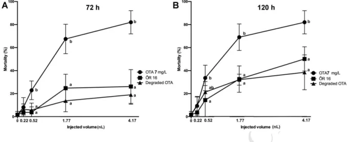
696
Fig 5 Effects of 7 mg/L Ochratoxin A (OTA 7 mg/L), bacterial metabolites (ŐR16) and 697
OTA degradation products (degraded OTA) derived from the biodegradation 698
experiment with Cupriavidus basilensis ŐR16 strain on the survival of 72 (A) and 120 699
(B) hpf zebrafish embryos. At 72 hpf mortality in the non-injected control was below 10%.
700
Statistical significant differences were observed between OTA and degraded OTA (p < 0.05), 701
OTA and ŐR16 (p < 0.05) in case of 0.52 nL, OTA and degraded OTA (p < 0.01), OTA and 702
ŐR16 (p < 0.01) in case of 1.77 nL, and OTA and degraded OTA (p < 0.01), OTA and ŐR16 703
(p < 0.01) in case of 4.17 nL. At 120 hpf mortality in the non-injected control was below 704
10%. Statistical significant differences were observed between OTA and ŐR16 (p < 0.05) in 705
case of 0.52 nL, OTA and degraded OTA (p < 0.05), OTA and ŐR16 (p < 0.01) in case of 706
1.77 nL, and OTA and degraded OTA (p < 0.001), OTA and ŐR16 (p < 0.01) in case of 4.17 707
nL.
708
709 710
M AN US CR IP T
AC CE PT ED
711
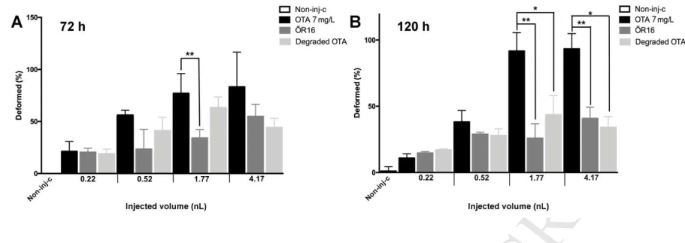
712
Fig 6 Effects of 7 mg/L Ochratoxin A (OTA 7 mg/L), bacterial metabolites (ŐR 16) and 713
OTA degradation products (Degraded OTA) derived from the biodegradation 714
experiment with Cupriavidus basilensis ŐR16 strain on the frequency of developmental 715
deformities in 72 (A) and 120 (B) hpf zebrafish embryos. At 72 hpf, the highest frequency 716
of morphological disorders was detected in the 7 mg/L OTA group. Statistically significant 717
difference was only observed between the 1.77 nl OTA 7 mg/L and 1.77 nL ŐR16 (p < 0.01) 718
groups. In 120 hpf embryos, the ratio of deformed embryos was the highest in the groups 719
treated with OTA from 0.52 nL and above. Statistically significant difference was observed 720
between OTA 7 mg/L and ŐR16 (p < 0.01) and OTA 7 mg/L and degraded OTA injected in 721
1.77 nL (p < 0.05), OTA 7 mg/L and degraded OTA 7 mg/L and ŐR16 (p < 0.01) and OTA 7 722
mg/L and degraded OTA (p < 0.05) injected in 4.17 nL. No significant difference was 723
detected between the deformation frequencies in the groups injected with the bacterial 724
metabolites and OTA degradation products.
725
726
M AN US CR IP T
AC CE PT ED
727
728
Fig 7 Representative development dysfunctions in zebrafish embryos following injection.
729
Ochratoxin A (OTA 7 mg/L), bacterial metabolites (ŐR16) and OTA degradation products 730
(Degraded OTA) derived from biodegradation experiment with Cupriavidus basilensis ŐR16 731
strain were injected in 1.7, 1.77 and 4.17 nL volumes and disorders were examined following 732
72 and 120 hours of injection (A). Non-injected control embryos (Non inj. control) are shown 733
on Figure 8B. Scale bar: 500µm.
734
735
M AN US CR IP T
AC CE PT ED
HIGHLIGHTS:
• ŐR16 degrades OTA to nontoxic products, however bacteria have intrinsic toxicity
• Toxicity differences between test solutions are detectable after 3 days of exposure
• Injection volume variations and control mortality correspond with OECD TG 236
• Microinjection is proper for qualifying the toxin-degrading properties of microbes
• The method helps in selecting the most effective, safe strains for detoxification