Contents lists available atScienceDirect
Mechanisms of Ageing and Development
journal homepage:www.elsevier.com/locate/mechagedev
Original Article
Genetic deletion of TRPA1 receptor attenuates amyloid beta- 1-42 (A β
1-42)- induced neurotoxicity in the mouse basal forebrain in vivo
M. Payrits
a,b,1, E. Borbely
a,b,1, S. Godo
b,c, D. Ernszt
b,c, A. Kemeny
a,b,d, J. Kardos
e, E. Szoke
a,b, E. Pinter
a,b,*
aDepartment of Pharmacology and Pharmacotherapy, Medical School, University of Pécs, Hungary
bCentre for Neuroscience, Szentágothai Research Center, University of Pécs, Pécs, Hungary
cInstitute of Physiology, University of Pécs, Pécs, Hungary
dDepartment of Medical Biology and Central Electron Microscope Laboratory, University of Pécs, Hungary
eELTE NAP Neuroimmunology Research Group, Department of Biochemistry, Eötvös Loránd University, Budapest, Hungary
A R T I C L E I N F O
Keywords:
TRPA1 Amyloid beta Cholinergic cell loss Memory loss
A B S T R A C T
Amyloidβ1–42 peptide (Aβ1–42) accumulates in Alzheimer's disease (AD) that is toxic to the basal forebrain cholinergic (BFC) neurons in substantia innominata-nucleus basalis magnocellularis complex (SI-NBM).
Transient Receptor Potential Ankyrin1 (TRPA1) receptor is present in murine brain, however its role in neu- rotoxic processes is unclear. We investigated the Aβ1–42-induced neurotoxicity in TRPA1 wild-type (TRPA1+/+) and knockout (TRPA1−/−) mice.
Expression and neuroanatomical localization of TRPA1 receptor were examined using RT qPCR. Cholinergic fibre loss was determined on acetylcholinesterase (AChE) stained brain slices, and choline acetyltransferase (ChAT) immunohistochemistry was used to assess the cholinergic cell loss. Novel object recognition (NOR), radial arm maze (RAM) and Y-maze tests were used to investigate memory loss.
Aβ1–42-injected WT mice showed marked loss of cholinergicfibres and cell bodies, which was significantly attenuated in TRPA1−/−animals. According to the NOR and RAM tests, pronounced memory loss was detected in Aβ1–42-injected TRPA1+/+mice, but not in TRPA1−/−group.
Ourfindings demonstrate that TRPA1 KO animals show substantially reduced morphological damage and memory loss after Aβ1–42injection in the SI-NBM. We conclude that TRPA1 receptors may play an important deteriorating role in the Aβ1–42-induced cholinergic neurotoxicity and the consequent memory loss in the murine brain.
1. Introduction
One of the major pathomorphological hallmarks of Alzheimer’s disease (AD) is the significant neuronal death in the cortex, hippo- campus and basal forebrain. The cholinergic system of the basal fore- brain (BFC) is the pivotal source of cholinergic connections to the cortex and hippocampus, which plays a major role in attention and memory (Zaborszky et al., 1999;Beninger et al., 2001). Accumulation of neurotoxicβ-amyloid peptides, such as Aβ1–42contributes to neu- ronal loss in BFC in AD (Maccioni et al., 2001). The Aβactivates as- trocytes and microglial cells, also it has direct neurotoxic effect by li- gand-like interaction with the N-methyl-D-aspartate (NMDA) glutamate receptors (Harkany et al., 2000). Aβ1–42microinjection into the nucleus
basalis magnocellularis (NBM) results in severe damage of neurons in BFC and memory deficits in rodents (Harkany et al., 2000, 1998;
Giovannelli et al., 1995;Kwakowsky et al., 2016).
There is an increasing evidence that neuro-immune interactions may play a critical role in the pathomechanism of several neurode- generative diseases (Candore et al., 2007;Masters et al., 2015, Sághy et al., 2016) and regulatory role of the transient receptor potential (TRP) ion channels is strongly suggested in these processes. TRP re- ceptors form non-selective cation channels, with preferentially high calcium ion permeability. Calcium influx triggers several intracellular pathways. TRP channels are sensitive for various stimuli including mechanical, thermal triggers or chemical ligands. Considering these interactions, they are likely to be sensors for several physiological or
https://doi.org/10.1016/j.mad.2020.111268
Received 23 January 2020; Received in revised form 16 May 2020; Accepted 20 May 2020
⁎Corresponding author at: Department of Pharmacology and Pharmacotherapy, Medical School, University of Pécs, Szigeti u.12. H-7624, Pécs, Hungary.
E-mail addresses:payrits.maja@gmail.com(M. Payrits),eva.borbely@aok.pte.hu(E. Borbely),soma.godo@gmail.com(S. Godo),ernszt.david@pte.hu(D. Ernszt), agnes.kemeny@aok.pte.hu(A. Kemeny),kardos@elte.hu(J. Kardos),eva.szoke@aok.pte.hu(E. Szoke),erika.pinter@aok.pte.hu(E. Pinter).
1M.P. and É.B. contributed equally to this work.
Mechanisms of Ageing and Development 189 (2020) 111268
Available online 28 May 2020
0047-6374/ © 2020 The Author(s). Published by Elsevier B.V. This is an open access article under the CC BY license (http://creativecommons.org/licenses/BY/4.0/).
T
pathophysiological stimuli (Khalil et al., 2018). One of the TRP re- ceptors, the transient receptor potential ankyrin 1 (TRPA1) is best known as a sensor for environmental irritants participating in pain, cold and itch sensation. TRPA1 has a wide range of ligands; specifically it is sensitive to low and high temperature, osmotic changes, natural and synthetic irritants. Several TRPA1 agonists are reactive electrophilic ligands (Zimova et al., 2018). Channel gating effect of these compounds is based on covalent modification of cysteine and lysine residues within the N-terminus and transmembrane domains of the receptor (Zygmunt and Högesättt, 2014). Since TRPA1 can be triggered by endogenous compounds generated by tissue injury and inflammation, it can be a potential target for anti-nociceptive and anti-inflammatory drug can- didates (Nishida et al., 2015;Horváth et al., 2016). Recent data showed that TRPA1 receptors are also expressed in the brain and play mod- ulatory role in neurodegenerative disorders and neuroinflammation, such as multiple sclerosis and AD (Sághy et al., 2016,Lee et al., 2016;
Bölcskei et al., 2018; Dalenogare et al., 2020; Ritter et al., 2020).
TRPA1 channels have also been found on astrocytes of corpus callosum (Sághy et al., 2016), also in oligodendrocytes of the cerebellum (Hamilton et al., 2016) and in the endothelium of cerebral arteries (Sullivan et al., 2015; Pires and Earley, 2018). Nowadays, Lee and colleagues have provided immunohistochemical evidence that hippo- campal astrocytes express TRPA1 in wild-type mice; and im- munoreactivity of the receptor protein also occurs in the cortical neu- rons of amyloid precursor protein/presenilin1 (APP/PS1) transgenic mice (Lee et al., 2016). However, only fewin vivodata are available to corroborate the function of TRPA1 in the brain. The principal aims of the present study were to investigate the morphological and the func- tional aspects of TRPA1-mediated events in BFC using Aβ1–42-induced neurotoxicity model in TRPA1 knockout mice.
2. Materials and methods
2.1. Animals
Experiments were carried out on 3–4 month-old male C57BL/6, TRPA1 knockout mice (TRPA1−/−) and their wild-type siblings (TRPA1+/+) (obtained from Prof. P. Geppetti, University of Florence, Italy). The animals were bred and kept in the Animal House of the Department of Pharmacology and Pharmacotherapy of the University of Pécs at 24 °C and provided with standard rodent food and water ad libitum. Mice were housed under conditions of 12-h light/dark cycle in groups of 3–5 in polycarbonate cages (530 cm2 floor space, 14 cm height) on wood shavings bedding. The animals had a 60-min accli- matization period prior to each behavioural experiment. Six animal groups were created, stated as untreated wild-type (TRPA1+/+naïve) and TRPA1 receptor gene-deficient mice (TRPA1−/−naïve), vehicle- injected wild-type (TRPA1+/+Veh) and TRPA1 knockout (TRPA1−/−
Veh), β-amyloid-treated wild-type (TRPA1+/+ Aβ1–42), and TRPA1 knockout (TRPA1−/− Aβ1–42). Eight TRPA1+/+ and seven TRPA1-/- animals were used for AChE histochemistry. Eight TRPA1+/+and six TRPA1-/- mice prepared for ChAT immunohistochemistry. In beha- vioural measurements the number of animals was 12−12 in TRPA1+/+
naïve and TRPA1-/-naïve groups, 8−8 in TRPA1+/+Veh and TRPA1-/- Veh groups, 10 in TRPA1+/+Aβ1–42group and 12 in TRPA1-/-Aβ1–42
group. All experimental procedures were approved by the Animal Welfare Committee of University of Pécs, licence no.: BA 02/2000−24/
2016, BA 02/2000−38/2016.
2.2. Preparation of Aβ1–42peptide samples
Aβ1–42 was expressed recombinantly in E. coli and purified ac- cording to Ikenoue and colleagues (Ikenoue et al., 2014). Briefly, the DNA sequence coding the human Aβ1–42peptide was artificially con- structed using codons preferred byE. coliwith an extra Met residue at the N-terminal of the peptide. The DNA construct was inserted into
pAED4 vector (Doering and Matsudaira, 1996) and the protein ex- pression was carried out inE. coliBL21 (DE3) pLysS strain (Novagen, Inc., Madison, WI), using 1 mM IPTG for induction. The peptide accu- mulated in the inclusion bodies of the bacterial cells. The purified in- clusion bodies were solubilized in 20 mM NaOH and the peptide was further purified by repeated cycles of amyloid growth at low pH and monomerization in hexafluoroisopropanol combined with centrifuga- tion steps. The monomeric lyophilized peptide wasfirst dissolved in 10 mM NaOH on ice, then it was diluted into appropriate amount of TBS buffer resulting in afinal concentration of 300μM peptide and a pH of 7.4. The solution was aged for 5 days at room temperature. Before administration, the solution was spun down in a tabletop centrifuge at 15,000gfor 2 min to remove larger aggregates.
2.3. Aβ1–42injection
Mice were anesthetized with isoflurane, mounted in a stereotaxic apparatus, and slowly injected (0.1μl/min) with 1μl of aged Aβ1–42
diluted in Tris-buffered saline (TBS) into the SI-NBM of the right hemisphere for the morphological studies or bilaterally for the beha- vioural studies. Aβ1–42was injected at the stereotaxic coordinates re- lative to Bregma at anteroposterior (−0.7 mm) and mediolateral (−2 mm), and dorsoventral from dura mater (−3.75 and−4.75 mm, 0.5μl at both coordinates). Based on previously published results the 300μM Aβ1–42 dose and the 12-day survival time were chosen (Kwakowsky et al., 2016) for morphological and behavioural experiments (Fig. 1).
2.4. Measurement of Trpa1 expression
Brains and trigeminal ganglia of TRPA1+/+(n = 6) and TRPA1−/−
mice (n = 6) were quickly dissected after decapitation. The samples were immediately frozen in liquid nitrogen and stored at−80 °C. Then, brains were sliced by razor blades using a coronal brain matrix (cat.nr.
15050, Ted Pella, Redding, CA) to obtain 1 mm coronal sections (Palkovits et al., 1978). A microdissection needle (cat.nr. 15091, Ted Pella) of 1 mm diameter was used to collect micropunches containing the following brain areas in the marked coronal planes (the distances of coronal sections from the Bregma were indicated in brackets: SI-NBM (0-(−1 mm)), hippocampus and somatosensory cortex (−1 mm-(−2 mm)) (Paxinos and Franklin, 2001). Total RNA content was isolated using Direct-zol RNA MiniPrep kit (Zymo Research, cat.nr: R2060), according to the manufacturer’s instruction. Samples were diluted to equal amount of total RNA and cDNA pools were generated by reverse transcription using Maxima First Strand cDNA Synthesis kit (Thermo Fisher Scientific, cat.nr: K1971). The expression level ofTrpa1mRNA was determined with Stratagene Mx3000 qPCR instrument using custom-designed Trpa1 TaqMan assay (forward primer: atgccttcag- caccccattg, reverse primer: gacctcagcaatgtccccaa, probe: 56FAMtggg- cagctZENtattgccttcacaat3IABkFQ) and normalised with the predesigned hypoxanthine phosphoribosyltransferase (Hprt) TaqMan assay (Mm.PT.39a.22214828). The thermal cycling was initiated at 95 °C for 5 min followed by 45 cycles of 35 s at 95 °C and 30 s at 62 °C and 1 min synthesis at 72 °C. Primers and probes were obtained from IDT, Leuven, Belgium.
2.5. Immunohistochemistry
To examine the cholinergic cell loss in the SI-NBM, ChAT im- munohistochemistry was accomplished with free-floating technique (Kwakowsky et al., 2016). To determine the rate of cholinergicfibre loss in the somatosensory cortex, AChE histochemistry with silver ni- trate intensification was performed (Koszegi et al., 2011). On day 13, Aβ1–42-injected and non-injected control animals were deeply an- esthetized and perfused as described above. Dissected brains were postfixed for 4 h at room temperature, then incubated overnight in 30
% sucrose diluted in phosphate buffer. Four sets of 30 um thick coronal
sections were cut on a freezing microtome (Leica, Germany).
2.6. Behavioural experiments and analysis 2.6.1. Novel object recognition test (NOR)
NOR tests the recognition memory (Li et al., 2011; Bevins and Besheer, 2006;D’Souza et al., 2015). We applied a 3-day-long protocol.
On day 1, mice were allowed to move around in the 45 × 45 × 30 cm wooden box for 5 min. On day 2, animals were let freely explore two identical objects for 5 min. On day 3 (24 h later), the animals were presented to one familiar and one novel object, which was similar in size but different in color and shape compared to the familiar one (Fig. 4A). Mice could explore the objects freely for 5 min. Behaviour of animals was video-recorded and analysed with Ethovision XT 11 soft- ware (Noldus Information Technology, Netherlands). The obtained data are calculated and represented as discrimination index (DI) = differ- ence in time exploring the novel and familiar object/total exploration time.
2.6.2. Radial arm maze test (RAM)
RAM is a suitable tool for the assessment of both short and long- term memory. Mice on normal diet were trained for three days (habi- tuation and learning period) to find food pellets (Dustless Precision Pellets 45 mg, Sucrose; BioServ, US) in four selected arms of the eight- arm radial maze (5 × 35 cm for each arm and 5 cm in diameter for the central platform). The sessions lasted until the animals collected all the four food pellets or 5 min, whichever camefirst. The learning ability of the animals was assessed on the fourth day of the experiment.
Behaviour was video-recorded and analysed with EthoVision XT 11 Software (Noldus Information Technology, Netherlands). The obtained data are calculated and represented as: reference memory error = en- tries into unbaited arms (Zhang et al., 2012;Li et al., 2011).
2.6.3. Y-maze test
Spatial working memory was measured after placing the animals in one of the arms of a Y-shaped maze (arms 5 cm wide x 35 cm long x 10 cm high) for 5-mins (D’Souza et al., 2015). The track of the animals was obtained with Ethovision XT 11 software (Noldus Information
Technology, Netherlands). The sequence and number of arm entries were used to determine the percentage of spontaneous alternation by the following formula: the sequence of visiting the 3 arms (A, B and C) consecutively in any order was considered as correct alternating be- haviour (ABC, ACB, BAC, BCA, CAB, CBA) and was divided by the number of arm entries minus two (representing all possible alternation sequences). Total number of arm entries was also measured for the assessment of locomotor activity.
2.6.4. Statistical analysis
Data in all experiments were expressed as mean ± SEM. Data were analysed by factorial ANOVA followed by Bonferroni’s post hoc test with a value of p < 0.05 considered significant. In case of cell/fibre loss, Mann-Whitney test was used. Learning curve was analysed by repeated measures two-way ANOVA followed by Bonferroni’s post hoc test. All statistical analysis was performed using GraphPad Prism Software 6.0, except for the factorial ANOVA analysis that was per- formed using STATISTICA Software (TIBCO Software Inc., CA).
3. Results
3.1. Trpa1 mRNA expression in the brain
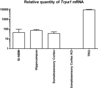
To elucidate whetherTrpa1is expressed in brain regions relevant to this work, we performed RT-qPCR in micropunched areas obtained from TRPA1+/+or TRPA1−/−animals. Trpa1 expression became sig- nificant at 35–36 cycles.Trpa1mRNA was detectable in SI-NBM, hip- pocampus, somatosensory cortex and trigeminal ganglion isolated from TRPA1+/+mice. The Trpa1 expression level of the TRPA1+/+samples was compared to their TRPA1-/-counterparts (Fig. 2).
3.2. Aβ1–42-induced cholinergic cell body andfibre loss is significantly attenuated in TRPA1−/−animals
Cholinergicfibre loss and cholinergic cell loss were detected in the ipsilateral somatosensory cortex (layer IV-V) and SI-NBM, in WT mice after Aβ1–42injection into SI-NBM. The cholinergicfibre loss labelled with AChE immunohistochemistry was 30.56 ± 1.39 % in TRPA1+/+
Fig. 1.Chronologicalflowchart for immunohistochemistry (A) and behavioural studies (B).
mice, while it was only 1.55 ± 0.91 % in TRPA1−/− animals. The ChAT-immuno-reactive cell loss in the SI-NBM was 28.68 ± 2.85 % in WT mice, and it was 2.19 ± 1.70 % in the TRPA1−/−mice (Figs. 3A and B).
3.3. The effect of Aβ1–42injection on cognitive functions is attenuated in TRPA1−/−mice
In the NOR test Aβ1–42injection had no effect on the locomotion of mice during habituation (Fig. 4B). There was no side preference during the exploration phase of the test in all studied groups (Fig. 4C). How- ever, during the test phase the discrimination ability was significantly attenuated in Aβ1–42–injected TRPA1+/+ mice (-0.02 ± 0.11) com- pared to the naïve controls (0.43 ± 0.08, p = 0.015703). This differ- ence was not observed between the Aβ1–42-treated and control
TRPA1−/− groups (DI: 0.36 ± 0.07 and 0.47 ± 0.14; respectively) showing intact discrimination abilities. This resulted in a significant difference between Aβ1–42treated TRPA1+/+and TRPA1−/−mice (p
= 0.037785). Vehicle injection had no significant effect (TRPA1+/+: 0.20 ± 0.11 and TRPA1−/−: 0.42 ± 0.09) (Fig. 4D).
In the RAM test animals showed learning ability during the ex- perimental days except for Aβ1–42injected TRPA1+/+mice (Fig. 5A).
There was no change in reference memory error of Aβ1–42 injected TRPA1+/+mice over the time (4.0 ± 0.63 on day 2 and 4.0 ± 0.26 on day 4), while in the vehicle-treated group there was a decrease (4.33 ± 1.76 on day 2 and 2.50 ± 0.86 on day 4). The same tendency could be detected in the TRPA1−/− groups (Aβ1–42-injected group:
4.0 ± 2.0 on day 2 and 2.6 ± 0.49 on day 4; vehicle-treated group:
3.75 ± 0.48 on day 2 and 2.4 ± 0.51 on day 4). This means, that Aβ1–42-injected TRPA1−/−animals made significantly less errors (p = 0.019726) on day 4 compared to Aβ1–42-treated TRPA1+/+ mice (Fig. 5B). There was no significant difference between the two naïve groups and there was no detectable change in learning ability over time in these groups (TRPA1+/+: 2.67 ± 0.33 and TRPA1−/−: 1.4 ± 0.24 on the day 4).
In the Y-maze test there was no difference in locomotion between groups reflected by the total number of arm entries. Examining the spontaneous alterations, each group showed intact working memory (naïve TRPA1+/+: 15.71 ± 1.91 and 68.42 % ± 2.32 and TRPA1−/−: 17 ± 1.19 and 66.31 % ± 3.61, Aβ1–42-treated TRPA1+/+: 16.4 ± 1.69 and 67.8 % ± 2.79; TRPA1−/−: 15.6 ± 2.4 and 58.92 % ± 4.73) (Figs. 5C and D).
4. Discussion
Our study provides thefirst evidence for the putative role of TRPA1 receptor in neuronal loss and cognitive impairment in Aβ1–42-induced murine neurotoxicity model. With the help of neuroanatomical eva- luation andin vivofunctional studies we have demonstrated that ge- netic deletion of TRPA1 receptor attenuates Aβ1–42 toxicity in choli- nergic neurons in the SI-NBM.
There are several dementia models available: spontaneous, chemi- cally induced (e.g. Aβ, scopolamine, okadaic acid) and transgenic (e.g.
APP/PS1 knockout mice, tau-related) models (Tayebati, 2006; Neha Sodhi et al., 2014). None of them is able to show all pathophysiological features of the human AD. Transgenic models are widely used, because non-invasive procedures are needed and intracellular Aβ-toxicity can Fig. 2. Relative quantity ofTrpa1mRNA in the brain and the trigeminal
ganglion.The expression level ofTrpa1mRNA was determined in the following brain regions: SI-NBM, hippocampus, somatosensory cortex of TRPA1+/+and TRPA1−/−mice along with trigeminal ganglion by RT qPCR. Samples collected from the somatosensory cortex of TRPA1−/−animals served as negative tissue control.
Fig. 3. Morphological changes in the brain of TRPA1+/+ and TRPA1−/− mice after Aβ1-42injection.Representative pictures (A) and numerical values (B) show the cholinergic fibre loss in the ipsilateral somatosensory cortex (layer IV-V) detected with acet- ylcholinesterase histochemistry. Scale bar: 200 μm, insert scale bar: 20 μm. Representative pictures (C) and numerical values (D) show the cholinergic cell body loss in the SI-NBM de- tected with choline acetyltransferase im- munohistochemistry. Scale bar: 1000 μm, in- sert scale bar: 250μm. Data were presented as the mean ± SEM (n = 6-10) and were ana- lysed with Mann-Whitney test (*** p < 0.001).
also be triggered (LaFerla and Green, 2012). However, in gene-ma- nipulated animals, the massive loss of neurons andfibres, as one of the most typical changes of human disease is not characteristic. In contrast to the genetic models of AD (e.g. APP/PS1 knockout mice) (Lee et al., 2016), our experimental approach with intracerebral injection of Aβ acutely generate AD-like neurotoxicity and cognitive deficits. Early recognition of the AD is almost impossible because of the atypical symptoms of the slow progression. The current treatment focuses on the maintenance of the remaining cholinergic functions applying choli- nesterase inhibitors and NMDA receptor antagonists (Talesa, 2001;
Eleti, 2016). Despite of some moderately effective drugs, the pharma- cotherapy of the AD is still an unsolved problem. Therefore, useful animal models that properly mimic the pathophysiological character- istics of AD are still essential for the discovery of novel therapeutic targets. Aβmicroinjection elicits significant loss of neuronal elements with remarkable memory impairment that can be detected with clas- sical behavioural tests (Morris water maze, Y/T-maze, NOR). This model represents the later phase of the disease when patients’relatives realize the memory problems.
TRPA1 is a widely studied non-selective cation channel, expressed by the primary afferent neurons regulating pain sensation (Nishida et al., 2015;Jardín et al., 2017). In addition, increasing evidence has been published about its function in the CNS: TRPA1 can be found in neurons of the rodent hippocampus, magnocellular neurosecretory cells, the visceral afferent pathway and cortex (Nilius et al., 2012;
Kheradpezhouh et al., 2017). Also its expression has been investigated in glial cells and its putative role in the pathophysiology of AD (Lee et al., 2016, Shigetomi et al., 2013). Since it is well-known, that TRPA1 is a key regulating receptor of calcium homeostasis, it may influence cell death or survival (Bosson et al., 2017, Stueber et al., 2017).
Aβ1–42 stereotaxic microinjection caused robust damage of choli- nergic neurons andfibres in the WT animals, which indicates that this method is suitable model of the typical pathomorphological changes of cholinergic neurotoxicity. Interestingly, in TRPA1−/− animals the Aβ1–42-induced cell andfibre loss of cholinergic neurons were nearly absent. These results suggest pivotal role of TRPA1 in the neurode- generative processes of the brain, especially in the basal forebrain cholinergic neurons. Morphological changes evoked by Aβ injection Fig. 4. Effects of vehicle or Aβ1-42injection on the performance in the novel object re- cognition (NOR) task in TRPA1+/+ and TRPA1−/−mice.On day 1 of the 3-day-long NOR test (A) distance moved (B), on day 2 location preference (C) was measured. On day 3 memory function was determined as dis- crimination index (D). Data are presented as the mean ± SEM (n = 6-10) and were ana- lysed by factorial ANOVA followed by Bonferroni’s post hoc test and location pre- ference with one samplet-test in comparison to 50.
Fig. 5. Effects of vehicle and Aβ1-42injec- tion on the performance in the radial arm maze (RAM) and Y-maze task in TRPA1+/+
and TRPA1−/−mice.Learning curve on the basis of reference memory error (A) and the reference memory error on day 4 (B) was de- termined in RAM. Spontaneous alternation (C) and average entries (D) was measured in Y- maze test. Data are presented as the mean ± SEM (n = 6-10). Learning curve was analysed by repeated measures two-way ANOVA followed by Bonferroni’s post hoc test, and reference memory error and average en- tries were analysed by factorial ANOVA fol- lowed by Bonferroni’s post hoc test (*p < 0.05).
have also been supported by functional investigations. Bilateral ad- ministration of Aβ1–42induced significantly impaired memory function in TRPA1+/+mice. These changes could be detected in both NOR and RAM, but Y-maze was not suitable for the demonstration this type of cognitive impairment. NOR test is one of the most appropriate methods assessing memory loss in rodents. It has been used in numerous dif- ferent (transgenic and non-transgenic) AD models. It has several ad- vantages, such as short testing period, no food restriction is needed, and it is based on the natural exploratory behaviour of the mice (Zhang et al., 2012;Grayson et al., 2015). RAM is also frequently used in AD mouse models (Webster et al., 2014), and it is a suitable for evaluation of both short- and long-term memory (Puzzo et al., 2014). Despite of the Aβtreatment, the memory performance of the TRPA1−/−animals was comparable with the naïve mice in these tests. This means, that the measured morphological changes are manifested at the behavioural level. These results verify the observations of the former transgenic model, that genetic lack of TRPA1 can significantly attenuate the memory loss (Lee et al., 2016), nevertheless not all of the tests showed the same results. Our Y-maze methodology was slightly different from the data of the above mentioned research group. Lee et al. divided Y- maze into start, familiar and novel arms, and they measured the visiting activity of mice in the novel arm, which is very similar to the aspect of NOR. On the contrary, we considered the arms as equal, and the al- ternation activity was assessed during the experiments. In this case we gain information about the spatial memory.
Our findings showed that genetic deletion of TRPA1 receptors substantially inhibited the Aβ1–42-induced cholinergic cell body loss andfibre loss. We demonstrated that Aβ1–42injection into the SI-NBM caused significantly reduced neuronal damage and memory loss in the TRPA1 KO animals. The exact molecular mechanism, how TRPA1 re- ceptors contribute to the Aβ1–42-induced cholinergic cell loss is still not fully elucidated. Previously, Harkany and colleagues published that Aβ1–42induces cholinergic cell death via NMDA receptor mediated in- tracellular Ca2+increase in SI-NBM (Harkány et al., 2000). We spec- ulate that TRPA1 receptor-mediated calcium influx may enhance the neurotoxic effect of the Aβ1–42due to the cytotoxic Ca2+overload in cholinergic neurons in SI-NBM. This theory warrants further in- vestigation.
5. Conclusion
Our study has presented thefirst evidence that the genetic lack of TRPA1 receptor significantly attenuates the Aβ1–42-induced cholinergic neurotoxicity in the SI-NBM. We also demonstrated that TRPA1 KO animals show substantially reduced memory loss after Aβ1–42 lesion compared to the wild-type counterparts. Based on our findings, in- hibition of the TRPA1 receptors might be a novel promising prospect in the pharmacotherapy of AD. The limitation of our study is, that its main conclusions are based exclusively on morphological and functional data provided by experiments on TRPA1 wildtype and KO mice.
Ethical approval and consent to participate
All experimental procedures were performed according to the 1998/
XXVIII Act of the Hungarian Parliament on Animal Protection, Consideration Decree of Scientific Procedures of Animal Experiments (243/1988), Hungarian regulations (40/2013, II.14.) and Directive 2010/63/EU of the European Parliament. The studies were approved by the Ethics Committee on Animal Research of University of Pécs ac- cording to the Ethical Codex of Animal Experiments and licence was given (licence no.: BA 02/2000−24/2016; BA 02/2000−38/2016).
Availability of data and material
The datasets used and/or analysed during the current study are available from the corresponding author on reasonable request.
Author’s contribution
All authors were involved in drafting the article or revising it cri- tically for important intellectual content, and all authors read, revised and approved thefinal version of the submitted manuscript. EP had full access to all of the data in the study and take responsibility for the integrity of the data and the accuracy of the data analysis. Study con- ception and design: MP, ÉB, EP. Acquisition of data: MP, ÉB, SG, DE, ÁK, EK, ÉSZ, JK. Analysis and interpretation of data: MP, ÉB, SG, ÁK, EK, JK, ÉSZ, EP.
Funding
The research was supported by ÚNKP-17−3-VI New National Excellence Program of the Ministry of Human Capacities, Gedeon Richter Talentum Foundation, Gedeon Richter Research Grant RG-IPI- 2016 TP10/042, Molecular Neuroendocrinology Research Group and ELTE-NAP Neuroimmunology Research Group (2017-1.2.1-NKP-2017- 00002); OTKA120391, The role of neuro-inflammation in neurode- generation: from molecules to clinics (EFOP-3.6.2−16-2017−00008);
Hungarian Brain Research Program 2. 2017−1.2.1-NKP- 2017−00002), 20765−3/2018 FEKUTSRAT, Janos Bolyai Scholarship of the Hungarian Academy of Sciences to ÉB.
Declaration of Competing Interest
The authors declare no conflict of interest.
Acknowledgements
The authors are grateful to Professor István Ábrahám for the in- troduction of the amyloidβ1-42peptide (Aβ1-42) microinjection-induced neurotoxicity model in our lab, Professor Zsuzsanna Helyes for the worthful help and useful advice relating the methodological improve- ment of the study. Dr. Dóra Zelena for her valuable contribution to the behavioural studies. Dr Klaudia Barabás for her help in the im- munohistochemical methods and Ildikó Udvarácz for the professional technical assistance.
References
Beninger, R.J., Wirsching, B.A., Jhamandas, K.B.R., 2001. Animal studies of brain acet- ylcholine and memory. Arch. Gerontol. Geriatr. 1, 71–89.
Bevins, R.A., Besheer, J., 2006. Object recognition in rats and mice: A one-trial non- matching-to-sample learning task to study‘recognition memory’. Nat. Protoc. 1, 1306–1311.
Bölcskei, K., Kriszta, G., Sághy, É, Payrits, M., Sipos, É., Vranesics, A., Berente, Z., Ábrahám, H., Ács, P., Komoly, S., Pintér, E., 2018. Behavioural alterations and morphological changes are attenuated by the lack of TRPA1 receptors in the cupri- zone-induced demyelination model in mice. J. Neuroimmunol. 320, 1–10.
Bosson, A., Paumier, A., Boisseau, S., Jacquier-Sarlin, M., Buisson, A., Albrieux, M., 2017.
TRPA1 channels promote astrocytic Ca2+ hyperactivity and synaptic dysfunction mediated by oligomeric forms of amyloid-βpeptide. Mol. Neurodegener. 12 53.
Candore, G., Balistreri, C.R., Grimaldi, M.P., Listì, F., Vasto, S., Chiappelli, M., Licastro, F., Colonna-Romano, G., Lio, D., Caruso, C., 2007. Polymorphisms of pro-inflammatory genes and Alzheimer’s disease risk: a pharmacogenomic approach. Mech. Ageing Dev.
128, 67–75.
D’Souza, Y., Elharram, A., Soon-Shiong, R., Andrew, R.D., Bennett, B.M., 2015.
Characterization of Aldh2-/-mice as an age-related model of cognitive impairment and Alzheimer’s disease. Mol. Brain 8, 27.
Dalenogare, D.P., Theisen, M.C., Peres, D.S., Fialho, M.F.P., Lückemeyer, D.D., Antoniazzi, C.T.D., Kudsi, S.Q., Ferreira, M.A., Ritter, C.D.S., Ferreira, J., Oliveira, S.M., Trevisan, G., 2020. TRPA1 activation mediates nociception behaviors in a mouse model of relapsing-remitting experimental autoimmune encephalomyelitis.
Exp. Neurol. 328, 113241.
Doering, D.S., Matsudaira, P., 1996. Cysteine scanning mutagenesis at 40 of 76 positions in villin headpiece maps the F-actin binding site and structural features of the do- main. Biochemistry 35, 12677–12685.
Eleti, S., 2016. Drugs in Alzheimer’s disease Dementia: an overview of current pharma- cological management and future directions. Psychiatr. Danub. 28, 136–140.
Giovannelli, L., Casamenti, F., Scali, C., Bartolini, L., Pepeu, G., 1995. Differential effects of amyloid peptidesβ-(1-40) andβ-(25-35) injections into the rat nucleus basalis.
Neuroscience 66, 781–792.
Grayson, B., Leger, M., Piercy, C., Adamson, L., Harte, M., Neill, J.C., 2015. Assessment of disease-related cognitive impairments using the novel object recognition (NOR) task in rodents. Behav. Brain Res. 285, 176–193.
Hamilton, N.B., Kolodziejczyk, K., Kougioumtzidou, E., Attwell, D., 2016. Proton-gated Ca2+-permeable TRP channels damage myelin in conditions mimicking ischaemia.
Nature 529, 523.
Harkany, T., O’Mahony, S., Kelly, J.P., Soós, K., Törõ, I., Penke, B., Luiten, P.G., Nyakas, C., Gulya, K., Leonard, B.E., 1998.β-amyloid(Phe(SO3H)24)25-35 in rat nucleus basalis induces behavioral dysfunctions, impairs learning and memory and disrupts cortical cholinergic innervation. Behav. Brain Res. 90, 133–145.
Harkany, T., Abrahám, I., Timmerman, W., Laskay, G., Tóth, B., Sasvári, M., Kónya, C., Sebens, J.B., Korf, J., Nyakas, C., Zarándi, M., Soós, K., Penke, B., Luiten, P.G., 2000.
β-Amyloid neurotoxicity is mediated by a glutamate-triggered excitotoxic cascade in rat nucleus basalis. Eur. J. Neurosci. 12, 2735–2745.
Horváth, Á., Tékus, V., Boros, M., Pozsgai, G., Botz, B., Borbély, É., Szolcsányi, J., Pintér, E., Helyes, Z., 2016. Transient receptor potential ankyrin 1 (TRPA1) receptor is in- volved in chronic arthritis: in vivo study using TRPA1-deficient mice. Arthritis Res.
Ther. 18, 6.
Ikenoue, T., Lee, Y.H., Kardos, J., Saiki, M., Yagi, H., Kawata, Y., 2014. Goto Y.COld denaturation ofα-synuclein amyloidfibrils. . Angew. Chemie - Int. Ed. 53, 4502–4512.
Jardín, I., López, J.J., Diez, R., Sánchez-Collado, J., Cantonero, C., Albarrán, L., Woodard, G.E., Redondo, P.C., Salido, G.M., Smani, T., Rosado, J.A., 2017. TRPs in pain sen- sation. Front. Physiol. 8, 382.
Khalil, M., Alliger, K., Weidinger, C., Yerinde, C., Wirtz, S., Becker, C., Engel, M.A., 2018.
Functional role of transient receptor potential channels in immune cells and epithelia.
Front. Immunol. 9, 174.
Kheradpezhouh, E., Choy, J.M.C., Daria, V.R., Arabzadeh, E., 2017. TRPA1 expression and its functional activation in rodent cortex. Open Biol. 7, 160314.
Koszegi, Z., Szego, É.M., Cheong, R.Y., Tolod-Kemp, E., Ábrahám, I.M., 2011. Postlesion estradiol treatment increases cortical cholinergic innervations via estrogen receptor- αdependent nonclassical estrogen signaling in vivo. Endocrinology 152, 3471–3482.
Kwakowsky, A., Potapov, K., Kim, S., Peppercorn, K., Tate, W.P., Ábrahám, I.M., 2016.
Treatment of beta amyloid 1–42 (Aβ1–42)-induced basal forebrain cholinergic da- mage by a non-classical estrogen signaling activator in vivo. Sci. Rep. 6, 21101.
LaFerla, F.M., Green, K.N., 2012. Animal models of alzheimer’s. Cold Spring Harb.
Perspect. Med. 2, a006320.
Lee, K.I., Lee, H.T., Lin, H.C., Tsay, H.J., Tsai, F.C., Shyue, S.K., Lee, T.S., 2016. Role of transient receptor potential ankyrin 1 channels in Alzheimer’s disease. J.
Neuroinflammation 12, 92.
Li, Y.F., Cheng, Y.F., Huang, Y., Conti, M., Wilson, S.P., O’Donnell, J.M., Zhang, H.T., 2011. Phosphodiesterase-4D knock-out and RNA interference-mediated knock-down enhance memory and increase hippocampal neurogenesis via increased cAMP sig- naling. J. Neurosci. 31, 172–183.
Maccioni, R.B., Muñoz, J.P., Barbeito, L., 2001. The molecular bases of Alzheimer’s
disease and other neurodegenerative disorders. Arch. Med. Res. 32, 367–381.
Masters, C.L., Bateman, R., Blennow, K., Rowe, C.C., Sperling, R.A., 2015. Cummings J.L.
Alzheimer’s disease. Nat. Rev. Dis. Prim. 1, 15056.
Neha Sodhi, R.K., Jaggi, A.S., Singh, N., 2014. Animal models of dementia and cognitive dysfunction. Life Sci. 109, 733–786.
Nilius, B., Appendino, G., Owsianik, G., 2012. The transient receptor potential channel TRPA1: from gene to pathophysiology. Pflugers Arch. Eur. J. Physiol. 464, 425–458.
Nishida, M., Kuwahara, K., Kozai, D., Sakaguchi, R., Mori, Y., 2015. TRP Channels: Their Function and Potentiality as Drug Targets. Innov. Med. Basic Res. Dev. 195–218.
Palkovits, M., Graf, L., Hermann, I., Borvendeg, J., Acs, Z.S., Lang, T., 1978. Regional Distribution of Enkephalins, Endorphins and ACTH in the Central Nervous System of Rats Determined by Radioimmunoassay. Endorphins’78, Acad. Press, Budapest, pp.
187–195.
Paxinos, G., Franklin, K.B.J., 2001. Mouse Brain in Stereotaxic Coordinates. Academic Press.
Pires, P.W., Earley, S., 2018. Neuroprotective effects of TRPA1 channels in the cerebral endothelium following ischemic stroke. Elife. 7, e35316.
Puzzo, D., Lee, L., Palmeri, A., Calabrese, G., Arancio, O., 2014. Behavioral assays with mouse models of Alzheimer’s disease: practical considerations and guidelines.
Biochem. Pharmacol. 88, 450–467.
Ritter, C., Dalenogare, D.P., de Almeida, A.S., Pereira, V.L., Pereira, G.C., Fialho, M.F.P., Lückemeyer, D.D., Antoniazzi, C.T., Kudsi, S.Q., Ferreira, J., Oliveira, S.M., Trevisan, G., 2020. Nociception in a progressive multiple sclerosis model in mice is dependent on spinal TRPA1 channel activation. Mol. Neurobiol. 57, 2420–2435.
Sullivan, M.N., Gonzales, A.L., Pires, P.W., Bruhl, A., Leo, M.D., Li, W., Oulidi, A., Boop, F.A., Feng, Y., Jaggar, J.H., Welsh, D.G., Earley, S., 2015. Localized TRPA1 channel Ca2+ signals stimulated by reactive oxygen species promote cerebral artery dilation.
Sci. Signal. 8 ra2.
Talesa, V.N., 2001. Acetylcholinesterase in alzheimer’s disease. Mech. Ageing Dev. 122, 1961–1969.
Tayebati, S.K., 2006. Animal models of cognitive dysfunction. Mech. Ageing Dev. 127, 100–108.
Webster, S.J., Bachstetter, A.D., Nelson, P.T., Schmitt, F.A., Van Eldik, L.J., 2014. Using mice to model Alzheimer’s dementia: an overview of the clinical disease and the preclinical behavioral changes in 10 mouse models. Front. Genet. 5, 88.
Zaborszky, L., Pang, K., Somogyi, J., Nadasdy, Z., Kallo, I., 1999. The basal forebrain corticopetal system revisited. Ann. N. Y. Acad. Sci. 877, 339–367.
Zhang, R., Xue, G., Wang, S., Zhang, L., Shi, C., Xie, X., 2012. Novel object recognition as a facile behavior test for evaluating drug effects in AβPP/PS1 Alzheimer’s disease mouse model. J. Alzheimers Dis. 31, 801–812.
Zimova, L., Sinica, V., Kadkova, A., Vyklicka, L., Zima, V., Barvik, I., Vlachova, V., 2018.
Intracellular cavity of sensor domain controls allosteric gating of TRPA1 channel. Sci.
Signal. 11 eaan8621.
Zygmunt, P.M., Högesättt, E.D., 2014. TRPA1. Handb. Exp. Pharmacol. 222, 583–630.