1 Journal of Crohn's and Colitis, 2021, 1–6
https://doi.org/10.1093/ecco-jcc/jjab158 Advance Access publication September 8, 2021 Original Article
© The Author(s) 2021. Published by Oxford University Press on behalf of European Crohn’s and Colitis Organisation. All rights reserved.
For permissions, please email: journals.permissions@oup.com
Original Article
Orofacial Granulomatosis Associated with Crohn’s Disease: a Multicentre Case Series
Frank Phillips,
a,Bram Verstockt,
b,Malgorzata Sladek,
cNanne de Boer,
dKonstantinos Katsanos,
eKonstantinos Karmiris,
fAhmad Albshesh,
g,Carl Erikson,
h,Daniel Bergemalm,
hTamas Molnar,
iPierre Ellul
j; ECCO CONFER Investigators
aNIHR Nottingham Digestive Diseases Biomedical Research Centre, Nottingham University Hospitals, Nottingham, UK bUniversity Hospitals Leuven, Gastroenterology and Hepatology, KU Leuven, Chronic Diseases, Metabolism and Ageing, TARGID-IBD unit, Leuven, Belgium cDepartment of Pediatrics, Gastroenterology and Nutrition, Jagiellonian University Medical College, Krakow, Poland dAmsterdam UMC, Department of Gastroenterology and Hepatology, Amsterdam UMC, Vrije Universiteit Amsterdam, AGEM Research Institute, Amsterdam, The Netherlands eDivision of Gastroenterology, Department of Internal Medicine, Faculty of Medicine, University of Ioannina School of Health Sciences, Ioannina, Greece fDepartment of Gastroenterology, Venizeleio General Hospital, Heraklion, Greece
gDepartment of Gastroenterology, Sheba Medical Centre, Tel Hashomer, Sackler School of Medicine, Tel-Aviv University, Tel-Aviv, Israel hDepartment of Gastroenterology, Faculty of Medicine and Health, Örebro University, Örebro, Sweden iFirst Department of Medicine, University of Szeged, Szeged, Hungary jDepartment of Medicine, Division of Gastroenterology, Mater Dei hospital, Msida, Malta
Corresponding author: Dr Frank Phillips, MRCP, NIHR Nottingham Digestive Diseases Biomedical Research Centre, Nottingham University Hospitals, Nottingham, UK. Tel.: 0115 969 1169; fax: 0115 840 5812; email: frankmphilllips@hotmail.com
Abstract
Background: Orofacial granulomatosis [OFG] is a rare syndrome that may be associated with Crohn’s disease [CD]. We aimed to characterise this relationship and the management options in the biologic era.
Methods: This multicentre case series was supported by the European Crohn’s and Colitis Organisation [ECCO], and performed as part of the Collaborative Network of Exceptionally Rare case reports [CONFER] project. Clinical data were recorded in a standardised collection form.
Results: This report includes 28 patients with OFG associated with CD: 14 males (mean age of 32 years, ±12.4 standard deviation [SD]) and 14 females [40.3 years, ±21.0 SD]. Non-oral upper gastrointestinal tract involvement was seen in six cases and perianal disease in 11. The diagnosis of OFG was made before CD diagnosis in two patients, concurrently in eight, and after CD diagnosis in 18. The distribution of OFG involved the lips in 16 cases and buccal mucosa in 18. Pain was present in 25 cases, with impaired swallowing or speaking in six. Remission was achieved in 23 patients, notably with the use of anti-tumour necrosis factors [TNFs] in nine patients, vedolizumab in one, ustekinumab in one, and thalidomide in two. A further five cases were resistant to therapies including anti-TNFs.
Conclusions: OFG associated with CD may occur before, concurrently with, or after the diagnosis of CD. Perianal and upper gastrointestinal [UGI] disease are common associations and there is a significant symptom burden in many. Remission can be obtained with a variety of immunosuppressive treatments, including several biologics approved for CD.
Key Words: Orofacial granulomatosis; oral Crohn’s disease; inflammatory bowel disease; Crohn’s disease; ulcerative colitis
Downloaded from https://academic.oup.com/ecco-jcc/advance-article/doi/10.1093/ecco-jcc/jjab158/6366377 by 81728827 user on 15 December 2021
1. Introduction
Inflammatory bowel diseases [IBD], including ulcerative colitis and Crohn’s disease [CD], are characterised by chronic relapsing in- flammation of the intestines. They are commonly associated with extra-intestinal manifestations [EIMs], with approximately a third developing an EIM in their disease course.1 Although the oral cavity is a common extra-intestinal site of involvement in IBD, orofacial granulomatosis [OFG] is rare in CD.
OFG was first described by Wiesenfeld et al. in 1985 and is in- creasingly recognised.2 It is characterised by persistent or recurrent enlargement of the tissues of the orofacial area, secondary to an underlying non-caseating granulomatous inflammatory process. The most frequent sites of involvement are the lips, followed by oral ul- cers, hyperplastic gingivitis, and buccal mucosal swelling.3,4 It should be noted that biopsy specimens of characteristic lesions may not al- ways reveal granulomas on histology.5
OFG is considered by some to be a distinct entity, a condition without an identified systemic cause.6 Others, including this report, use it as a descriptive term to identify the collection of features due to a granulomatous process which may have an identifiable asso- ciated condition, such as CD or sarcoidosis.5 The relationship be- tween OFG and CD is unclear. Concurrent intestinal CD has been described in 20–50% of adult patients with OFG.7 However, many patients with OFG will have no intestinal disease, and others may have evidence of intestinal disease that is asymptomatic. Sanderson et al. found that endoscopic and histological intestinal abnormalities occurred in up to 60% of patients with OFG and without gastro- intestinal symptoms.8 Although there are obvious similarities in the orofacial manifestations and histopathology of the two conditions, the human leukocyte antigen [HLA] profiles of idiopathic OFG may be different from those in Crohn’s disease. A small study of 16 patients with idiopathic OFG showed possible consistent HLA genotypes; in particular, HLA-A2 was seen in 50% but carries a comparative risk of CD of only 1.25, whereas HLA-A11 was seen in 25% and has an inverse association with CD.9 More recently, an analysis of NOD-2 variants has shown association with OFG associ- ated with CD [OFG + CD] but not OFG alone.10 The inflammatory infiltrate also appears divergent, with patients with OFG only having a higher number of CD3-expressing T cells and CD11c-expressing dendritic cells in the connective tissues.11 Campbell et al. studied the clinical features between the two entities, and found that compared with OFG only, OFG + CD was more likely to have buccal sulcus in- volvement, oral ulceration, raised C-reactive protein, and abnormal full blood count.4 The clinical response to azathioprine [AZA] also appears to be different, with one study including 60 OFG patients treated with AZA showing response to AZA at 12 months of 23.6%
[n = 8/38] in OFG only cases versus 59.1% [n = 13/22] of cases of OFG + CD.12 In another study with 14 patients [seven with OFG only and seven with OFG + CD] treated with infliximab [IFX], the presence of intestinal CD did not predict response to IFX.13
There are few case series describing OFG + CD. This case series aims to further characterise this relationship and the management options in the biologic era. This is important, as OFG can be difficult to treat, with systemic steroids reported to provide remission in only half of patients, and carries a high psychological morbidity.6
2. Methods
2.1. Study designThe European Crohn’s and Colitis Organisation [ECCO] CONFER [COllaborative Network For Exceptionally Rare case reports]
projects are based on an initiative introduced by ECCO to support individual investigators in identifying, assembling, and reporting together rare IBD cases of clinical relevance, which are otherwise seldom reported. The core of CONFER methodology is selecting certain topics worthy of investigation out of case proposals sub- mitted by physicians involved with IBD. The Steering Committee makes an initial selection, identifying those cases with the highest scientific interest and proximity to the purpose of CONFER project.
A Feasibility Network, comprising 30–35 high-volume IBD centres around the globe, is asked to identify similar cases, and the final deci- sion is again taken by the Steering Committee based on the outcome of networking. This topic then becomes a CONFER project. ECCO supports dissemination of a call to identify similar cases encountered by IBD physicians worldwide using several tools: announcements in the ECCO Annual Congress and in national and international IBD meetings across Europe; emailing to all ECCO members; posting in ECCO website and ECCO eNews; flyers; and personal communi- cation between ECCO members. Physicians are then prompted to report their case[s] using a predetermined standardised case report form. The call for the present case series was entitled ‘Orofacial granulomatosis associated with Crohn’s disease’. No financial sup- port or input into the collection or analysis or publication of the data collected is provided by ECCO. ECCO and/or any of its staff mem- bers may not be held liable for any information published in good faith in the ECCO CONFER articles.
2.2. Patients and procedures
Patients with a diagnosis of OFG either before, concurrently with, or after IBD diagnosis were eligible for inclusion in this study. Diagnosis of OFG was made clinically by oral medicine or dermatology spe- cialists, with the support of histology in half of the cases. Diagnosis of Crohn’s disease was made by gastroenterology specialists. Data were collected using a case report form, which was divided into two main sections. Section 1 included demographics, Montreal classifica- tion, and previous medication. Section 2 included time of diagnosis of OFG, location and associated symptoms of OFG, and treatments and outcomes for the OFG. Data were collected and analysed an- onymously, and handled according to local regulations. Informed consent was obtained where obligatory.
2.3. Statistical analysis
All statistical analyses [frequencies, descriptive statistics] were done using a Microsoft® Excel® spreadsheet.
3. Results
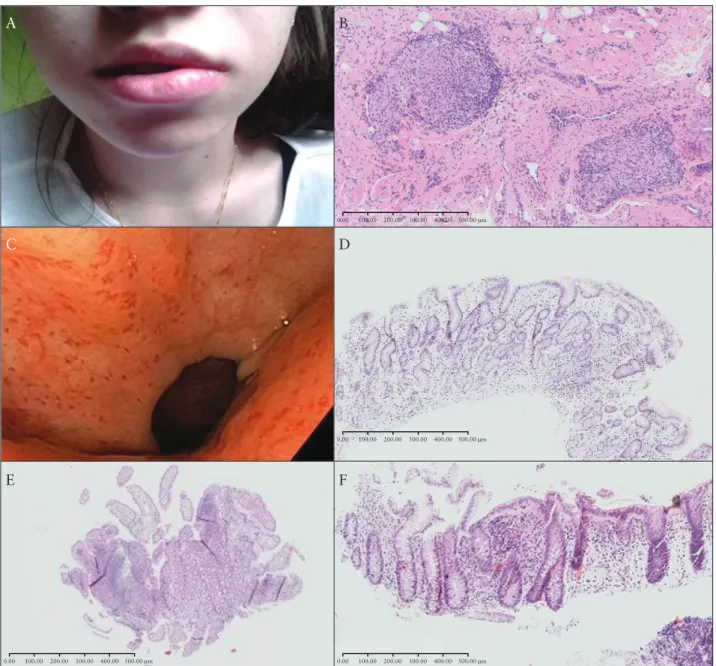
This report includes 28 patients with OFG + CD from nine dif- ferent centres; 14 males with mean age of 32 years (±12.4 standard deviation [SD], range 14–60) and 14 females with mean age of 40.3 years [±21.0 SD, range 11–79]. Baseline characteristics are de- scribed in Table 1, and Table 2 gives details of OFG manifestations and outcomes. Figure 1 includes images related to one of the patients in this series.
Only six cases had a smoking history, 19 had never smoked, and a further three were undocumented. Very few had systemic conditions [diabetes mellitus in one case, coeliac disease in two, Hermansky‐Pudlak syndrome in one] or a positive family history of IBD [five cases]. CD location was ileal in six cases, colonic in six, and ileocolonic in 15. Upper gastrointestinal tract [UGI] dis- ease occurred in six patients, with one of them having isolated UGI disease. Eleven patients had perianal disease. Eight had other EIMs
Downloaded from https://academic.oup.com/ecco-jcc/advance-article/doi/10.1093/ecco-jcc/jjab158/6366377 by 81728827 user on 15 December 2021
[six cases of peripheral arthropathy including two also having ery- thema nodosum, one of ankylosing spondylitis, and one of anterior uveitis]. CD phenotype was inflammatory in 16 cases, stricturing in seven, and penetrating in five. Eight patients had undergone intes- tinal resection [ileal resection in one case, ileocaecal resection in two, hemicolectomy in two, proctocolectomy in two, and anterior resec- tion in one].
Only half of the patients [14 patients] had a biopsy, and in two of these patients, granulomas were not present on histology. In the rest of the group, OFG was diagnosed clinically. OFG was diag- nosed prior to CD in two cases [12 and 24 months beforehand], concurrently [within 12 months] with CD in eight, and after CD diagnosis in 18 [mean of 11.4 years after, range 12‐396 months].
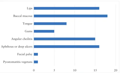
For the 26 patients with CD diagnosed before or concurrently with OFG, luminal activity was quiescent in 13 patients and active in 13, as judged by the treating physician. C-reactive protein [CRP] was elevated in 14 cases, although 10 of these also had active luminal disease, and 10 cases had a normal CRP. Ten patients were treated with an anti-tumour necrosis factor [TNF] agent [with or without immunomodulator combination] and five with azathioprine mono- therapy before or at diagnosis of OFG. Figure 2 shows the distri- bution of OFG in the patients in this series. In addition, pain was present in 25 cases, with impaired swallowing or speaking in six.
A number of different treatments led to remission, based on clin- ical assessment. The most common of these were therapies that in- cluded an anti-TNF alpha [infliximab and adalimumab] [n = 9]; of these, six patients started anti-TNF alpha specifically to treat OFG and three started due to luminal disease. Vedolizumab [VDZ, n = 1], ustekinumab [UST, n = 1], and thalidomide [n = 2] were also used as successful rescue therapies, and were all started specifically to treat OFG. In particular, one patient initially responded to ciclosporin, but this had to be stopped due to side effects; remission was restored and maintained with thalidomide for at least 11 years. Another patient had no response to infliximab [IFX] but gained remission with thal- idomide, although this subsequently had to stop due to side effects.
Another patient achieved remission with adalimumab but had to discontinue due to side effects; this resulted in treatment with VDZ, which restored and maintained remission over a 5-year follow-up.
Finally, another patient lost response to IFX, exhibited primary non- response to VDZ, and achieved remission with UST. Recurrence occurred in five patients [including those described above]: after anti-TNF stopped in three cases; and mild breakthrough symptoms on anti-TNF in two cases. Remission was also achieved with topical- only therapies in four patients [defaztan gel in one case, topical sodium tetraborate in two, hydrocortisone lozenges and antiseptic mouthwashes in one], local corticosteroid injections [triamcinolone]
in two, and exclusive enteral nutrition [EEN] in one.
A further five cases were resistant to anti-TNF based therapies [three of whom were already on anti-TNF at the onset of OFG], assessed by unchanged OFG signs and symptoms. Of these, CD distribution was ileocolonic in three cases and colonic in two;
perianal disease was present in two cases and none of these had an- other EIM. All five cases had quiescent luminal disease. Three of Table 1. Demographic and disease characteristics of patients at
baseline.
N %
Demographics Males [mean age 32 yr] 14 50.0 Females [mean age 40.3 yr] 14 50.0 Comorbidities Systemic comorbidities 4 14.3
Family history 5 17.9
Smoking history Current or former 6 21.4
Never 19 67.9
Unknown 3 10.7
Crohn’s distribution Ileocolonic 15 53.6
Ileal 6 21.4
Colonic isolated 6 21.4
UGI involvement 6 21.4
Perianal involvement 11 39.3
Crohn’s phenotype Inflammatory 16 57.1
Stricturing 7 25.0
Penetrating 5 17.9
Extraintestinal EIMs 8 28.6
manifestations No EIMs 20 71.4
UGI, upper gastrointestinal; EIM, extraintestinal manifestation; yr, years..
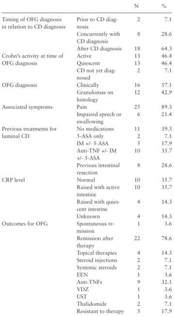
Table 2. Characteristics of orofacial granulomatosis.
N %
Timing of OFG diagnosis in relation to CD diagnosis
Prior to CD diag- nosis
2 7.1
Concurrently with CD diagnosis
8 28.6
After CD diagnosis 18 64.3 Crohn’s activity at time of
OFG diagnosis
Active 13 46.4
Quiescent 13 46.4
CD not yet diag- nosed
2 7.1
OFG diagnosis Clinically 16 57.1
Granulomas on histology
12 42.9
Associated symptoms Pain 25 89.3
Impaired speech or swallowing
6 21.4
Previous treatments for luminal CD
No medications 11 39.3
5-ASA only 2 7.1
IM +/- 5-ASA 5 17.9
Anti-TNF +/- IM +/- 5-ASA
10 35.7
Previous intestinal resection
8 28.6
CRP level Normal 10 35.7
Raised with active intestine
10 35.7
Raised with quies- cent intestine
4 14.3
Unknown 4 14.3
Outcomes for OFG Spontaneous re- mission
1 3.6
Remission after therapy
22 78.6
Topical therapies 4 14.3
Steroid injections 2 7.1
Systemic steroids 2 7.1
EEN 1 3.6
Anti-TNFs 9 32.1
VDZ 1 3.6
UST 1 3.6
Thalidomide 2 7.1
Resistant to therapy 5 17.9 OFG, orofacial granulomatosis; CD, Crohn’s disease; 5-ASA, 5-aminosalicylic acid; IM, immunomodulator; anti-TNF, anti-tumour ne- crosis factor; EEN, exclusive enteral nutrition; VDZ, vedolizumab; UST, ustekinumab; CRP, C-reactive protein.
Downloaded from https://academic.oup.com/ecco-jcc/advance-article/doi/10.1093/ecco-jcc/jjab158/6366377 by 81728827 user on 15 December 2021
these cases were followed up for ≥5 years and the other two were managed for 6 months and then discharged back to the referring physician. A single case had spontaneous remission without therapy [Table 2]. Follow-up occurred for an average of 5.32 years [range 5–180 months].
A sub-analysis of the 12 patients who had histologically proven granulomatous disease included seven females and five males. with a mean age of 23.2 years [SD 11.9, range 13–60]. OFG was diag- nosed prior to CD in two cases, concurrently in five, and after CD diagnosis in five. CD distribution was ileal in two, colonic in two, ileocolonic in seven, and isolated UGI in one. Perianal disease oc- curred in five, UGI disease in two, and EIMs other than OFG in four. OFG distribution involved the lips in eight. Two cases were
refractory to anti-TNFs, and 10 achieved remission [with steroids in three, EEN and AZA in one, infliximab in five, ustekinumab in 1].
4. Discussion
This case series describes the clinical features and course of patients with CD + OFG. By the nature of this study, all those included had a diagnosis of CD, but it is important to note that OFG without CD is much more common: Campbell et al. looked at 207 patients with OFG, and 46 [22%] had intestinal CD.4 In similarity to other series, there is no gender predilection. The average age here is higher than previously reported, although five patients were included aged 16 years or younger, compared with approximately a third of those
A B
C D
F
0.00 100.00 200.00 300.00 400.00 500.00 μm
0.00 100.00 200.00 300.00 400.00 500.00 μm
0.00 100.00 200.00 300.00 400.00 500.00 μm
E
0.00 100.00 200.00 300.00 400.00 500.00 μm
Figure 1. Images from a 20 year-old female, diagnosed initially with OFG age 15 years, then pan-enteric Crohn’s 7 months later. Remission was achieved with mesalamine, azathioprine, and exclusive enteral nutrition. A] Cheilitis granulomatosa. B]. H&E staining [10x magnification] of lower lip biopsy showing infiltration of lymphocytes, plasmocytes, and non-caseating granuloma. C] Gastroscopy images showing patchy erythema in the gastric body and antrum. D]
H&E staining [10x magnification] of pyloric antrum biopsy showing focal, chronic lymphocytic infiltration. E] H&E staining [2x magnification] of terminal ileum biopsy showing mild infiltration of lymphocytes and plasmocytes. F] H&E staining [10x magnification] of descending colon biopsy showing focal lymphocytosis, basal plasmocytosis, and crypt distortion. Courtesy of Malgorzata Sladek. OFG, orofacial granulomatosis; H&E, haematoxylin and eosin.
Downloaded from https://academic.oup.com/ecco-jcc/advance-article/doi/10.1093/ecco-jcc/jjab158/6366377 by 81728827 user on 15 December 2021
with OFG in Campbell et al.’s study.4 Smoking is not associated, as most patients had never smoked. Few cases reported other EIMs, and the presence of EIMs was not associated with a treatment- refractory disease course. There does appear to be a more severe intestinal disease, with high numbers with stricturing or penetrating disease and eight cases with previous intestinal resection. There was no predilection for OFG to occur with different locations of intes- tinal Crohn’s, with a fair spread between small bowel, ileocolonic, and colonic disease. However, there was an over-representation of both UGI and perianal CD. Dupuy et al. have previously described the higher prevalence of UGI involvement in patients presenting with oral manifestations of CD.14 One study identified UGI involvement in 72 of 940 CD patients [7.6%],15 whereas our series identified six of 28 patients [21.4%]. Harty et al. had noted a higher incidence of concomitant perianal CD in those patients also presenting with oral disease.16 However, a limiting factor is that the numbers are small in these studies.
In this series, the diagnosis of OFG occurred in 64.3% at 12 months to 33 years after the diagnosis of CD, whereas there were also eight cases of OFG diagnosed within a year of CD [28.6%]
and two cases of OFG diagnosed prior to CD. This contrasts with Campbell et al.’s study where 42.5% [17/40] patients with OFG + CD developed OFG prior to CD [median 2 years, range 1–39], 20 devel- oped OFG after CD [median 10 years, range 1–45], and three had OFG and CD diagnosed concurrently in the same year.4
Similar to previous reports, the patients in our series usually had multiple areas of the orofacial area affected, mostly involving the lips and buccal mucosa, as well as the tongue and gums commonly.
Angular cheilitis and aphthous or deep ulcers were very common ac- companying features, and other common associated symptoms were pain and impaired swallowing or speaking. This shows how disabling OFG can be and the need to address these problems separately. Only half the patients in our series had biopsy of their OFG lesion, with two out of these 10 having no granulomas detected. This may represent sampling error, as granulomas are often located deep within the oral mucosa, meaning shallow specimens can give false-negative results.17
The management of OFG can often be difficult, which is re- flected in the 25% of patients resistant to anti-TNF based thera- peutic regimens. Most patients require medical intervention, which can range from local treatments such as intralesional corticosteroid
injections,18 systemic treatments for CD such as systemic cortico- steroids and anti-TNF biologics,19 to rarer medications like the anti- leprosy drug clofazimine and thalidomide.20 Elimination diets such as the cinnamon and benzoate-free diets have been successful in some patients.21,22 In this series, a variety of different treatment op- tions led to remission. Notably, anti-TNF biologics led to remission in 32.1% of cases and there were also cases of both vedolizumab and ustekinumab [UST] leading to OFG remission. This appears to be the first reported case of VDZ leading to remission, and adds to the two recently published cases of UST inducing remission in OFG + CD.23,24 This shows the importance of targeting the underlying intestinal CD in treating OFG. Interestingly, UST has also recently been shown to be useful for another extraintestinal granulomatous condition, meta- static CD [MCD]: nine cases of MCD were treated with UST, and all responded with the first or second dose, with five achieving remis- sion in a median time of 5 months.25 This may also have implications for the treatments of idiopathic OFG, particularly when therapeutic options appear to be exhausted.
The limitations of this study are that reporting is retrospective and histology was not available in all cases, although it is common for such diagnoses and assessment of response or remission to be made on clinical grounds. Patients included are from a select cohort, so caution must be exercised in comparing features with larger un- selected cohorts in other studies. Furthermore, given the nature of a multicentre study of a rare condition, there was no standardisation of diagnosis or management.
To conclude, in this multicentre case series of selected patients with OFG + CD, the diagnosis of OFG mostly occurred after that of CD and can be many decades after, but may also occur before or concurrently with diagnosis of intestinal disease. Perianal disease and UGI disease were common associations, and there was a sig- nificant symptom burden due to pain and impaired swallowing in many. Lastly, remission was obtained with a variety of treatments, including topical and intralesional agents, thalidomide, and different biologic agents, including VDZ and UST, although many remained resistant to multiple treatments.
Funding
No specific funding has been received for this project.
0 5 10 15 20
Pyostomatitis vegetans Facial palsy Aphthous or deep ulcers Angular cheilitis Gums Tongue Buccal mucosa Lips
Figure 2: Bar chart showing number of cases with different areas of involvement by OFG. Note that there were 10 cases which had both lips and oral cavity affected. OFG, orofacial granulomatosis.
Downloaded from https://academic.oup.com/ecco-jcc/advance-article/doi/10.1093/ecco-jcc/jjab158/6366377 by 81728827 user on 15 December 2021
Author Contributions
FP: principal investigator for the study, conceived the study idea, contributed one case and prepared the manuscript. BV: contributed 12 cases. MS: con- tributed six cases. NdB: contributed two cases. KKat: contributed two cases.
KKar: contributed one case. AA: contributed one case. CD: contributed one case. DM: contributed one case. TM: contributed one case. PE: case manager for the study and supervised the project. All authors reviewed the manuscript and approved the final version. Presented at the 15th Congress of ECCO, February 2020, Vienna, Austria.
Conflict of Interest
BV: financial support for research from Pfizer; lecture fees from Abbvie, Ferring, Takeda Pharmaceuticals, Janssen, and R Biopharm; consultancy fees from Janssen and Sandoz. MS: received grant support/lecture fee/advisory board payment from AbbVie, Biogen, Egis, Ferring, Nestle, Nutricia. NdB:
speaker for AbbVie and MSD; consultant and/or principal investigator for TEVA Pharma BV and Takeda; research grant from Dr Falk, TEVA Pharma BV, Takeda, and MLDS. KKat: honoraria for consulting services [educa- tional services, scientific articles, participation in advisory boards, clinical trials, lecture fees, other] from AbbVie, Amgen, Enorasis, Epsilon Health, Falk, Ferring, Genesis, Grifols S.A., Janssen, MSD, Mylan, Shire, Takeda, Vianex. CE: received grant support/lecture fee/advisory board payment from Takeda, Janssen Cilag, Pfizer. DB: personal fees from Ferring, Pfizer, Takeda, Janssen. TM: speaker’s honoraria from MSD, AbbVie, Egis, Goodwill Pharma, Takeda, Pfizer, Richter, Fresenius Kabi, and Teva. PE: educational grants from Abbvie and MSD. All conflicts of interest stated here are outside the submitted work.
References
1. Vavricka SR, Brun L, Ballabeni P, et al. Frequency and risk factors for extraintestinal manifestations in the Swiss inflammatory bowel disease co- hort. Am J Gastroenterol 2011;106:110–9.
2. Wiesenfeld D, Ferguson MM, Mitchell DN, et al. Oro-facial granuloma- tosis – a clinical and pathological analysis. Q J Med 1985;54:101–13.
3. Leão JC, Hodgson T, Scully C, Porter S. Review article: orofacial granu- lomatosis. Aliment Pharmacol Ther 2004;20:1019–27.
4. Campbell H, Escudier M, Patel P, et al. Distinguishing orofacial granu- lomatosis from Crohn’s disease: two separate disease entities? Inflamm Bowel Dis 2011;17:2109–15.
5. Marcoval J, Penín RM. Histopathological features of orofacial granu- lomatosis. Am J Dermatopathol 2016;38:194–200.
6. Grave B, McCullough M, Wiesenfeld D. Orofacial granulomatosis – a 20-year review. Oral Dis 2009;15:46–51.
7. Zbar AP, Ben-Horin S, Beer-Gabel M, Eliakim R. Oral Crohn’s disease: is it a separable disease from orofacial granulomatosis? A review. J Crohns Colitis 2012;6:135–42.
8. Sanderson J, Nunes C, Escudier M, et al. Oro-facial granulomatosis:
Crohn’s disease or a new inflammatory bowel disease? Inflamm Bowel Dis 2005;11:840–6.
9. Gibson J, Wray D. Human leucocyte antigen typing in orofacial granu- lomatosis. Br J Dermatol 2000;143:1119–21.
10. Mentzer A, Nayee S, Omar Y, et al. Genetic association analysis reveals differences in the contribution of NOD2 variants to the clinical pheno- types of orofacial granulomatosis. Inflamm Bowel Dis 2016;22:1552–8.
11. Gale G, Ostman S, Saalman R, Telemo E, Jontell M, Hasséus B.
Immunophenotype in orofacial granulomatosis with and without Crohn’s disease. Med Oral Patol Oral Cir Bucal 2014;19:e584–91.
12. Mentzer A, Goel R, Elliott T, et al. Azathioprine is effective for oral in- volvement in Crohn’s disease but not for orofacial granulomatosis alone. J Oral Pathol Med 2016;45:312–8.
13. Elliott T, Campbell H, Escudier M, et al. Experience with anti-TNF-α therapy for orofacial granulomatosis. J Oral Pathol Med 2011;40:14–9.
14. Dupuy A, Cosnes J, Revuz J, Delchier JC, Gendre JP, Cosnes A. Oral Crohn disease: clinical characteristics and long-term follow-up of 9 cases.
Arch Dermatol 1999;135:439–42.
15. Wagtmans MJ, Verspaget HW, Lamers CB, van Hogezand RA. Clinical aspects of Crohn’s disease of the upper gastrointestinal tract: a comparison with distal Crohn’s disease. Am J Gastroenterol 1997;92:1467–71.
16. Harty S, Fleming P, Rowland M, et al. A prospective study of the oral mani- festations of Crohn’s disease. Clin Gastroenterol Hepatol 2005;3:886–91.
17. Hullah EA, Escudier MP. The mouth in inflammatory bowel disease and aspects of orofacial granulomatosis. Periodontol 2000 2019;80:61–76.
18. Mignogna MD, Fortuna G, Leuci S, Amato M, Massimo A. Oral Crohn’s disease: a favorable clinical response with delayed-release triamcinolone acetonide intralesional injections. Am J Gastroenterol 2008;103:2954–5.
19. Cardoso H, Nunes AC, Carneiro F, Tavarela Veloso F. Successful infliximab therapy for oral Crohn’s disease. Inflamm Bowel Dis 2006;12:337–8.
20. Hegarty A, Hodgson T, Porter S. Thalidomide for the treatment of recal- citrant oral Crohn’s disease and orofacial granulomatosis. Oral Surg Oral Med Oral Pathol Oral Radiol Endod 2003;95:576–85.
21. Wray D, Rees SR, Gibson J, Forsyth A. The role of allergy in oral mucosal diseases. QJM 2000;93:507–11.
22. White A, Nunes C, Escudier M, et al. Improvement in orofacial granu- lomatosis on a cinnamon- and benzoate-free diet. Inflamm Bowel Dis 2006;12:508–14.
23. Gilmore R, Li Wai Suen CFD, Elliott T, De Cruz P, Srinivasan A. Using ustekinumab to treat Crohn’s disease-related orofacial granulomatosis:
two birds, one stone. Inflamm Bowel Dis 2020;26:e79–80.
24. Taxonera C, Alba C, Colmenares M, Olivares D, Rey E. Recurrent granu- lomatous cheilitis associated with Crohn’s disease successfully treated with ustekinumab: case report and literature review. Therap Adv Gastroenterol 2020;13:1756284820934327.
25. Phillips FM, Verstockt B, Sebastian S, et al. Inflammatory cutaneous lesions in inflammatory bowel disease treated with vedolizumab or ustekinumab: an ECCO CONFER multicentre case series. J Crohns Colitis 2020;14:1488–93.
Downloaded from https://academic.oup.com/ecco-jcc/advance-article/doi/10.1093/ecco-jcc/jjab158/6366377 by 81728827 user on 15 December 2021