Nodal-Stage Classification in Invasive Lobular Breast Carcinoma: Influence of Different Interpretations of the pTNM Classification
Carolien H.M. van Deurzen, Gabor Cserni, Simonetta Bianchi, Vania Vezzosi, Riccardo Arisio, Jelle Wesseling, Martin Asslaber, Maria P. Foschini, Anna Sapino, Isabella Castellano, Grace Callagy, Daniel Faverly, Maria-Dolores Martin-Martinez, Cecily Quinn, Isabel Amendoeira, Janina Kulka, Angelika Reiner-Concin, Alicia Cordoba, Cornelis A. Seldenrijk, and Paul J. van Diest
From the Department of Pathology, Univer- sity Medical Center Utrecht, Cancer Center, Utrecht; Department of Pathology, St Antonius Hospital, Nieuwegein; and the Netherlands Cancer Institute, Amsterdam, the Netherlands; Department of Pathology, Ba´cs-Kiskun County Teaching Hospital, Kecskeme´t, Hungary; Department of Human Pathology and Oncology, University of Florence, Azienda Ospedaliero- Universitaria Careggi, Firenze; Department of Hematology and Oncology L. and A.
Seragnoli Section of Anatomic Pathology, Bellaria Hospital, University of Bologna, Bologna; and Department of Pathology, Sant’Anna Hospital; and Department of Biological Science and Human Oncology, University of Turin, Turin, Italy; Institute of Pathology, Medical University Graz, Graz;
and Institute of Pathology, Donauspital, Wien, Austria; Department of Pathology, NUI Galway, Clinical Science Institute, Galway; and Department of Pathology, St Vincent’s Hospital, Elm Park, Donnybrook, Dublin, Ireland; CMP Pathology, Brussels, Belgium; Instituto de Patologia e Imunolo- gia da Universidade do Porto and Hospital de S. Joa˜o, Rua Roberto Frias s/n, Porto, Portugal; Second Department of Pathology, Semmelweis University, Budapest, Hunga- ry; and Department of Pathologic Anatomy, Hospital de Navarra, Navarra, Spain.
Submitted February 1, 2009; accepted September 25, 2009; published online ahead of print at www.jco.org on January 19, 2010.
Authors’ disclosures of potential conflicts of interest and author contributions are found at the end of this article.
Corresponding author: Paul J. van Diest, MD, PhD, University Medical Center Utre- cht, PO Box 85500, 3508 GA Utrecht, the Netherlands; e-mail: p.j.vandiest@
umcutrecht.nl.
© 2010 by American Society of Clinical Oncology
0732-183X/10/2806-999/$20.00 DOI: 10.1200/JCO.2009.22.0723
A B S T R A C T
Purpose
Application of current nodal status classification is complicated in lobular breast carcinoma metastases. The aim of this study was to define the optimal interpretation of the pTNM classification in sentinel node (SN) –positive patients to select patients with limited or with a high risk of non-SN involvement.
Patients and Methods
SN metastases of 392 patients with lobular breast carcinoma were reclassified according to interpretations of the European Working Group for Breast Screening Pathology (EWGBSP) and guidelines by Turner et al, and the predictive power for non-SN involvement was assessed.
Results
Reclassification according to definitions of EWGBSP and Turner et al resulted in different pN classification in 73 patients (19%). The rate of non-SN involvement in the 40 patients with isolated tumor cells according to Turner et al and with micrometastases according to EWGBSP was 20%, which is comparable to the established rate for micrometastases. The rate of non-SN involvement in the 29 patients with micrometastases according to Turner et al and with macrometastases according to EWGBSP was 48%, which is comparable to the established rate for macrometasta- ses. Therefore, the EWGBSP method to classify SN tumor load better reflected the risk of non-SN involvement than the Turner et al system.
Conclusion
Compared with the guidelines by Turner et al, the EWGBSP definitions better reflect SN metastatic tumor load and allow better differentiation between patients with lobular breast carcinoma who have a limited or a high risk of non-SN metastases. Therefore, we suggest using the EWGBSP definitions in these patients to select high-risk patients who may benefit from additional local and/or systemic therapy.
J Clin Oncol 28:999-1004. © 2010 by American Society of Clinical Oncology
INTRODUCTION
The introduction of the sentinel node (SN) proce- dure in patients with breast cancer has led to an increased detection rate of small lymph node metas- tases1,2because of an intensive pathology protocol that includes step sectioning and immunohisto- chemistry.3This phenomenon is reflected by the current definitions of the International Union Against Cancer (UICC) Tumor Lymph Node Me- tastasis (TNM) Classification of Malignant Tumors4 and the sixth edition of the American Joint Commit- tee on Cancer (AJCC) Staging Manual,5 which distinguish between isolated tumor cells (ITCs), mi-
crometastases, and macrometastases on the basis of metastatic cluster size.
Although there is no consensus whether the distinction between ITCs and micrometastases has prognostic significance6-8it is important, as it directly affects decisions with regard to completion axillary lymph node dissection and adjuvant sys- temic therapy. Patients with SN ITC are gener- ally considered and treated as node negative (ie, pN0[i⫹]), whereas patients with micrometastases (ie, pN1mi) and macrometastases (pN1) are staged as node-positive. These patients commonly receive additional axillary treatment and, in certain circum- stances, adjuvant systemic therapy.
J
OURNAL OFC
LINICALO
NCOLOGY O R I G I N A L R E P O R TCurrently, the UICC and AJCC definitions of ITC, micrometas- tases, and macrometastases are imprecise; therefore, different inter- pretations of the definitions exists, which has resulted in suboptimal reproducibility of nodal staging.9-13Although both the UICC and AJCC systems use size of the largest metastatic cluster, the UICC also considers some qualitative features of metastatic deposits (ie, prolifer- ation and extravasation). Furthermore, there is no generally accepted definition for a cluster, which complicates size measurement in case of multiple clusters and/or cells. This is particularly important in patients with invasive lobular breast carcinoma, because these metastases fre- quently consider multiple, scattered, small clusters and/or single cells in both the sinus and the parenchyma.
Thus, new pathologic nodal staging criteria should be introduced to arrive at better reproducibility and, consequently, optimal thera- peutic decision making. The European Working Group for Breast Screening Pathology (EWGBSP) offered some refinements of the cur- rent nodal staging definitions,9which resulted in improved (although still suboptimal) reproducibility (, 0.49). Turner et al11recently re- ported alternate interpretations of the same definitions, which re- sulted in enhanced interobserver agreement (, 0.92), even in the classification of lobular carcinoma SN metastases. However, a poten- tial disadvantage of the refinements by Turner et al11is the classifica- tion of high SN tumor load dispersed pattern metastases as ITC, which might not optimally reflect the prediction of prognosis. This pattern is especially seen in patients with lobular breast carcinoma, which is the second most common histologic subtype of breast cancer. A compar- ative study on predictive value of the EWGBSP9and Turner et al11 classifications of SN tumor load in patients with a lobular carcinoma has not been conducted before. Although probably only long-term follow-up studies may reveal the most optimal nodal staging defini- tion, non-SN involvement can be regarded as a good surrogate prog- nostic indicator, which can be assessed instantaneously. The objective of this study, therefore, was to compare the rates of non-SN involve- ment associated with SN metastases (according to the classification by
the EWGBSP9 and Turner et al11) in patients with lobular breast carcinoma to select the most optimal method that differentiates be- tween patients with a limited risk and those patients with a high risk of non-SN involvement.
PATIENTS AND METHODS
Data Acquisition
Members of the EWGBSP that contributed to a previous multicenter study14were asked to participate in this study. This study group was updated and supplemented with patient cases from several other members of this working group, which resulted in an overlap of 10% and made this study independent from this previous study. All contributors were asked to extract data from their own archives and to reclassify the positive SN metastases of the tumors according to the interpretations defined by the EWGBSP9and Turner et al.11A detailed, step-wise protocol; a data-extraction form (via Microsoft Excel sheet); and digital training programs (via Microsoft PowerPoint) were distributed to all contributors. None of those patient cases evaluated in these training programs were included in this study. The two main differences between these two classifications are the definition of a cluster (which results in different diameters of metastases) and the role of the microanatomic location of tumor deposits. Details of these two interpretations are listed in Table 1.
The following criteria for study inclusion had to be fulfilled. First, pa- tients had to be diagnosed with any type of invasive lobular breast carcinoma.
Second, patients had to be diagnosed previously with a tumor-positive SN (ie, ITC, micrometastases, or macrometastases), followed by an axillary lymph node dissection (includingⱖsix lymph nodes, additionally denoted as non- SNs). Data extracted from the original reports included primary tumor fea- tures (ie, diameter, lymphovascular invasion, unifocality, or multifocality), the number of (tumor-positive) SNs, and the number of (tumor-positive) non- SNs. All contributors reclassified tumor-positive SNs according to both the EWGBSP9and Turner et al11classifications, and they were blinded to the status of the non-SN. The detection method of these metastases (ie, hematox- ylin and eosin [H&E] or immunohistochemistry) and the presence or absence of extranodal extension also was recorded. In occurrences of discordance between both classifications, the reason (either diameter or microanatomic location) was recorded to evaluate the effect of these features separately.
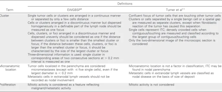
Table 1.Main Differences in the EWGBSP9and Turner et al11Definitions to Classify SN Metastases
Term
Definitions
EWGBSP9 Turner et al11
Cluster Single tumor cells or clusters are arranged in a continuous manner or separated by only a few cells distance
Cells or clusters arranged in a discontinuous manner but dispersed homogeneously in a definable part of the lymph node should be measured as one focus
Cells, clusters, or foci arranged in a discontinuous manner and dispersed unevenly should be considered as one if the distance between clusters or foci is smaller than the smallest cluster or focus; if the distance between these cells, clusters, or foci is larger than the smallest cluster or focus, it should be characterized by the size of the largest cluster or focus Three-dimensional information is used: single cluster in
corresponding areas of two consecutive sections at⬎0.2 mm interval is measured as one
Confluent focus of tumor cells that are touching other tumor cells Clusters or cells separated by a single benign cell or a spatial gap
are measured as separate clusters, except when fibroblastic reaction of the tumor has caused this separation
Single cells represent ITC; densely crowded cells when contiguous/touching are measured and classified according to the largest group of contiguous/touching cells
Only the two-dimensional image of the microscopic section is considered
Microanatomic location
Tumor cells localized in the parenchyma are considered micrometastases (except with ⱕfive single cells), even if the largest diameter is⬍0.2 mm
Metastatic cells in extranodal lymph vessels should not be recorded as nodal involvement
Microanatomic location is not a factor in classification; ITC may be found in nodal parenchyma
Metastatic cells in extranodal lymph vessels are classified as nodal disease on the basis of size of deposit
Proliferation Mitotic activity is considered as a feature reflecting malignant/metastatic activity
Mitotic activity is not considered
Abbreviations: EWGBSP, European Working Group for Breast Screening Pathology; ITC, isolated tumor cell.
SN Processing
The pathologic work-up of SNs was somewhat heterogeneous, but all laboratories used a multilevel assessment and performed immunohistochem- istry for patient cases with a negative SN on H&E staining, which was generally performed on multiple levels. Non-SNs were evaluated by a single to a few H&E–stained sections without the routine use of immunohistochemistry.
Statistical Analysis
The 95% CIs were calculated by use of the Rothman spreadsheet.15 Univariate analysis with the2test was used to correlate covariates with non-SN involvement. The Spearman correlation coefficient was used as a measure of correlation between the total number of SNs and the number of positive non-SNs. In those patient cases with more than one SN involved, the largest deposit was used for statistical analysis. Statistical analyses were per- formed by using SPSS for Windows (version 13.0; SPSS, Chicago, IL). Two- sidedPvalues less than .05 were considered significant.
RESULTS
Clinicopathologic Features
Overall, 392 patients with invasive lobular breast carcinoma and a positive SN were included in this study, and the median tumor size was 2.2 cm. The median numbers of SNs and non-SN assessed per patient were two (range, one to seven) and 15 (range, six to 50), respectively. The overall rate of non-SN involvement was 183 (47%;
95% CI, 41.8 to 51.6) of 392 patients.
SN Staging
The originally diagnosed SN metastases were classified as ITC (n⫽31; 6%), micrometastases (n⫽132; 26%), or macrometastases (n⫽274; 54%), or they remained unclassified (n⫽74; 14%). The
majority of these metastases (ie, 286 [73%] of 392) were detected by H&E staining.
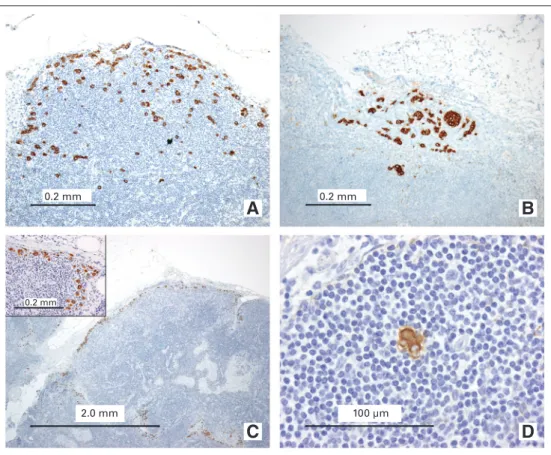
Reclassification according to the definitions of the EWGBSP9 and Turner et al11resulted in a discordance in nodal staging in 86 (22%) of 392 patients because of a difference in either diameter (n⫽42; 49%), microanatomic location (n ⫽ 29; 34%), or both (n⫽15; 17%). This resulted in a different pN classification in 73 patients (19%). Overall, the rates of SN ITC, micrometastases, and macrometastases were 7%, 27%, and 66% according to the interpre- tation of the EWGBSP,9compared with 18%, 25%, and 57% on the basis of the definitions by Turner et al.11Figure 1 shows some exam- ples of metastatic deposits causing discordance between both staging definitions. The majority of these discordantly staged cases (40 [55%]
of 73 patients) were classified as micrometastases according to the interpretation of the EWGBSP,9whereas they were classified as ITC according to the interpretation by Turner et al11(Table 2). The rest of these discrepancies were caused by a difference between micrometas- tases (Turner et al11) versus macrometastases (EWGBSP9; 40%) or between ITC (Turner et al11) versus macrometastases (EWGBSP9; 5%). Taking the total number of patients into account revealed a discordance in 40 (37%) of 107 of all patients with micrometastases according to the EWGBSP.9The rate of discordances in the group of patients classified as macrometastases according to EWGBSP was only 13% (33 of 258 patients).
Non-SN Involvement
Univariate analysis showed that the diameter of the primary tumor and the presence of lymphovascular invasion were significantly
A B
C D
0.2 mm
0.2 mm
0.2 mm
2.0 mm 100 µm
Fig 1.Examples of sentinel node (SN) metastases from lobular breast carcinoma that caused difficulty in nodal classifica- tion and discordance between different interpretations. SNs with multiple single cells and clusters arranged in a discontin- uous manner but dispersed homoge- neously in a definable part of the lymph node, classified as (A, B) micrometastases or (C) macrometastasis according to the European Working Group for Breast Screen- ing Pathology (EWGBSP) interpretations9 versus ITC according to Turner et al.11(D) SN with a tumor cluster less than 0.2 mm located in the parenchyma, classified as a micrometastasis according to the EWGBSP interpretation9versus isolated tumor cells according to Turner et al.11
associated with non-SN involvement (P⫽.002 andP⫽.001, respec- tively). SN features that were significantly associated with non-SN involvement included the detection method (P⬍.001), the presence of extracapsular extension (P⬍.001), SN staging according to the EWGBSP9, as well as Turner et al11(P⬍.001 for both; Table 3). There was no significant correlation between the total number of SNs and the number of positive non-SNs (P⫽.32).
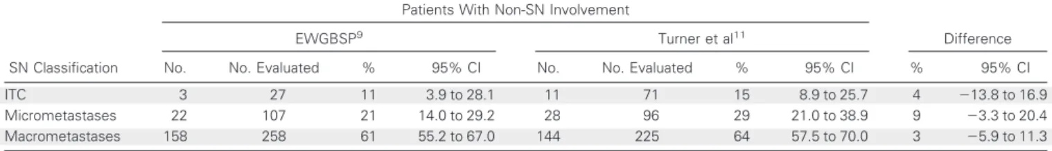
The rates of non-SN involvement in patients with SN ITC, mi- crometastases, and macrometastases were 11%, 21%, and 61% ac- cording to the interpretation of the EWGBSP,9compared with 15%, 29% and 64% on the basis of the definitions by Turner et al11(Table 4).
None of these differences were statistically significant (P ⫽ .82, P⫽.21, andP⫽.6, respectively). SN metastases with a diameter of less than 0.2 mm that were classified as micrometastases on the basis of microanatomic location according to the EWGBSP9were associated with non-SN involvement in four (16%; 25 patient cases; 95% CI, 6.4% to 34.7%), compared with three patient cases (11%; 95% CI, 3.9% to 28.1%) with metastases with a diameter of less than 0.2 mm located in the sinus, classified as ITC.
Table 5 shows the rates of non-SN involvement in those 73 patient cases with a discrepant classification between EWGBSP9 and Turner et al.11 The rate of non-SN involvement in the 40 patients with ITC according to Turner and micrometastases ac- cording to EWGBSP was 20%, which is comparable to the estab- lished rate for micrometastases. The rate of non-SN involvement in the 29 patients with micrometastases according to Turner et al11 and macrometastases according to EWGBSP9was 48%, which is comparable to the established rate for macrometastases. However, none of those four patient cases classified as having macrometas- tases according to EWGBSP9versus ITC according to Turner et al11 had detected non-SN involvement (Fig 1C). Overall, the EWGBSP method to classify SN tumor load thereby better reflected the risk of non-SN involvement than the Turner et al11system in these discrepant patients.
DISCUSSION
Lymph node metastases from lobular invasive breast cancer often show a pattern with multiple scattered clusters and/or single cells in both sinus and parenchyma of (sentinel) lymph nodes, which complicates nodal staging. This study is the first to evaluate nodal staging in this specific group of patients by assessing the frequency
of non-SN involvement for two different interpretations of nodal staging guidelines.
Overall, the rates of non-SN involvement in patients with a lob- ular breast carcinoma and SN ITC, micrometastases, or macrometas- tases were consistent with results from previous breast cancer studies including all histologic subtypes.16,17Several features of the primary tumor (ie, diameter, lymphovascular invasion) and the SN (ie, num- ber of involved SNs, extracapsular extension) were also significantly associated with non-SN involvement, which is consistent with previ- ous breast cancer studies.18-21
Table 2.Comparison of SN Staging According to the Interpretations of EWGBSP9and Turner et al11Criteria in a Group of 392 Patients With
Invasive Lobular Breast Cancer EWGBSP
Criteria9
Turner et al11Criteria
ITC Micrometastases Macrometastases Total
ITC 27 0 0 27
Micrometastases 40 67 0 107
Macrometastases 4 29 225 258
Total 71 96 225 392
Abbreviations: EWGBSP, European Working Group for Breast Screening Pathology; ITC, isolated tumor cell.
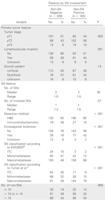
Table 3.Comparison of Clinicopathologic and SN Characteristics in Patients With Lobular Breast Carcinoma Without and With Non-SN Involvement
Variable
Patients by SN Involvement
P Non-SN
Negative (n⫽209)
Non-SN Positive (n⫽183)
No. % No. %
Primary tumor feature Tumor stage
pT1 107 51 62 34 .002
pT2 89 43 102 56
pT3 13 6 19 10
Lymphovascular invasion .001
No 138 66 93 51
Yes 58 28 81 44
Unknown 13 6 9 5
Growth pattern .14
Unifocal 115 55 87 48
Multifocal 78 37 81 44
Unknown 16 8 15 8
SN feature No. of SNs
Median 2 2 .92
Range 1-5 1-5
No. of involved SNs .07
Median 1 1
Range 1-3 1-5
Detection method ⬍.001
H&E 130 62 156 85
Immunohistochemistry 79 38 27 15
Extracapsular extension ⬍.001
No 159 76 103 56
Yes 38 18 77 42
Unknown 12 6 3 2
SN classification according
to EWGBSP9 ⬍.001
ITC 24 10 3 2
Micrometastases 85 41 22 12
Macrometastases 100 48 158 86
SN classification according
to Turner et al11 ⬍.001
ITC 62 30 11 6
Micrometastases 66 32 28 15
Macrometastases 81 39 144 79
No. of non-SNs .069
ⱕ10 39 19 25 14
⬎10 toⱕ15 81 39 59 32
⬎15 89 43 99 54
Abbreviations: SN, sentinel node; H&E, hematoxylin and eosin; ITC, isolated tumor cell; EWGBSP, European Working Group for Breast Screening Pathology.
SN reclassification according to the definitions of the EWGBSP9and Turner et al11resulted in a different pN classifica- tion in 73 patients (19%). Not all discrepancies resulted in a differ- ent pN classification, because patients were staged according to the largest metastatic deposit; for example, a patient with two macro- metastases according to the EWGBSP interpretation9was classified as having one macrometastases and one micrometastasis according to Turner et al.11
In general, the EWGBSP9interpretation designates more often metastases into a higher category than the Turner et al11system. This is because metastatic cell clusters that have a size compatible with ITC are yet classified as micrometastases when the microanatomic location is in the SN parenchyma. Second, a dispersed pattern of generally small tumor clusters or single metastatic cells is considered ITC or micrometastases in the Turner et al11classification depending on size of the largest cluster, whereas these are often designated micrometas- tases or macrometastases in the EWGBSP system.9Of 107 patient cases classified as micrometastases according to the EWGBSP defini- tions,9 40 patients (37%) were staged as having ITC according to Turner et al.11This is higher than the discordance rate of 24% previ- ously reported by Cserni et al,14which was expected, as we considered only lobular breast cancers whereas the study of Cserni et al14included all breast cancer subtypes. The discrepancy between micrometastases (Turner et al11) versus macrometastases (EWGBSP9) or between ITC (Turner et al11) versus macrometastases (EWGBSP9), conversely, was relatively limited. The main explanation for this finding is that a substantial proportion of macrometastases show a compact pattern of single cells and/or a fibroblastic reaction, which precludes a discrep- ancy between both interpretations.
The risk of non-SN involvement in patients with SN ITC overall did not differ significantly between both interpretations, a finding
similar to that of Cserni et al14and dealing with all histologic breast cancer types and only low volume metastases. We demonstrated that the interpretation of Turner et al11is associated with a higher positive predictive value of having non-SN metastases after finding either ITC or micrometastases in the SN. However, a high positive predictive value is not desirable in these groups of patients, because the objective is to select those patients with a limited risk of non-SN involvement, which correlates better with results of the EWGBSP definitions.9In those patients with a discordant classification between both defini- tions, the EWGBSP method to classify SN tumor load better reflected the risk of non-SN involvement than the Turner system, which leads to the conclusion that the EWGBSP method is better for the subgroup of invasive lobular carcinomas. Second, the rate of non-SN metastases in patients with SN metastases less than 0.2 mm but located in the parenchyma was 16%, which is relatively high compared with the risk for non-SN metastases in patients with SN metastases less than 0.2 mm located in the sinus (11%). These clusters located in the paren- chyma were designated micrometastases according to EWGBSP,9 whereas they are regarded as ITC by Turner et al.11Although the limited number of patients and the wide CIs do not allow definitive conclusions, these seem to support the hypothesis that extravasation of metastatic tumor deposits from the sinus into the parenchyma is a sign of the potential to generate additional metastatic spread. One must realize that the absence of a capsule deeper in the tissue block, immediately behind these deposits that seem to be located purely in the parenchyma, cannot be excluded. However, the potential risk of overstaging these patient cases was reduced by serial sectioning of the SN.
It was interesting to note that none of the four patient cases classified as SN ITC according to Turner et al11and as macrometasta- ses according to EWGBSP9had non-SN metastases. This could imply that in these patient cases, the Turner11classification may perform better, although it is more likely that the main underlying cause is the number of patient cases studied being too low to draw any conclu- sions. In addition, non-SN metastases with a dispersed pattern may have remained undetected in these cases, since non-SNs were not routinely examined by immunohistochemistry.
Another difference between both classifications is the inter- pretation of metastatic cells in extranodal lymph vessels without nodal involvement. These patient cases were classified as having nodal disease according to Turner et al,11whereas they were clas- sified as node negative according to the EWGBSP9interpretation.
The effect of this difference with regard to the rate of non-SN involvement is probably limited. In this study, it was not evaluated, because these patient cases were not selected as having a positive
Table 4.Frequency and Comparison of Non-SN Involvement According to Two Different Interpretations of the N Staging System
SN Classification
Patients With Non-SN Involvement
Difference
EWGBSP9 Turner et al11
No. No. Evaluated % 95% CI No. No. Evaluated % 95% CI % 95% CI
ITC 3 27 11 3.9 to 28.1 11 71 15 8.9 to 25.7 4 ⫺13.8 to 16.9
Micrometastases 22 107 21 14.0 to 29.2 28 96 29 21.0 to 38.9 9 ⫺3.3 to 20.4
Macrometastases 158 258 61 55.2 to 67.0 144 225 64 57.5 to 70.0 3 ⫺5.9 to 11.3
Abbreviations: SN, sentinel node; EWGBSP, European Working Group for Breast Screening Pathology; ITC, isolated tumor cell.
Table 5.Frequency of Non-SN Involvement for 73 Invasive Lobular Carcinoma Cases With Dissimilar N Classification According to Two
Different Interpretations of the N Staging System
Turner et al11
Criteria EWGBSP Criteria9
No. of Patients
Patients With Non-SN Involvement
No. %
ITC Micrometastases 40 8 20
ITC Macrometastases 4 0 0
Micrometastases Macrometastases 29 14 48
Abbreviations: SN, sentinel node; EWGBSP, European Working Group for Breast Screening Pathology; ITC, isolated tumor cell.
SN. The capsular and subcapsular lymphatic space on the other hand is regarded as part of the lymph node according to both definitions. These metastatic deposits are classified as either ITC or micrometastases, depending on size.
The definitions by Turner et al11are superior to those by the EWGBSP9 regarding reproducibility in small metastases (,0.92 v 0.49). However, they tend to understage high-volume metastases.
Because the EWGBSP definitions9reflect the total metastatic tumor load, especially in patients with lobular breast carcinoma, we suggest using these definitions at least in patients with lobular breast carci- noma for nodal staging to select high-risk patients that may benefit from additional local and/or systemic therapy. Clarifications regard- ing the measurement of nodal metastases, supplemented by visual examples, should be included in future staging manuals (or well worked out on Web sites) to achieve reproducible nodal staging.
Although an improved interobserver agreement does not necessarily mean better treatment, it is required to make a better prognostic separation. Findings from large studies assessing the effect of ITC and micrometastases on survival will provide greater clinical evidence re- garding policies on local and systemic treatment in patients with either ITC or micrometastases in their SNs.
AUTHORS’ DISCLOSURES OF POTENTIAL CONFLICTS OF INTEREST
The author(s) indicated no potential conflicts of interest.
AUTHOR CONTRIBUTIONS
Conception and design:Carolien H.M. van Deurzen, Gabor Cserni, Paul J. van Diest
Provision of study materials or patients:Carolien H.M. van Deurzen, Gabor Cserni, Simonetta Bianchi, Vania Vezzosi, Riccardo Arisio, Jelle Wesseling, Martin Asslaber, Maria P. Foschini, Anna Sapino, Isabella Castellano, Grace Callagy, Daniel Faverly, Maria-Dolores
Martin-Martinez, Cecily Quinn, Isabel Amendoeira, Janina Kulka, Angelika Reiner-Concin, Alicia Cordoba, Cornelis A. Seldenrijk, Paul J. van Diest
Collection and assembly of data:Carolien H.M. van Deurzen, Gabor Cserni, Simonetta Bianchi, Vania Vezzosi, Riccardo Arisio, Martin Asslaber, Maria P. Foschini, Anna Sapino, Isabella Castellano, Grace Callagy, Daniel Faverly, Maria-Dolores Martin-Martinez, Cecily Quinn, Isabel Amendoeira, Janina Kulka, Angelika Reiner-Concin, Alicia Cordoba Data analysis and interpretation:Carolien H.M. van Deurzen, Gabor Cserni, Paul J. van Diest
Manuscript writing:Carolien H.M. van Deurzen, Gabor Cserni, Simonetta Bianchi, Vania Vezzosi, Riccardo Arisio, Jelle Wesseling, Martin Asslaber, Maria P. Foschini, Anna Sapino, Isabella Castellano, Grace Callagy, Daniel Faverly, Maria-Dolores Martin-Martinez, Cecily Quinn, Isabel Amendoeira, Janina Kulka, Angelika Reiner-Concin, Alicia Cordoba, Cornelis A. Seldenrijk, Paul J. van Diest
Final approval of manuscript:Carolien H.M. van Deurzen, Gabor Cserni, Simonetta Bianchi, Vania Vezzosi, Riccardo Arisio, Jelle Wesseling, Martin Asslaber, Maria P. Foschini, Anna Sapino, Isabella Castellano, Grace Callagy, Daniel Faverly, Maria-Dolores
Martin-Martinez, Cecily Quinn, Isabel Amendoeira, Janina Kulka, Angelika Reiner-Concin, Alicia Cordoba, Cornelis A. Seldenrijk, Paul J. van Diest
REFERENCES
1. Cserni G, Amendoeira I, Apostolikas N, et al:
Pathological work-up of sentinel lymph nodes in breast cancer: Review of current data to be consid- ered for the formulation of guidelines. Eur J Cancer 39:1654-1667, 2003
2. van der Heiden-van der Loo M, Bezemer PD, Hennipman A, et al: Introduction of sentinel node biopsy and stage migration of breast cancer. Eur J Surg Oncol 32:710-714, 2006
3. van Diest PJ: Histopathological workup of sentinel lymph nodes: How much is enough? J Clin Pathol 52:871-873, 1999
4. Sobin LH WC: International Union Against Cancer TNM Classification of Malignant Tumours (ed 6). New York, NY, Wiley-Liss, 2002
5. Singletary SE, Greene FL, Sobin LH: Classifi- cation of isolated tumor cells: Clarification of the 6th edition of the American Joint Committee on Cancer Staging Manual. Cancer 98:2740-2741, 2003
6. Cox CE, Kiluk JV, Riker AI, et al: Significance of sentinel lymph node micrometastases in hu- man breast cancer. J Am Coll Surg 206:261-268, 2008
7. de Boer M, van Deurzen CH, van Dijck J:
Micrometastases or isolated tumor cells and the outcome of breast cancer. N Engl J Med 361:653- 663, 2009
8.Pugliese MS, Beatty JD, Tickman RJ, et al:
Impact and Outcomes of Routine Microstaging of Sentinel Lymph Nodes in Breast Cancer: Signifi- cance of the pN0(i⫹) and pN1mi Categories. Ann Surg Oncol 16:113-120, 2009
9.Cserni G, Bianchi S, Boecker W, et al: Improv- ing the reproducibility of diagnosing micrometasta- ses and isolated tumor cells. Cancer 103:358-367, 2005
10.Cserni G, Sapino A, Decker T: Discriminating between micrometastases and isolated tumor cells in a regional and institutional setting. Breast 15:347- 354, 2006
11.Turner RR, Weaver DL, Cserni G, et al: Nodal stage classification for breast carcinoma: Improving interobserver reproducibility through standardized histologic criteria and image-based training. J Clin Oncol 26:258-263, 2008
12.Roberts CA, Beitsch PD, Litz CE, et al: Inter- pretive disparity among pathologists in breast senti- nel lymph node evaluation. Am J Surg 186:324-329, 2003
13.de Mascarel I, MacGrogan G, Debled M, et al:
Distinction between isolated tumor cells and micro- metastases in breast cancer: Is it reliable and use- ful? Cancer 112:1672-1678, 2008
14.Cserni G, Bianchi S, Vezzosi V, et al: Variations in sentinel node isolated tumour cells/micrometas- tasis and non-sentinel node involvement rates ac- cording to different interpretations of the TNM definitions. Eur J Cancer 44:2185-2191, 2008
15.Rothman KJ: Epidemiology: An Introduction.
New York, NY, Oxford University Press, 2002 16.van Deurzen CH, de Boer M, Monninkhof EM, et al: Non-sentinel lymph node metastases associated with isolated breast cancer cells in the sentinel node. J Natl Cancer Inst 100:1574-1580, 2008
17.Viale G, Maiorano E, Pruneri G, et al: Predict- ing the risk for additional axillary metastases in patients with breast carcinoma and positive sentinel lymph node biopsy. Ann Surg 241:319-325, 2005
18.Bolster MJ, Peer PG, Bult P, et al: Risk factors for non-sentinel lymph node metastases in patients with breast cancer: The outcome of a multi-institutional study. Ann Surg Oncol 14:181- 189, 2007
19.Degnim AC, Griffith KA, Sabel MS, et al:
Clinicopathologic features of metastasis in nonsen- tinel lymph nodes of breast carcinoma patients.
Cancer 98:2307-2315, 2003
20.Orvieto E, Maiorano E, Bottiglieri L, et al:
Clinicopathologic characteristics of invasive lobular carcinoma of the breast: Results of an analysis of 530 cases from a single institution. Cancer 113:
1511-1520, 2008
21.van Deurzen CH, van Hillegersberg R, Hobbelink MG, et al: Predictive value of tumor load in breast cancer sentinel lymph nodes for second echelon lymph node metastases. Cell Oncol 29:497- 505, 2007
■ ■ ■