genes
G C A T T A C G G C A T
Article
Headcase is a Repressor of Lamellocyte Fate in Drosophila melanogaster
Gergely I. B. Varga1, Gábor Csordás1,†, Gyöngyi Cinege1, Ferenc Jankovics2 , Rita Sinka3, Éva Kurucz1, István Andó1,* and Viktor Honti1,*
1 Laboratory of Immunology, Institute of Genetics, Biological Research Centre of the Hungarian Academy of Sciences, 6726 Szeged, Hungary; varga.gergely@brc.mta.hu (G.I.B.V.); cgabor@uni-koeln.de (G.C.);
cinege.gyongyi@brc.mta.hu (G.C.); kurucz.eva@brc.mta.hu (É.K.)
2 Laboratory ofDrosophilaGerm Cell Differentiation, Institute of Genetics, Biological Research Centre of the Hungarian Academy of Sciences, 6726 Szeged, Hungary; jankovics.ferenc@brc.mta.hu
3 Department of Genetics, Faculty of Science and Informatics, University of Szeged, 6726 Szeged, Hungary;
rsinka@bio.u-szeged.hu
* Correspondence: ando.istvan@brc.mta.hu (I.A.); honti.viktor@brc.mta.hu (V.H.); Tel.: +36-62-599-677 (I.A.);
+36-62-599-448 (V.H.)
† Present address: Institute for Genetics and Cologne Excellence Cluster on Cellular Stress Responses in Aging-Associated Diseases (CECAD), University of Cologne, 50931 Cologne, Germany.
Received: 11 December 2018; Accepted: 1 March 2019; Published: 5 March 2019 Abstract:Due to the evolutionary conservation of the regulation of hematopoiesis,Drosophilaprovides an excellent model organism to study blood cell differentiation and hematopoietic stem cell (HSC) maintenance. The larvae ofDrosophila melanogasterrespond to immune induction with the production of special effector blood cells, the lamellocytes, which encapsulate and subsequently kill the invader.
Lamellocytes differentiate as a result of a concerted action of all three hematopoietic compartments of the larva: the lymph gland, the circulating hemocytes, and the sessile tissue. Within the lymph gland, the communication of the functional zones, the maintenance of HSC fate, and the differentiation of effector blood cells are regulated by a complex network of signaling pathways. Applying gene conversion, mutational analysis, and a candidate based genetic interaction screen, we investigated the role of Headcase (Hdc), the homolog of the tumor suppressor HECA in the hematopoiesis of Drosophila. We found that naive loss-of-functionhdcmutant larvae produce lamellocytes, showing that Hdc has a repressive role in effector blood cell differentiation. We demonstrate thathdcgenetically interacts with the Hedgehog and the Decapentaplegic pathways in the hematopoietic niche of the lymph gland. By adding further details to the model of blood cell fate regulation in the lymph gland of the larva, our findings contribute to the better understanding of HSC maintenance.
Keywords:Drosophila; hemocyte; blood cell; innate immunity; differentiation; hematopoiesis; niche
1. Introduction
Hematopoiesis is a sequence of strictly regulated events, during which mature blood cells differentiate from hematopoietic stem cells (HSCs) [1,2]. The ability of HSCs to self-renew is maintained by a complex network of signaling pathways in a specific microenvironment, the hematopoietic niche [3–5]. As improper regulation of HSC function leads to development of leukemias [6,7], the maintenance of the homeostatic balance between HSC self-renewal and blood cell differentiation is essential for the survival of the organism.
Due to the similarities of its immune system to that of vertebrates, Drosophilais extensively used as a model system, via which to study hematopoiesis. Similar to their vertebrate counterparts, the blood cells ofDrosophila, the hemocytes, differentiate in several waves throughout ontogenesis
Genes2019,10, 173; doi:10.3390/genes10030173 www.mdpi.com/journal/genes
Genes2019,10, 173 2 of 17
and are localized in dedicated hematopoietic compartments in each developmental stage. Such hemocyte compartments in the larva are the lymph gland, the sessile tissue, and the circulation [8–11].
The circulating hemocytes of the larva are classified into three categories: the phagocytic plasmatocytes, the melanizing crystal cells, and the encapsulating lamellocytes [9], which are distinguished by the expression of cell-type specific antigens and transgenic markers [12–18]. Lamellocytes are not present in naive larvae, but they vigorously differentiate in response to various stress stimuli, including infestation by the parasitoid waspLeptopilina boulardi, and eliminate the invader by forming a multilayered melanizing capsule around the parasite egg [19–21].
The lamellocyte pool that arises upon immune induction originates from all three hematopoietic compartments [9,22]. Lamellocytes differentiate from two separate cell types: plasmatocytes and dedicated precursors [22–27]. Plasmatocytes located in the sessile tissue and in the circulation are capable of transdifferentiating into lamellocytes as a result of genetic reprogramming or immune induction [22,24,26,28,29]. So far, dedicated precursor hemocytes, which are predestined to become lamellocytes, were found exclusively in the lymph gland [25]. Precursor hemocytes are located in the medullary zone of the lymph gland and are under the control of the posterior signaling center (PSC), a group of cells distinguished by the expression of the transcription factor Collier [30]. Cells of the PSC emanate cytonemes into the medullary zone. Signaling from the PSC is essential to maintain the prohemocyte state of medullary zone cells, therefore the PSC is regarded as a genuine hemocyte niche [31,32]. Concomitantly, alteration of PSC structure or activity was described to trigger precocious differentiation of the medullary zone population into various effector cell types [31–35]. However, recent results demonstrated that the PSC is not directly required for the maintenance of undifferentiated cells in the medullary zone (MZ), rather it controls their proper differentiation upon an immune challenge through relaying both activating and suppressive signals toward the progenitor cells [36,37].
In the lymph gland, prohemocyte state and effector hemocyte differentiation are controlled by a complex signaling network. Major components of this network that contribute to hemocyte fate choice are the Hedgehog (Hh), the Decapentaplegic (Dpp), and the JAK/STAT signaling pathways [11,32,38–40].
Hh and Dpp signaling originates from the PSC and is required to maintain the undifferentiated state of the MZ non-autonomously [32,41]. Active JAK/STAT signaling is required in the MZ cells to block their premature differentiation. Concomitantly, JAK/STAT activity is downregulated in these cells upon immune challenge to enable the differentiation of effector hemocytes [42–44]. Furthermore, JAK/STAT has been also implicated to act in the CZ cells to control plasmatocyte differentiation [45].
In unchallenged larvae, the lymph gland remains separated from the circulation until pupation, i.e., no hemocyte can enter or leave the organ [22,46]. However, following immune induction, effector hemocytes, including lamellocytes, differentiate from the progenitor cells and enter the circulation [22,23].
During lamellocyte differentiation, hemocytes gradually change their morphology and gene expression patterns [24,28,29]. A portion of the genes that becomes activated or silenced during the differentiation process contributes to the effector function of the cells, e.g., genes encoding phagocytosis receptors such as NimC1 and Eater [22,27,47] are downregulated. Other markers that are expressed in different hemocyte types represent regulator genes, such as transcription and epigenetic factors that directly determine the fate of the given cell type [9].
In this study, we investigated the role of Headcase (Hdc), theDrosophila homolog of HECA (Hdc homolog, cell cycle regulator), in the regulation of hematopoiesis. HECA interacts with cyclins and acts as a suppressor of several types of tumors in humans [48–51]. Our previous results [28], in accord with earlier literature data [52], showed thathdcis expressed in the cells of the lymph gland.
However, the effector hemocytes that differentiate and exit the lymph gland upon immune induction showed nohdc-LacZexpression [28], suggesting that Hdc activity may be related to the differentiation state of hemocytes. Hdc was originally described as a factor that blocks premature differentiation of imaginal tissues [52], and it was later shown to be active as a maintenance factor in the stem cell niche
Genes2019,10, 173 3 of 17
of the testis [53]. Hdc was also identified as a marker of intestinal stem cells and enteroblasts [54].
Nevertheless, the molecular function of Hdc is still unknown, and no characteristic domains were identified in the protein. Our results show thathdcexpression in the lymph gland is developmentally regulated, and Hdc plays an indispensable role in blocking premature lamellocyte differentiation. Hdc acts in the PSC, upstream of the Hedgehog and Decapentaplegic regulatory pathways, which normally maintain the prohemocyte state of medullary zone cells.
2. Materials and Methods
2.1. Drosophila Stocks
The following Drosophila lines were used in the study: w1118 (BSC#9505), w; P{GawB}5015 (BSC#2721), w; hdcB5 (a gift from Christos Samakovlis), w; hdc19-Gal4/TM3, Kr>GFP(this study), w; hdc43/TM6, Tb (BSC#64063),w; hdc∆84/TM3, Kr>GFP(this study),w; Hml∆-Gal4, UAS-GFP[55],w;
UAS-hdcRNAi (VDRC#v45069),w; Hml∆-Gal4, UAS-GFP, UAS-hdcRNAi,w; Dot-Gal4(BSC#67608),y, w, UAS-FLP; Dot-Gal4, AFG, UAS-GFP[22],w; Dot-Gal4,UAS-hdcRNAi,w; Pcol85-Gal4/CyO, GFP[31],w;
Pcol85-Gal4, UAS-hdcRNAi/CyO, GFP,y, w; UAS-2xEGFP (BSC#6658),w; Sp/CyO; UAS-hh(M4)/TM6b (a gift from Tamás Matusek), w; UAS-dpp (BSC#1486), w; UAS-hepRNAi (VDRC#v47507), w;
UAS-hopRNAi(VDRC#v102830), w; UAS-hdc.S(a gift from Christos Samakovlis),w; UAS-hdc.S(2nd chromosomal insertion, generated by the remobilization of the P element inBSC#6658),w; hdc19-Gal4, UAS-hdc.S/TM3(this study).
The flies were kept on a standard cornmeal-yeast diet at 25◦C. All crosses were performed at 25◦C.
2.2. Antibodies
Lamellocytes were detected with a mixture of L1a, L1b, and L1c (L1) mouse monoclonal antibodies [15]. Plasmatocytes were stained with the mixture of P1a and P1b (P1) antibodies [14].
PSC cells were stained with anti-Collier antibody [30], a kind gift from Michele Crozatier. The bound primary antibodies were visualized with CF 568 conjugated goat anti-mouse immunoglobulin (Biotium, Cat: 20100).
2.3. P Element Conversion
The exchange of the enhancer trapping P element was carried out according to Sepp and Auld [56].
Briefly,w; P{GawB}/SM6b; P{LacZ}hdcB5/TM3∆2-3jumpstarter virgins were crossed tow1118in order to increase the likelihood of the successful conversion events. Singlew; SM6b/+; P{GawB}hdc/+male progeny were crossed tow1118virgins to map the insertions to chromosomes based on the segregation of markers. Insertions segregating irrespectively ofSM6band sex chromosomes were regarded as third chromosomal. Candidate males were crossed individually toy, w; UAS-2xEGFPvirgins to verify the Gal4 activity and expression pattern.
2.4. X-GAL Staining
Lymph glands and imaginal discs were dissected from wandering larvae in PBT on ice, then were fixed and stained as described by Jankovics et al. [57].
2.5. PCR Mapping of the P Element Insertions
The localization of theP{LacZ}hdcB5element was determined by screening with a set of forward primers covering thehdcgenomic region and a reverse primer specific for the P element. Genomic DNA was isolated from female adult flies using the GenEluteTM Mammalian Genomic DNA Miniprep Kit (Sigma). PCR reactions of 100 ng genomic DNA template and the primer setInsertion forward (5’-CGAGCCGCAACGAAAGTG-3’) andInsertion reverse(5’-CCACCTTATGTTATTTCATCATG-3’) were found to amplify a 701 bp DNA fragment.
Genes2019,10, 173 4 of 17
The fragment was isolated and sequenced using a BigDye Terminator v3.1 Cycle Sequencing Kit (Invitrogen) and a 3500-Genetic Analyzer (Applied Biosystems), with the primer FarHdc fw (5’-TGAAGAAGTGCGGAAAATCGG-3’). The sequencing revealed that theP{LacZ}hdcB5insertion is localized 1017 bp upstream of thehdcstart codon.
A similar strategy was used to determine the position of theP{GawB}hdcinsertions in three independent convertants (hdc19-Gal4, hdc31-Gal4, hdc55-Gal4). PCR reactions using the hdc rev (5’-TCCCACCACTCGAAGCACTC-3’) andPGawB end(5’-GCTATGACCATGATTACGCCAAG-3’) primer pair amplified a 1748 bp DNA fragment. The fragment was isolated and sequenced with the PGawB endprimer. In each tested line, theP{GawB}insertion was localized 1025 bp upstream of thehdc start codon.
2.6. Generation of hdc Alleles and Breakpoint Mapping
Jumpstarter males (w; hdc19-Gal4/TM3,∆2-3, ry+) were crossed toTM6/TM3virgins. Candidates were selected for the loss of the miniwhitemarker gene and for lethality in combination with the hdc31-Gal4 insertion, an independent hypomorphic mutation isolated in our previous P element conversion screen.
We carried out a PCR screening on the candidate lines with the Excision forward (5’-ACCAATCTCGGTTAGAAACCCACT-3’) and theExcision reverse(5’-TCCCACCACTCGAAGCAC TC-3’) primers, which amplify an 823 bp long fragment overlapping the translation start codon of hdc, to isolate amorphic alleles, in which the protein coding region of the hdcgene is affected by the deficiency. The candidate that did not yield the expected PCR fragment was further analyzed with various primer sets. The breakpoints of the deficiency were identified with the Upstream forward(5’-ACCAAATTCTGGCCTACAGTGG-3’) andUpstream reverse(5’-CCACCTTATGT TATTTCATCATG-3’) primers for the upstream breakpoint, andDownstream forward(5’-TGGCATCATT GAAACAGCAAGG-3’) andDownstream reverse(5’-GGATATCTCGCCACTGGA CTG-3’) primers for the downstream breakpoint.
2.7. Immunostaining of Circulating Hemocytes, Imaging, and Counting
Larvae were dissected in 30µl Schneider’s medium (Lonza) supplemented with 5% FBS (Gibco) and n-Phenylthiourea (PTU, Sigma-Aldrich) on a multispot microscope slide (Hendley-Essex SM011).
The isolated hemocytes were allowed to adhere at room temperature for one hour. The samples were fixed with acetone for 6 min or with 2% paraformaldehyde for 10 min, washed three times with PBS and blocked for 20 min in PBS containing 0.1% BSA (Roche Diagnostics GmbH), incubated with primary antibodies for 45 min at room temperature, washed three times in PBS, and stained with CF 568-conjugated goat anti-mouse antibody (Biotium) for 45 min. Nuclei were visualized with DAPI (Sigma-Aldrich). The samples were mounted with 1:1 PBS-glycerol or Fluoromount-G (SouthernBiotech). For quantifications, hemocytes were identified with DAPI staining, lamellocyte and plasmatocyte ratios were determined with ImageJ, based on lamellocyte specific L1 staining and plasmatocyte specific P1 staining, respectively. Statistical analysis and graph assembly were performed using GraphPad Prism 6.
2.8. Preparation and Immunostaining of the Lymph Gland
Lymph glands of the larvae were dissected, fixed, and blocked as described by Márkus et al. [28].
The samples were incubated with respective monoclonal antibodies overnight at 4◦C, washed three times in PBS, and stained with CF 568-conjugated goat anti-mouse antibody at room temperature for 45 min. Nuclei were stained with DAPI (Sigma-Aldrich). Samples were mounted with Fluoromount-G (SouthernBiotech).
Genes2019,10, 173 5 of 17
3. Results
3.1. hdc is Expressed in the Lymph Gland, but Not in the Other Hemocyte Compartments
To enable the thorough analysis ofhdcexpression and functional studies in vivo, we performed a P element conversion screen, in which theLacZcontaining P element (P{LacZ}hdcB5) in thehdcB5 allele was exchanged with aGal4containingP{GawB}enhancer trap element. This was achieved by the simultaneous mobilization of the two P elements [56]. Of the severalhdc-Gal4lines, we selected hdc19-Gal4for further analysis. As detected by X-Gal staining,hdc19-Gal4did not show LacZ expression in any tissues or organs of the larva, confirming that theP{LacZ}hdcB5element was completely removed from the genome. Molecular analysis revealed that theP{GawB}insertion in thehdc19-Gal4allele was located 8 bp upstream of the originalP{LacZ}insertion (1025 bp upstream of the translation start codon of the gene) (Figure1A).
Genes 2019, 10, x FOR PEER REVIEW 5 of 18
3.1. hdc is Expressed in the Lymph Gland, but Not in the Other Hemocyte Compartments
To enable the thorough analysis of hdc expression and functional studies in vivo, we performed a P element conversion screen, in which the LacZ containing P element (P{LacZ}hdcB5) in the hdcB5 allele was exchanged with a Gal4 containing P{GawB} enhancer trap element. This was achieved by the simultaneous mobilization of the two P elements [56]. Of the several hdc-Gal4 lines, we selected hdc19-Gal4 for further analysis. As detected by X-Gal staining, hdc19-Gal4 did not show LacZ expression in any tissues or organs of the larva, confirming that the P{LacZ}hdcB5 element was completely removed from the genome. Molecular analysis revealed that the P{GawB} insertion in the hdc19-Gal4 allele was located 8 bp upstream of the original P{LacZ} insertion (1025 bp upstream of the translation start codon of the gene) (Figure 1A).
Figure 1. Genomic locations of hdc alleles and the expression pattern of hdc>GFP. (A) Molecular map of the hdc gene region. The positions of the original hdcB5 insertion, the hdc19-Gal4 insertion, and the breakpoints of the hdc∆84 deletion are indicated; (B–E) hdc19-Gal4>GFP (green) was expressed in the imaginal discs (B), the abdominal histoblasts (C), the genital disc (D), and in the lymph gland (E) of the larva. Images were taken with Leica TCS SP5 Confocal Microscope, original magnification ×20 (B,E) and ×40 (C,D), and processed with LAS AF Lite (Leica Microsystems CMS GmbH), ImageJ and Adobe Photoshop CS2 software. Scale bars: 25 µm.
hdc19-Gal4>GFP larvae (hdc19-Gal4/UAS-2xEGFP) expressed GFP in the imaginal discs, the abdominal histoblasts, the genital disc, and the lymph gland (Figure 1B–E), similarly to the LacZ expression of the hdcB5 larvae. No GFP expression was detected in adult flies, which underlines that Hdc is required in the larval stages for the formation of adult tissues [52]. These results indicate that the P{GawB}hdc19-Gal4 element traps the same enhancers as the P{LacZ}hdcB5 insertion. GFP expression was not detected in the circulating and sessile hemocytes of the hdc19-Gal4>GFP larvae, showing that hdc is not expressed in hemocytes outside of the lymph gland.
3.2. hdc Enhancer Trap Insertions are Hypomorphic hdc Alleles
Although hdcB5 was homozygous viable, hdc19-Gal4 showed pupal lethality both in homozygous condition, and in combination with two other P{GawB}hdc insertions (hdc31-Gal4, hdc55-Gal4), which were isolated in the same conversion screen and were located in the same molecular position.
Similarly, hdc19-Gal4 was lethal in combination with hdc43, a previously described null mutant allele [52], but it was viable in combination with hdcB5. These findings suggest that hdc19-Gal4 is a stronger
Figure 1.Genomic locations ofhdcalleles and the expression pattern ofhdc>GFP. (A) Molecular map of thehdcgene region. The positions of the originalhdcB5insertion, thehdc19-Gal4insertion, and the breakpoints of thehdc∆84deletion are indicated; (B–E)hdc19-Gal4>GFP(green) was expressed in the imaginal discs (B), the abdominal histoblasts (C), the genital disc (D), and in the lymph gland (E) of the larva. Images were taken with Leica TCS SP5 Confocal Microscope, original magnification×20 (B,E) and×40 (C,D), and processed with LAS AF Lite (Leica Microsystems CMS GmbH), ImageJ and Adobe Photoshop CS2 software. Scale bars: 25µm.
hdc19-Gal4>GFP larvae (hdc19-Gal4/UAS-2xEGFP) expressed GFP in the imaginal discs, the abdominal histoblasts, the genital disc, and the lymph gland (Figure1B–E), similarly to the LacZ expression of thehdcB5larvae. No GFP expression was detected in adult flies, which underlines that Hdc is required in the larval stages for the formation of adult tissues [52]. These results indicate that theP{GawB}hdc19-Gal4element traps the same enhancers as theP{LacZ}hdcB5insertion. GFP expression was not detected in the circulating and sessile hemocytes of thehdc19-Gal4>GFPlarvae, showing that hdcis not expressed in hemocytes outside of the lymph gland.
3.2. hdc Enhancer Trap Insertions are Hypomorphic hdc Alleles
AlthoughhdcB5was homozygous viable,hdc19-Gal4showed pupal lethality both in homozygous condition, and in combination with two otherP{GawB}hdcinsertions (hdc31-Gal4, hdc55-Gal4), which were isolated in the same conversion screen and were located in the same molecular position. Similarly, hdc19-Gal4was lethal in combination withhdc43, a previously described null mutant allele [52], but it
Genes2019,10, 173 6 of 17
was viable in combination withhdcB5. These findings suggest thathdc19-Gal4is a stronger hypomorphic allele thanhdcB5. The most obvious explanation for this phenomenon may be the possibly stronger enhancer trapping activity of theP{GawB}hdc19-Gal4than that of the originalP{LacZ}hdcB5element.
Homozygous lethality of thehdc19-Gal4insertion was rescued by the expression ofhdc from aUAS-hdctransgene (hdc19-Gal4/hdc19-Gal4, UAS-hdc.S), indicating that lethality is not caused by a second-site mutation. Additionally, it confirmed that Gal4 expression in thehdc19-Gal4recapitulates the endogenoushdcexpression pattern (Figure1B–E). RNA-interference mediated silencing ofhdc with thehdc19-Gal4(hdcRNAi/+; hdc19-Gal4/+) resulted in 100% pupal lethality, which is typical tohdc loss-of-function [52].
3.3. hdc Loss-Of-Function Mutant Larvae Produce Lamellocytes without Immune Induction
We analyzed the hematopoietic phenotype ofhdcby staining the circulating hemocytes of third instar homozygoushdc19-Gal4larvae for the P1 marker of plasmatocytes [14] and the L1 marker for lamellocytes [15]. Immunostainings revealed the precocious differentiation of lamellocytes in homozygoushdc19-Gal4larvae with 19% penetrance (Figure2A).
Genes 2019, 10, x FOR PEER REVIEW 6 of 18
hypomorphic allele than hdcB5. The most obvious explanation for this phenomenon may be the possibly stronger enhancer trapping activity of the P{GawB}hdc19-Gal4 than that of the original P{LacZ}hdcB5 element.
Homozygous lethality of the hdc19-Gal4 insertion was rescued by the expression of hdc from a UAS- hdc transgene (hdc19-Gal4/hdc19-Gal4, UAS-hdc.S), indicating that lethality is not caused by a second- site mutation. Additionally, it confirmed that Gal4 expression in the hdc19-Gal4 recapitulates the endogenous hdc expression pattern (Figure 1B–E). RNA-interference mediated silencing of hdc with the hdc19-Gal4 (hdcRNAi/+; hdc19-Gal4/+) resulted in 100% pupal lethality, which is typical to hdc loss- of-function [52].
3.3. hdc Loss-Of-Function Mutant Larvae Produce Lamellocytes without Immune Induction
We analyzed the hematopoietic phenotype of hdc by staining the circulating hemocytes of third instar homozygous hdc19-Gal4 larvae for the P1 marker of plasmatocytes [14] and the L1 marker for lamellocytes [15]. Immunostainings revealed the precocious differentiation of lamellocytes in homozygous hdc19-Gal4 larvae with 19% penetrance (Figure 2A).
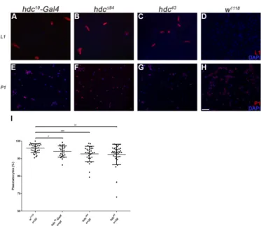
Figure 2. Circulating hemocytes of the hdc mutant larvae. (A–D) Lamellocytes were present in the circulation of hdc19-Gal4 (2.61% (n = 42)) (A), hdc∆84 (2.15% (n = 24)) (B) and hdc43 (6.07% (n = 52)) (C) homozygous mutant larvae. Lamellocytes were rarely observed in control w1118 larvae (0.07% (n = 24)) (D). Lamellocytes were stained with L1 antibody (red); (E–H) Reduced plasmatocyte ratio was observed in hdc19-Gal4 (E), hdc∆84 (F), and hdc43 (G) homozygous mutant larvae compared to control w1118 (H). Plasmatocytes were stained with P1 antibody (red). Nuclei were stained with DAPI (blue).
Hemocytes were visualized on a Zeiss Axioskope 2MOT epifluorescent microscope, original magnification ×10 and processed with ImageJ and Adobe Photoshop CS2 softwares. Scale bar: 20 µm.
(I) Quantification of plasmatocytes in the hdc mutant larvae. Error bars represent the standard deviation of the mean. ***: p < 0.001; **: p < 0.01; *: p < 0.05. Significance was determined by unpaired Student’s t-test.
To elucidate the effects of complete loss of hdc, we generated loss-of-function hdc mutants with the remobilization of the P element in the hdc19-Gal4 line. Of the 13 isolated lethal alleles, molecular mapping of the hdc locus uncovered one deletion, hdc∆84, in which the start codon was deleted together with a part of the first exon, spanning 1014 bp upstream and 971 bp downstream of the ATG site (Figure 1A). Considering the original position of the P{GawB} element, we conclude that the P element underwent a local hop of 11 bp before generating the excision. The newly generated hdc∆84 allele was pupal lethal both in homozygous condition and in interallelic combination with hdc43,
Figure 2.Circulating hemocytes of thehdcmutant larvae. (A–D) Lamellocytes were present in the circulation ofhdc19-Gal4(2.61% (n = 42)) (A),hdc∆84 (2.15% (n = 24)) (B) andhdc43(6.07% (n = 52)) (C) homozygous mutant larvae. Lamellocytes were rarely observed in controlw1118larvae (0.07%
(n = 24)) (D). Lamellocytes were stained with L1 antibody (red); (E–H) Reduced plasmatocyte ratio was observed inhdc19-Gal4(E),hdc∆84(F), andhdc43 (G) homozygous mutant larvae compared to controlw1118 (H). Plasmatocytes were stained with P1 antibody (red). Nuclei were stained with DAPI (blue). Hemocytes were visualized on a Zeiss Axioskope 2MOT epifluorescent microscope, original magnification×10 and processed with ImageJ and Adobe Photoshop CS2 softwares. Scale bar:
20µm. (I) Quantification of plasmatocytes in thehdcmutant larvae. Error bars represent the standard deviation of the mean. ***:p< 0.001; **:p< 0.01; *:p< 0.05. Significance was determined by unpaired Student’st-test.
To elucidate the effects of complete loss ofhdc, we generated loss-of-functionhdcmutants with the remobilization of the P element in thehdc19-Gal4line. Of the 13 isolated lethal alleles, molecular mapping of thehdclocus uncovered one deletion,hdc∆84, in which the start codon was deleted together with a part of the first exon, spanning 1014 bp upstream and 971 bp downstream of the ATG site (Figure1A). Considering the original position of theP{GawB}element, we conclude that the P element underwent a local hop of 11 bp before generating the excision. The newly generatedhdc∆84 allele was pupal lethal both in homozygous condition and in interallelic combination withhdc43, which
Genes2019,10, 173 7 of 17
was described as a null allele [52]. Based on these data,hdc∆84can be also regarded as a null allele of the gene.
Immunostaining of circulating larval hemocytes for the L1 marker showed that both hdc∆84 andhdc43homozygous larvae contained lamellocytes with 83% and 100% penetrance, respectively (Figure2B–D). Lamellocyte differentiation was also observed in the lymph gland (Figure3). The ratio of plasmatocytes was significantly reduced in larvae carrying homozygoushdcalleles (Figure2E–I), which may be due to the appearance of lamellocytes in the circulation.
Genes 2019, 10, x FOR PEER REVIEW 7 of 18
which was described as a null allele [52]. Based on these data, hdc∆84 can be also regarded as a null allele of the gene.
Immunostaining of circulating larval hemocytes for the L1 marker showed that both hdcΔ84 and hdc43 homozygous larvae contained lamellocytes with 83% and 100% penetrance, respectively (Figure 2B–D). Lamellocyte differentiation was also observed in the lymph gland (Figure 3). The ratio of plasmatocytes was significantly reduced in larvae carrying homozygous hdc alleles (Figure 2E–I), which may be due to the appearance of lamellocytes in the circulation.
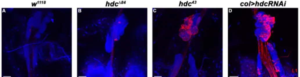
Figure 3. Lamellocyte differentiation in the lymph gland of hdc mutant and hdc silenced larvae. (A) There are no lamellocytes present in the lymph gland of w1118 control larvae; (B–C) Lamellocytes are detected with staining for the L1 marker (red) in the lymph gland of hdc∆84 (B) and hdc43 (C) larvae;
(D) Silencing of hdc with the col-Gal4 driver (Pcol85-Gal4, UAS-hdcRNAi/+) also resulted in the differentiation of lamellocytes (red) in the lymph gland. Nuclei were stained with DAPI (blue).
Confocal images were generated with a Leica TCS SP5 Confocal Microscope, original magnification
×20, and processed with LAS AF Lite (Leica Microsystems CMS GmbH), ImageJ and Adobe Photoshop CS2 softwares. Scale bars: 50 µm.
3.4. Hdc Exerts its Effect in the Lymph Gland
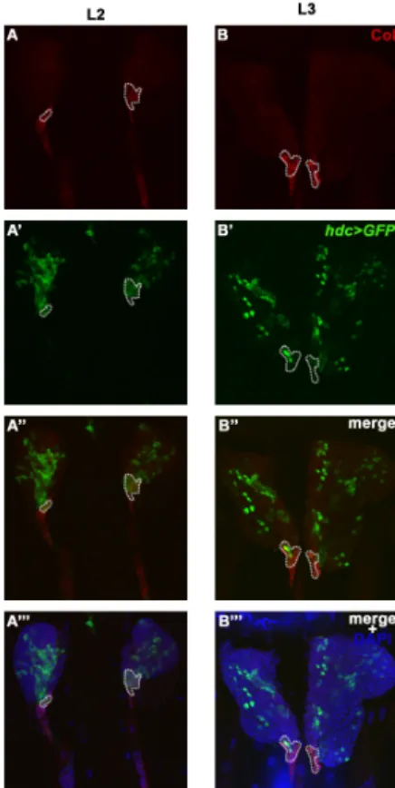
To locate the focus of the hdc mutation in relation to its hematopoietic phenotype, first we analyzed the hdc19-Gal4>GFP expression in the lymph gland at different larval stages. In L2 larvae, hdc19-Gal4>GFP was broadly expressed in the primary lobes of the lymph gland. The expression pattern partially overlapped with the anti-Collier staining in the PSC and in the secondary lymph gland lobes (Figure 4A–A’’’ and Figure A1A–A’’). In early L3 larvae, the expression pattern was greatly altered as compared to the L2 stage, the hdc19-Gal4>GFP being expressed in less hemocytes of the primary lobes, and only in a few of the cells in the PSC (Figure 4B–B’’’ and Figure A1B–B’’). In wandering L3 larvae, the number of GFP expressing hemocytes was further reduced in the primary lobes, and no GFP expression was detected in the PSC. However, hdc19-Gal4>GFP was still detectable in a subset of the cells in the secondary lymph gland lobes (Figure A1C–C’’).
Figure 3. Lamellocyte differentiation in the lymph gland ofhdc mutant andhdcsilenced larvae.
(A) There are no lamellocytes present in the lymph gland ofw1118control larvae; (B–C) Lamellocytes are detected with staining for the L1 marker (red) in the lymph gland of hdc∆84 (B) and hdc43 (C) larvae; (D) Silencing ofhdcwith thecol-Gal4driver (Pcol85-Gal4, UAS-hdcRNAi/+) also resulted in the differentiation of lamellocytes (red) in the lymph gland. Nuclei were stained with DAPI (blue).
Confocal images were generated with a Leica TCS SP5 Confocal Microscope, original magnification
×20, and processed with LAS AF Lite (Leica Microsystems CMS GmbH), ImageJ and Adobe Photoshop CS2 softwares. Scale bars: 50µm.
3.4. Hdc Exerts its Effect in the Lymph Gland
To locate the focus of the hdc mutation in relation to its hematopoietic phenotype, first we analyzed thehdc19-Gal4>GFPexpression in the lymph gland at different larval stages. In L2 larvae, hdc19-Gal4>GFPwas broadly expressed in the primary lobes of the lymph gland. The expression pattern partially overlapped with the anti-Collier staining in the PSC and in the secondary lymph gland lobes (Figure4A–A”’ and Figure A1A–A”). In early L3 larvae, the expression pattern was greatly altered as compared to the L2 stage, thehdc19-Gal4>GFPbeing expressed in less hemocytes of the primary lobes, and only in a few of the cells in the PSC (Figure4B–B”’ and FigureA1B–B”). In wandering L3 larvae, the number of GFP expressing hemocytes was further reduced in the primary lobes, and no GFP expression was detected in the PSC. However,hdc19-Gal4>GFPwas still detectable in a subset of the cells in the secondary lymph gland lobes (FigureA1C–C”).
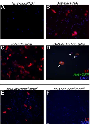
To determine the subgroup of hemocytes in which Hdc is required, we silencedhdcwith various drivers. SilencinghdcwithHemolectin-Gal4(Hml∆-Gal4) [55], a general plasmatocyte specific driver, which is active in all three hemocyte compartments, but inactive in the PSC, did not result in lamellocyte differentiation. However, silencinghdcwith the lymph gland specificDorothy-Gal4(Dot-Gal4) [58] or col-Gal4(Pcol85-Gal4) [31] led to the appearance of lamellocytes in the circulation. As the overlapping domain of expression of these two drivers is the PSC, we concluded that lamellocyte differentiation is triggered upon loss ofhdcfunction in the hematopoietic niche of the lymph gland (Figure5A–C).
Importantly, overexpression ofhdcwith thecol-Gal4driver rescued the hematopoietic phenotype of hdc43homozygous larvae (Figure5E,F).
GenesGenes 2019, 10, x FOR PEER REVIEW 2019,10, 173 8 of 188 of 17
Figure 4. Overlap of hdc>GFP and Collier expression in the posterior signaling center (PSC) of the lymph gland. (A–B) PSC was labeled with anti-Collier staining (red) of the lymph gland of L2 (A) and L3 (B) larvae; (A’–B’) The expression of hdc was monitored by the expression of the GFP marker (green) in second- (A’) and third instar (B’) hdc19-Gal4/UAS-2xEGFP larvae; (A’’–B’’) hdc>GFP (green) and Collier (red) expressions overlap in the PSC of the lymph gland; (A’’’–B’’’) DAPI staining (blue) of lymph gland cells. Images were taken with a Leica TCS SP5 Confocal Microscope, original magnification ×20, and processed with LAS AF Lite (Leica Microsystems CMS GmbH), ImageJ and Adobe Photoshop CS2 softwares. Scale bars: 25 µm.
To determine the subgroup of hemocytes in which Hdc is required, we silenced hdc with various drivers. Silencing hdc with Hemolectin-Gal4 (HmlΔ-Gal4) [55], a general plasmatocyte specific driver, which is active in all three hemocyte compartments, but inactive in the PSC, did not result in lamellocyte differentiation. However, silencing hdc with the lymph gland specific Dorothy-Gal4 (Dot- Gal4) [58] or col-Gal4 (Pcol85-Gal4) [31] led to the appearance of lamellocytes in the circulation. As the overlapping domain of expression of these two drivers is the PSC, we concluded that lamellocyte differentiation is triggered upon loss of hdc function in the hematopoietic niche of the lymph gland (Figure 5A–C). Importantly, overexpression of hdc with the col-Gal4 driver rescued the hematopoietic phenotype of hdc43 homozygous larvae (Figure 5E,F).
As these findings suggested that Hdc has a non-cell-autonomous function in the PSC to regulate the fate of the medullary zone hemocytes, we knocked down hdc with RNA interference in the Dot hemocyte lineage, which is composed of cells from all the three functional zones of the lymph gland [22,59], and scored circulating immune cells for cell-type specific markers. We found that 68% of the lamellocytes (n = 66) appearing in third instar larvae were derived from cells that did not belong to the Dot-lineage (Figure 5D), showing that lamellocyte differentiation mostly occurred in a non- lineage-autonomous fashion. These results, together with the appearance of lamellocytes when hdc is knocked down specifically in the PSC, suggest that Hdc is required in the signaling center for its capacity to negatively regulate lamellocyte differentiation in the lymph gland.
Figure 4.Overlap ofhdc>GFPand Collier expression in the posterior signaling center (PSC) of the lymph gland. (A–B) PSC was labeled with anti-Collier staining (red) of the lymph gland of L2 (A) and L3 (B) larvae; (A’–B’) The expression ofhdcwas monitored by the expression of the GFP marker (green) in second- (A’) and third instar (B’)hdc19-Gal4/UAS-2xEGFPlarvae; (A”–B”)hdc>GFP(green) and Collier (red) expressions overlap in the PSC of the lymph gland; (A”’–B”’) DAPI staining (blue) of lymph gland cells. Images were taken with a Leica TCS SP5 Confocal Microscope, original magnification×20, and processed with LAS AF Lite (Leica Microsystems CMS GmbH), ImageJ and Adobe Photoshop CS2 softwares. Scale bars: 25µm.
As these findings suggested that Hdc has a non-cell-autonomous function in the PSC to regulate the fate of the medullary zone hemocytes, we knocked down hdc with RNA interference in the Dothemocyte lineage, which is composed of cells from all the three functional zones of the lymph gland [22,59], and scored circulating immune cells for cell-type specific markers. We found that 68%
of the lamellocytes (n = 66) appearing in third instar larvae were derived from cells that did not belong to theDot-lineage (Figure5D), showing that lamellocyte differentiation mostly occurred in a non-lineage-autonomous fashion. These results, together with the appearance of lamellocytes when hdcis knocked down specifically in the PSC, suggest that Hdc is required in the signaling center for its capacity to negatively regulate lamellocyte differentiation in the lymph gland.
GenesGenes 2019, 10, 173 2019,10, 173 9 of 179 of 18
Figure 5. The effect of hdc silencing and the rescue of the hematopoietic phenotype. (A) Silencing hdc with the plasmatocyte specific Hml-Gal4 (HmlΔ-Gal4, UAS-GFP, UAS-hdcRNAi) did not result in the differentiation of lamellocytes; (B) Silencing hdc with the lymph gland specific Dot-Gal4 driver (Dot- Gal4/UAS-hdcRNAi) resulted in the differentiation of lamellocytes (red); (C) Silencing hdc in the PSC of the lymph gland with the col-Gal4 driver (Pcol85-Gal4/UAS-hdcRNAi) led to lamellocyte differentiation (red); (D) Silencing of hdc in the entire Dot-lineage (y, w, UAS-Flp; Dot-Gal4, AFG/UAS- hdcRNAi) resulted in the appearance of lamellocytes in the circulation (red). These lamellocytes fell into two classes. The lamellocytes, which derive from the Dot-lineage, expressed both GFP (green) and the lamellocyte specific L1 antigene (red). The lineage-independent lamellocytes expressed only the L1 marker (red). Arrowhead indicates a GFP- lamellocyte, while arrows point to double positive lamellocytes; (E–F) Lamellocytes (red) were always present in the circulation of hdc43 homozygous larvae (Pcol85-Gal4/+; hdc43/hdc43) (2.85% (n = 12)) (E), while their proportion was reduced when hdc was overexpressed with the col-Gal4 driver (Pcol85-Gal4/UAS-hdc.S; hdc43/hdc43) (0.25% (n = 12)) (F).
Nuclei were stained with DAPI (blue). Hemocytes were visualized on a Zeiss Axioskope 2MOT epifluorescent microscope, original magnification ×20 (A–D) and x10 (E,F) and processed with ImageJ and Adobe Photoshop CS2 softwares. Scale bars: 20 µm.
3.5. Hdc Interacts with the Hedgehog and the Decapentaplegic Pathways
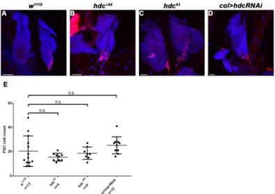
As dysfunction of the PSC leads to unprovoked differentiation of lamellocytes in the lymph gland [11,30], we investigated whether the loss of hdc affects the integrity or Col expression of the PSC. We found that the size of both the lymph gland and the PSC was unchanged in both the hdc43 and the hdcΔ84 mutant larvae, compared to that of the controls. Similarly, no difference was observed in the expression pattern of Col either in the primary or secondary lymph gland lobes as Col was expressed at a high level in the PSC and in the secondary lobes, and at a low level in the medullary zone of the primary lobes (n = 13 in each genotype). These findings suggest that Hdc is not required in PSC cells to gain or maintain their identity and their Col expression level (Figure 6 and Figure A2).
Figure 5. The effect ofhdcsilencing and the rescue of the hematopoietic phenotype. (A) Silencing hdcwith the plasmatocyte specificHml-Gal4(Hml∆-Gal4, UAS-GFP, UAS-hdcRNAi) did not result in the differentiation of lamellocytes; (B) Silencing hdc with the lymph gland specific Dot-Gal4 driver (Dot-Gal4/UAS-hdcRNAi) resulted in the differentiation of lamellocytes (red); (C) Silencing hdc in the PSC of the lymph gland with the col-Gal4 driver (Pcol85-Gal4/UAS-hdcRNAi) led to lamellocyte differentiation (red); (D) Silencing ofhdcin the entireDot-lineage (y, w, UAS-Flp; Dot-Gal4, AFG/UAS-hdcRNAi) resulted in the appearance of lamellocytes in the circulation (red). These lamellocytes fell into two classes. The lamellocytes, which derive from theDot-lineage, expressed both GFP (green) and the lamellocyte specific L1 antigene (red). The lineage-independent lamellocytes expressed only the L1 marker (red). Arrowhead indicates a GFP- lamellocyte, while arrows point to double positive lamellocytes; (E–F) Lamellocytes (red) were always present in the circulation of hdc43homozygous larvae (Pcol85-Gal4/+; hdc43/hdc43) (2.85% (n = 12)) (E), while their proportion was reduced whenhdcwas overexpressed with thecol-Gal4driver (Pcol85-Gal4/UAS-hdc.S; hdc43/hdc43) (0.25% (n = 12)) (F). Nuclei were stained with DAPI (blue). Hemocytes were visualized on a Zeiss Axioskope 2MOT epifluorescent microscope, original magnification×20 (A–D) and×10 (E,F) and processed with ImageJ and Adobe Photoshop CS2 softwares. Scale bars: 20µm.
3.5. Hdc Interacts with the Hedgehog and the Decapentaplegic Pathways
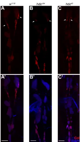
As dysfunction of the PSC leads to unprovoked differentiation of lamellocytes in the lymph gland [11,30], we investigated whether the loss ofhdcaffects the integrity or Col expression of the PSC.
We found that the size of both the lymph gland and the PSC was unchanged in both thehdc43and the hdc∆84mutant larvae, compared to that of the controls. Similarly, no difference was observed in the expression pattern of Col either in the primary or secondary lymph gland lobes as Col was expressed at a high level in the PSC and in the secondary lobes, and at a low level in the medullary zone of the primary lobes (n = 13 in each genotype). These findings suggest that Hdc is not required in PSC cells to gain or maintain their identity and their Col expression level (Figures6andA2).
Genes2019,10, 173 10 of 17
Genes 2019, 10, x FOR PEER REVIEW 10 of 18
Figure 6. Integrity of the lymph gland and the PSC in hdc mutants. (A,A’) Anti-Collier staining (red) of the lymph gland of the w1118 control larva; (B–C’) Anti-Collier staining (red) of the lymph gland of the hdc∆84 (B,B’) and hdc43 (C,C’) mutant larvae. The size and morphology of the lymph gland and the PSC in the hdc mutants did not differ from the control. Nuclei were stained with DAPI (blue) (A’,B’,C’). Arrows point to the PSC of the lymph gland. Images were taken with Leica TCS SP5 Confocal Microscope, original magnification ×20, and processed with LAS AF Lite (Leica Microsystems CMS GmbH), ImageJ and Adobe Photoshop CS2 softwares. Scale bars: 50 µm.
In first instar larvae, Dpp signaling maintains niche dependent hematopoietic stem cells (HSCs) in the lymph gland [41], while in later stages, Hh activity is required for the maintenance of the precursor state of medullary zone prohemocytes [32]. To gain an insight into the connection of Hdc to these signaling networks, we silenced hdc with the col-Gal4 driver, and simultaneously, activated Hh and Dpp signaling. The overexpression of both Hh and Dpp significantly reduced the number of circulating lamellocytes in hdc-silenced larvae as compared with the controls, indicating that Hdc acts upstream to both signal transduction pathways in the PSC cells (Figure 7A–C,F).
Interestingly, we found that the effect of hdc was also partially rescued by the simultaneous silencing of hopscotch (hop) (Figure 7D,F), a gene coding for the JAK kinase of the JAK/STAT pathway [60]. This pathway was not previously described to be active in the cells of the PSC. Our results, however, suggest that JAK/STAT interacts with Hdc in the regulation of hematopoiesis in the hematopoietic niche of the lymph gland. Silencing of hemipterous (hep), a serine/threonine protein kinase involved in JNK signaling [61], did not significantly reduce the number of lamellocytes (Figure 7E,F).
Figure 6.Integrity of the lymph gland and the PSC inhdcmutants. (A,A’) Anti-Collier staining (red) of the lymph gland of thew1118control larva; (B–C’) Anti-Collier staining (red) of the lymph gland of thehdc∆84(B,B’) andhdc43(C,C’) mutant larvae. The size and morphology of the lymph gland and the PSC in thehdcmutants did not differ from the control. Nuclei were stained with DAPI (blue) (A’,B’,C’). Arrows point to the PSC of the lymph gland. Images were taken with Leica TCS SP5 Confocal Microscope, original magnification×20, and processed with LAS AF Lite (Leica Microsystems CMS GmbH), ImageJ and Adobe Photoshop CS2 softwares. Scale bars: 50µm.
In first instar larvae, Dpp signaling maintains niche dependent hematopoietic stem cells (HSCs) in the lymph gland [41], while in later stages, Hh activity is required for the maintenance of the precursor state of medullary zone prohemocytes [32]. To gain an insight into the connection of Hdc to these signaling networks, we silencedhdcwith thecol-Gal4driver, and simultaneously, activated Hh and Dpp signaling. The overexpression of both Hh and Dpp significantly reduced the number of circulating lamellocytes inhdc-silenced larvae as compared with the controls, indicating that Hdc acts upstream to both signal transduction pathways in the PSC cells (Figure7A–C,F).
Interestingly, we found that the effect ofhdcwas also partially rescued by the simultaneous silencing of hopscotch (hop) (Figure 7D,F), a gene coding for the JAK kinase of the JAK/STAT pathway [60]. This pathway was not previously described to be active in the cells of the PSC. Our results, however, suggest that JAK/STAT interacts with Hdc in the regulation of hematopoiesis in the hematopoietic niche of the lymph gland. Silencing ofhemipterous(hep), a serine/threonine protein kinase involved in JNK signaling [61], did not significantly reduce the number of lamellocytes (Figure7E,F).
Genes2019,Genes 2019, 10, x FOR PEER REVIEW 10, 173 11 of 1811 of 17
Figure 7. The interaction of Hdc with the signal transduction pathways of the lymph gland. (A) Silencing of hdc with the Pcol85-Gal4 driver in the PSC of the lymph gland resulted in differentiation of lamellocytes in naive larvae (Pcol85-Gal4, UAS-hdcRNAi/+; UAS-2xEGFP/+); (B–E) Overexpression of hh (B) and dpp (C) with the col-Gal4 driver significantly reduced the proportion of lamellocytes (red) in the circulation of hdc-silenced larvae (Pcol85-Gal4, UAS-hdcRNAi/UAS-hh(M4), Pcol85-Gal4, UAS- hdcRNAi/UAS-dpp, respectively). Silencing of hop with RNA interference (Pcol85-Gal4, UAS- hdcRNAi/UAS-hopRNAi) (D) also resulted in the rescue of the hdc phenotype, while silencing of hep (Pcol85-Gal4, UAS-hdcRNAi/UAS-hepRNAi) (E) did not affect the phenotype significantly. Nuclei were stained with DAPI (blue). Hemocytes were visualized on a Zeiss Axioskope 2MOT epifluorescent microscope, original magnification x10 and processed with ImageJ and Adobe Photoshop CS2 softwares. Scale bar: 20 µm. (F) Quantification of lamellocyte numbers in the hdc-silenced and the rescued larvae. Error bars represent the standard deviation of the mean. **: p < 0.01; *: p < 0.05; ns:
non-significant. Significance was determined by unpaired Student’s t-test using GraphPad Prism 6.
4. Discussion
In this report, we show that Hdc, the Drosophila homolog of the human HECA, is a key component in the regulation of HSC function and hematopoiesis in the fruit fly. Of the three larval hemocyte compartments, hdc activity is confined to the lymph gland, suggesting that Hdc acts specifically in this hematopoietic organ. It was also observed that lamellocytes downregulate hdc expression when leaving the lymph gland [28]. These findings suggest that Hdc may exert a repressive role during hematopoiesis, which is also corroborated by the observation that hdc alleles suppress the disruption of the sessile compartment induced by Toll10b [62].
We found that hdc>GFP is active in the majority of the cells of the lymph gland of second instar larvae, and its expression is gradually switched off at later stages; therefore, we hypothesized that Hdc plays a role in maintaining the non-differentiated state of lymph gland hemocytes. This hypothesis is further underlined by the finding that lamellocytes differentiate in hdc loss-of-function mutants, as well as in hdc-silenced larvae in a naive state. Although we observed a decreased ratio of plasmatocytes in the circulation in hdc mutant larvae, this may correspond to the appearance of lamellocytes, a cell type not observed in control larvae. According to these results, Hdc may play a role in repressing the lamellocyte fate in the lymph gland. It was shown that Hdc is required for the maintenance of the hub cells in the testis niche of larvae [53]. This, together with our data, suggests that Hdc may have a role in maintaining the hemocyte niche function in this organ. While the precise role of the PSC in the developmental control of the lymph gland has recently been challenged [36,37], it is still considered to orchestrate the differentiation of lamellocytes from precursor cells upon immune induction. According to a recent study, the HSC and prohemocyte state of the medullary zone hemocytes is regulated by the consecutive action of the Dpp and the Hh signaling. The Dpp Figure 7. The interaction of Hdc with the signal transduction pathways of the lymph gland.
(A) Silencing of hdc with the Pcol85-Gal4 driver in the PSC of the lymph gland resulted in differentiation of lamellocytes in naive larvae (Pcol85-Gal4, UAS-hdcRNAi/+; UAS-2xEGFP/+);
(B–E) Overexpression ofhh(B) anddpp(C) with thecol-Gal4driver significantly reduced the proportion of lamellocytes (red) in the circulation ofhdc-silenced larvae (Pcol85-Gal4, UAS-hdcRNAi/UAS-hh(M4), Pcol85-Gal4, UAS-hdcRNAi/UAS-dpp, respectively). Silencing ofhopwith RNA interference (Pcol85-Gal4, UAS-hdcRNAi/UAS-hopRNAi) (D) also resulted in the rescue of thehdcphenotype, while silencing of hep(Pcol85-Gal4, UAS-hdcRNAi/UAS-hepRNAi) (E) did not affect the phenotype significantly. Nuclei were stained with DAPI (blue). Hemocytes were visualized on a Zeiss Axioskope 2MOT epifluorescent microscope, original magnification×10 and processed with ImageJ and Adobe Photoshop CS2 softwares. Scale bar: 20µm. (F) Quantification of lamellocyte numbers in the hdc-silenced and the rescued larvae. Error bars represent the standard deviation of the mean. **:p< 0.01; *:p< 0.05; ns:
non-significant. Significance was determined by unpaired Student’st-test using GraphPad Prism 6.
4. Discussion
In this report, we show that Hdc, theDrosophilahomolog of the human HECA, is a key component in the regulation of HSC function and hematopoiesis in the fruit fly. Of the three larval hemocyte compartments,hdcactivity is confined to the lymph gland, suggesting that Hdc acts specifically in this hematopoietic organ. It was also observed that lamellocytes downregulatehdcexpression when leaving the lymph gland [28]. These findings suggest that Hdc may exert a repressive role during hematopoiesis, which is also corroborated by the observation thathdcalleles suppress the disruption of the sessile compartment induced byToll10b[62].
We found thathdc>GFPis active in the majority of the cells of the lymph gland of second instar larvae, and its expression is gradually switched off at later stages; therefore, we hypothesized that Hdc plays a role in maintaining the non-differentiated state of lymph gland hemocytes. This hypothesis is further underlined by the finding that lamellocytes differentiate inhdcloss-of-function mutants, as well as inhdc-silenced larvae in a naive state. Although we observed a decreased ratio of plasmatocytes in the circulation inhdcmutant larvae, this may correspond to the appearance of lamellocytes, a cell type not observed in control larvae. According to these results, Hdc may play a role in repressing the lamellocyte fate in the lymph gland. It was shown that Hdc is required for the maintenance of the hub cells in the testis niche of larvae [53]. This, together with our data, suggests that Hdc may have a role in maintaining the hemocyte niche function in this organ. While the precise role of the PSC in the developmental control of the lymph gland has recently been challenged [36,37], it is still considered to orchestrate the differentiation of lamellocytes from precursor cells upon immune induction. According to a recent study, the HSC and prohemocyte state of the medullary zone hemocytes is regulated by the consecutive action of the Dpp and the Hh signaling. The Dpp signaling is active only in first instar larva [41], and subsequently its suppressive function is taken over by the Hh pathway [32].
Additionally, it was shown that feeding the larvae with an inhibitor of Smoothened (a key mediator of the Hh signaling pathway) leads to the exit from quiescence in hemocyte progenitors [63]. As the
Genes2019,10, 173 12 of 17
hematopoietic phenotype ofhdcwas suppressed by the overexpression of Hh and Dpp, we speculated that Hdc acts upstream of both these signaling routes. Surprisingly, we also found that Hdc interacts with the JAK/STAT pathway in the PSC (Figure8). The JAK/STAT pathway was known to regulate lamellocyte differentiation in the larva [40,64,65], however, its effect was not shown previously in the hematopoietic niche of the lymph gland. Our results suggest a complex network of interactions among signaling pathways in the PSC cells, in which JAK/STAT might act antagonistically to Hh and Dpp signaling. Besides the interaction of Hdc with the Hh and Dpp pathways, one possible underlying mechanism of thehdcloss-of-function phenotype in the lymph gland may be the generation of reactive oxygen species (ROS), which was previously associated with lamellocyte formation [66]. However, ROS-induced lamellocyte differentiation was also described to cause the disruption of the lymph gland [67], which was not observed in the case ofhdcloss.
Genes 2019, 10, x FOR PEER REVIEW 12 of 18
signaling is active only in first instar larva [41], and subsequently its suppressive function is taken over by the Hh pathway [32]. Additionally, it was shown that feeding the larvae with an inhibitor of Smoothened (a key mediator of the Hh signaling pathway) leads to the exit from quiescence in hemocyte progenitors [63]. As the hematopoietic phenotype of hdc was suppressed by the overexpression of Hh and Dpp, we speculated that Hdc acts upstream of both these signaling routes.
Surprisingly, we also found that Hdc interacts with the JAK/STAT pathway in the PSC (Figure 8).
The JAK/STAT pathway was known to regulate lamellocyte differentiation in the larva [40,64,65], however, its effect was not shown previously in the hematopoietic niche of the lymph gland. Our results suggest a complex network of interactions among signaling pathways in the PSC cells, in which JAK/STAT might act antagonistically to Hh and Dpp signaling. Besides the interaction of Hdc with the Hh and Dpp pathways, one possible underlying mechanism of the hdc loss-of-function phenotype in the lymph gland may be the generation of reactive oxygen species (ROS), which was previously associated with lamellocyte formation [66]. However, ROS-induced lamellocyte differentiation was also described to cause the disruption of the lymph gland [67], which was not observed in the case of hdc loss.
Figure 8. The proposed model of Hdc function in the regulation of signaling pathways in the PSC of the lymph gland. Genetic interactions suggest that Hdc regulates the Hh, the Dpp and the JAK/STAT signaling pathways in the cells of the PSC, thereby maintaining the precursor state of medullary zone (MZ) prohemocytes (left panel). In the lack of Hdc (right panel), the downregulation of the Hh and the Dpp pathways results in the differentiation of lamellocytes in the cortical zone (CZ) without immune induction.
An early study found Hdc to act in a non-cell-autonomous manner to block the formation of branches during trachea development [68]. Likewise, we found that hdc silencing in the Dot-lineage [22] resulted in the differentiation of lamellocytes from both lineages-traced and lineage-independent precursors. The most rational explanation to this finding is that Hdc activity feeds into communication between the PSC and the precursor cells of the medullary zone.
There is very little information available on how the hdc gene is regulated. During tracheal branching, the main regulator of hdc expression is escargot, a transcription factor acting in stem cells in various Drosophila tissue types [68]. It was shown that buttonhead and Sp1 activates hdc during the development of ventral imaginal discs [69]. The regulatory region of the hdc gene has not yet been characterized, and whether it has a PSC specific enhancer element is unknown. The generated hdc- Gal4 insertions and their excisions may facilitate the future characterization of these regions.
Figure 8.The proposed model of Hdc function in the regulation of signaling pathways in the PSC of the lymph gland. Genetic interactions suggest that Hdc regulates the Hh, the Dpp and the JAK/STAT signaling pathways in the cells of the PSC, thereby maintaining the precursor state of medullary zone (MZ) prohemocytes (left panel). In the lack of Hdc (right panel), the downregulation of the Hh and the Dpp pathways results in the differentiation of lamellocytes in the cortical zone (CZ) without immune induction.
An early study found Hdc to act in a non-cell-autonomous manner to block the formation of branches during trachea development [68]. Likewise, we found thathdcsilencing in theDot-lineage [22]
resulted in the differentiation of lamellocytes from both lineages-traced and lineage-independent precursors. The most rational explanation to this finding is that Hdc activity feeds into communication between the PSC and the precursor cells of the medullary zone.
There is very little information available on how thehdc gene is regulated. During tracheal branching, the main regulator ofhdcexpression isescargot, a transcription factor acting in stem cells in variousDrosophilatissue types [68]. It was shown thatbuttonheadandSp1activateshdcduring the development of ventral imaginal discs [69]. The regulatory region of thehdcgene has not yet been characterized, and whether it has a PSC specific enhancer element is unknown. The generatedhdc-Gal4 insertions and their excisions may facilitate the future characterization of these regions.
Hdc is a cytoplasmic protein [52] with a yet unknown molecular function and domain structure.
InDrosophila, Hdc forms a complex with Unkempt, and functions as a downstream regulator of the InR/mTOR pathway [70]. Our study ascribes a novel function to Hdc in the regulation of hemocyte cell fate via its interaction with the Dpp and Hh signal transduction pathways in the lymph gland. These all suggest that Hdc is an emerging key regulator of HSC maintenance and blood cell differentiation.
Genes2019,10, 173 13 of 17
Author Contributions: G.I.B.V., G.C. (Gábor Csordás), G.C. (Gyöngyi Cinege) and V.H. performed the experiments; F.J andÉ.K. helped to establish the methodology; V.H. and I.A. analyzed the data, G.I.B.V. and G.C.
(Gábor Csordás) prepared the figures; R.S.,É.K. and F.J. reviewed the manuscript; A.I. and V.H. designed the research and wrote the manuscript.
Funding: This research was supported by grants from the Hungarian National Science Foundation OTKA PD-115534 (V.H.), OTKA NN 118207 (I.A.), and GINOP-2.3.2-15-2016-00001 (I.A.). This research was supported by theEuropean Unionand theState of Hungary, co-financed by the European Social Fundin the framework of TÁMOP 4.2.4.A/2-11-1-2012-0001 ‘National Excellence Program’ (V.H. and G.I.B.V.).
Acknowledgments: We are grateful to Olga Kovalcsik, Anita Balázs, AnikóKépíró, and Erika Gábor for the technical help. We thank Ferhan Ayaydin and the members of the Cellular Imaging Facility (HAS BRC) for help with confocal microscopy. We are grateful to Michele Crozatier, Dan Hultmark, Christos Samakovlis, and Tamás Matusek forDrosophilalines and reagents. We thank Navodita Maurice for helpful comments on the manuscript.
Conflicts of Interest:The authors declare no conflict of interest.
Appendix A
Genes 2019, 10, x FOR PEER REVIEW 13 of 18
Hdc is a cytoplasmic protein [52] with a yet unknown molecular function and domain structure.
In Drosophila, Hdc forms a complex with Unkempt, and functions as a downstream regulator of the InR/mTOR pathway [70]. Our study ascribes a novel function to Hdc in the regulation of hemocyte cell fate via its interaction with the Dpp and Hh signal transduction pathways in the lymph gland.
These all suggest that Hdc is an emerging key regulator of HSC maintenance and blood cell differentiation.
5. Appendix
Figure A1. hdc>GFP expression in the lymph gland. (A–C’’) Confocal images of lymph glands of second instar (A,A’,A’’), early third instar (B,B’,B’’) and wandering (C,C’,C’’) hdc19-Gal4/UAS-2xEGFP larvae; (A) hdc19-Gal4>GFP (green) was expressed throughout the primary lymph gland lobes in 2nd instar larvae; (A’) Anti-Collier staining (red) marked the PSC of the lymph gland, and most of the secondary lobe cells; (A’’) The GFP expression (green) partially overlapped with anti-Collier staining (red); (B) In early 3rd instar larvae, the number of hdc19-Gal4>GFP expressing cells (green) were greatly reduced in the primary lobes of the lymph gland; (B’,B’’) The overlap of hdc19-Gal4>GFP (green) and anti-Collier staining (red) was observable only in the secondary lymph gland lobes, but not in the PSC; (C) In wandering 3rd instar larvae, the number of hdc19-Gal4>GFP expressing lymph gland cells was further reduced; (C’,C’’) Virtually no overlap of hdc19-Gal4>GFP (green) and Collier expressing (red) cells was detected. Dashed lines indicate the primary lobes of the lymph glands. Images were taken with Leica TCS SP5 Confocal Microscope, original magnification ×40 (A–A’’,B–B’’) and ×20 (C–
C’’), and processed with LAS AF Lite (Leica Microsystems CMS GmbH), ImageJ and Adobe Photoshop CS2 softwares. Scale bars: 25 µm.
Figure A1. hdc>GFPexpression in the lymph gland. (A–C”) Confocal images of lymph glands of second instar (A,A’,A”), early third instar (B,B’,B”) and wandering (C,C’,C”)hdc19-Gal4/UAS-2xEGFP larvae; (A)hdc19-Gal4>GFP(green) was expressed throughout the primary lymph gland lobes in 2nd instar larvae; (A’) Anti-Collier staining (red) marked the PSC of the lymph gland, and most of the secondary lobe cells; (A”) The GFP expression (green) partially overlapped with anti-Collier staining (red); (B) In early 3rd instar larvae, the number ofhdc19-Gal4>GFPexpressing cells (green) were greatly reduced in the primary lobes of the lymph gland; (B’,B”) The overlap ofhdc19-Gal4>GFP(green) and anti-Collier staining (red) was observable only in the secondary lymph gland lobes, but not in the PSC;
(C) In wandering 3rd instar larvae, the number ofhdc19-Gal4>GFPexpressing lymph gland cells was further reduced; (C’,C”) Virtually no overlap ofhdc19-Gal4>GFP(green) and Collier expressing (red) cells was detected. Dashed lines indicate the primary lobes of the lymph glands. Images were taken with Leica TCS SP5 Confocal Microscope, original magnification×40 (A–A”,B–B”) and×20 (C–C”), and processed with LAS AF Lite (Leica Microsystems CMS GmbH), ImageJ and Adobe Photoshop CS2 softwares. Scale bars: 25µm.