PREDICTABILITY OF ADVERSE REACTIONS TO BIOPHARMACEUTICALS
PhD thesis Vid Stanulović
Doctoral School of Pharmaceutical Sciences Semmelweis University
Supervisors:
Dr. Romána Zelkó, D. Sc.
Dr. Sándor Kerpel-Fronius, MD, D.Sc.
Official reviewers:
Dr. Tamás Török, D.Sc.
Dr. Gábor Halmos, Ph.D.
Head of the Final Examination Committee:
Dr. Kornélia Tekes, D.Sc
Members of the Final Examination Committee:
Dr. Valéria Kecskeméti, D.Sc.
Dr. Tamás Paál, D.Sc.
Budapest, 2014
1
CONTENTS1 Contents ... 1
2 Abbreviations ... 3
3 Introduction (review of literature) ... 5
3.1 Background on pharmacovigilance of biopharmaceuticals... 5
3.1.1 Current trends in pharmacovigilance: focus on safety prediction ... 5
3.1.2 Immunogenicity and pharmacovigilance of biopharmaceuticals ... 6
3.1.3 Factors influencing the immunogenicity of biopharmaceuticals ... 9
3.1.4 Regulatory guidance ... 15
3.1.5 Biosimilars or similar biotherapeutic products ... 17
3.2 Tests used for prediction of immunogenicity ... 18
3.2.1 Skin tests ... 20
3.2.1.1 Systemic reactions from skin testing ... 23
3.2.1.2 Interpretation of skin-test results ... 24
3.2.1.3 Skin testing with biopharmaceuticals ... 25
3.2.2 In-vitro tests ... 26
3.2.2.1 Antibody determination ... 27
3.2.2.2 In vitro diagnostic tests of cell-mediated immunity ... 30
3.2.2.3 Alternatives to the conventional antibody determination strategies ... 34
3.3 Product quality assessment ... 35
3.4 Further challenges: biosimilars ... 36
4 Objectives ... 38
5 Methods ... 39
5.1 Regulatory authority medical product database search ... 39
5.2 Kaplan-Meier analysis ... 41
6 Results ... 43
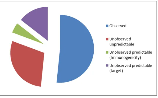
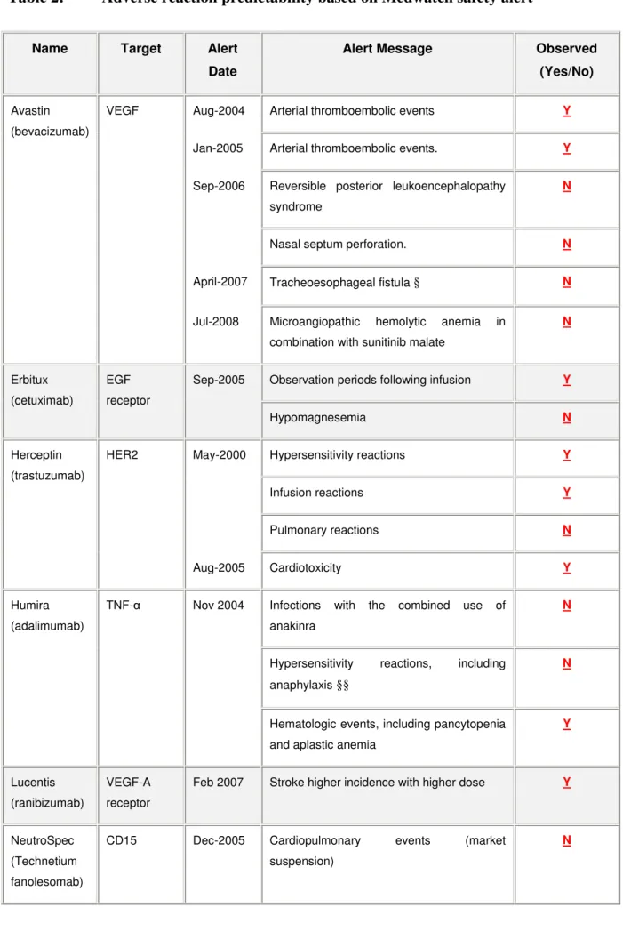
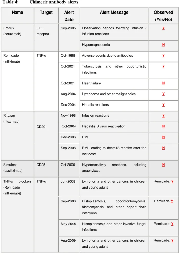
6.1 Predictability of serious adverse reaction alerts for monoclonal antibodies ... 43
6.2 Time to safety alerts ... 54
7 Discussion ... 63
7.1 Predictability of serious adverse reaction alerts for monoclonal antibodies ... 63
7.1.1 Source and reliability of data ... 63
7.1.2 Pattern of adverse reactions ... 64
7.2 Kaplan-Meier “survival analysis” ... 65
7.3 Limitations ... 66
7.3.1 Limitations of predictability evaluation ... 66
7.3.2 Limitations of Kaplan-Meier estimate ... 67
7.4 Predictive methods ... 68
7.5 Rechallenge ... 71
8 Conclusion ... 78
9 Summary ... 80
10 Összefoglalás ... 81
11 References ... 82
12 Own publications ... 92
13 Acknowledgements ... 94
Table of figures
Figure 1: Product-related factors affecting the immunogenicity of biopharmaceuticals ... 11 Figure 2: Process-related actors affecting the immunogenicity of biopharmaceuticals ... 12 Figure 3: Patient-related factors affecting the immunogenicity of biopharmaceuticals ... 13 Figure 4: Disease and treatment-related factors affecting the immunogenicity of
biopharmaceuticals ... 14 Figure 5: Adverse reaction predictability based on Medwatch safety alert : taking into account alerts predictable based on structure and target. ... 44 Figure 6: Kaplan-Meier survival curve of time to Medwatch safety alert ... 57 Figure 7: Points to consider in clinical decision making in the setting of intentional
rechallenge ... 77
2 ABBREVIATIONS
ACE Angiotensin converting enzyme
ADA Anti-drug antibodies
ADR Adverse drug reactions αGal Galactose-α-1,3-Galactose BAT Basophil activation test;
CD Cluster of differentiation
CI Confidence interval
CIOMS Council of International Organization of Medical Sciences CSF Colony-stimulating factor
DNA Deoxyribonucleic acid
DPT Drug provocation test
EGF Epidermal growth factor
ELISA Enzyme-linked immunosorbent assay ELISPOT Enzyme-linked immunospot
EMA European Medicines Agency
FDA Food and Drug Administration
HER Human epidermal growth
HLA Human leukocyte antigen
HSR Hypersensitivity reaction
GM Granulocyte macrophage
IFN Interferon
Ig Immunoglobulin
IL Interleukin
LAT Lymphocyte activation test LTT Lymphocyte transformation test NSAIDs Non-steroidal anti-inflammatory drugs
mAb Monoclonal antibody
MAH Marketing authorization holder
MGDF Megakaryocyte growth and development factor MHC Major histocompatibility complex
PBMC Peripheral blood mononuclear cells
PEG Polyethylene glycol
PD Pharmacodynamic
PK Pharmacokinetic
PML Progressive multifocal leukoencephalopathy Risk MAP Risk minimization action plan
RMP Risk management plan
ROC Receiver-operated curves RSV Respiratory syncytial virus
SD Standard deviation
SPT Skin prick tests
TNF Tumour Necrosis Factor
VEGF Vascular endothelial growth factor US United States (of America)
WHO World Health Organisation
3 INTRODUCTION (REVIEW OF LITERATURE)
3.1 Background on pharmacovigilance of biopharmaceuticals 3.1.1 Current trends in pharmacovigilance: focus on safety prediction
Pharmacovigilance is changing. It is no longer a passive discipline of awaiting and detecting adverse reactions, but active in predicting and managing risks. This approach is taken both by the European authorities in the form of risk management plans (RMP) and the United States Food and Drug Administration (US FDA) in Risk Minimization Action Plans (RiskMAPs).
Even the very definition of pharmacovigilance is maybe no longer appropriate. It was not long ago in 2002 that the World Health Organization proposed the definition of pharmacovigilance as: “The science and activities relating to the detection, assessment, understanding and prevention of adverse effects or any other drug related problem”. Pharmacovigilance today must aim for more proactive approach to predict adverse effects and avoid them, and only detect and assess them where the predictive model fails or proves to be insufficient. While prediction is necessarily performed with a certain degree of uncertainty, a predictive model is indirectly recommended in RMPs and RiskMAPs. The level of uncertainty decreases with accumulating safety data throughout the product lifecycle and the predictive model is continuously refined. In lay terms, predictive model can be described as “educated guesswork” based on the thorough evaluation of available pre-clinical and clinical data on the medicinal product as well as the product class.
Industrial drug discovery aims at identifying drug candidates with the highest possible chance of completing clinical trials, reaching the market, and establishing themselves as efficacious and well-tolerated, safe medicines. Such drug candidates require a balance of favorable pharmacological, pharmacokinetic and physicochemical properties. Prediction of both efficacy (target effect) and safety (absence of off-target effect) starts in-silico. The absence of unintended pharmacological promiscuity, that is, the absence of interactions with non- therapeutic ‘off-targets’, is one important aspect of that balance. Pharmacological promiscuity can lead to adverse drug reactions (ADRs) and has been linked to preclinical findings of toxicity (2). ADRs and animal toxicity account for 30% of all drug candidate termination (3), and the proportion of promiscuous compounds decreases with advancing clinical development (4). The mainstay of pharmacological promiscuity assessment, however, is still the conventional screening against large panels of safety-relevant targets (2).
3.1.2 Immunogenicity and pharmacovigilance of biopharmaceuticals
Pharmacovigilance of biopharmaceuticals deals with all the complexities of conventional small molecule drugs, and on top of that, takes into account its own specificities. For biological drugs the task is, therefore, multiple-fold more complex.
Immunogenicity is the most typical adverse action of biopharmaceuticals. For some biopharmaceuticals this is not the most important adverse action (as demonstrated in the examples provided below), but essentially all biopharmaceuticals have been shown to exhibit some immune mediated adverse effect. Adverse reactions can be immediate – such as infusion reactions, or delayed – resulting from non-immediate action of anti-drug antibody (ADA) formation.
Infusion reactions are most commonly associated with a complex of chills, fever, nausea, asthenia, headache, skin rash, pruritus, etc. (5). However, infusion reactions may also present with a variety of signs and symptoms of severe hypersensitivity reaction. The mechanisms by which biopharmaceuticals elicit infusion reactions are multi-factorial. In addition to immune- mediated reactions, cytokine mediated effects are reported. For oncological therapy, tumour lysis syndrome should be considered in differential diagnosis of immune-mediated reactions.
It is a syndrome in which the destruction of large numbers of rapidly proliferating tumour cells gives rise to hyperuricemia, hyperphosphatemia, and other metabolic abnormalities usually within 24 hours of infusion (5).
Monoclonal antibodies may interact with their molecular targets on circulating blood cells, tumour cells, or effector cells recruited to the tumour site (e.g., rituximab with cluster of differentiation (CD)20), thereby promoting the release of inflammatory cytokines. When released into the circulation, cytokines can produce a wide range of symptoms characteristic of infusion reactions (5). Because a cytokine-dependent mechanism does not depend on prior sensitization, it may contribute to infusion reactions that occur with the first infusion of a mAb. Massive cytokine release may precipitate life-threatening infusion reactions leading to multi organ failure, as in the case of a novel anti-CD28 monoclonal antibody (mAb). No evidence of anaphylaxis was seen (6).
Rituximab is characterised by an outstandingly high induction of infusion reactions compared to other biopharmaceuticals. During the first infusion to patients with relapsed B-cell chronic lymphocytic leukaemia or low-grade B-cell lymphoma, serum levels of Tumour
Necrosis Factor-α (TNF-α) and interleukin-6 (IL-6) peaked at 90 minutes and were accompanied by fever, chills, nausea, vomiting, hypotension, and dyspnoea. The severity of the infusion reaction was related to the number of circulating lymphocytes (7). It seems likely that the infusion reactions typical for rituximab are not due to its immunogenicity but due direct cytokine release.
Immediate-type (Type 1 or) hypersensitivity reactions are generally mediated by immunoglobulin E (IgE), leading to release of histamine, leukotrienes, and prostaglandins.
These pro-inflammatory mediators induce smooth muscle contraction, capillary dilation, and vascular permeability, leading to the development of urticaria, rash, angioedema, bronchospasm, and hypotension. Anaphylaxis, the most severe form of immediate hypersensitivity, is a life-threatening condition that may appear within minutes of starting an infusion. It is characterized by respiratory distress, laryngeal edema, and severe bronchospasm, which may be accompanied by cutaneous and gastrointestinal symptoms, and may lead to a hypotensive crisis (8).
Because prior sensitization is required for immune-mediated hypersensitivity, it would not be expected to occur with the first administration. However, pre-existing IgE that cross-reacts with the drug may be responsible (9), as discussed below for cetuximab.
On the other hand, immunogenicity may not lead to immediate manifestations. Unwanted immunogenicity of erythropoietin leads to formation of neutralizing antibodies without demonstration of immediate type hypersensitivity (10).
It is well established that repeated injection of even native human proteins can result in a break in immune tolerance to self-antigens in some patients leading to a humoral response against the protein that is enhanced when the protein is aggregated or partially denatured (11).
Although in some cases an immune response to a biopharmaceutical has limited clinical impact, ADAs may pose a number of potential risks for the patient. Firstly, an ADA response can adversely affect the pharmacokinetics and bioavailability of a drug thereby reducing the efficacy of treatment. But more importantly, ADAs can also adversely affect the safety of treatment and cause immune complex disease, allergic reactions and, in some cases, severe autoimmune reactions. Serious and life-threatening adverse events can occur when ADAs cross react with an essential non-redundant endogenous protein such as erythropoietin or thrombopoietin. Thus, several cases of pure red cell aplasia were associated with the development of antibodies to recombinant erythropoietin following a change in formulation
(10). Similarly, the development of antibodies to pegylated megakaryocyte growth and development factor (MGDF) cross reacted with endogenous MGDF resulting in several cases of severe thrombocytopenia (12).
In silico models have been used with several notable published successes in predicting immunogenicity of pharmaceuticals. In silico methods are based on the ability of T-helper cell recognition of antigenic epitopes. T-helper cells, a subset of T-lymphocytes specifically recognize epitopes presented by antigen presenting cells in the context of major histocompatibility complex (MHC) Class II molecules. T-helper cells, are the major drivers of the mature antibody response. Protein therapeutics that express MHC Class II restricted T- helper epitopes are likely to elicit more frequent and mature antibody responses with IgG as predominant isotypes. These T-helper epitopes can be represented as linear sequences comprising 8 to 12 contiguous amino acids that fit into the MHC Class II binding groove. A number of computer algorithms have been developed and used for detecting Class II epitopes within protein molecules of various origins. Such “in silico” predictions of T-helper epitopes have already been successfully applied in attempts to increase immunogenicity and efficacy of vaccines (13).
The relationship between T-cell epitopes and immune response has also been the subject of a number of investigations in the field of protein therapeutics. In some cases, therapeutic proteins have also been screened for T-helper epitopes in an attempt to evaluate their potential immunogenicity. Obviously, reliable in silico prediction of helper epitopes would be of significant value in development of protein therapeutics. Such predictions would make it possible to meaningfully rank candidates at the pre-clinical stage of drug development or to reengineer proteins to make them less immunogenic. Furthermore, individuals at higher risk of developing T-cell-driven antibody responses to the protein therapeutic could be identified prospectively using human leukocyte antigen (HLA) typing, if certain HLA can be associated with T-cell response and higher neutralizing antibody titres, as recently described by Barbosa et al (14).
Some in silico algorithms are freely available for public use on the internet.
(http://www.pharmfac.net/allertop/, http://www.pharmfac.net/EpiTOP/). The validity of in- silico and other prediction methods still needs to be demonstrated on a wider scale even for small molecular entities. The use of these methods in the context of clinical trials of protein therapeutics is rather recent and deserves further exploration.
Animal data are considered not to be predictive for immunogenicity assessment of biopharmaceuticals, but they may be useful to detect major differences in immune response.
For example, animal models may in some cases be of value for the comparative immunogenicity assessment of new product candidates. Such an example is chemically modified human factor VIII products for the treatment of haemophilia A developed with the aim of extending the half-life of Factor VIII. Because any chemical or molecular modification of a protein might create new immunogenic epitopes or generate structures that could stimulate the innate immune system, it is reasonable to compare their potential immunogenicity to the non-modified factor VIII molecule before entering clinical development. New mouse models of haemophilia were specifically designed for comparative immunogenicity assessment during preclinical development of modified factor VIII proteins.
One of these models expresses a human factor VIII complementary deoxyribonucleic acid (c- DNA) as a transgene which causes the development of immunological tolerance to native human factor VIII. When immune-tolerant mice are treated with a modified human factor VIII that expresses new immunogenic epitopes, tolerance breaks down and antibodies against human factor VIII develop. Therefore, this model allows for the exclusion of high-risk candidates early during pre-clinical development. The other mouse model of haemophilia expresses a human MHC-class II protein that is associated with an increased risk for the development of antibodies against factor VIII in patients. As all murine MHC-class II genes are completely knocked out in this model, factor VIII peptides that drive anti-factor VIII immune responses are presented by the human MHC-class II protein. Although such models have their limitations, e.g. the human MHC-class II complex is usually highly polymorphic not consisting of only one or two haplotypes, they might help to identify high-risk candidates before entering clinical development (15). The final immunogenicity assessment, as in any predictive model, still requires clinical studies.
3.1.3 Factors influencing the immunogenicity of biopharmaceuticals
Currently available techniques do not permit one to predict with a sufficient degree of accuracy whether a biopharmaceutical will be immunogenic and if so, to what extent (16). It is also difficult to predict which patients will develop an immune response to a particular drug, and at what time during treatment an immune response will occur.
There are, however, a number of both drug-related and patient-related factors that are known
to influence the immunogenicity of biopharmaceuticals, as presented in Figure 1, . Drug- related factors include the presence of nonhuman sequences or novel epitopes generated by amino acid substitution designed to enhance stability, or novel epitopes created at the junction of fusion proteins. Molecular structure, and in particular, changes in glycosylation, can also influence the immunogenicity of a biopharmaceutical. Thus, the absence of glycosylation or an altered pattern of glycosylation can expose cryptic B-cell and T-cell epitopes in the protein, or cause the protein to appear foreign to the immune system (17).
Carbohydrate moieties present upon biopharmaceuticals can elicit the production of IgE antibodies that can cause serious adverse reactions including anaphylaxis even upon the first treatment exposure. Pre-existing antibodies against galactose-a-1,3-galactose (αGal) have been shown to be responsible for IgE-mediated anaphylactic reactions in patients treated with cetuximab (9). Pegylation can reduce the immunogenicity of some proteins although patients produce antibodies to the polyethylene glycol (PEG) residue adversely affecting efficacy (18).
In addition to attributes that can induce a classical immune response, repeated administration of even authentic human proteins such as albumin can under certain circumstances cause a break in immune tolerance leading to the development of an immune response. Thus, the presence of degradation products resulting from oxidation or deamination of the protein, aggregates, or the intrinsic immunomodulatory properties of the molecule can also influence the immunogenicity of a biopharmaceutical. Protein aggregation in particular has long been associated with increased immunogenicity, although the mechanisms underlying this effect remain poorly understood. It has been suggested that aggregated proteins form repetitive arrays that can lead to efficient cross-linking of B-cell receptors, leading to B-cell activation in the absence of T-cell help, thereby resulting in a break in immune tolerance to self-proteins (19).
The relatively high incidence of ADAs in patients treated with recombinant granulocyte macrophage colony-stimulating factor (GM-CSF) may be related at least in part to the immunostimulatory properties of the molecule itself (18). Thus, GM-CSF can recruit antigen- presenting cells to the site of antigen processing, stimulate the maturation of myeloid dendritic cells, and enhance an antigen-specific CD8+ T-cell response, suggesting that repeated administration of GM-CSF may function as an adjuvant. Indeed, GM-CSF has been used as an immunological adjuvant in a number of vaccination protocols designed to elicit an immune response to self-antigens (20).
Figure 1: Product-related factors affecting the immunogenicity of biopharmaceuticals
Product-related factors
Foreign sequences Amino acid substitution
Aggregates Glycolysation changes
Pegylation . Native protein
Increasingimmunigenicity
Process-related impurities, including traces of residual DNA or proteins from the expression system, or contaminants that leach from the product container, can also influence the immunogenicity of recombinant biopharmaceuticals (22).
Figure 2: Process-related actors affecting the immunogenicity of biopharmaceuticals
Process-related factors
Presence of host-cell proteins and DNA
Processing reagents
Container derived material (silicone, glass, metal)
Storage conditions
Increasingimmunigenicity
Patient-related factors, such as genetic makeup, age, gender, disease status, concomitant medication, and route of administration, can also influence the immune response to a particular biopharmaceutical. For example, a common MHC class II allele, DRB1*0701, is associated with the antibody response to interferon-β in multiple sclerosis patients (17).
Disease state and immune competency also influence an individual’s immune response to a treatment with a biopharmaceutical. Thus, development of antibodies to pegylated MGDF is less frequent in cancer patients who tend to be immunosuppressed than in healthy individuals (12).
Figure 3: Patient-related factors affecting the immunogenicity of biopharmaceuticals
Patient-related factors
Absence of endogenous protein (replacement therapy)
MHC haplotype
Ethnicity
Age, gender
Increasingimmunigenicity
Concomitant therapy with immunosuppressive drugs can also influence a patient’s immune response to a biopharmaceutical. Thus, administration of methotrexate together with the chimeric monoclonal antibody infliximab has been shown to reduce the immune response to infliximab and improve the clinical response in patients with rheumatoid arthritis (23). The duration of treatment and the route of administration also influence the immune response to a biopharmaceutical.
Typically, administration of a protein in a single dose results in the production of low-affinity IgM antibodies, while repeated administration results in the production of high-affinity and high-titer IgG antibodies, which may be neutralizing. Thus, in patients with multiple sclerosis treated with interferon-β neutralizing antibodies to IFNβ often do not appear until after several months of therapy (24).
The intravenous route of administration is considered to be least likely to generate an immune response to a biopharmaceutical compared with intramuscular or subcutaneous administration (15).
Figure 4: Disease and treatment-related factors affecting the immunogenicity of biopharmaceuticals
Disease and treatment-related factors
Autoimmune disease Repeated intermittent dosing
sc -> im -> iv administration Immunosuppressive disease Immunosuppressive therapy
Single administration
Increasingimmunigenicity
The complexity of the humoral response to biopharmaceuticals and the difficulty in establishing the effect on ADAs on drug efficacy is illustrated by the response of patients to treatment with IFNβ, for the treatment of relapsing remitting multiple sclerosis. Five products are currently available in the US and Europe as first-line disease-modifying agents for the treatment of relapsing remitting multiple sclerosis , IFNβ-1a (Avonex ® and Rebif®), IFNβ- 1b (Betaseron® and Betaferon®), and more recently, the IFNβ-1b biosimilar Extavia®.
Avonex® and Rebif® are both glycosylated forms of native human IFNβ-1a produced in Chinese hamster ovary cells. Betaseron® and Extavia® are a nonglycosylated form of IFNβ- 1a produced in Escherichia coli that has a serine substitution for the unpaired cystine at position 17 of the native protein. Most patients develop an antibody response to IFNβ products, and as many as up to 45% of patients develop neutralizing antibodies to IFNβ, in some cases as early as 3 months after initiation of therapy. Overall, some 25% of patients develop anti-IFNβ-neutralizing antibodies usually within 6 to 18 months. ADAs are more frequent in patients treated with IFNβ-1b than IFNβ-1a, while subcutaneous IFNβ-1a (Rebif®) is more immunogenic than intramuscular IFNβ-1a (Avonex®) (32). The immunogenicity of IFNβ varies among individuals, both as a function of the presence of particular MHC class II alleles, and as a function of IFN-receptor expression. Thus, patients who process the DRB1*0701 allele, or who express low levels of IFNAR2, one of the two
chains of the type I IFN receptor, upon initiation of treatment, have a significantly higher risk of developing anti-IFNβ neutralizing antibodies (33). Although it has been shown in numerous trials that patients who develop antibodies against IFNβ have higher relapse rates, increased number of lesions detected by MRI, and higher rates of disease progression, the significance of anti-IFNβ ADAs remains controversial (34). This is due to the difficulty in establishing a temporal correlation between the presence of anti IFNβ ADAs and the loss of drug efficacy due to the variable nature of the disease, the partial effectiveness of the drug, the delay between initiation of treatment and the detection of an effect of the drug on the course of the disease, and the difference in the immunogenicity of different IFNβ products. The lack of standardized ADA assays has also rendered direct comparisons of immunogenicity between different products and different studies difficult, which has contributed to the difficulty in establishing a correlation between ADAs and loss of drug efficacy.
The assessment of efficacy described above for multiple sclerosis is complex enough, but still comparatively well-grounded in quantifiable and comparable assessment of relapse rate and the number of inflammatory lesions. On the other hand, the assessment of safety takes into account all of the complexities described above for efficacy, and additionally needs to account the relatedness, relevance and severity of reactions in the background rate of adverse events in the given population. Singling out adverse reactions which may be due to development of ADAs and other immunological mechanisms from the reactions due to target effect of the drugs seems like an unachievable aim.
3.1.4 Regulatory guidance
Assessment of immunogenicity is an important component of drug safety evaluation in preclinical, clinical, and post-marketing studies. Draft Guidance for Industry Assay Development for Immunogenicity Testing of Therapeutic Proteins has been published by the US Food and Drug Administration (35). Similarly, guidelines on the immunogenicity assessment of biotechnology- derived therapeutic proteins established by the Committee for Medicinal Products for Human Use of the European Medicines Agency (EMA) came into effect in April 2008 (36). These guidelines provide a general framework for a systematic and comprehensive evaluation of immunogenicity that should be modified as appropriate, on a case-by-case basis. Although differences in approach and emphasis exist between the US, European Union, and Japanese regulatory authorities there is, nevertheless, a large degree of
consensus on the type of approach that should be adopted; namely, a risk-based approach that is clinically driven and takes into account pharmacokinetic data. Thus, biopharmaceuticals with no endogenous counterpart are considered to be of relative low risk, while drugs with a non-redundant endogenous counterpart are considered to present a high risk. A multi-tiered approach to testing samples is also recommended. This consists of an appropriate screening assay capable of detecting both IgM and IgG ADAs, the sensitivity of which is such that a percentage of false-- positive samples would be detected. The specificity of the samples that test positive in the screening assay are then re-assayed in a confirmatory assay usually by competition with an unlabelled drug using the same assay format as that used for the screening assay. Samples that test positive in the screening and confirmatory assays are then tested for the presence of neutralizing ADAs using a cell-based assay whenever possible.
The prediction of both incidence and clinical significance of immunogenicity is still problematic. Therefore, the recommended approach is to apply suitably sensitive bioanalytical methods to detect host responses to the drug product and to relate these to clinical correlates of pharmacokinetics (PK), efficacy and safety. The current, commonly used, and in most cases recommended bioanalytical approach, is a three-stage process consisting of screening, confirmation, and characterisation: If blood samples are found to be positive for ADA during screening, these samples are then subjected to a confirmatory assay (e.g. competitive inhibition enzyme-linked immunosorbent assay (ELISA)) ensuring that ADA are binding specifically. Having established that positive findings do not result from non-specific interactions such as with materials in the assay milieu (e.g. plastic, other proteins), ADA need to be characterised. Typically, this characterisation includes assessment of ADA neutralising capacity. Furthermore, assays for relevant biomarkers and/or pharmacokinetic measurements should complement ADA characterisation and analysis of their in vivo impact (17).
Clinical consequences of immunogenicity may be comprised of acute consequences, such as anaphylaxis or infusion reactions, non-acute consequences (e.g. loss of efficacy), cross- reactivity with and neutralisation of natural endogenous counterparts, and delayed hypersensitivity. Therefore, the clinical outcome of unwanted immune responses differ widely depending on the affinity, class, amount and persistence of ADA generated, the epitope recognised by the biotherapeutic protein, and the ability of ADA to activate complement. This diversity of causes and consequences underlines the importance of the systematic evaluation of immunogenicity during clinical trials (15).
3.1.5 Biosimilars or similar biotherapeutic products
Biosimilar products present a specific challenge not only for immunogenicity assessment, but for safety and efficacy assessment overall. The potential for altered immunogenicity needs to be considered even if comparative physicochemical and biological data on product quality do not indicate any difference. Because the predictability of non-clinical studies for the evaluation of immunogenicity is low, routine monitoring of patient samples might be required during clinical trials. In this respect, the extent of immunogenicity studies (clinical evaluation prior to or after authorisation of the change) might be based on a risk analysis that pays regard to both the nature of any observed differences and their potential clinical impact. Emerging technologies might provide additional data for the further evaluation of potential immunogenicity induced by the change introduced in the process.
The current understanding is that the biosimilar and the innovator product should be identical on the aminoacid level and any difference needs to be justified, e.g. in posttranslational modifications. However, this approach is being challenged and current recommendations call for accepting highly similar to the reference medicinal product in physicochemical and biological terms. (36) This is formulated in draft guidelines released for public consultation by the EMA in 2013, but is subject to possible modification. For assessment of immunogenicity, human data are always required. Animal data, even if potentially useful to detect major differences in immune response, are considered not to be predictive. An optimised antibody testing strategy with detailed sampling protocols, a sensitive validated screening assay and further characterisation of ADA, if detected, is requested. (15)
Immunogenicity data (on both the biosimilar and the reference product) are usually required for interpretation of the results. The pre-licensing immunogenicity database is expected to exclude excessive immunogenicity of the biosimilar relative to the reference product (39) but further data may be requested post-marketing. According to the European Union guidelines on the immunogenicity assessment of biotechnology- derived therapeutic proteins: further systematic immunogenicity testing might become necessary after marketing authorization, and may be included in the RMP. This may be applicable in particular in situations when rare and serious ADA-related adverse reactions have been encountered with the reference product or the substance class. Because of these regulations, biosimilars in the European Union show close resemblance to their reference product with respect to quality, efficacy and safety. In this respect, global consistency is needed because ‘copy versions’ of innovator biologicals are
licensed in various other countries without a clear regulatory pathway and based on different data requirements.
It is important to use appropriate terminology for biosimilars. “Biosimilar” should be used in the context of a product meeting the regulatory requirements for a biosimilar within the given legislation. It should not be used for nay product which is similar or bears resemblance, but this has not been approved by a regulatory authority. Whereas the term biosimilar is used both by European legislator and the US FDA, it has to be emphasized that the term may be used only if such a product has gained approval. Therefore biosimilar according to European Union may not necessarily be biosimilar according to US standards, despite the harmonisation efforts which are being undertaken. In addition, various other legislations and organisations apply different terminologies. In Canada, for example, the term used is “subsequent entry biological” (40) whereas World Health Organisation (WHO) uses the term “similar biotherapeutic products” (41).
Due to the general lack of standardised assays, comparative immunogenicity data (on both the biosimilar and the reference product) are usually required for interpretation of the results. The pre-licensing immunogenicity database is expected to exclude excessive immunogenicity of the biosimilar relative to the reference product but further data may be requested post- marketing, especially when rare and serious ADA-related adverse reactions have been encountered with the reference product or the substance class. Because of these regulations, biosimilars in the European Union show close resemblance to their reference product with respect to quality, efficacy and safety. In this respect, global consistency is needed because
‘copy versions’ of innovator biologicals are licensed in various other countries without a clear regulatory pathway and based on different data requirements. Moreover, such non-innovator products are often called “biosimilars” despite the lack of a (thorough) comparison with the original product and even in the presence of clear differences. Therefore, WHO has developed the ‘Guidelines on evaluation of similar biotherapeutic products’ which in principle is in line with the European Union requirements (41).
3.2 Tests used for prediction of immunogenicity
In vivo and in vitro tests are used in diagnosis of drug hypersensitivity. In vivo skin tests such as prick, patch, and intra-dermal tests are the most readily available tools. Most readily refers to the ease of performing with the minimal need for use of specialized equipment and
reagents. Determination of specific IgE levels is still the most common in vitro method for diagnosing immediate reactions. New diagnostic tools, such as the basophil activation test, the lymphocyte activation test, and enzyme-linked immunospot assays for analysis of the frequency of antigen-specific, cytokine-producing cells, have been developed for evaluating either immediate or non-immediate reactions (42).
However, neither specificity nor sensitivity of allergologic tests is100%. Therefore in selected cases provocation tests (i.e. rechallenge with primarily diagnostic purpose) are necessary. As provocation testing caries a significant risk, it should be performed only when necessary.
In selecting diagnostic tests it is important to consider whether the reaction is immediate or non-immediate. The tests which were identified as currently most frequently used are summarized in Table 1 (42).
Table 1: Diagnostic tests of hypersensitivity reactions to drugs
Immediate reactions occur within the first hour after the last drug administration and are manifested clinically by urticaria, angioedema, rhinitis, bronchospasm, and anaphylactic shock. Non-immediate reactions occur more than 1 hour after the last drug administration.
The main non-immediate reactions are maculo-papular eruptions and delayed-appearing urticarial exanthema. Immediate allergic reactions are thought to be IgE-mediated and have been extensively studied, whereas the mechanisms involved in non-immediate reactions seem
Type of reaction Type of tests
Immediate In vitro Specific IgE assays
Flow cytometric Basophil activation tests In vivo Skin tests
Provocation tests
Nonimmediate In vitro Lymphocyte transformation tests or Lymphocyte activation tests
Enzyme-linked immunospot assays for analysis of antigen-specific, cytokine- producing cells
In vivo Delayed-reading intradermal tests Patch tests
Provocation tests
to be heterogeneous. However, clinical and laboratory studies indicate that a T cell–mediated pathogenic mechanism is often involved in macula-papular rashes. This mechanism has also been demonstrated in other non-immediate reactions, such as urticarial manifestations, angioedematous manifestations, or both; toxic epidermal necrolysis; bullous exanthems; drug reaction with eosinophilia and systemic symptoms; and acute generalized exanthematous pustulosis (43).
In non-allergic hypersensitivity reactions to drugs, inflammatory mediators are released by nonspecific immunologic mechanisms. The drugs most frequently responsible for such reactions are traditional small molecule drugs (e.g. non-steroidal anti-inflammatory drugs), with biological agents increasingly involved (44).
3.2.1 Skin tests
There is only a small number of drugs for which skin testing can provide useful information e.g. penicillin, muscle relaxants and carboplatin skin testing. However, for most drugs the relevant immunogen (intermediate metabolite) is unknown and therefore the predictive value of skin testing remains undetermined. Both false-positive and false-negative results may occur (45).
Skin prick tests (SPTs) for specific IgE-mediated drug reactions are useful for the diagnosis of reactions with both low molecular weight and high molecular weight agents. Tests are normally carried out at therapeutic concentrations unless the drug possesses intrinsic histamine-releasing activity in which case a dilution may be appropriate to avoid false- positive results (45).
Skin tests have to be applied according to the suspected pathomechanism of the drug hypersensitivity. An IgE-mediated reaction can be demonstrated by a positive skin prick and/or intradermal test after 20 min. On the other hand, non-immediate reactions to β-lactams manifesting by cutaneous symptoms occurring more than one hour after last drug intake, are often T-cell mediated and a positive patch test and/or a late-reading intradermal test is found after several hours or days. Moreover, skin tests have the additional capability to give insights concerning the immunologic pathomechanism (46).
There are other diseases where immunological reactions to drugs could be involved, but skin testing has generally not been found helpful. For example, renal or hepatic manifestations may occur as a part of a generalized allergic reaction (e.g., in “drug reaction with eosinophilia and systematic symptoms”). This is an obvious demonstration of the inappropriateness of the
“one size fits all” approach, and the necessity of tailoring the testing strategy according to the specific drug and specific pathology (46).
The negative predictive value of skin tests is generally low. This may be partly due to the fact that physiologic metabolites rather than the active drug itself is responsible for the reaction and because many drugs are haptens, which have to be conjugated with a carrier protein before becoming an allergen. Thus, a negative skin test to a drug alone is unreliable for ruling out drug allergy. In the case of a negative skin test, one should consider proceeding to more hazardous drug provocation tests after carefully evaluating the risks and the benefits in the specific patient. On the contrary, even when a proper technique and proper drug material are employed, a positive skin test result does normally indicate the diagnosis. The positive predictive value of a skin test tends to be high, provided that a sufficient number of controls have been tested negative with exactly the same methodology (46).
Skin tests, such as patch, prick, and intracutaneous tests are the most readily available form of allergy testing for physicians, but often do not yield positive reactions, even in patients with well documented histories. Provocation tests i.e. intentional diagnostic drug rechallenge are considered to be the gold standard, but they are not well accepted by patients, because of the risk of severe reactions and are therefore restricted to certain specialist centres with resuscitative equipment. Moreover, for delayed reactions provocation tests are not standardized and a single dose may exclude an IgE-mediated reaction, but not a delayed reaction, which may appear after a higher dose and longer treatment. (46)
A SPT is done by pricking the skin percutaneously with a prick needle through an allergen solution. It is the safest and easiest test, but only moderately sensitive for immediate drug reactions. An intra-dermal test is accomplished by injecting 0.02–0.05 ml of an allergen intra- dermally, raising a small bleb measuring 3 mm in diameter. The intra-dermal test is more sensitive than the SPT, but also carries a higher risk for inducing an irritative, falsely positive reaction and might even lead to an anaphylactic reaction in IgE-dependent reactions. Certain drugs have to be discontinued prior to skin testing (antihistamines, glucocorticoids). The
patient should be free of infectious diseases, fever or inflammatory reactions at the time of testing, unless the skin test is urgently needed. The intake of β-adrenergic blocking agents should be discontinued (usually for 48 h,) according to their half-life of elimination, if the drug to be tested had induced an anaphylactic reaction, as these drugs may interfere with treatment of a possible systemic reaction elicited by the skin test. SPT should be performed on the volar aspect of the forearm. If this is negative after 15–20 min, an intra-dermal test can be performed on the volar forearm, although other regions can be tested (however, there is no comparison for drug allergens). The pain of intra-dermal tests may limit their use in young children.
Normally these tests are well tolerated, but in highly IgE sensitized patients generalized symptoms (urticaria and anaphylaxis) might appear.
Readings should be taken after 15–20 min if immediate reactions are analysed, and after 24 and 72 h for evaluation of non-immediate (late) reactions. In selected cases, additional readings (e.g., after 96 h) are sometimes recommended, as time intervals between testing and positive test reactions may vary. Immediate reactions are documented by measuring the mean diameter of the wheal (and erythema) of the test preparations and the negative control directly after the injection and after 15–20 min. In order to compare the results, a morphological score should be applied as well, enabling a later comparison of different scoring systems. The preferred documentation manner is outlining the size of the injected area and of the reaction at 15–20 min on a translucent cellophane tape. A body of experience has been gained using skin tests in small-molecule drugs, while specific recommendation for biologicals are lacking.
Even within the field of small molecule drugs, a certain level of extrapolation is performed from the most commonly performed tests, such as penicillin tests, and the principles well established for penicillin are applied to skin testing with other drugs. As a criterion for positivity, it is current recommended to employ the criteria used in the diagnosis of penicillin allergy. Reactions are considered positive when the size of the initial wheal increases by 3 mm or greater in diameter after 15–20 min and is associated with a flare (46). Complicating the evaluation is, not only the variability between individual drugs, be they small molecule or biological, but also the multiple possible reactions a drug can cause. Even for one same drug, it is possible that different mechanism may be involved and that the same drug demonstrates different types of immune reactions (47).
In addition to immediate reactions, late reactions, such as delayed or late-phase reactions, should always be examined. They are documented by the diameter of erythema, papulation/infiltrate and morphological description, such as erythematous swelling, erythematous infiltrate, erythema only, eczema with papulation and/or vesicles. Any infiltrated erythema is considered to represent a positive reaction (46).
There is a consensus of opinion that skin tests should be performed after a time interval which allows resolution of clinical symptoms, clearance from the circulation of the incriminated drugs and anti-allergic medications. However, it is not known whether the reactivity might be higher (e.g., cellular hyper-reactivity) or lower (e.g., initial histamine depletion of mast cells or tolerance) if skin tests are performed directly after the reaction (within the next few days).
It is also not known to what extent the sensitization to a drug decreases over time. Thus, many groups carry out tests after some minimal time interval of, for example, three weeks, but not after more than three months, if possible (46). This testing strategy, however, appears to be based on common practice and common sense, rather than evidence-based. In particular, this has not been evaluated based on clinical trials.
3.2.1.1 Systemic reactions from skin testing
There are some patients experiencing systemic reactions after skin testing. Patients who had a life-threatening drug hypersensitivity are at risk, even if there is a long time interval between the drug hypersensitivity and skin testing (51). Even fatal outcomes were reported, ass presented in a review published in 1987 (52). However, it is argued that these events were associated with biologic products that are no longer used, such as horse serum-derived tetanus or dyphtheria toxins or pneumococcal antiserum. In the last thirty years the occurrence of systemic reactions, at least with SPT for inhalant allergens extracts, has decreased dramatically. The recent surveys suggest that the overall risk of inducing anaphylactic reactions by SPT is less than 0.02 %, whereas intra-dermal test is more likely to induce systemic reactions. (53) Given the lower specificity and increased risks, intra-dermal test is no longer recommended as first-choice, but for selected diagnostic procedures (49).
Due to the rarity of severe reactions following skin tests, it is difficult to clearly identify all the possible risk factors in the general population, but some basic recommendations can be suggested. Case-control studies gathering data from different centres are needed to evaluate precisely the exact risk factors. In high-risk patients a risk-benefit analysis has to be done: is
the skin test necessary? Are all precautions taken in case of some reactions occur? The risk- benefit analysis has to be made in regard to the clinical reaction, the possibilities of treatment for a possible adverse reactions, the risk for the patient and the importance of the drug. If ever possible, pregnant women should not been tested. The drug should initially be tested with a higher dilution of the test preparations. The next concentration step has to be applied only if the higher dilution has yielded a negative result. In severe, non-immediate reactions it has to be considered to extend the time interval between tests and not to perform intra-dermal test with the highest concentration before performing patch tests (46).
3.2.1.2 Interpretation of skin-test results
Reliable skin test procedures for the diagnosis of drug hypersensitivity are generally missing and test concentrations are unknown or poorly validated for most drugs. For drugs suspected of causing severe reactions or where literature/experience is lacking, skin tests should use nonirritant concentrations of the drug. This can be established using different dilutions of increasing drug concentration (48).
In a large number of patients presenting Hypersensitivity reaction (HSR), no positive findings on either in vivo or in vitro tests is demonstrated. This may be either due to the lack of adequate test reagents or procedures, or may indicate a non-immune pathological mechanism.
The negative predictive value of skin tests is generally low. This may be partly due to the fact that physiologic metabolites rather than the active drug itself is responsible for the reaction and because many drugs are haptens, which have to be conjugated with a carrier protein before becoming an allergen. Thus, a negative skin test to a drug alone is unreliable for ruling out drug allergy. In the case of a negative skin test, one should consider proceeding to more hazardous drug provocation tests after carefully evaluating the risks and the benefits in the specific patient (46).
On the contrary, even when a proper technique and proper drug material are employed, a positive skin test result does normally indicate the diagnosis. The positive predictive value of a skin test tends to be high, provided that a sufficient number of controls have been tested negative with exactly the same methodology (46).
The effect of concomitant drugs should s be taken in consideration in the interpretation of the results. Most significant are drugs used for systemic immunosuppression. The effect of
systemic corticosteroid therapy on allergic patch test reactions has been researched. A recent randomized, double blind study involved patch testing individuals with a known nickel allergy to a nickel sulphate dilution series, both during treatment with a 20 mg daily dose of prednisolone and with placebo. Twenty milligrams of oral prednisolone significantly decreased the total number of positive nickel patch tests, increased the threshold concentration for eliciting reactions, and shifted the degree of reactivity towards weaker reactions (54).
Patients with suspected allergies but taking immunosuppressive agents may not always be investigated due to the assumption that positive results would be suppressed. Many patients are heavily dependent on their drugs to control their underlying condition and stopping them before skin tests might be unethical or may lead to a disease flare making testing impossible.
There are only few patients in this group and the clinical question remains whether they can be reliably tested or not. A small recent series showed that positive reactions can be seen in patients taking azathioprine, ciclosporin, methotrexate, mycophenolate mofetil, tacrolimus, infliximab, adalimumab, and etanercept (55). The relevance of reactions in this cohort of patients were varied, with some being significant to their presentation and others being of old or uncertain relevance. This study could not, however, shed light on what degree some allergic reactions may have been suppressed by particular immunomodulating drugs.
Importantly, in any situation where the mechanism of ADR is unknown a negative result is unreliable.
3.2.1.3 Skin testing with biopharmaceuticals
The experience with skin testing with small molecule drugs is extensive. It has generally focussed on drugs known to induce anaphylactic reactions such as beta-lactam antibiotics, but also a range of other drugs. The use of skin tests for diagnosis of biopharmaceutical-induced immunogenicity is more recent. There are some notable examples of successful use (56). Skin testing has been successfully used alone or in combination with anti-drug antibody assessment. It has been found useful in predicting serious immune reactions and in assessing the risk and need for desensitisation.
Specific characteristic of biopharmaceutical is that they are essentially all administered parenterally: generally either intravenously or subcutaneously. Every drug administration is therefore an equivalent to a skin test and characteristics of injection site reactions can be
correlated with systemic reactions. As for small molecule drugs, irritative and immune- mediated injection site reactions may be distinguished. The formation of anti-drug antibodies (ADAs) may promote immune-mediated injection site reactions that likely represent either anaphylactic type I reactions, or cutaneous Arthus-like type III reactions according to the Coombs and Gell classification (57). A differentiation between these reactions by clinical course and skin testing may help to decide if treatment should be stopped to avoid the development of more severe ADRs if injection site reactions occur.
The methodology does not vary significantly and the principles tests for small molecule drugs may be applied. With all the limitations discussed above, skin testing may and should be considered as a part of a developmental RMP for biopharmaceuticals in pre-authorization phase. European regulations i.e. the EMA Guideline on Immunogenicity Assessment of Biotechnology-Derived. Therapeutic Proteins suggests that any test which is found useful can be applied i.e. that ongoing consideration should be given to the use of emerging technologies (novel in vivo, in vitro and in silico models) (58). With the advances in standardization of testing, even the old traditional methods such as skin testing may be suitable and useful for modern drugs.
Understandably, skin testing may not always be appropriate, or may not be necessary in for low-risk drugs. On the other hand, the ease of use in the sense of ready availability makes it an appropriate test to consider. As always, the standardization and validation in sufficient number of samples is required for any test. This is an additional reason why a structured and planned testing strategy may be recommended for a large number of drugs in development.
The use of skin tests in post-authorization phase can equally be considered as part of post- authorization RMPs or as part of routine clinical use. Of course, depending on the particular circumstance and assessment procedure-associated risk, patient inconvenience and cost versus benefit in terms of adverse reaction risk reduction.
3.2.2 In-vitro tests
In vitro tests would be a safe procedure for patients, avoiding possible disadvantages of in- vivo and provocation tests: new or recall sensitizations to the drug and risk of severe adverse reactions. Moreover, they provide deeper insight into the pathomechanisms involved in drug
hypersensiticity and allow simultaneous assessment of immune responses to multiple drugs.
Nevertheless, it is necessary to be aware of the limitations of these tests: they are partly still research tools, standardization is not done for each drug with each test, as exposed controls are missing; They are useful only for certain type of drug allergies (not class II/III reactions according to Gell and Coombs classification) and reflect a sensitization, which is just a risk factor for a symptomatic immune reaction after re-exposure to the drug. However, similar limitations apply to skin tests and provocation tests (59).
3.2.2.1Antibody determination
The incidence and magnitude of antibody formation depend on a balance of ‘foreignness’ and the tolerance to the protein. Immune responses to protein drugs that are foreign (‘non-self’) proteins, or contain portions of a foreign protein, resemble immune responses to vaccines: in most cases, neutralizing antibodies appear that bind to the active site of the biologic and inhibit (‘neutralize’) its potency by preventing target binding. Protein products that are foreign originate from, or are expressed from, bacteria, plants and nonhuman mammalian systems, such as streptokinase, staphylokinase and the mAb OKT3 (Orthoclone; Ortho Biotech, Bridgewater, NJ). Neutralizing antibodies to such products could also bind to an unrelated site and hinder activity by inducing conformational change. Non-neutralizing antibodies bind to sites on the drug molecule without affecting target binding. Non-neutralizing antibodies are often incorrectly referred to as ‘binding antibodies’; but all ADAs (including neutralizing antibodies) are inherently binding antibodies. Although non-neutralizing antibodies do not abolish target binding, they can lower drug bioavailability by increasing the rate of clearance, resulting in an outcome (lowered drug efficacy) that is clinically similar to that observed with neutralizing antibodies. Thus, non-neutralizing antibodies that lower pharmacokinetic parameters are sometimes considered ‘clinically neutralizing’, but such ADAs are better classified as ‘clearing antibodies’ (60).
Immune responses to drugs that are structurally identical to human proteins (‘self ’) are induced by a different mechanism that is based on breaking immune tolerance. How tolerance is induced or broken is not completely understood, but it has been observed that the repetitive administration of proteins and the dose level affect it. Breaking tolerance led to the generation of ADAs against human IFNs, interleukin 2, GM-CSF, erythropoietin and thrombopoietin. It can also occur when a protein is denatured or modified, creating a new antigenic determinant (for example, fusion proteins), when a contaminant is introduced during by
formulation changes (for example, erythropoietin) or when a human protein is given along with a potent adjuvant to enhance its immunogenicity (for example, tumor antigens). In most such instances, patients initially produced ADAs with undetectable neutralizing ability but ultimately developed detectable neutralizing antibodies (61).
Because most therapeutic mAbs developed today are human or humanized, the most likely target for ADAs are the hypervariable or complementarity-determining regions (CDRs) that provide the majority of binding contacts. The immunogenicity of CDRs often leads to the production of neutralizing antibodies, but non-neutralizing antibodies to these sequences or other parts of the mAb may also be elicited.
Presumably, the incidence of ADAs and neutralizing antibodies within the drug development phase predicts anti-drug immune response incidences in clinical practice (post-marketing).
Yet the incidence of ADAs and neutralizing antibodies in controlled clinical studies may not reliably estimate that seen during the post-approval stage, with larger numbers of exposed subjects, more concomitant medications, repeated drug re-exposures and reduced patient treatment compliance. Nonetheless, measuring drug-induced ADAs during drug development is important. To do this, and to provide context to immunogenicity data, it is vital to understand the test methods used and their caveats.
Two types of platform technologies exist:
(i) immunoreactivity assays such as radioimmunoassay, surface plasmon resonance or enzyme-based solid-phase immunoassays, to detect ADAs; and
(ii) functional cell-based bioassays or target binding (receptor recognition) inhibition- based immunoassays for the characterization of the neutralizing antibodies subset of ADAs.
Both assay types can be used together for the complete characterization of the antibody response against a drug molecule. The ADA immunoreactivity assays can be further divided into three subtypes that include:
first, a sensitive screening immunoassay to identify samples potentially positive for ADAs;
second, a specificity confirmation immunoassay that eliminates false positives; and
third, an immunoassay to obtain a relative measure or titre of the ADA concentration in serum.
When appropriate, cell-based neutralizing antibody bioassays or target binding inhibition– based neutralizing antibody immunoassays are also conducted to characterize the neutralizing ability of the ADAs. Samples can also be characterized for ADA isotyping by immunoassay, but the value of this approach may be limited. Sensitive detection assays combined with appropriate characterization of the ADAs can provide helpful information directly related to patient safety and treatment as well as overall understanding of the humoral immune response to therapeutic proteins.
Whereas the development and validation of sensitive and reproducible methods should be the goal for ADA bioanalysis, at present, standardized assays are not available and reference standards are rarely available, which make it difficult to compare results obtained from different laboratories and different studies. The incidence of ADA may also be limited by the assay method used—for example, low-affinity ADAs by surface plasmon resonance versus immunoassays that use multiple wash steps. Similarly, some additional limitations of ADA test methods must also be understood.
First, the ‘sensitivity’ of a method is dependent on the affinity of the positive control used to characterize it, making it inappropriate to compare across test methods employing different positive controls, and even more so for ADA test methods of different products.
Second, the therapeutic protein often interferes with ADA assays, and this ‘drug tolerance limit’ is generally characterized; in such instances, it is a common malpractice to apply the drug tolerance limit in deciding a subject’s ADA status (that is, when ADAs are undetectable and the drug level in that sample is below the drug tolerance limit, it is reported as ADA negative). Because the tolerance limit, like sensitivity, is dependent on the affinity of the individual ADA and drug, it cannot be represented by the tolerance limit of the assay positive control. Thus, drug tolerance limits should not be used in determining ADA status; instead, study designs should allow for the collection of data from at least one time point where drug has been fully cleared from the circulation. The assessment of treatment-emergent ADAs should be made per individual subject and should use a prospective decision tree to characterize the subject appropriately.
Third, neutralizing antibody assays—whether cell-based bioassays or target binding inhibition–based immunoassays—are also limited by sensitivity, and lack of neutralizing activity in these assays does not confirm that the ADA is a non-neutralizing antibody (60).
For all these reasons, it is inappropriate to compare ADA incidence rates between different drug products, and certainly between products from different companies. In fact, the US Food and Drug Administration (FDA) has required that biological product package inserts explicitly state that comparisons can be misleading.
3.2.2.2In vitro diagnostic tests of cell-mediated immunity
As in vitro tests rely on the presence of drug-reactive immune cells in blood of drug-sensitised patients, persistence and frequency of these cells has a crucial impact on in vitro diagnosis of drug hypersensitivity. Beeler et al (15) demonstrated that 1:250 – 1:10 000 of T cells in the peripheral blood of patients in the remission react to the relevant drug. This study also showed that T cells can persist as memory cells in peripheral blood of drug-allergic patients for up to 12 years after disease outcome. On the contrary some patients can lose reactivity 1–3 years after the original treatment with drugs that caused hypersensitivity reaction (62). At present, it is impossible to predict how long the reactivity of drug-specific T cells in an individual patient will persist.
In vitro tests are normally done during remission of disease, because peripheral blood mononuclear cells (PBMC) obtained ex vivo from acute drug-allergic patients are strongly activated. This could lead to high background proliferation and difficulties in detecting an enhanced proliferation after drug stimulation. The time interval between acute stage and test performance allows washing out the incriminated drugs and any anti allergic drugs, which may suppress the immune response in vitro. Thus, according to common opinion, in vitro tests should be performed after a minimal time interval of 3 weeks after the DHR. An analysis in the first 6 months or minimally first year is recommended, but later tests may still be positive due to the long-persisting T cells specific for the drug (59).