PROINFLAMMATORY ACTIVATION OF ENDOTHELIAL CELLS
Ph.D. Thesis
Veronika Makó
Semmelweis University
PhD School of Molecular Medical Sciences
Supervisor: Zoltán Prohászka, MD, D.Sc.
Opponents: Péter Hamar, MD, Ph.D.
István Krizbai, MD, Ph.D.
Head of doctoral committee: Imre Oláh, MD, D.Sc.
Members of doctoral committee: Éva Pállinger, Ph.D Gábor Réz, Ph.D.
Budapest 2011.
1. INTRODUCTION
The inflammatory response of endothelial cells is of critical importance in various diseases, such as (septic or anaphylactic) shock, thrombosis, atherosclerosis and chronic venous insufficiency.
Controlling inflammation at the level of endothelial cells may efficiently retard or reverse the pathogenetic process; therefore, understanding the differences among the signaling pathways of inflammatory factors is indispensable. Inflammatory signals from exogenous and endogenous sources may stimulate endothelial cells to escalate inflammation through the production of cytokines/chemokines (IL-1β, TNFα, IL-6, IL-8, MCP-1, etc.) and adhesion molecules (ICAM-1, VCAM-1, E-Selectin).
The three most cited factors that provide inflammatory signals to endothelial cells are lipopolysaccharide (LPS), tumor-necrosis factor-α (TNFα), and interleukin-1-β (IL-1β).
All these three factors use the nuclear-factor-κ-B (NFκB) signaling pathway to induce or shut off several genes with NFκB- binding sites; activation of p38 MAPK, JNK and PI-3-kinase/Akt pathways has also been described in context with LPS, TNFα, and IL-1β in endothelial cells. Although it is highly important to comprehend inflammatory signaling in endothelial cells, a number of questions are still unanswered or controversial.
Response of endothelial cells to different exogenous factors is a highly complex reaction. Thus, the effects of the stimuli can be
2
comparable only if they are examined in exactly the same circumstances using multiparametric, pattern-based approach.
Although there are numerous publications studying the effects of IL- 1β, TNFα and LPS, none of them have used such an approach.
The complement system is a part of the innate immune system, since it can recognize, label, and eliminate invading pathogens as well as altered host cells, but it also bridges innate and adaptive immunity. The pattern-recognition molecules of the lectin pathway are mannose-binding lectin (MBL) and ficolins (H-, L- and M- ficolin), which form supramolecular complexes with the MASPs (MBL-associated serine proteases). Three MASPs have been identified: MASP-1, MASP-2, and MASP-3. MASP-2 has an unambiguous physiological role: it can autoactivate and cleave C4 and C2 subcomponents, which form the C3-convertase complex (C4b2a)
MASP-1 is the most abundant protease of the lectin pathway of the complement system. Its serum concentration (about 70 nM) considerably exceeds that of MASP-2 (about 5 nM), the enzyme responsible for initiating the proteolytic cascade. Although MASP-1 was the first enzyme identified as a protease member of the lectin pathway, its exact physiological role has not been fully clarified yet.
An interesting observation was that MASP-1 is responsible for limited coagulation, because it has several thrombin-like properties, including the cleavage of major thrombin substrates such as fibrinogen.
2. AIMS
The first aim of my dissertation was to compare the LPS-, TNFα-, and IL-1β-dependent proinflammatory responses of endothelial cells using activation pattern, based on multifactorial cytometric analysis.
Changes in gene expression induced by IL-1β, TNFα and LPS are similar, but not identical. Dissimilarity in gene expression can result from signaling differences; however, no comprehensive study has been conducted, therefore our questions were:
- Are there any differences between LPS, IL-1β and TNFα signaling at the level of NFκB nuclear translocation and activation of p38, JNK and Akt pathways? Our aims were to compare the kinetics and maximum response of the three factors in terms of signaling pathways.
- What causes can stand behind of these differences?
- Are there any differences among the LPS-, TNFα-, and IL-1β- induced ICAM-1, E-Selectin, IL-6, IL-8 and MCP-1 expression?
Responding to these questions stand out a comparative activation pattern of 10 inflammation-related molecules.
In first part of my work I worked with three well-known inflammatory factors, in the second part I was wondering the effect of a poorly studied serine protease, MASP-1. Since MASP-1 is a thrombin-like protease and thrombin can activate several
4
intracellular pathways in endothelial cells, so we observed the following questions:
- Can MASP-1 cause intracellular Ca2+-mobilization like thrombin?
- Can MASP-1 contribute to the initialization of proinflammatory pathways? Can MASP-1 initiates NFκB and p38 MAPK signaling in HUVECs?
- Can MASP-1 induce adhesion molecules or cytokines expression?
- Is MASP-1 a proinflammatory factor in term of endothelial cells?
3. METHODS
Human umbilical vein endothelial cells (HUVEC) were harvested from
fresh umbilical cords of normally delivered healthy neonates by collagenase digestion.
NFκκκκB nuclear translocation and intracellular Ca2+-signaling was
measured by fluorescence microscopy. Samples were observed and images were recorded using an Olympus IX-81 inverted fluorescence microscope mounted with an Olympus DP70 digital camera. Images were analyzed with analySIS v3.2 software.
p38, JNK, and Akt activation was analyzed by Western blotting.
Densitometric analysis was performed using Syngene GeneTools software.
TLR4 localozation and LPS internalization was measured by
fluorescence microscopy. In case of colocalization experiments Immunofluorescence was analyzed with a Olympus Fluoview 500 confocal microscope, equipped with 40x and 60x objectives. Pearson’s Co- localization Index (CI) was calculated with the Image Correlator Plus plugin of ImageJ 1.34i software.
Expression of adhesion molecules was detected by cellular ELISA. E-
selectin and ICAM-1 expression was measured after 6 and 24 hours treatment, respectively.
Cytokine/chemokine production was analyzed by ELISA from supernatants of treated cells for ICAM-1 cellular ELISA.
Statistical analyses were done with GraphPad Prism v4.02. Significant differences were assumed if p<0.05.
4. RESULTS
4.1. Comparing the effect of LPS, TNFα and IL-1β proinlammatory factors
Kinetics of NFκB nuclear translocation
First, we determined the dose of LPS, TNFα, and IL-1β capable of inducing maximal NFκB nuclear translocation, than we used these concentrations in the following experiments. Were treated the cells with 1 µg/ml LPS, 10 ng/ml TNFα or 1 ng/ml IL-1β for 3.75, 7.5, 15, 30, 60, 120 or 240 minutes. LPS induced significant nuclear translocation of NFκB only after 30 minutes, and translocation reached maximum at 120 minutes. Both TNFα and IL-1β elicited NFκB nuclear translocation with an onset at 7.5 minutes, and a peak at 30 minutes.
Kinetics of p38, JNK and Akt phosphorylation
HUVECs were treated with LPS, TNFα, and IL-1β for 10, 30 or 60 minutes in the same concentrations used in NFκB activation measurements, to ensure comparability. Then, we analyzed the phosphorylation of p38, JNK and Akt by Western blot. In case of p38 and JNK LPS reached its maximum effect only after 60 minutes, and its effect decreased after 120 minutes, whereas TNFα and IL-1β reached this maximum after 10 and 30 minutes, respectively. Similar to the induction of NFκB translocation, there were differences – in addition to kinetics – also in the maximum effect of the three factors.
MAPK phosphorylation level was the highest after IL-1β treatment, whereas the effect of TNFα and LPS was smaller. In contrast to the MAPKs and NFkB pathways, only slight phosphorylation of Akt by the three factors was observed, with the same kinetics.
Ca2+-signaling
Intracellular Ca2+-mobilization was studied by fluorescence microscopy, using Fluo-4-AM labeled HUVECs. Compared to vehicle, thrombin and bradykinin (used as positive controls) induced an intense Ca2+-response. By contrast, none of the three inflammatory factors elicited any elevation of intracellular Ca2+- concentration.
Localization of TLR4 in HUVECs
It was described earlier in human coronary endothelial and intestinal epithelial cells, TLR4 is not a cell surface receptor of LPS and its intracellular localization might influence the signaling pathways mediated by TLR4. Thus, we examined the localization of TLR4 in HUVECs by fluorescence microscopy, which clearly revealed that TLR4 is predominantly located in the perinuclear region and not in the cell membrane. Using BODIPY-FL C5 Ceramide as a Golgi marker to identify the cytoplasmatic compartment in which TLR4 is localized, we found that TLR4 is strongly co-localized with the Golgi complex.
8 Kinetics of LPS internalization
We treated the cells with biotinylated LPS for 2, 10, 30 or 60 minutes and after incubation with streptavidin-conjugated Alexa488, investigated the localization of LPS and its co-localization with the plasma membrane. After 2 minutes, LPS showed a diffuse cell membrane staining and co-localization with PECAM-1 membrane protein. At 10 minutes, the co-localization decreased significantly, after 30 and 60 minutes, LPS was almost completely internalized and could be found mainly around the nucleus.
Expression of adhesion molecules and cytokines
We asked whether the dissimilar kinetics and/or efficiency of signaling pathways induced by LPS, TNFα and IL-1β could result in a different pattern of adhesion molecules and cytokines. E-Selectin expression of HUVECs could not be stimulated by LPS as efficient as by TNFα or IL-1β. In contrast, ICAM-1 expression did not differ significantly at 24 hours after stimulation by LPS, TNFα or IL-1β.
IL-6 production was minimally induced by TNFα, LPS provoked stronger IL-6 secretion, whereas IL-1β was the superior. IL-8 was induced most efficiently by IL-1β, while LPS and TNFα had less effect. Production of MCP-1 was highly and similarly enhanced by all the three inflammatory molecules.
4.2.
Effect of MASP-1 on endothelial cells
MASP-1 elicits Ca2+-responseApplication of MASP-1 caused a significant increase in cytosolic [Ca2+], in a concentration-dependent manner. Comparing the Ca2+- signal of MASP-1 to that of thrombin as positive control showed similar kinetics of the Ca2+-response; however, MASP-1 consistently elicited a lower signal, compared to thrombin. In contrast to MASP- 1, its cognate molecule, MASP-2 (the physiological function of which has largely been elucidated), was unable to trigger any Ca2+- signal in the cells – even at a very high concentration.
Activation of NFκB and p38 MAPK pathways
We tested whether MASP-1 can contribute to the initialization of proinflammatory pathways. HUVECs treated with MASP-1 for 1 h induced NFκB nuclear translocation in a dose-dependent manner, although their overall effects were smaller, than that of LPS.
HUVECs treated with MASP-1 for 30 min induced strong, dose- dependent phosphorylation of p38 MAPK.
Expression of adhesion molecules and cytokines
E-selectin, IL-6 and IL-8 expression could not be stimulated by MASP-1 (860 nM) as efficient as by LPS or IL-1β, but MASP-1 provoked stronger IL-6 and IL-8 secretion than TNFα.
10
5. DISCUSSION
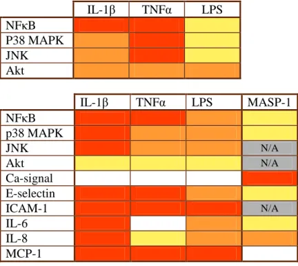
We describe for the first time the differences and similarities of LPS, TNFα, and IL-1β in the induction of endothelial inflammatory response by using a pattern of 10 inflammation-related molecules (Figure 1.).
• We demonstrated that signaling of LPS was less effective than that of IL-1β, and was significantly slower than that of TNFα and IL-1β, which can be partially explained by the special localization of Toll-like receptor 4 (TLR4). We showed that TLR4 is mainly localized in Golgi apparatus in HUVEC.
• The proinflammatory capacity of TNFα was similar to that of IL-1β in inducing NFκB nuclear translocation, while IL-1β was the strongest activator of MAPK pathways
• We showed that the delay in the LPS-induced NFκB and MAPK signaling pathways does not seem to result from Ca2+- or Akt- signaling.
• Expression of E-Selectin, IL-6 and IL-8 induced by LPS, TNFα and IL-1β were also dissimilar, whereas we did not find such a difference in ICAM-1 and MCP-1 expression. Due to the higher induction of E-selectin and IL-8, IL-1β might have more important role in neutrophil recruitment than LPS and TNFα.
IL-1β TNFα LPS NFκB
P38 MAPK JNK
Akt
IL-1β TNFα LPS MASP-1
NFκB p38 MAPK
JNK N/A
Akt N/A
Ca-signal E-selectin
ICAM-1 N/A
IL-6 IL-8 MCP-1
Figure 1. Pattern of the kinetics (upper panel) and maximal (lower panel) responses induced by LPS, TNFαααα, IL-1ββββ and MASP-1.
Upper: We compare the kinetics of signaling pathways stimulated by the three factors. Red: the factor reached its maximal effect within 15 minutes; orange: the maximal effect was around at 30 minutes; yellow: maximal effect was at 60 minutes or later.
Lower: We compared the relative maximal effect of the three factors for each parameter applying two-way ANOVA. Red: the most efficient activators in each line. Orange: significantly weaker activation than that of the strongest factor. Yellow:
significantly weaker activation than that of the second strongest factor. Grey, N/A: Not available.
.
12
• By above mentioned parameters we identified a signaling and expression pattern for the three proinflammatory molecules. This pattern illustrates how complex a proinflammatory process can be, and may enable us to predict and compare the pathomechanism of various inflammatory diseases.
In contrast to the three well-known proinflammatory factors, the effect of MASP-1 is poorly defined. Activation of endothelial cells by MASP-1 leads to proinflammatory phenotype and points to a novel connection between the complement- and coagulation systems during the inflammatory process.
• MASP-1, at its plasma concentration, triggered significant Ca2+- response in HUVECs and the kinetics of the responses elicited by MASP-1 and thrombin were similar. MASP-2, the other important protease of the lectin pathway failed to induce Ca2+- response even at a very high concentration.
• MASP-1 induced both NFκB nuclear translocation and p38 MAPK phosphorylation in a dose-dependent manner, as well as secretion of IL-6 and IL-8 cytokines. Activation of these pathways leads to a complex change in the phenotype of HUVECs.
• This well-characterized proinflammatory phenotype enhances leukocyte adhesion and transmigration, smooth muscle cell proliferation, and remodeling of the extracellular matrix.
6. LIST OF PUBLICATIONS
Publications related to the thesis1. Makó V, Czúcz J, Weiszhár Z, Herczenik E, Matkó J, Prohászka Z, Cervenak L. Proinflammatory activation pattern of human umbilical vein endothelial cells induced by IL-1β, TNF-α, and LPS.
Cytometry A. 2010 Oct;77(10):962-70.
IF: 3,032 2. Megyeri M*, Makó V*, Beinrohr L, Doleschall Z, Prohászka Z, Cervenak L, Závodszky P, Gál P. Complement protease MASP-1 activates human endothelial cells: PAR4 activation is a link between complement and endothelial function.
J Immunol. 2009 Sep 1;183(5):3409-16. IF: 5,646
*Megosztott elsıszerzıs
Publication related to the topic of the thesis
1. Kiszel P, Makó V, Prohászka Z, Cervenak L. Interleukin-6 -174 promoter polymorphism does not influence IL-6 production after LPS and IL-1 beta stimulation in human umbilical cord vein endothelial cells.
Cytokine. 2007 Oct;40(1):17-22.
IF: 2,169 2. Keltai K, Cervenak L, Makó V, Doleschall Z, Zsáry A, Karádi I.Doxorubicin selectively suppresses mRNA expression and production of endothelin-1 in endothelial cells.
Vascul Pharmacol. 2010 Nov-Dec;53(5-6):209-14.
IF: 2,044
14 Other publications
1. Stenczer B, Rigó J Jr, Prohászka Z, Derzsy Z, Lázár L, Makó V, Cervenak L, Balogh K, Mézes M, Karádi I, Molvarec A. Plasma osteopontin concentrations in preeclampsia - is there an association with endothelial injury? Clin Chem Lab Med. 2010 Feb;48(2):181-7.
IF: 1,886
2. Lazar L, Rigó J Jr, Nagy B, Balogh K, Makó V, Cervenak L, Mézes M, Prohászka Z, Molvarec A. Relationship of circulating cell- free DNA levels to cell-free fetal DNA levels, clinical characteristics and laboratory parameters in preeclampsia. BMC Med Genet. 2009
Nov 21;10:120. IF: 2,84
3. Gombos T, Makó V, Cervenak L, Papassotiriou J, Kunde J, Hársfalvi J, Förhécz Z, Pozsonyi Z, Borgulya G, Jánoskuti L, Prohászka Z. Levels of von Willebrand factor antigen and von Willebrand factor cleaving protease (ADAMTS13) activity predict clinical events in chronic heart failure. Thromb Haemost. 2009
Sep;102(3):573-80. IF: 4,451
4. Molvarec A, Derzsy Z, Kocsis J, Bıze T, Nagy B, Balogh K, Makó V, Cervenak L, Mézes M, Karádi I, Prohászka Z, Rigó J Jr.
Circulating anti-heat-shock-protein antibodies in normal pregnancy and preeclampsia. Cell Stress Chaperones. 2009 Feb 11.
IF: 2,167 5. Herczenik E, Varga Z, Eros D, Makó V, Oroszlán M, Rugonfalvi- Kiss S, Romics L, Füst G, Kéri G, Orfi L, Cervenak L. Protein kinase inhibitor-induced endothelial cell cytotoxicity and its
prediction based on calculated molecular descriptors. J Recept Signal Transduct Res. 2009;29(2):75-83. IF: 1,517 6. Molvarec A, Rigó J Jr, Bõze T, Derzsy Z, Cervenak L, Makó V, Gombos T, Udvardy ML, Hársfalvi J, Prohászka Z. Increased plasma von Willebrand factor antigen levels but normal von Willebrand factor cleaving protease (ADAMTS13) activity in preeclampsia.
Thromb Haemost. 2009 Feb;101(2):305-11. IF: 4,451 7. Molvarec A, Rigó J Jr, Lázár L, Balogh K, Makó V, Cervenak L, Mézes M, Prohászka Z. Increased serum heat-shock protein 70 levels reflect systemic inflammation, oxidative stress and hepatocellular injury in preeclampsia. Cell Stress Chaperones. 2009 Mar;14(2):151
IF: 2,16