BGP-15, a PARP-inhibitor, prevents imatinib-induced
cardiotoxicity by activating Akt and suppressing JNK and p38 MAP kinases
Zsolt Sarszegi• Eszter Bognar•Balazs Gaszner• Attila Ko´nyi• Ferenc Gallyas Jr.•Balazs Sumegi• Zoltan Berente
Received: 3 October 2011 / Accepted: 27 January 2012 / Published online: 14 February 2012 ÓSpringer Science+Business Media, LLC. 2012
Abstract In this study, we investigate the cardiotoxic effects of the well-known cytostatic agent imatinib mesy- late (Gleevec), and presented evidence for the cardiopro- tective effect of BGP-15 which is a novel insulin sensitizer.
The cardiotoxic effect of imatinib mesylate was assessed in Langendorff rat heart perfusion system. The cardiac high- energy phosphate levels (creatine phosphate (PCr) and ATP) were monitored in situ by 31P NMR spectroscopy.
The protein oxidation, lipid peroxidation, and the activa- tion of signaling pathways were determined from the freeze-clamped hearts. Prolonged treatment of the heart with imatinib mesylate (20 mg/kg) resulted in cardiotox- icity, which were characterized by the depletion of high- energy phosphates (PCr and ATP), and significantly increased protein oxidation and lipid peroxidation. Imatinib mesylate treatment-induced activation of MAP kinases (including ERK1/2, p38, and JNK) and the phosphorylation of Akt and GSK-3beta. BGP-15 (200lM) prevented the imatinib mesylate-induced oxidative damages, attenuated the depletion of high-energy phosphates, altered the sig- naling effect of imatinib mesylate by preventing p38 MAP kinase and JNK activation, and induced the phosphoryla- tion of Akt and GSK-3beta. The suppressive effect of BGP- 15 on p38 and JNK activation could be significant because these kinases contribute to the cell death and inflammation in the isolated perfused heart.
Keywords Imatinib mesylateCardiotoxicityBGP-15 Oxidative stress p38JNK AktGSK-3beta
Abbreviations
PI3-K Phosphatidylinositol 3-kinase
Akt Phospho-specific Akt-1/protein kinase B-a Ser473
ERK Extracellular signal regulated kinase p38-MAPK Thr183–Tyr185, phospho-specific p38 mitogen-
activated protein kinase
JNK Thr180–Gly–Tyr182, phospho-specific c-Jun N-terminal kinase
GSK-3b Phospho-specific glycogen synthase kinase ER stress Endoplasmic reticulum stress response GIST Gastrointestinal stromal tumor
BGP-15 O-(3-piperidino-2-hydroxy-1-propyl) nicotinic-amidoxime
ATP Adenosine triphosphate ROS Reactive oxygen species PARP Poly (ADP-ribose) polymerase
MDA Malondialdehyde
TBA Thiobarbituric acid TCA Trichloroacetic acid
TBARS Thiobarbituric acid-reactive substances NMR Nuclear magnetic resonance
IRE1 Inositol requiring enzyme 1
Ask1 Apoptosis signal-regulating kinase 1 BAX Bcl-2-associated X protein
PTEN Phosphatase and tensin homolog located on chromosome 10
Introduction
Imatinib mesylate (Gleevec), a potent and specific inhibitor of the Bcr-Abl tyrosine kinase, has been used successfully Z. Sarszegi (&)B. GasznerA. Ko´nyi
Department of Cardiology, Heart Institute, University of Pecs, Medical School, Ifjusag 13, Pecs 7624, Hungary
e-mail: sarszegizsolt@gmail.hu
E. BognarF. Gallyas Jr.B. SumegiZ. Berente
Department of Biochemistry and Medical Chemistry, University of Pecs, Medical School, Ifjusag 13, Pecs 7624, Hungary DOI 10.1007/s11010-012-1252-8
for the treatment of advanced-phase chronic myeloid leu- kemia (CML) [1–3]. Bcr-Abl is a constitutively active tyrosine kinase in leukemic cells and activates several signaling pathways, such as mitogen-activated protein kinase (MAPK) and phosphatidylinositol 3-kinase (PI3-K/
Akt). BCR-ABL also activates pro-survival pathways in leukemic cells including Jak-STAT and Grb2-ERK sig- naling pathways [4], which can lead to increased sponta- neous and DNA damage-induced proliferation frequency.
Although the detection of imatinib-related cardiotoxicity has been based largely on physical examination and med- ical history, the laboratory-based evidence is still missing.
A comprehensive study was published by Kerkela et al. [5], showing that imatinib-treated patients developed chronic heart failure, which could be modelled on C57BL6 mice as well after prolonged administration of imatinib. The underlying mechanisms of cardiotoxicity involved the activation of endoplasmic reticulum (ER) stress response, the collapse of the mitochondrial membrane potential, the release of cythochrome c into the cytosol, a reduction in cellular ATP content and cell death [5]. Further evidence of congestive heart failure development has been reported by another clinical study, which investigated the toxicity of imatinib in sixteen CML and GIST patients [6], and strengthened the presumption of cardiotoxicity [7].
In vitro experiments revealed that at least two different pathways, one involving caspase activation and another one is PARP-1 enzyme-mediated pathway, coexisted in imatinib-induced apoptosis [8]. The treatment of BaF3BA cells with a broad caspase inhibitor alone was not sufficient to completely block imatinib-induced apoptosis, however, co-administration with PARP-inhibitor PJ34 resulted in an increased cytoprotection [8].
Consequences of pathophysiological PARP-1 enzyme activation in cardiomyocytes has been well established [9,10].
Over-activation of PARP-1 enzyme can induce rapid cellular NAD? and ATP pool depletion leading to mitochondrial dysfunction and can suppress the activity of the PI-3-kinase–
Akt pathway resulting in necrotic or apoptotic cell death.
Mitochondrial dysfunction in turn can further impair energy metabolism and increase mitochondrial ROS production manifesting in lipid peroxidation, protein oxidation, and DNA damage [11]. Earlier studies demonstrated the beneficial effect of BGP-15 on oxidative stress [11,12]. BGP-15 is a nicotinic-amidoxime derivate (Fig.1), which was originally developed against insulin resistance. BGP-15 is a potent insulin sensitizer [13]. Data generated by our group showed that multitarget agent BGP-15 successfully inhibited the activation of PARP-1 enzyme and protected the mitochondria from oxidative damage under condition of ischemia–reper- fusion on a Langendorff rat heart model [11,12].
The aim of our study was to identify the mechanisms by which imatinib induces cardiotoxicity in a Langendorff
perfused rat heart model and to determine how PARP inhibitor and antioxidant agent BGP-15 can modulate these processes. We investigated whether Imatinib mesylate administration could lead to oxidative stress and alterations in cardiac energy metabolism and the ability of BGP-15 to counteract these effects. We studied the possible role of JNK and p38 MAP kinase activation in imatinib-induced car- diotoxicity, and the potential beneficial effect of BGP-15 on these processes.
Materials and methods
Chemicals
4-[(4-Methyl-1-piperazinyl)methyl]-N-[4-methyl-3-[[4-(3- pyridinyl)-2-pyrimidinyl] amino]-phenyl] benzamide meth- anesulfonate (Imatinib mesylate, Gleevec) was donated by the Department of Internal Medicine, University of Pecs.
BGP-15 was synthesised by N-Gene Research and Devel- opment, Ltd., Hungary. Antibodies against phospho-specific extracellular signal regulated kinase (ERK1/2) Thr183– Tyr185, phospho-specific p38 mitogen-activated protein kinase (p38-MAPK) Thr180–Gly–Tyr182, phospho-specific c-Jun N-terminal kinase (JNK), phospho-specific Akt-1/
protein kinase B-a Ser473, and phospho-specific glycogen synthase kinase (GSK)-3bSer9were purchased from Cell Signalling Technology, Kvalitex Co., Budapest, Hungary.
Antibody against N-terminal domain of actin was obtained from Sigma-Aldrich Co, Budapest, Hungary. Anti-PARP was obtained from Alexis Biotechnology, London, U.K. All other highly purified reagents were commercially available.
Experimental animals
Male Wistar rats weighting 300–350 g were used for this study. The investigation conformed to the Guide for the Care and Use of Laboratory Animals published by the U.S.
National Institutes of Health (NIH Publication No. 85-23, revised 1996).
Heart perfusion
Rats were anticoagulated with sodium heparin (100 U, i.p.) and were anesthetized using ketamine (200 mg/kg, i.p.).
Hearts were immediately removed and arrested in ice-cold
Fig. 1 Chemical structure of BGP-15 (O-(2-hydroxy-3-piperidine- propyl)-pyridine-carbonic acid-amidoxime dihydrochloride)
perfusion buffer. Each heart was then cannulated through the aorta and perfused at 37°C by the Langendorff method at a constant perfusion pressure equivalent to 70 Hgmm.
Retrograde aortic perfusion was maintained with a modi- fied phosphate free-Krebs–Henseleit (KH) buffer, con- taining 118 mM NaCl, 5 mM KCl, 1.2 mM MgSO4, 25 mM NaHCO3, 1.25 mM CaCl2, 1.8 mM octanoic acid, and 11 mM glucose. The KH buffer was filtered through a 0.22lM Millipore filter to remove any particulate con- taminants and was saturated with a mixture of 95% O2and 5% CO2resulting in a pH of 7.4. After a non-recirculating washout period of 10 min, hearts underwent 60 min per- fusion. Imatinib and/or BGP-15 were administered into the perfusion medium at the beginning of baseline perfusion.
Heart perfusion models were distributed into four groups with ten perfused heart model in each group: 1. normoxic control group, 2. normoxic perfusion with BGP-15, 3.
normoxic perfusion with imatinib, and 4. normoxic perfu- sion with BGP-15 and imatinib. Hearts were freeze- clamped at the end of each perfusion.
NMR spectroscopy and data analysis
Cardiac energy metabolism was monitored in situ during the perfusion by31P NMR spectroscopy through quantification of creatine phosphate (d=0.0 ppm). NMR spectra were recor- ded with a VarianUNITYINOVA 400 WB instrument (Varian Inc., Palo Alto, CA, USA).31P measurements (161.90 MHz) of the perfused hearts were run at 37°C in a ZSPEC 20 mm broadband probe (Nalorac Co., Martinez, CA, USA), applying WALTZ proton decoupling (cB2=2 kHz) during the acqui- sition only. Field homogeneity was adjusted following the1H signal (w1/2=10–15 Hz). Spectra were collected with a time resolution of 3 min by accumulating 120 transients in each FID. 45°flip angle pulses were employed after a 1.25 s recycle delay, and transients were acquired over a 10 kHz spectral width in 0.25 s, and the acquired data points (5000) were zero- filled to 16 K. Under the above mentioned circumstances the relative concentrations of the species can be taken proportional to the peak areas. Peak areas were determined by deconvolu- tion of simulated spectra fitted to experimental spectra obtained after referencing (d=0 for creatine phosphate) and baseline correction using Vnmr 6.1C software (Varian Inc., Palo Alto, CA, USA). Amounts of individual metabolite levels in each spectrum were expressed as their ratio to the first cre- atine phosphate (PCr) amount.
Lipid peroxidation
Determination of thiobarbituric acid-reactive substances (TBARs) in the heart was performed according to a modified method of Serbinova et al. [14]. Malondialdehyde, formed by the breakdown of polyunsaturated fatty acids, served as an
indicator of the extent of lipid peroxidation. Malondialde- hyde reacted with thiobarbituric acid to give a red species absorbing at 535 nm. Amount of 50 mg frozen cardiac tissue was homogenized in 6.5% trichloroacetic acid (TCA) and in a reagent containing 15% TCA, 0.375% thiobarbituric acid (TBA), and 0.25% HCl. Homogenates were then heated for 15 min in a boiling water bath, cooled, centrifuged, and the absorbance of the supernatant was compared at 535 nm with a blank sample that contained all the reagents except the tissue homogenate. Using malondialdehyde standard, TBARS were calculated as nmol/g wet tissue [14].
Protein oxidation
Fifty milligrams of freeze-clamped perfused heart tissue were homogenized with 2 ml 10% trichloroacetic acid (TCA) and was divided into two equal parts. After 10 min centrifugation at 3,000 g, 2 ml 2 N hydrochloric acid containing 0.2% dinitrophenyl hydrazine was given to the pellets. The pellets were then vortexed, treated with 50%
TCA and the protein content was collected by centrifuga- tion. The protein carbonyl content was determined by means of the 2,4-dinitrophenyl-hydrazine method [15].
Western blot analysis
Fifty milligram of heart samples were homogenized in ice- cold Tris buffer (50 mM, pH 8.0) and harvested in 29con- centrated SDS-polyacrylamide gel electrophoretic sample buffer. Proteins were separated on 12% SDS-polyacrylamide gel and transferred to nitrocellulose membranes. After blocking (2 h with 3% non-fat milk in Tris-buffered saline) membranes were probed overnight at 4°C with antibodies (1:1,000 dilution) recognizing the following antigens: GAP- DH, pAkt, pGSK-3b, pERK1/2, pp38, pJNK, and anti-PARP.
Membranes were washed six times for 5 min in Tris-buffered saline (pH 7.5) containing 0.2% Tween (TBST) before addi- tion of anti-rabbit horseradish peroxidase conjugated sec- ondary antibody (1:3,000 dilution; Bio-Rad, Budapest, Hungary). Membranes were washed six times for 5 min in TBST. The antibody–antigen complexes were visualized by means of enhanced chemiluminescence on conventional films. Quantification of band intensities (E540) of the blots was performed by a DU-62 spectrophotometer equipped with a densitometry attachment (Beckman Coulter Inc., Fullerton, CA) and ImageJ (public domain) software. Data representing three-independent experiments are expressed as percentage of the untreated control (mean±SEM).
Statistical analysis
Statistical analysis was performed using ANOVA and all data were expressed as mean±SEM. Two-way repeated
measures ANOVA was used to evaluate the statistical significance of differences among groups for levels of PCI.
Bonferroni post hoc analysis was used for specific com- parisons, when significant differences were detected for the treatment-by-time interactions, differences were consid- ered statistically significant at the level ofp\0.05.
Results
Effect of BGP-15 on myocardial energy metabolism of Langendorff perfused rat hearts in the presence of imatinib
Under our experimental conditions creatine phosphate levels of untreated Langendorff perfused rat hearts showed a slight decrease during the 60-min perfusion. Cardiac PCr levels were decreased by 18% compared to untreated hearts when 20 mg/l imatinib was added to the perfusate. Co- administration of imatinib and BGP-15 revealed that BGP- 15 in a concentration of 200 mg/l was able to prevent the decrease of imatinib-induced PCr levels. Administration of BGP-15 alone increased cardiac PCr levels over the normoxic values (Fig.2). The change of ATP concentra- tion followed a similar pattern to the change in creatine phosphate level in our investigated time period. Imatinib- treated hearts showed significant decrease in ATP levels whereas co-administration with BGP-15 successfully pre- vented this effect. ATP levels in the presence of BGP-15 alone were not significantly different from untreated, control cases (Fig.3). Inorganic phosphate showed mod- erate increase during the perfusion reaching 15–20% of the normoxic PCr level after 30 min (data not shown).
Effect of BGP-15 on myocardial oxidative damages induced by imatinib
Determination of lipid peroxidation after 1 h normoxic perfusion showed that administration of imatinib signifi- cantly (p\0.01) increased thiobarbituric reactive sub- stance (TBARS) formation compared to untreated hearts.
On the other hand, when both imatinib and BGP-15 were present in the perfusate, the formation of TBARS was significantly diminished. BGP-15 alone did not alter TBARS formation (Fig.4). In the case of protein oxidation we observed that the presence of imatinib markedly ele- vated the level of protein-bound aldehyde groups as com- pared to untreated hearts. This phenomenon was significantly inhibited (p\0.01) by the coadministration
Fig. 2 Effect of BGP-15 and/or Imatinib on cardiac creatine phosphate levels in Langendorff perfused rat hearts. Creatine phosphate amounts, determined by in situ31P NMR spectroscopy, are expressed as their ratio to initial creatine phosphate amount.
Values are given as means±SEM (p\0.01) (n=10 in each group)
Fig. 3 Effect of BGP-15 and/or Imatinib on cardiac ATP levels in Langendorff perfused rat hearts. ATP amounts, determined by in situ
31P NMR spectroscopy, are expressed as their ratio to initial ATP amount. Values are given as means±SEM (p\0.01) (n=10 in each group)
Fig. 4 Effect of BGP-15 on imatinib-induced lipid peroxidation in Langendorff perfused rat hearts. The figure demonstrates the quantity of thiobarbituric reactive substances in various animal groups. Values are given as means±SEM (***p\0.001 compared to normoxic levels,???p\0.001 compared to imatinib-treated levels) (n=10 in each group)
of imatinib and BGP-15. However, BGP-15 alone had no significant influence on the level of protein oxidation (Fig.5).
Effects of imatinib and BGP-15 in signaling pathways Akt, GSK-3ß, ERK, JNK, and p38-MAPK phosphorylation were examined by Western blot in the following samples:
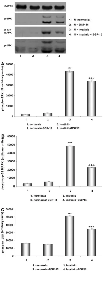
without treatment (normoxia) (1), treatment with BGP-15 (2), imatinib (3), and with imatinib?BGP-15 (4). Imati- nib induced the activation of MAP kinases (ERK1/2, p38, and JNK). JNK and p38 MAP kinase contribute to cell death [16], and inflammatory reactions [17], therefore could play a significant role in the imatinib-induced car- diotoxicity. BGP-15 in the presence of imatinib suppressed the activation of JNK and p38 MAP kinase, and this effect of BGP-15 could be significant in its protective role (Fig.6).
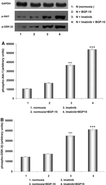
Imatinib-induced the phosphorylation of Akt and GSK- 3ß, but in the presence of imatinib BGP-15 further induced the phosphorylation of these kinases (Fig.7). Phosphory- lation and activation of Akt in the myocardium may play a protective role under stress conditions by maintaining mitochondrial membrane integrity [18] and by phosphor- ylation and inactivation of GSK-3ß[19].
Effects of imatinib and BGP-15 on PARP activation One hour perfusion with imatinib resulted roboust PARP-1 activation. BGP-15 administered alone had no effect of PARP activation, but when co-administered with imatinib successfully reduced the effect of imatinib on PARP-1 activation (Fig.8).
Discussion
As of date our study is a pioneering work to elaborate the effect of BGP-15 on imatinib-induced energy run-down, oxidative damages, and signaling mechanisms on an iso- lated Langendorff perfused rat heart model. BGP-15 is a nicotinic-amidoxime derivate originally developed against insulin resistance. Studies investigating the effects of BGP- 15 revealed that this multitarget agent is a potent cardio- protectant under conditions of ischemia–reperfusion by inhibiting overactivation of PARP-1 enzyme and stabiliz- ing the mitochondria, thus preventing ROS generation along the mitochondrial respiratory chain [11, 20, 21].
Furthermore hepatoprotective [22], nephroprotective [23], and neuroprotective [24] effects of BGP-15 were also published. Recently BGP-15 has been introduced as a potent insulin sensitizer [13,25].
Earlier studies performed on myocardium cell cultures confirmed that imatinib treatment might lead to the col- lapse of mitochondrial membrane potential and the loss of membrane integrity [5]. The significant decrease of mito- chondrial membrane potential plays a pivotal role in the development of cardiotoxicity [4]. Earlier data clearly defined the structural damage of mitochondria detected by electron microscopy both in human myocardial histological samples and animal models after imatinib treatment. The connection between the deterioration of mitochondrial function and ATP depletion is also well demonstrated. A well-known cytostatic agent, Herceptin causes 35%
decrease in myocardial ATP level, contrary to imatinib, which generates 65% decline in ATP concentration. The reduction of ATP level may significantly run down many cellular processes. Earlier studies show that both necrotic and apoptotic cell death coexist in cultured cardiomyo- cytes. This fact may introduce apoptosis as an ATP- dependent process.
Our study is a pioneering work in that it has elaborated energy run-down in isolated, perfused Langendorff rat heart model. Using31P NMR spectroscopy, we managed to show significant ATP and creatine-phosphate depletion in perfused rat heart in situ. The detection of energy depletion after long-time imatinib administration has been well established by earlier reports. By perfusing the heart imatinib-containing solution we have shed light on a new aspect of a relatively quick energy run-down. Accordingly, it is worth to note that not only long-term administration but also its ‘‘acute phase’’ effect of imatinib can deteriorate cellular reactions in general. Energy depletion may com- promise the cardiac function and can lead to a compensa- tory remodelling and heart failure [26]. ATP depletion may also compromise ion transports, mainly increase the cyto- plasmic free Ca2?level. The increased level of calcium in cytoplasm causes an increase in the level of calcium in Fig. 5 Effect of BGP-15 on imatinib-induced protein oxidation in
Langendorff perfused rat hearts. The figure demonstrates the protein carbonyl content in various groups. Values are given as means±SEM (***p\0.001 compared to normoxic levels, ???p\0.001 com- pared to imatinib-treated levels) (n=10 in each group)
mitochondria, which can lead to mitochondrial perme- ability pore formation and enhanced mitochondrial ROS production, which then causes enhanced oxidative damages [27]. Mitochondrial damages can lead to the activation of apoptotic pathways, which can result in the loss of cardiomyocytes in the damaged heart regions [28]. The increase in ROS production and the imbalance of ion concentrations can lead to the rupture of plasma membrane and necrotic cell death. BGP-15 protects the mitochondrial membrane system against oxidative damages [22]. This effect of BGP-15 can be crucial in the heart, because mitochondrial energy production is predominant [29], and the protection of the mitochondrial membrane system as well as the membrane potential is prerequisite of active mitochondrial ATP production. Therefore, stress condi- tions were induced by different mechanisms, in all these cases BGP-15 decreased oxidative damages. These data suggested that BGP-15 had significant mitochondrial pro- tecting effects [11,12,22], regarding to protection against mitochondria-related apoptotic pathway [30]. Our data show that the imatinib-induced significant oxidative dam- age in the heart (Figs.4,5), could be reversed by BGP-15, and this observed effect is in good agreement with the mechanism found in ischemic-reperfusion models [11, 12,22].
Kerkala et al. [5] showed in cultured cells that imatinib treatment induced the activation of the endoplasmic retic- ulum (ER) stress response, the collapse of the mitochon- drial membrane potential, the release of cytochrome c into the cytosol, a reduction in cellular ATP content and cell death. In our perfused heart system we found oxidative damages, ATP depletion and the effect of imatinib treat- ment on signaling pathways, which can lead to cell death (Figs.2,3,4,5,6,7). The activation of JNK and p38 MAP kinase as a consequence of imatinib treatment can play significant role in the mitochondrial depolarization and the activation of mitochondrial-related apoptotic pathway [31].
The role of p38 and Akt activation in the cardiotoxic effect is controversial, because these signaling pathways were not dysregulated in other studies (e.g., Kerkela et al. [5]. This discrepancy should be lain on the different model system.
Furthermore the dose of imatinib was higher than the regular plasma concentration in humans, therefore the direct toxic effect of imatinib must be considered.
Fig. 6 Phosphorylation of ERK, JNK, and p38-MAPK in Lange- ndorff perfused (60 min) rat hearts under normal conditions (nor- moxia), after treatment with BGP-15, with imatinib and with BGP- 15?imatinib. Representative Western blot analysis of ERK, p38-MAPK, JNK phosphorylation, and densitometric evaluations of ERK (a), p38-MAPK (b), and JNK (c) are shown. Values are given as means±SEM (***p\0.001 compared to normoxic levels,
???p\0.001 compared to imatinib-treated levels) (n=3 in each group)
c
However, Akt activation could be explained theoretically because excessive ROS generation inactivates intracellular PTEN which normally inhibits PI3-kinase–Akt pathway.
Therefore, inhibition of PTEN could activate Akt pathway.
In our model p38 pathway was activated also. Earlier studies clearly defined that imatinib treatment induces ER stress response which enhances JNK and p38 activation via IRE1-ASK1 pathway. During the imatinib-induced oxida- tive stress oxidation of the Ask1 inhibitor thioredoxins lead to the activation of Ask1 kinase, which is an upstream
kinase of JNK and p38 MAP kinases [32]. A consequence, the modulation of these kinases can provide a mechanism by which we could protect the heart during imatinib treatment. Here, we showed that by combining BGP-15 with imatinib we could prevent the imatinib-induced acti- vation of JNK and p38 MAP kinase in the heart and pre- vent the oxidative stress and as well as ATP depletion (Figs. 3,4,5,6). The protective mechanism of BGP-15 is likely mediated by mitochondrial protection, because our previous data indicated that BGP-15 did not interfere with the ER stress response [22], while our other data indicated a mitochondrial protective effect of BGP-15 [11,12, 22].
Furthermore, we found that BGP-15-activated PI-3-kinase Akt pathway, which is a well-known cytoprotective path- way. Akt activation protects cells by preventing the col- lapse of mitochondrial membrane system in oxidative stress [18] which is the further evidence of BGP-15-med- iated mitochondria protecting effect. In this article we used the well-known cytostatic agent Imatinib mesylate to induce cardiotoxicy in perfused Langendorff rat hearts, and tried to identify mechanism of cardiotoxicity in situ. Our data showed for the first time that imatinib-induced oxi- dative stress compromised energy metabolism and the activation of potentially cell death inducing kinases (JNK and p38 MAP kinases). These data raised the possibility that the modulation of these pathways could prevent the toxic cardiac effect of Imatinib mesylate.
Fig. 7 Phosphorylation of Akt1 and GSK-3b in Langendorff per- fused (60 min) rat hearts under normal conditions (normoxia) and after treatment with BGP-15, with imatinib and with BGP-15? imatinib. Representative Western blot analysis of p-Akt1, and p-GSK- 3bphosphorylation and densitometric evaluation are shown (aAkt1, b GSK-3). Values are given as means±SEM (***p\0.001 compared to normoxic levels, ???p\0.001 compared to imati- nib-treated levels) (n=3 in each group)
Fig. 8 Effect of BGP-15 on imatinib-induced PARP-1 activation under normal conditions (normoxia) and after treatment with BGP-15, with imatinib, and with BGP-15?imatinib. Representative Western blot analysis of PARP and densitometric evaluation (a) are shown.
Values are given as means±SEM (***p\0.001 compared to normoxic levels,???p\0.001 compared to imatinib-treated levels) (n=3 in each group)
Imatinib treatment leads to a rapid increase in poly(ADP- ribosyl)ation (PAR), preceding the loss of mitochondrial membrane integrity and DNA fragmentation. It is also important to note that the inhibition of PAR in imatinib-treated cells partially prevented cell death to an extent comparable to that observed after caspase inhibition [8]. Previous data showed that BGP-15 protected cells in oxidative stress by protecting the mitochondrial membrane system [11, 22], which suppressed the activation of nuclear poly ADP-ribo- sylation [2]. Our data support the earlier findings that imatinib treatment induces PARP activation. Furthermore, we dem- onstrated that BGP-15 was protected against PARP activation and the toxic cardiac effect of imatinib in situ.
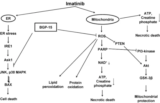
The mechanism by which BGP-15 prevented the imatinib-induced cardiotoxicity is likely regarded to its mitochondrial protective role, because BGP-15 counter- acted the oxidative stress-induced effect of imatinib and prevented the Imatinib mesylate-induced activation of JNK and p38 MAP kinase. Furthermore, BGP-15 attenuated PARP-1 activation and induced the activation of PI-3- kinase—Akt pathway, which can also contribute to the mitochondrial protection (Fig.9).
Conclusion
According to our study, BGP-15 successfully prevented Imatinib mesylate-induced cardiac oxidative damages,
attenuated high-energy phosphate depletion and reduced p38 MAP kinase and JNK activation. Furthermore BGP-15 enhanced the activation of prosurvival PI-3 kinase–Akt pathway. Suppression of imatinib-induced p38 MAP kinase and JNK activation by BGP-15 may be of great importance regarding the role of these kinases in myocardial cell death and inflammation. Thorough investigation of imatinib- related cardiotoxic effects using already well known car- dioprotectants like BGP-15 would still be necessary, since patients with underlying cardiovascular diseases may be at greater risk for developing the cardiovascular abnormali- ties associated with imatinib therapy.
References
1. Deninger M, Buchdunger E, Duker BJ (2005) The development of imatinib as a therapeutic agent for chronic myeloid leukemia.
Blood 105:2640–2653
2. Cohen MH, Williams G, Johnson JR, Duan J et al (2002) Approval summary of imatinib mesylate capsules in the treatment of chronic myelogenous leukemia. Clin Cancer Res 8:935–942 3. Czechowska A, Poplawski T, Drzewoski J, Blasiak J (2005)
Imatinib (STI571) induces DNA damage in BCR/ABL-express- ing leukemic cells but not in normal lymphocytes. Chemico-Biol Interact 152:139–150
4. Van Etten RA (2004) Mechanism of transformation by the BCR- ABL oncogene: new perspectives in the post-imatinib era. Leuk Res 28(suppl 1):S21–S28
Fig. 9 Molecular mechanisms of Imatinib mesylate-induced cell death and possible regulatory points of BGP-15. Imatinib induces ER stress leading to activation IRE1-Ask1 pathway and further activation of JNK and p38 MAPK. Releasing of BAX and imatinib itself induce mitochondrial membrane depolarization leading to ATP depletion, cytochrome c (Cyt c) release, and features of necrotic and apoptotic
cell death. During mitochondrial damage excessive ROS generation enhances PARP-1 activation leading to NAD? and ATP depletion, which eventually cause necrosis. Furthermore ROS-mediated PTEN inactivation could activate PI3-Akt pathway. Inhibition of PARP-1 activity suppresses both processes leading to attenuation of NAD? depletion, mitochondrial protection, and cell survival
5. Kerkela R, Grazette L, Yacobi R, Iliescu C, Patten R, Beahm C, Walters B, Shevtsov S, Pesant S, Clubb FJ, Rosenzweig A, Sal- omon RN, Van Etten RA, Alroy J, Durand J-B, Force T (2006) Cardiotoxicity of the cancer therapeutic agent imatinib mesylate.
Nat Med 12:908–916
6. Park YH, Park HJ, Kim BS, Ha E, Jung KH, Yoon SH, Yim SV, Chung JH (2006) BNP as a marker of the heart failure in the treatment of imatinib mesylate. Cancer Lett 243:16–22 7. Orphanos GS, Ioannidis GN, Ardavanis AG (2009) Cardiotox-
icity induced by tyrosine kinase inhibitors. Acta Oncol 48:964–
970
8. Moehring A, Wohlbold L, Aulitzky WE, Kuip H (2005) Role of poly(ADP- ribose) polymerase activity in imatinib mesylate- induced cell death. Cell Death Differ 12:627–636
9. Szabo´ C (2005) Cardioprotective effects of poly(ADP-ribose) polymerase inhibition. Pharmacol Res 52:34–43
10. Pacher P, Szabo´ C (2007) Role of poly(ADP-ribose) polymerase 1 (PARP-1) in cardiovascular diseases: the therapeutic potential of PARP inhibitors. Cardiovasc Drug Rev 25:235–260
11. Halmosi R, Berente Z, Osz E, Toth K, Literati-Nagy P, Sumegi B (2001) Effect of poly(ADP-ribose) polymerase inhibitors on the ischemia-reperfusion-induced oxidative cell damage and mito- chondrial metabolism in Langendorff heart perfusion system.
Mol Pharmacol 59:1497–1505
12. Szabados E, Literati-Nagy P, Farkas B, Sumegi B (2000) BGP- 15, a nicotinic amidoxime derivate protecting heart from ische- mia-reperfusion injury trough modulation of poly(ADP-ribose) polymerase. Biochem Pharmacol 59:937–945
13. Litera´ti-Nagy B, Kulcsa´r E, Litera´ti-Nagy E, Buday B, Peterfai E, Horvath T, Tory K, Kolonics A, Fleming A, Mandl J, Koranyi L (2009) Improvement of insulin sensitivity by a novel drug, BGP- 15, in insulin-resistant patients. A proof of concept randomized double-blind clinical trial. Horm Metab Res 41:374–380 14. Serbinova EA, Kadiiska MB, Bakalova RA et al (1989) Lipid
peroxidation activation and cytochrome P-450 decrease in rat liver endoplasmic reticulum under oxidative stress. Toxicol Lett 47:119–123
15. Butterfield DA, Howard BJ, Yatin S, Allen KL, Carney JM (1997) Free radical oxidation of brain proteins in accelerated senescence and its modulation by N-tert-butyl-alpha-phenylni- trone. Proc Natl Acad Sci USA 21(94):674–678
16. Baines CP, Molkentin JD (2005) STRESS signaling pathways that modulate cardiac myocyte apoptosis. J Mol Cell Cardiol 38:47–62
17. Gil A, Marı´a Aguilera C, Gil-Campos M, Can˜ete R (2007) Altered signalling and gene expression associated with the immune system and the inflammatory response in obesity. Br J Nutr 98(Suppl 1):S121–S126
18. Tapodi A, Debreceni B, Hanto K, Bognar Z, Wittmann I, Gallyas F Jr, Varbiro G, Sumegi B (2005) Pivotal role of Akt activation in mitochondrial protection and cell survival by poly(ADP- ribose)polymerase-1 inhibition in oxidative stress. J Biol Chem 280:35767–35775
19. Liang J, Slingerland JM (2003) Multiple roles of the PI3K/PKB (Akt) pathway in cell cycle progression. Cell Cycle 2:339–345 20. Toth A, Halmosi R, Kovacs K, Deres P, Kalai T, Hideg K, Toth
K, Sumegi B (2010) Akt Activation induced by an antioxidant compound during ischemia-reperfusion. Free Rad Biol Med 35:1051–1063
21. Kovacs K, Toth A, Deres P, Kalai T, Hideg K, Sumegi B (2004) Myocardial protection by selective poly(ADP-ribose) polymerase inhibitors. Exp Cardiol 9:1–4
22. Nagy G, Szarka A, Lotz G, Do´czi J, Wunderlich L, Kiss A, Jemnitz K, Veres Zs, Ba´nhegyi G, Schaff Zs, Su¨megi B, Mandl J (2010) BGP-15 inhibits caspase-independent programmed cell death in acetaminophen-induced liver injury. Toxicol Appl Pharmacol 243:96–103
23. Ra´cz I, Tory K, Gallyas F Jr, Berente Z, Osz E, Jaszlits L, Ber- nath S, Sumegi B, Rabloczky Gy, Literati-Nagy P (2002) BGP- 15—a novel poly (ADP-ribose) polymerase inhibitor—protects against nephrotoxicity of cisplatin without compromising its antitumor activity. Biochem Pharmacol 63:1099–1111
24. Ba´rdos G, Mo´ricz K, Jaszlits L, Rabloczky G, Tory K, Ra´cz I, Berna´th S, Su¨megi B, Farkas B, Litera´ti-Nagy P (2003) BGP-15, a hydroximic acid derivative, protects against cisplatin- or taxol- induced peripheral neuropathy in rats. Toxicol Appl Pharmacol 190:9–16
25. Litera´ti-Nagy B, E´ i Pe´terfa, Kulcsa´r E, Litera´ti-Nagy Zs, Buday B, Tory K, Mandl J, Su¨megi B, Fleming A, Roth J, Kora´nyi L (2010) Beneficial effect of the insulin sensitizer (HSP inducer) BGP-15 on olanzapine induced metabolic disorders. Brain Res Bull 83:340–344
26. Palfi A, Toth A, Hanto K, Deres P, Szabados E, Szereday Z, Kulcsar G, Kalai T, Hideg K, Gallyas F Jr, Sumegi B, Toth K, Halmosi R (2006) PARP inhibition prevents postinfarction myocardial remodeling and heart failure via the protein kinase C/glycogen synthase kinase-3beta pathway. J Mol Cell Cardiol 41:149–159
27. Murphy E, Steenbergen C (2008) Mechanisms underlying acute protection from cardiac ischemia-reperfusion injury. Physiol Rev 88:581–609
28. Subramanian S, Kalyanaraman B, Migrino RQ (2010) Mitoc- hondrially targeted antioxidants for the treatment of cardiovas- cular diseases. Recent Pat Cardiovasc Drug Discov 5:54–65 29. Balaban RS (2009) The role of Ca(2?) signaling in the coordi-
nation of mitochondrial ATP production with cardiac work.
Biochim Biophys Acta 787:1334–1341
30. Hori M, Nishida K (2009) Oxidative stress and left ventricular remodelling after myocardial infarction. Cardiovasc Res 81:
457–464
31. Duplain H (2006) Salvage of ischemic myocardium: a focus on JNK. Adv Exp Med Biol 588:157–164
32. Matsuzawa A, Ichijo H (2008) Redox control of cell fate by MAP kinase: physiological roles of ASK1-MAP kinase pathway in stress signaling. Biochim Biophys Acta 80:1325–1336