Research Article
Tyrosine Kinase Inhibitor Imatinib Mesylate Alters
DMBA-Induced Early Onco/Suppressor Gene Expression with Tissue-Specificity in Mice
Péter Attila Gergely ,
1Balázs Murnyák ,
2János Bencze ,
3Andrea Kurucz,
4Timea Varjas ,
5Katalin Gombos,
6and Tibor Hortobágyi
2,71Institute of Forensic Medicine, University of Debrecen, 4012 Debrecen, Hungary
2MTA-DE Cerebrovascular and Neurodegenerative Research Group, Department of Neurology, University of Debrecen, 4012 Debrecen, Hungary
3Institute of Pathology, Faculty of Medicine, University of Debrecen, 4012 Debrecen, Hungary
4Department of Pharmacology and Pharmacotherapy, University of Debrecen, 4012 Debrecen, Hungary
5Department of Public Health Medicine, Medical School, University of P´ecs, 7624 P´ecs, Hungary
6Department of Laboratory Medicine, Clinical Center, University of P´ecs, 7624 P´ecs, Hungary
7Institute of Pathology, University of Szeged, 6725 Szeged, Hungary
Correspondence should be addressed to P´eter Attila Gergely; 4n6medical@gmail.com and Tibor Hortob´agyi;
hortobagyi.tibor@med.u-szeged.hu
Received 6 September 2018; Revised 28 December 2018; Accepted 13 January 2019; Published 7 February 2019
Academic Editor: Takashi Yazawa
Copyright © 2019 P´eter Attila Gergely et al. This is an open access article distributed under the Creative Commons Attribution License, which permits unrestricted use, distribution, and reproduction in any medium, provided the original work is properly cited.
Tyrosine kinases play crucial roles in cellular development and tumorigenesis. Tyrosine kinase inhibitors (TKIs) are effective and widely used drug molecules in targeted cancer therapies. Altered expressions of protooncogenes and tumor suppressor genes after DMBA (7,12-dimethylbenz[a]anthracene) treatment have been described as early markers of tumor induction; however their tissue-specific effects remain still unclear. Our study was aimed at examining the short-term possible antineoplastic and chemopreventive effects of a TKI compound (imatinib mesylate) on a DMBA-induced mouse tumor model. In addition, we also investigated the tissue-specific expressions ofHras, Kras, Myc,andTrp53genes in the brain, bone marrow, spleen, liver, abdominal lymph nodes, thymus, lungs, and kidneys, respectively. 24 hours after the imatinib mesylate injection, we observed significantKras downregulation in the bone marrow and lung of the DMBA-treated mice. Moreover, the mRNA expression ofMycwas also found to be decreased significantly in the spleen. Interestingly, whileTrp53expression was significantly increased in the lung, it was decreased in the other tissues. However, there was also a tendency in the decreasedMyclevel in the bone marrow, brain, kidneys, lungs, and lymph nodes and in the decreasedHraslevel in the bone marrow, kidneys, and lungs, although no significant differences were observed. Our findings indicate rapid tissue-specific impact of imatinib mesylate on DMBA-induced gene expressionin vivo, supporting the chemopreventive potential of imatinib mesylate in cancer.
1. Introduction
Protein kinases (PKs) play pivotal roles in cellular pro- cesses such as metabolism, proliferation, apoptosis, immune response, or nervous system functions. PKs regulate enzyme activity by phosphorylating cellular proteins [1] and their dys- regulation may lead to pathological conditions, i.e., different types of cancers or inflammatory diseases. Therefore, PKs
have become one of the most extensively investigated drug targets in the past two decades [2]. To date, the human PK gene family consists of 518 members and can be categorized into nine groups. Among them, tyrosine kinases (TKs)—and their inhibitor molecules—are the most promising targets of cancer studies [3]. TKs are classified as receptor and non- receptor tyrosine kinases. Receptor tyrosine kinases (RTKs) are transmembrane proteins consisting of an extracellular
Volume 2019, Article ID 8670398, 12 pages https://doi.org/10.1155/2019/8670398
ligand-binding domain and an intracellular kinase domain [4]. Nonreceptor tyrosine kinases can be found in the cytosol and nucleus or in the inner part of the plasma membrane, participating in the regulation of cell proliferation or differ- entiation [5]. The activation of TKs is under tight control.
Their kinase activity is low in nonproliferating cells. On the contrary, TK expression is extremely increased in cancer cells, caused by ligand or receptor overexpression by various mechanisms [6–11].
Imatinib was the first small-molecule TKI that accom- plished a remarkable clinical success in the treatment of chronic myeloid leukemia (CML). Imatinib mesylate inhibits the constitutively active BCR-Abl protein kinase that is responsible for the constant proliferation of myeloid cells [12]. Druker et al. reported that imatinib produced a 92- 98% decrease in the number of colonies from BCR-Abl cells, while having minimal effect on normal cells [13]. Imatinib targets further protein kinases, including the stem cell fac- tor receptor (c-kit) and the platelet-derived growth factor receptor (PDGFR), whose inhibition might have potential implications for the treatment of several malignancies [14].
Imatinib treatment is usually well-tolerated; however, side effects may develop, e.g., edema, nausea, skin rash or moder- ate myelosuppression [15]. Resistance to imatinib can occur within months or years after the beginning of the treatment.
Several mechanisms of resistance have been discovered, categorized as BCR-Abl-dependent (like point mutation in the protein kinase domain of Abl, amplification, or overex- pression of the gene) [16]) or independent (decreased drug uptake, increased efflux, or upregulation of secondary signal transduction pathway elements, such as Ras-Raf-MEK-ERK) [17]).
Other tyrosine kinase inhibitors include sunitinib for metastatic renal cell carcinoma [18], sorafenib for clear-cell renal carcinoma [19], gefitinib for advanced non-small cell lung cancer [20], erlotinib for the treatment of pancreatic cancer [21], lapatinib for women with advanced breast cancer [22], pazopanib for locally advanced or metastatic renal cell carcinoma [23], vandetanib for advanced non-small-cell lung cancer [24], and axitinib as a second line therapy for metastatic renal cell carcinoma [25]. This class of small- molecule drugs offers enormous promise for targeted man- agement of malignant diseases. A growing body of evidence suggests that suppressing the secondary signal transduction pathway intensity by TKI-s might be promising target in antitumor therapy [26]. Oncogenes and tumor suppressor genes play essential roles in tumorigenesis. The ‘classical’
mammalian RAS protooncogenes (HRAS, KRAS,andNRAS), the MYC protooncogene, and the tumor suppressor TP53 gene are of great relevance in tumorigenesis. Ras proteins are small GTP-ase transcription factors that play a regulatory role in MAPK and PI3K secondary signal transduction pathways.
Their disturbed functions result in cell proliferation and death [27]. Mutant Ras proteins are constitutively active, leading to uncontrolled cell proliferation, and can be associated with nearly one-third of human cancers such as pancreatic, epidermal, lung, colorectal cancers, or multiple myeloma [28].Mycis a member of the MYC oncogene family (Myc, Mycn,andMycl) that encodes a phosphoprotein being able
to transform cells through multiple pathways [29, 30]. Asso- ciated with almost 70% of human cancers [31],Mycis a master regulator of tumorigenesis and development through mod- ulating the activity of genes in cell proliferation, apoptosis, tumor suppression, DNA repair, angiogenesis, and invasion [32]. P53 is the most extensively studied tumor suppressor protein, since its gene is mutated nearly in half of the human tumors [33]. The majority of mutations occur in the DNA- binding domain; however, mutations may be observed in every region of the humanTP53gene [34, 35]. Two forms of TP53mutation exist: ‘loss-of-function’ and ‘gain-of-function’
mutations [36]. ‘Loss-of-function’ mutations lead to loss of oncosuppressive activity, while ‘gain-of-function’ mutations may result in numerous different effects including enhanced tumor cell invasion and motility [37], chemoresistance [38], proliferation [39], and enhanced cell survival [40].
In a previous study, we investigated the antineoplastic and chemopreventive properties of four tyrosine kinase molecules in the liver, lung, bone marrow, and kidney of a DMBA (7,12-dimethylbenz[a]anthracene) induced mouse preclinical tumor model by examining the expression ofHras andTrp53genes. DMBA is a widely used polycyclic aromatic hydrocarbon chemical carcinogen that initiates chemical carcinogenesis by inducing various oncogenic mutations resulting in lung tumor, squamous cell carcinoma, and vas- cular tumors (hemangiomas), as well as intestinal, mammary, uterine, or hematologic tumors [41, 42]. The results suggested that chalcone analogues, as intermediary compounds of the flavonoid biosynthetic pathway, and plant derivatives may possess potential chemopreventive effects [43].
In this study, we assessed the short-term tissue-specific effects of imatinib mesylate on the expression ofHras, Kras, andMycandTrp53genes in the bone marrow, brain, kidneys, liver, lungs, lymph nodes, spleen, and thymus of DMBA- treated mice.
2. Materials and Methods
2.1. Experimental Animals. Six- to eight-week-old (25±5 g) conventionally raised NMRI inbred mice (n=12, 6 males and 6 females in each group) were involved in our study, which was approved by the Animal Experiment Committee of University of P´ecs (BA 02/2000-16/2011). The mice were housed six animals per cage at an ambient temperature under a 12h:12h light:dark cycle withad libitumaccess to chow food and water.
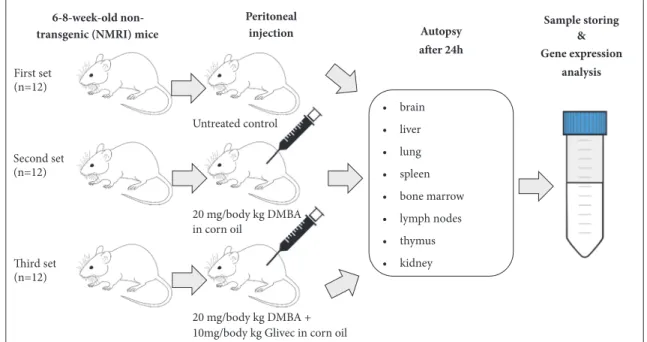
2.2. Treatment Group Assignment. Three experimental sets were created for the experimental agents (Figure 1). The first set of animals was treated intraperitoneally (i.p.) with vehicle (corn oil) and served as a negative control group. The second set of mice (positive control) was treated i.p. with a 20 mg/kg dose of DMBA dissolved in corn oil (both compounds were purchased from Sigma Aldrich, Budapest, Hungary). In the third group (experimental set), animals were simultaneously treated i.p. with 10 mg/kg imatinib mesylate (4-[(4-methyl- 1-piperazinyl)methyl]-N-[4-methyl-3-[[4-(3-pyridinyl)-2- pyrimidinyl]amino]-phenyl]benzamide methanesulfonate,
Sample storing
&
Gene expression analysis First set
(n=12)
Second set (n=12)
20 mg/body kg DMBA + 10mg/body kg Glivec in corn oil
• brain
• liver
• lung
• spleen
• bone marrow
• lymph nodes
• thymus
• kidney Autopsy after 24h
Third set (n=12)
6-8-week-old non- transgenic (NMRI) mice
in corn oil
Peritoneal injection
Untreated control
20 mg/body kg DMBA
Figure 1:Experimental design. Six- to eight-week-old (n=36) conventionally raised NMRI inbred mice were divided randomly into three sets:
the negative control group was i.p. treated with the vehicle (corn oil) (1st set, n=12), the positive control group (2nd set, n=12) was treated i.p.
with a 20 mg/kg body weight dose DMBA (7,12-dimethylbenz[a]anthracene), and the experimental group (3rd set, n=12) was treated i.p. with 10 mg/kg imatinib mesylate and 20 mg/kg DMBA. Animals were autopsied 24 hours after treatment, and organs were dissected and stored for further analysis.
Novartis Pharma GmbH product (Glivec), and 20 mg/kg DMBA dissolved in corn oil. Mice were sacrificed 24 hours after the injections, and organs (liver, spleen, kidney, lung, thymus, lymph node, bone marrow, and brain) were harvested and snap-frozen in liquid nitrogen and then stored at - 80∘C for further use.
2.3. RNA Extraction. mg tissue samples of each organ from the respective groups were homogenized in MagNA Lyzer Green Beads tubes (Roche (Hungary) Ltd.) using the MagNA Lyzer instrument (Roche (Hungary) Ltd.). Total RNA was isolated from the tissue lysates using the EXTRAzol RNA extraction kit (Invitrogen Life Technologies Magyarorsz´ag Kft). The RNA quality was assessed by absorption measurement at 260/280 nm (A260/A280 was>1.8).
2.4. Gene Expression Investigations. One-step PCR including reverse transcription and target amplification was performed using Kapa SYBR FAST One-step RTqPCR Kit (Kapa Biosystems) on a LightCycler 480 qPCR platform with a 96- well format. The specific primers (IDT) for mouse tumor sup- pressor genes (Hras, 5-AATTGGGGGAGCAAGGACAT- 3); (Kras, 5-TATCCTGCTTCCCATCAGTGTTC-3);
(Myc, 5-GTTGTGCTGGTGAGTGGAGA-3); (Trp53, 5- CTTCACTTGGGCCTTCAAAA-3) and for a housekeeping gene (Gapdh, 5-CACATTGGGGGTAGGAACAC-3) were used in the quantitative amplification.
RT-qPCR was initiated by 5 min. and 3 min. incubations at 42∘C and 95∘C, respectively, followed by 50 cycles (95∘C for 10 s, 55∘C for 20 s, and 72∘C for 20 s) with a fluorescent reading
taken at the end of each cycle. Each run was completed with a melting curve analysis (95∘C for 5 s, 65∘C for 60 s, and 97∘C
∞) to confirm the specificity of amplification. Fluorescent values were calculated following theΔΔCp method on Exor 4 software (Roche (Hungary) Ltd.) and gene expressions are reflected as relative quantification results.
2.5. Data Analysis. Statistical analyses were performed using R software (http://www.r-project.org) and SPSS 21.0 software (SPSS Inc., IL, USA). The differences in mRNA expression levels were calculated using a two-tailed Student’s t-test and were considered to be significant when p<0.05. Gene- gene interaction networks to demonstrate the relationship between genes in different organs/experimental sets were generated by the GeneMania Cytoscape 3.4.0 application.
Physical, coexpression, and gene-gene interactions were evaluated [44]. Heat map was constructed using Gene-E ver- sion 3.0.204 (http://www.broadinstitute.org/cancer/software/
GENE-E/index.html).
3. Results
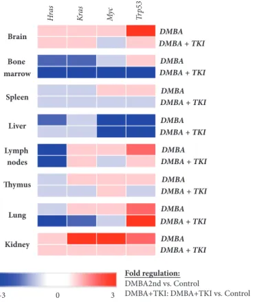
3.1. Gene Expression. Gene expression patterns of the three experimental sets are shown on Figures 2 and 3. Importantly, we found no gender-specific differences in the gene expres- sion patterns.
3.1.1. Bone Marrow. In the bone marrow, DMBA injection decreased the expressions of Hras, Kras, and Myc, respec- tively, and increased Trp53 expression. DMBA+imatinib
Fold regulation:
DMBA2nd vs. Control
DMBA+TKI: DMBA+TKI vs. Control Brain
Bone marrow
Spleen
Liver Lymph
nodes Thymus
Lung
Kidney
Hras Kras Myc Trp53
DMBA DMBA + TKI
DMBA DMBA + TKI DMBA DMBA + TKI
DMBA DMBA + TKI DMBA DMBA + TKI
DMBA DMBA + TKI DMBA DMBA + TKI
DMBA DMBA + TKI
-3 0 3
w
s
y
Figure 2:Heat map of gene expression patterns compared to the negative control. Blue boxes represent negative (down) regulation, while red boxes indicate positive (up)regulation of the gene expression.
mesylate administration further decreased the Hras, Kras, and Myc expressions. Compared to the negative control, significantly lowerKrasexpressions were found in the second (p<0.05)and third sets of mice(p<0.05).The combined treat- ment also decreased the expression of the tumor suppressor Trp53to a significant extent (p<0.05), first (control) versus third (DMBA + imatinib mesylate) set.
3.1.2. Brain. Compared to the negative controls, DMBA administration resulted in increased gene expressions in the brain; however, these changes were found to be non- significant. Combined administration of DMBA and imatinib mesylate decreased the expressions of the studied genes;
however, these alterations were not significant either.
3.1.3. Kidney. DMBA increased the expressions of theHras, Kras,andMyc,respectively, and the expression of theTrp53, as well. The simultaneous administration of DMBA and TKI reduced the expression of all the investigated genes.
3.1.4. Liver. In the liver, DMBA administration lowered the expressions of Hras, Kras, Myc,and Trp53, respectively. As a result of the combined DMBA+TKI administration, the decrease in the expression of these genes became reduced.
3.1.5. Lung. In the lung, mRNA expressions of the Kras (p<0.05), Myc, and Trp53 genes were increased, while the
Hrasexpression was decreased following the DMBA injec- tion. Simultaneous treatment with DMBA and TKI led to decreased the expression of protooncogenes (Hras, Kras,and Myc) and increasedTrp53mRNA levels.
3.1.6. Lymph Nodes. In the lymphoid tissues, DMBA decreased the Hras expression and increased the Kras and Trp53 expressions, that remained unchanged after the combined administration with DMBA+TKI. However, the expression ofMycwas increased by DMBA and decreased as a result of DMBA+TKI combination. However, this change in mRNA expression was not statistically significant.
3.1.7. Spleen. HrasandKrasgene expressions were decreased after DMBA injection, although they did not change after DMBA+TKI administration. In turn, DMBA induced increased expressions ofMyc(p<0.05) and decreasedTrp53 expressions after treatment (DMBA+TKI).
3.1.8. Thymus. In the thymus, DMBA increased the expres- sions ofKras, MycandTrp53, respectively, while decreasing theHras expression. As a result of combined administration of DMBA+imatinib mesylate, the expressions ofKrasandTrp53 were found to be reduced compared to the negative control.
Additionally, the expression ofMycshowed an increase, while the expression ofHrasremained unaltered after the combined injections.
Brain Bone marrow
Spleen Liver
Thymus Lymph nodes
Kidney Lung
DMBA DMBA + TKI DMBA DMBA + TKI
DMBA DMBA + TKI DMBA DMBA + TKI
DMBA DMBA + TKI DMBA DMBA + TKI
DMBA DMBA + TKI DMBA DMBA + TKI
Hras Myc
Kras Trp53
DMBA: DMBA vs Control
DMBA+TKI: DMBA+TKI vs Control
∗
∗
∗
∗
∗
∗
−2
−1 0 1 2 3 4
Fold change
−3
−2
−1 0 1 2 3
Fold change
−4
−2 0 2 4
Fold change
0 1 2 3 4 5
Fold change
−5 0 5 10
Fold change
−10
−5 0 5
Fold change
−8
−6
−4
−2 0
Fold change
−15
−10
−5 0 5
Fold change
Figure 3:Gene expression pattern of experimental groups in different organs.∗p<0,05.
3.2. Gene Network. Figure 4 shows the fold regulation of gene expressions in selected organs and their predicted interac- tions among the different regulatory genes. We observed sig- nificant alterations in gene expressions in the bone marrow, lung, and spleen. Our network analysis revealed thatHras,
Kras,andMycprotooncogenes andTrp53tumor suppressor gene have extensive connections to other regulatory genes.
Zhx2(also known asRAF) is a homodimeric transcription factor that belongs to the zinc fingers and homeoboxes gene family [45], Abi1 (abl interactor 1) is an adaptor protein
Lung (DMBA)
Lung (DMBA+TKI)
Bone m.
(DMBA)
Bone m.
(DMBA + TKI)
Spleen (DMBA)
Spleen (DMBA + TKI)
Fold regulation:
• DMBA: DMBA vs.
Control
• DMBA+TKI: DMBA+TKI vs. Control
-3.00 3.00
∗
∗
∗
∗ ∗
∗
Figure 4:A gene-gene interaction network presenting the correlation among the fold regulation of Kras, Hras, Myc, and Trp53 genes in the represented organs and their predicted interactions with 10 functionally related genes. The 10 correlated genes were obtained using the GeneMania application of Cytoscape; level of significance:∗(p<0.05).
that facilitates several signal transduction pathways, reg- ulates actin polymerization and cytoskeleton remodeling, and therefore has a role in cell proliferation [46]. Tcf4 (transcription factor 4) is essential for neuronal development [47], andTsc2(TSC complex subunit 2) gene codes a tumor suppressor protein (tuberin), mutation of which (together with mutation of hamartin, coded byTsc1) causes tuberous sclerosis complex [48].Huwe1encodes an E3 ubiquitin ligase protein that is responsible for ubiquitination and degradation of the antiapoptotic protein Mcl1 (myeloid cell leukemia sequence 1 (Bcl2-related)) [49].Cdkn2a (cyclin dependent kinase inhibitor 2a) is an important tumor suppressor gene, having at least three alternative spliced variants that code two CDK4 inhibitors and one p53 stabilizer protein, therefore playing a pivotal role in cell cycle G1 control [50].Nde1(nudE neurodevelopment protein 1) gene codes a protein that has essential role in microtubule organization, mitosis, and neu- ronal migration, mutation of which can be associated with lissencephaly [51].Kmt5a(lysine methyltransferase 5a) codes a protein that is a transcriptional repressor and is important for cell proliferation and chromatin condensation [52].Mcm4 (minichromosome maintenance complex component 4) gene codes a protein that is highly conserved and important for ini- tiation of eukaryotic genome replication [53].Eif4e(eukary- otic translation initiation factor 4E) functions as a protoonco- gene; its product helps the initiation of translation [54].
4. Discussion
Several studies have demonstrated the role of tyrosine kinases in human diseases [55, 56]. Consequently, tyrosine kinases
have become one of the main areas of pharmacological experiments intended to develop targeted drugs [57]. Protein tyrosine kinase inhibitors are small molecules that are able to diffuse through the cell membrane targeting cytoplasmic kinases or the intracellular domain of receptor tyrosine kinases. TKIs are currently booming and are widely used in cancer cure either in the form of monotherapy or in combination with other chemotherapeutic agents [58].
In our present study, we investigated the potential chemo- preventive effect of imatinib mesylate that is the first small- molecule tyrosine kinase inhibitor used in CML and gastroin- testinal stromal tumor (GIST) [59, 60]. To date, our study is among the first ones to examine the possible preventive effect of imatinib mesylate by studying the alterations in DMBA- induced gene expression levels and trying to put the results into the gene network of different protooncogenes (Hras, Kras, and Myc) and a tumor suppressor gene (Trp53) in a short-term experiment. The outcomes shown here suggest that imatinib mesylate might have a possible mitigating role in diseases beyond CML and GIST.
Major results of the present study include that short- term DMBA treatment (i) elevated the expression of all the three protooncogenes (Hras, Kras, and Myc) in the brain and kidneys; (ii) increased the level ofKrasandMycin the lung, lymph nodes and thymus; (iii) increased the expression of the tumor suppressor geneTrp53that can be considered an adaptive physiologic countermeasure in response to a chemical carcinogen. These phenomena have been previously described by several investigations, concluding that DMBA is a potent inducer of chemical carcinogenesis and can be used for studying different types of malignant tumors. DMBA is a
polyaromatic hydrocarbon similar to hydrocarbons to which humans can be exposed. DMBA causes point mutations in protooncogenes likeHrasthat is common in human carci- nomas [61]. In the bone marrow and liver, DMBA decreased the expression level ofHras, Kras,andMyc. This observation might be explained by the fact that DMBA is a carcinogenesis inducer, and it is usually applied simultaneously with a carcinogenesis promoter, e.g., 12-O-tetradecanoylphorbol 13- acetate (TPA) [62]. Therefore, in case of the bone marrow and liver, DMBA might not be enough for complete tumorige- nesis. In the spleen, the elevated level ofMycwas the only prominent and significant alteration in the gene expression pattern. Several studies have elucidated the role of Myc in tumorigenesis. Probably the best-established association is that nearly every case of Burkitt’s lymphoma involves rearrangement and therefore overexpression of Mycwith a regulatory element of immunoglobulin heavy or light chains or other nonrandom somatic mutations of the gene [63, 64].
The results of the aforementioned studies correlate with our finding of elevated expression of Myc in the spleen and lymph nodes as a consequence of DMBA treatment. The increased expression of the examined four genes gain more importance in the context of their extensive gene network.
Zhx2 (also known asRAF) has previously been associated with Hodgkin lymphoma [65] and hepatocellular carcinoma [66];Abi1(abl interactor 1) has a role in colorectal carcinoma development and invasion [67] and also in neuroblastoma propagation [68]. Aberrant function of Tcf4(transcription factor 4) has been reported in glioblastoma [69] and in colorectal tumors [70].Tsc2(TSC complex subunit 2) gene codes a tumor suppressor protein (tuberin), mutation of which have been associated with tumors in the brain, lungs, kidneys, skin, heart, uterus, and eyes [71, 72].Huwe1encodes an E3 ubiquitin ligase protein that is required for the development of colorectal carcinoma and ovarian tumors [73, 74]. Cdkn2a (cyclin dependent kinase inhibitor 2a) is an important tumor suppressor gene predisposing to several tumors, e.g., urothelial carcinoma, hereditary melanoma, pancreas cancer, or non-small-cell lung cancer [75–77].Nde1 (nudE neurodevelopment protein 1) gene codes a protein that has essential role in microtubule organization and mitosis, and recent studies have elucidated its potential role in acute or chronic myeloid leukemia [78, 79].Mcm4(minichromosome maintenance complex component 4) has been reported to be upregulated in ovarian cancer, skin cancer, or esophageal carcinoma [80–82]. Eif4e (eukaryotic translation initiation factor 4E) functions as a protooncogene; its product has been suggested to regulate expression of proteins that are crucial for cell cycle progression, cell survival, and motility. A growing body of evidence implicates this translational factor in cell transformation, tumorigenesis, or tumor progression, e.g., in case of prostate cancer, lymphomas, CML, or lung cancers [83].
As it is suggested by the extensive gene network of the examined genes, cancer development involves more than one transforming events and the interaction of several oncogenes and tumor suppressor genes. This network and series of events offers numerous opportunities to effectively influence the process of tumorigenesis.
In the lungs, the expression of protooncogenes (Hras, Kras,andMyc) and their connections to other genes coding transcription factors or cell proliferation regulators (e.g., Tcf4, Abi1, and Zhx2)prominently decreased as a result of the short-term combined DMBA+TKI treatment, while the expression ofTrp53gene increased. Comparing to the nega- tive control, the decrease inKrasexpression was significant.
In the bone marrow, DMBA+TKI combined treatment significantly decreased the expression and gene interactions of theKrasandTrp53.
DMBA+TKI treatment could significantly decrease the DMBA-induced increase in the expression and gene inter- actions ofMycprotooncogene. The expression of the tumor suppressor Trp53 also decreased following the combined treatment; however, this decrease was not significant.
Outcomes of our short-term experiment suggest that pro- tein tyrosine kinase inhibitor treatment (imatinib mesylate) simultaneously administered with the chemical carcinogen, DMBA, might have an impact on the expression pattern of the examined protooncogenes (Hras, Kras,andMyc)and tumor suppressor gene (Trp53), therefore on the tumorigenesis, controlled by these genes.
Imatinib mesylate is a well-known small-molecule inhibitor of tyrosine kinases. In our study, this drug was able to decrease significantly the expression ofKrasoncogene in the bone marrow and in the lung, as well as the expression of Myc oncogene in the spleen. Additionally, Myc mRNA expressions were tended to be lowered in the bone marrow, brain, kidneys, lungs, and lymph nodes and we also observed tendencies in theHrasmRNA expressions to be decreased in the bone marrow, kidneys, and lungs, although these changes were not statistically significant. The reduced expression of these oncogenes may be attributed to the kinase inhibitor effect of imatinib mesylate, as described by other recent studies. Among others, Lorri Puil et al. reported that BCR- Abl was able to activate Ras signaling in CML, by creating a direct link between Grb2 and mSos1 that are responsible for the conversion of inactive GDP-bound form of Ras into the active, GTP-bound form. Therefore, inhibiting BCR-Abl kinase activity may downregulate Ras signaling in CML [84].
Besides Ras signaling, BCR-Abl kinase can indirectly acti- vateMyceither through the Janus-activated kinase 2 (JAK2) pathway [85] or by the mitogen-activated protein kinase (MAPK) pathway [86]. It is tempting to speculate that ima- tinib might have decreased the expression ofMycwell before its DMBA-induced overexpression. Callahan R. et al. revealed that imatinib mesylate was able to induce complete regression of mammary tumor and restore lobuloalveolar development and lactation by inhibiting Notch4 andMycsignaling, which result also support the idea of therapeutic potential of imatinib mesylate, other than CML and GIST [87].
PDGF isoforms and their receptors (PDGFRs) are con- sidered as prototypes of growth factors and receptor tyrosine kinases for more than 25 years. They are essential for nor- mal gastrulation and cranial, neuronal, cardiac, pulmonary, intestinal, gonadal, hematological, skin, renal, and skeletal development, as well as for hematopoiesis, through the secondary signal transduction pathway, including activa- tion of Ras and the downstream Raf and MAPK cascades
[88]. However, overexpression or mutational events in the PGDFR gene may drive tumor development and progression [89]. Recent studies have elucidated the role of PDGFRs in the evolution of different nervous system tumors, i.e., glioblastoma [90], ependymoma [91], meningioma [92], and schwannoma (in which PDGFR mutation is usually associated with c-kit overactivation [93]). In addition to brain tumors, the role of mutant PDGFR has numerously been emphasized in other malignant diseases, like dermatofi- brosarcoma protuberans [94], gastrointestinal stromal tumor (GIST) [95], osteosarcoma [96], alveolar rhabdomyosarcoma [97], chronic myeloid leukemia (CML) [98], prostate cancer [99], liver cancer [100], non-small-cell lung cancer [101], and colorectal cancer [102] and in breast cancer [103]. There have been numerous attempts to inhibit the activity of PDGFRs, including tyrosine kinase inhibitors, like imatinib or sorafenib, and also several antibodies targeting the differ- ent PDGF isoforms or the receptors themselves to prevent their activation. In general, antibodies are much more specific therapeutic tools; however, their administration is expen- sive and sometimes inconvenient. Tyrosine kinase inhibitors are not specific; they have the potential to inhibit more kinases—and in this way have more adverse effects—as ima- tinib is able to inhibit PDGFRs, Abl kinases, and the stem cell receptor c-kit, but in cancer treatment, it can be advantageous to target more than one component of tumorigenesis [89].
Kras, Hras, and Myc are the executive elements of numerous oncogenic pathways, so they can be favorable to inhibit a common point of tumorigenesis by one molecule.
p53 is the best characterized tumor suppressor protein, as it is able to induce cell cycle arrest or cell death in response to hypoxia and incorrigible genetic mutations, while mutations of TP53 gene have been associated with more that 50%
of human tumors [104]. There is growing evidence that these mutations are ‘loss-of-function’ mutations; however, missense mutations may result in simultaneous gain of functions that have usually detrimental effect to the cell [105]. Numerous studies have reported that mutant p53 played a key role in tumor development, progression, and invasion of several cancer types, e.g., in case of breast cancer [106], lung cancer [107], colorectal cancer [108], different brain tumors, and gastric adenocarcinoma [109]. In our present study, short-term imatinib mesylate treatment administered simultaneously with DMBA resulted in a prominent increase in the Trp53 expression in the lung, while decreasing it in all the other tissues. These data indicate a possible ‘gain-of-function’ mutation in the gene of the tumor suppressor p53 protein and that imatinib mesylate attempted to decrease the level of this aberrant protein.
Based on our recent and previous findings we suggest that imatinib mesylate is a promising chemotherapeutic agent for prevention and management of several malignant tumors by decreasing the mRNA expression of the protooncogenes and the mutantTrp53gene.
5. Conclusion
The outcomes of the present study demonstrate that imatinib mesylate decreases the mRNA expressions ofHras, Kras, Myc,
andTrp53genes in certain organs after 24 hours of a single dose of TKI treatment in a DMBA-induced mouse tumor model. These results suggest its preventive and curative roles in malignant diseases.
Data Availability
The experimental analysis data used to support the findings of this study are available from the corresponding author upon request.
Disclosure
The current affiliation for Bal´azs Murny´ak is Center for Cran- iofacial Molecular Biology, University of Southern California, Los Angeles, CA 90033, USA.
Conflicts of Interest
The authors declare that they have no conflicts of interest.
Authors’ Contributions
P´eter Attila Gergely and Bal´azs Murny´ak are joint first authors and contributed equally to this work.
Acknowledgments
This study was supported by Institute of Forensic Medicine, University of Debrecen, Department of Public Health, Uni- versity of P´ecs, the Hungarian Brain Research Program (2017- 1.2.1-NKP-2017-00002), SZTE ´AOK-KKA 2018/Hortob´agyiT (TH), ´UNKP-18-3 New National Excellence Program of the Ministry of Human Capacities, and EFOP-3.6.3-VEKOP-16- 2017-00009 (JB). The authors are sincerely grateful to Norbert N´adasdi, Zsuzsa Bayer, and M´onika Herczeg for their help through the experimental procedures, to Istv´an Kiss, P´eter F¨ul¨op, and P´eter T¨or¨ok for their work in organizing, format- ting, and editing this article. This work isin memoriamof Professor Istv´an Ember (1952-2013).
References
[1] R. Roskoski, “A historical overview of protein kinases and their targeted small molecule inhibitors,”Pharmacological Research, vol. 100, pp. 1–23, 2015.
[2] P. Cohen, “Protein kinases—the major drug targets of the twenty-first century?”Nature Reviews Drug Discovery, vol. 1, no.
4, pp. 309–315, 2002.
[3] G. Manning, D. B. Whyte, R. Martinez, T. Hunter, and S.
Sudarsanam, “The protein kinase complement of the human genome,”Science, vol. 298, no. 5600, pp. 1912–1934, 2002.
[4] N. Volinsky and B. N. Kholodenko, “Complexity of receptor tyrosine kinase signal processing,”Cold Spring Harbor Perspec- tives in Biology, vol. 5, no. 8, Article ID a009043, 2013.
[5] D. S. Krause and R. A. van Etten, “Tyrosine kinases as targets for cancer therapy,”The New England Journal of Medicine, vol.
353, no. 2, pp. 172–187, 2005.
[6] K. Taniuchi, Y. Yamada, A. Nonomura, and K. Takehara,
“Immunohistochemical analysis of platelet-derived growth fac- tor and its receptors in fibrohistiocytic tumors,” Journal of Cutaneous Pathology, vol. 24, no. 7, pp. 393–397, 1997.
[7] U. Krishnamurti and J. F. Silverman, “HER2 in breast cancer: A review and update,”Advances in Anatomic Pathology, vol. 21, no.
2, pp. 100–107, 2014.
[8] F. A. Lagunas-Rangel and V. Ch´avez-Valencia, “FLT3–ITD and its current role in acute myeloid leukaemia,”Medical Oncology, vol. 34, no. 6, p. 114, 2017.
[9] W. Pao, V. Miller, M. Zakowski et al., “EGF receptor gene mutations are common in lung cancers from never smokers and are associated with sensitivity of tumors to gefitinib and erlotinib,”Proceedings of the National Academy of Sciences of the United States of America, vol. 101, no. 36, pp. 13306–13311, 2004.
[10] B. Murnyak, T. Csonka, K. Hegyi et al., “Occurrence and molecular pathology of high grade gliomas,”Ideggyogy Sz, vol.
66, no. 9-10, pp. 312–321, 2013.
[11] K. M. Smith, R. Yacobi, and R. A. Van Etten, “Autoinhibition of Bcr-Abl through its SH3 domain,”Molecular Cell, vol. 12, no. 1, pp. 27–37, 2003.
[12] B. J. Druker, “Imatinib mesylate in the treatment of chronic myeloid leukaemia,”Expert Opinion on Pharmacotherapy, vol.
4, no. 6, pp. 963–971, 2003.
[13] B. J. Druker, S. Tamura, E. Buchdunger et al., “Effects of a selective inhibitor of the Ab1 tyrosine kinase on the growth of Bcr-Ab1 positive cells,”Nature Medicine, vol. 2, no. 5, pp. 561–
566, 1996.
[14] E. Nadal and E. Olavarria, “Imatinib mesylate (Gleevec/Glivec) a molecular-targeted therapy for chronic myeloid leukaemia and other malignancies,”International Journal of Clinical Prac- tice, vol. 58, no. 5, pp. 511–516, 2004.
[15] M. W. N. Deininger and B. J. Druker, “Specific targeted therapy of chronic myelogenous leukemia with imatinib,”Pharmacolog- ical Reviews, vol. 55, no. 3, pp. 401–423, 2003.
[16] A. S. Corbin, P. La Ros´ee, E. P. Stoffregen, B. J. Druker, and M. W.
Deininger, “Several Bcr-Abl kinase domain mutants associated with imatinib mesylate resistance remain sensitive to imatinib,”
Blood, vol. 101, no. 11, pp. 4611–4614, 2003.
[17] M. Ohanian, J. Cortes, H. Kantarjian, and E. Jabbour, “Tyrosine kinase inhibitors in acute and chronic leukemias,” Expert Opinion on Pharmacotherapy, vol. 13, no. 7, pp. 927–938, 2012.
[18] R. J. Motzer, T. E. Hutson, P. Tomczak et al., “Sunitinib versus interferon alfa in metastatic renal-cell carcinoma,”The New England Journal of Medicine, vol. 356, no. 2, pp. 115–124, 2007.
[19] K. Tatsugami, M. Oya, K. Kabu, and H. Akaza, “Efficacy and safety of sorafenib for advanced renal cell carcinoma: real-world data of patients with renal impairment,”Oncotarget, vol. 9, no.
27, pp. 19406–19414, 2018.
[20] M. Fukuoka, Y.-L. Wu, S. Thongprasert et al., “Biomarker analyses and final overall survival results from a phase III, randomized, open-label, first-line study of gefitinib versus car- boplatin/paclitaxel in clinically selected patients with advanced non-small-cell lung cancer in Asia (IPASS),”Journal of Clinical Oncology, vol. 29, no. 21, pp. 2866–2874, 2011.
[21] M. J. Moore, D. Goldstein, J. Hamm et al., “Erlotinib plus gemcitabine compared with gemcitabine alone in patients with advanced pancreatic cancer: a phase III trial of the National Cancer Institute of Canada Clinical Trials Group,”Journal of Clinical Oncology, vol. 15, pp. 1960–1966, 2005.
[22] D. Cameron, M. Casey, M. Press et al., “A phase III randomized comparison of lapatinib plus capecitabine versus capecitabine alone in women with advanced breast cancer that has pro- gressed on trastuzumab: Updated efficacy and biomarker anal- yses,”Breast Cancer Research and Treatment, vol. 112, no. 3, pp.
533–543, 2008.
[23] R. J. Motzer, N. B. Haas, F. Donskov et al., “Randomized phase III trial of adjuvant pazopanib versus placebo after nephrectomy in patients with localized or locally advanced renal cell carcinoma,”Journal of Clinical Oncology, vol. 35, no.
35, pp. 3916–3923, 2017.
[24] R. S. Herbst, Y. Sun, W. E. E. Eberhardt et al., “Vandetanib plus docetaxel versus docetaxel as second-line treatment for patients with advanced non-small-cell lung cancer (ZODIAC):
A double-blind, randomised, phase 3 trial,”The Lancet Oncol- ogy, vol. 11, no. 7, pp. 619–626, 2010.
[25] B. Escudier, B. I. Rini, R. J. Motzer et al., “Genotype Correlations with Blood Pressure and Efficacy from a Randomized Phase III Trial of Second-Line Axitinib Versus Sorafenib in Metastatic Renal Cell Carcinoma,”Clinical Genitourinary Cancer, vol. 13, no. 4, pp. 328–337.e3, 2015.
[26] J. R. Woodburn, “The epidermal growth factor receptor and its inhibition in cancer therapy,”Pharmacology & Therapeutics, vol.
82, no. 2-3, pp. 241–250, 1999.
[27] M. Malumbres and M. Barbacid, “RAS oncogenes: the first 30 years,”Nature Reviews Cancer, vol. 3, no. 6, pp. 459–465, 2003.
[28] A. D. Cox, S. W. Fesik, A. C. Kimmelman, J. Luo, and C. J. Der,
“Drugging the undruggable RAS: Mission Possible?” Nature Reviews Drug Discovery, vol. 13, no. 11, pp. 828–851, 2014.
[29] M. Eilers, D. Picard, K. R. Yamamoto, and J. M. Bishop,
“Chimaeras of Myc oncoprotein and steroid receptors cause hormone-dependent transformation of cells,”Nature, vol. 340, no. 6228, pp. 66–68, 1989.
[30] R. A. Beckman and L. A. Loeb, “Genetic instability in cancer:
Theory and experiment,”Seminars in Cancer Biology, vol. 15, no.
6, pp. 423–435, 2005.
[31] C. E. Nesbit, J. M. Tersak, and E. V. Prochownik, “MYC oncogenes and human neoplastic disease,”Oncogene, vol. 18, no.
19, pp. 3004–3016, 1999.
[32] A. Kuzyk and S. Mai, “c-MYC-induced genomic instability,”
Cold Spring Harbor Perspectives in Medicine, vol. 4, no. 4, p.
a014373, 2014.
[33] C. Kandoth, M. D. McLellan, F. Vandin et al., “Mutational land- scape and significance across 12 major cancer types,”Nature, vol.
502, no. 7471, pp. 333–339, 2013.
[34] B. Leroy, J. L. Fournier, C. Ishioka et al., “The TP53 website: An integrative resource centre for the TP53 mutation database and TP53 mutant analysis,”Nucleic Acids Research, vol. 41, no. 1, pp.
D962–D969, 2013.
[35] B. Murnyak and T. Hortobagyi, “Immunohistochemical corre- lates of TP53 somatic mutations in cancer,”Oncotarget, vol. 7, no. 40, pp. 64910–64920, 2016.
[36] P. A. J. Muller and K. H. Vousden, “Mutant p53 in cancer: New functions and therapeutic opportunities,”Cancer Cell, vol. 25, no. 3, pp. 304–317, 2014.
[37] M. Adorno, M. Cordenonsi, and M. Montagner, “A Mutant- p53/Smad complex opposes p63 to empower TGFbeta-induced metastasis,”Cell, vol. 137, no. 1, pp. 87–98, 2009.
[38] C. Gaiddon, M. Lokshin, J. Ahn, T. Zhang, and C. Prives, “A subset of tumor-derived mutant forms of p53 down-regulate p63 and p73 through a direct interaction with the p53 core
domain,”Molecular and Cellular Biology, vol. 21, no. 5, pp. 1874–
1887, 2001.
[39] H. Song, M. Hollstein, and Y. Xu, “p53 gain-of-function cancer mutants induce genetic instability by inactivating ATM,”Nature Cell Biology, vol. 9, no. 5, pp. 573–580, 2007.
[40] C. Lin, Y. Liang, H. Zhu, J. Zhang, and X. Zhong, “R280T mutation of p53 gene promotes proliferation of human glioma cells through GSK-3𝛽/PTEN pathway,” Neuroscience Letters, vol. 529, no. 1, pp. 60–65, 2012.
[41] J. M. Ward, S. Rehm, D. Devor et al., “Differential carcinogenic effects of intraperitoneal initiation with 7,12-dimethylbenz(a) anthracene or urethane and topical promotion with 12-O- tetradecanoylphorbol-13-acetate in skin and internal tissues of female SENCAR and BALB/c mice,”Environmental Health Perspectives, vol. 68, pp. 61–68, 1986.
[42] T. Sugiyama, M. Osaka, K. Koami, S. Maeda, and N. Ueda, “7,12- DMBA-induced rat leukemia: A review with insights into future research,”Leukemia Research, vol. 26, no. 12, pp. 1053–1068, 2002.
[43] P. Gergely, F. Bud´an, G. Mezey et al., “Kinase inhibitors reduce 7,12-dimethylbenz a anthracene-induced onco-suppressor gene expression in short-term experiments,”European Journal of Oncology, vol. 17, pp. 11–21, 2012.
[44] J. Montojo, K. Zuberi, H. Rodriguez, G. D. Bader, and Q. Morris,
“GeneMANIA: Fast gene network construction and function prediction for Cytoscape,”F1000Research, vol. 3, p. 153, 2014.
[45] H. Kawata, K. Yamada, Z. Shou et al., “Zinc-fingers and homeoboxes (ZHX) 2, a novel member of the ZHX family, functions as a transcriptional repressor,”Biochemical Journal, vol. 373, no. 3, pp. 747–757, 2003.
[46] Z. Biesova, C. Piccoli, and W. T. Wong, “Isolation and charac- terization of e3B1, an eps8 binding protein that regulates cell growth,”Oncogene, vol. 14, no. 2, pp. 233–241, 1997.
[47] F. E. M. In’t Hout, B. A. van der Reijden, D. Monteferrario, J. H. Jansen, and G. Huls, “High expression of transcription factor 4 (TCF4) is an independent adverse prognostic factor in acute myeloid leukemia that could guide treatment decisions,”
Haematologica, vol. 99, no. 12, pp. e257–e259, 2014.
[48] L. Xu, C. Sterner, M. M. Maheshwar et al., “Alternative splicing of the tuberous sclerosis 2 (TSC2) gene in human and mouse tissues,”Genomics, vol. 27, no. 3, pp. 475–480, 1995.
[49] P. K. Jackson, A. G. Eldridge, E. Freed et al., “The lore of the RINGs: Substrate recognition and catalysis by ubiquitin ligases,”
Trends in Cell Biology, vol. 10, no. 10, pp. 429–439, 2000.
[50] T. Nobori, K. Miura, D. J. Wu, A. Lois, K. Takabayashi, and D. A.
Carson, “Deletions of the cyclin-dependent kinase-4 inhibitor gene in multiple human cancers,”Nature, vol. 368, no. 6473, pp.
753–756, 1994.
[51] M. Kitagawa, M. Umezu, J. Aoki, H. Koizumi, H. Arai, and K. Inoue, “Direct association of LIS1, the lissencephaly gene product, with a mammalian homologue of a fungal nuclear distribution protein, rNUDE,”FEBS Letters, vol. 479, no. 1-2, pp.
57–62, 2000.
[52] J.-F. Couture, E. Collazo, J. S. Brunzelle, and R. C. Trievel,
“Structural and functional analysis of SET8, a histone H4 Lys- 20 methyltransferase,”Genes & Development, vol. 19, no. 12, pp.
1455–1465, 2005.
[53] C. Musahl, D. Schulte, R. Burkhart, and R. Knippers, “A Human Homologue of the Yeast Replication Protein Cdc21: Interactions with Other Mcm Proteins,”European Journal of Biochemistry, vol. 230, no. 3, pp. 1096–1101, 1995.
[54] J. Pelletier, J. D. Brook, and D. E. Housman, “Assignment of two of the translation initiation factor-4E (EIF4EL1 and EIF4EL2) genes to human chromosomes 4 and 20,”Genomics, vol. 10, no.
4, pp. 1079–1082, 1991.
[55] M. Labots, J. C. Van der Mijn, H. Dekker et al., “Selection of Protein Kinase Inhibitors Based on Tumor Tissue Kinase Activity Profiles in Patients with Refractory Solid Malignancies:
An Interventional Molecular Profiling Study,”The Oncologist, vol. 23, no. 10, pp. 1135–e118, 2018.
[56] H. Patterson, R. Nibbs, I. Mcinnes, and S. Siebert, “Protein kinase inhibitors in the treatment of inflammatory and autoim- mune diseases,”Clinical & Experimental Immunology, vol. 176, no. 1, pp. 1–10, 2014.
[57] D. R. Shah, R. R. Shah, and J. Morganroth, “Tyrosine kinase inhibitors: Their on-target toxicities as potential indicators of efficacy,”Drug Safety, vol. 36, no. 6, pp. 413–426, 2013.
[58] A. Canonici, L. Ivers, N. T. Conlon et al., “HER-targeted tyrosine kinase inhibitors enhance response to trastuzumab and pertuzumab in HER2-positive breast cancer,”Investigational New Drugs, 2018.
[59] M. Miura, “Therapeutic drug monitoring of imatinib, nilotinib, and dasatinib for patients with chronic myeloid leukemia,”
Biological & Pharmaceutical Bulletin, vol. 38, no. 5, pp. 645–654, 2015.
[60] T. Nishida, K. Shirao, A. Sawaki et al., “Efficacy and safety profile of imatinib mesylate (ST1571) in Japanese patients with advanced gastrointestinal stromal tumors: A phase II study (STI571B1202),”International Journal of Clinical Oncology, vol.
13, no. 3, pp. 244–251, 2008.
[61] A. Balmain, M. Ramsden, G. T. Bowden, and J. Smith, “Acti- vation of the mouse cellular Harvey-ras gene in chemically induced benign skin papillomas,”Nature, vol. 307, no. 5952, pp.
658–660, 1984.
[62] M. Xiao, C. Wang, J. Zhang, Z. Li, X. Zhao, and Z.
Qin, “IFNgamma; promotes papilloma development by up- regulating th17-associated inflammation,”Cancer Research, vol.
69, no. 5, pp. 2010–2017, 2009.
[63] C. A. Spencer and M. Groudine, “Control of c-myc regulation in normal and neoplastic cells,”Advances in Cancer Research, vol.
56, pp. 1–48, 1991.
[64] J. M. Johnston and W. L. Carroll, “C-myc hypermutation in burkitt’s lymphoma,”Leukemia & Lymphoma, vol. 8, no. 6, pp.
431–439, 1992.
[65] S. Nagel, B. Schneider, C. Meyer, M. Kaufmann, H. G. Drexler, and R. A. F. MacLeod, “Transcriptional deregulation of home- obox gene ZHX2 in Hodgkin lymphoma,”Leukemia Research, vol. 36, no. 5, pp. 646–655, 2012.
[66] F. Luan, P. Liu, H. Ma et al., “Reduced nucleic ZHX2 involves in oncogenic activation of glypican 3 in human hepatocellular carcinoma,”The International Journal of Biochemistry & Cell Biology, vol. 55, pp. 129–135, 2014.
[67] K. Steinestel, S. Br¨uderlein, J. K. Lennerz et al., “Expression and Y435-phosphorylation of Abelson interactor 1 (Abi1) pro- motes tumour cell adhesion, extracellular matrix degradation and invasion by colorectal carcinoma cells,”Acta Veterinaria Scandinavica, vol. 13, no. 1, p. 145, 2014.
[68] X. Liu, H. Peng, W. Liao et al., “MiR-181a/b induce the growth, invasion, and metastasis of neuroblastoma cells through target- ing ABI1,”Molecular Carcinogenesis, vol. 57, no. 9, pp. 1237–1250, 2018.
[69] J.-X. Zhang, J. Zhang, W. Yan et al., “Unique genome-wide map of TCF4 and STAT3 targets using ChIP-seq reveals their
association with new molecular subtypes of glioblastoma,”
Neuro-Oncology, vol. 15, no. 3, pp. 279–289, 2013.
[70] M. B. Meyer, P. D. Goetsch, and J. W. Pike, “VDR/RXR and TCF4/𝛽-catenin cistromes in colonic cells of colorectal tumor origin: Impact on c-FOS and c-MYC gene expression,”
Molecular Endocrinology, vol. 26, no. 1, pp. 37–51, 2012.
[71] H. Prizant, A. Sen, A. Light et al., “Uterine-specific loss of Tsc2 leads to myometrial tumors in both the uterus and lungs,”
Molecular Endocrinology, vol. 27, no. 9, pp. 1403–1414, 2013.
[72] H. Northrup, M. K. Koenig, and D. A. Pearson, “Tuberous Sclerosis Complex,” in GeneReviews((R)), M. P. Adam, Ed., Seattle, Wash, USA, 1993.
[73] S. Peter, J. Bultinck, K. Myant et al., “Tumor cell-specific inhibition of MYC function using small molecule inhibitors of the HUWE1 ubiquitin ligase,”EMBO Molecular Medicine, vol.
6, no. 12, pp. 1525–1541, 2014.
[74] D. Yang, B. Sun, X. Zhang et al., “Huwe1 sustains normal ovarian epithelial cell transformation and tumor growth through the histone H1.3-H19 cascade,”Cancer Research, vol. 77, no. 18, pp.
4773–4784, 2017.
[75] P. H. Kim, E. K. Cha, J. P. Sfakianos et al., “Genomic predictors of survival in patients with high-grade urothelial carcinoma of the bladder,”European Urology, vol. 67, no. 2, pp. 198–201, 2015.
[76] F. Jouenne, I. C. de Beauchene, E. Bollaert et al., “Germline CDKN2A/P16INK4A mutations contribute to genetic deter- minism of sarcoma,”Journal of Medical Genetics, vol. 54, no. 9, pp. 607–612, 2017.
[77] D. P. Bradly, P. Gattuso, M. Pool et al., “CDKN2A (p16) Promoter hypermethylation influences the outcome in young lung cancer patients,”Diagnostic Molecular Pathology, vol. 21, no. 4, pp. 207–213, 2012.
[78] B. A. Van Der Reijden, M. Massop, A. Simons, T. De Witte, M.
Breuning, and J. H. Jansen, “The NDE1 gene is disrupted by the inv(16) in 90% of cases with CBFB-MYH11-positive acute myeloid leukemia,”Leukemia, vol. 24, no. 4, pp. 857–859, 2010.
[79] R. La Starza, R. Rosati, G. Roti et al., “A new NDE1/PDGFRB fusion transcript underlying chronic myelomonocytic leu- kaemia in Noonan Syndrome,”Leukemia, vol. 21, no. 4, pp. 830–
833, 2007.
[80] L. Xie, T. Li, and L. H. Yang, “E2F2 induces MCM4, CCNE2 and WHSC1 upregulation in ovarian cancer and predicts poor over- all survival,”European Review for Medical and Pharmacological Sciences, vol. 21, no. 9, pp. 2150–2156, 2017.
[81] Y. Ishimi and D. Irie, “G364R mutation of MCM4 detected in human skin cancer cells affects DNA helicase activity of MCM4/6/7 complex,”The Journal of Biochemistry, vol. 157, no.
6, pp. 561–569, 2015.
[82] B. Choy, A. LaLonde, J. Que, T. Wu, and Z. Zhou, “MCM4 and MCM7, potential novel proliferation markers, significantly cor- related with Ki-67, Bmi1, and cyclin E expression in esophageal adenocarcinoma, squamous cell carcinoma, and precancerous lesions,”Human Pathology, vol. 57, pp. 126–135, 2016.
[83] U. Laemmll, E. M¨olbert, M. Showe, and E. Kellenberger, “Form- determining function of the genes required for the assembly of the head of bacteriophage T4,”Journal of Molecular Biology, vol.
49, no. 1, pp. 99–113, 1970.
[84] L. Puil, J. Liu, G. Gish et al., “Bcr-Abl oncoproteins bind directly to activators of the Ras signalling pathway,”EMBO Journal, vol.
13, no. 4, pp. 764–773, 1994.
[85] S. Xie, H. Lin, T. Sun, and R. B. Arlinghaus, “Jak2 is involved in c-Myc induction by Bcr-Abl,”Oncogene, vol. 21, no. 47, pp.
7137–7146, 2002.
[86] M. Notari, P. Neviani, R. Santhanam et al., “A MAPK/HNRPK pathway controls BCR/ABL oncogenic potential by regulating MYCmRNA translation,”Blood, vol. 107, no. 6, pp. 2507–2516, 2006.
[87] R. Callahan, B. A. Chestnut, and A. Raafat, “Original Research:
Featured Article: Imatinib mesylate (Gleevec) inhibits Notch and c-Myc signaling: Five-day treatment permanently rescues mammary development,”Experimental Biology and Medicine, vol. 242, no. 1, pp. 53–67, 2017.
[88] J. Andrae, R. Gallini, and C. Betsholtz, “Role of platelet- derived growth factors in physiology and medicine,”Genes &
Development, vol. 22, no. 10, pp. 1276–1312, 2008.
[89] C. H. Heldin, “Targeting the PDGF signaling pathway in tumor treatment,”Cell Communication and Signaling, vol. 11, article 97, 2013.
[90] M. Nister, T. A. Libermann, C. Betsholtz et al., “Expression of messenger RNAs for platelet-derived growth factor and transforming growth factor-alpha and their receptors in human malignant glioma cell lines,”Cancer Research, vol. 48, no. 14, pp.
3910–3918, 1988.
[91] L. Moreno, S. Popov, A. Jury, S. Al Sarraj, C. Jones, and S.
Zacharoulis, “Role of platelet derived growth factor receptor (PDGFR) over-expression and angiogenesis in ependymoma,”
Journal of Neuro-Oncology, vol. 111, no. 2, pp. 169–176, 2013.
[92] P. Y. Wen, W. K. A. Yung, K. R. Lamborn et al., “Phase II study of imatinib mesylate for recurrent meningiomas (North American Brain Tumor Consortium study 01-08,”Neuro-Oncology, vol. 11, no. 6, pp. 853–860, 2009.
[93] J. Mukherjee, D. Kamnasaran, A. Balasubramaniam et al.,
“Human schwannomas express activated platelet-derived growth factor receptors and c-kit and are growth inhibited by gleevec (imatinib mesylate),”Cancer Research, vol. 69, no. 12, pp. 5099–5107, 2009.
[94] B. Malhotra and S. M. Schuetze, “Dermatofibrosarcoma protru- berans treatment with platelet-derived growth factor receptor inhibitor: A review of clinical trial results,”Current Opinion in Oncology, vol. 24, no. 4, pp. 419–424, 2012.
[95] M. C. Heinrich, C. L. Corless, A. Duensing et al., “PDGFR𝛼acti- vating mutations in gastrointestinal stromal tumors,”Science, vol. 299, no. 5607, pp. 708–710, 2003.
[96] C. Betsholtz, B. Westermark, B. Ek, and C.-H. Heldin, “Coex- pression of a PDGF-like growth factor and PDGF receptors in a human osteosarcoma cell line: Implications for autocrine receptor activation,”Cell, vol. 39, no. 3, pp. 447–457, 1984.
[97] E. Taniguchi, K. Nishijo, A. T. McCleish et al., “PDGFR-A is a therapeutic target in alveolar rhabdomyosarcoma,”Oncogene, vol. 27, no. 51, pp. 6550–6560, 2008.
[98] T. R. Golub, G. F. Barker, M. Lovett, and D. G. Gilliland,
“Fusion of PDGF receptor𝛽to a novel ets-like gene, tel, in chronic myelomonocytic leukemia with t(5;12) chromosomal translocation,”Cell, vol. 77, no. 2, pp. 307–316, 1994.
[99] Y. J. Ko, E. J. Small, and F. Kabbinavar, “A multi-institutional phase ii study of SU101, a platelet-derived growth factor receptor inhibitor, for patients with hormone-refractory prostate cancer,”
Clinical Cancer Research, vol. 7, no. 4, pp. 800–805, 2001.
[100] J. Gotzmann, A. N. Fischer, and M. Zojer, “A crucial function of PDGF in TGF-beta-mediated cancer progression of hepato- cytes,”Oncogene, vol. 25, no. 22, pp. 3170–3185, 2006.
[101] A. S. Tsao, W. Wei, E. Kuhn et al., “Immunohistochemical overexpression of platelet-derived growth factor receptor-beta (PDGFR-𝛽) is associated with PDGFRB gene copy number
gain in sarcomatoid non-small-cell lung cancer,”Clinical Lung Cancer, vol. 12, no. 6, pp. 369–374, 2011.
[102] G. Lindmark, C. Sundberg, B. Glimelius, and L. P˚ahlman, “Stro- mal expression of platelet-derived growth factor beta-receptor and platelet-derived growth factor B-chain in colorectal cancer,”
Laboratory Investigation, vol. 69, no. 6, pp. 682–689, 1993.
[103] M. D. Coltrera, J. Wang, P. L. Porter, and A. M. Gown,
“Expression of Platelet-derived Growth Factor B-Chain and the Platelet-derived Growth Factor Receptor𝛽Subunit in Human Breast Tissue and Breast Carcinoma,”Cancer Research, vol. 55, no. 12, pp. 2703–2708, 1995.
[104] A. J. Levine, “p53, the cellular gatekeeper for growth and division,”Cell, vol. 88, no. 3, pp. 323–331, 1997.
[105] M. P. Kim, Y. Zhang, and G. Lozano, “Mutant p53: Multiple Mechanisms Define Biologic Activity in Cancer,”Frontiers in Oncology, vol. 5, p. 249, 2015.
[106] P. Bertheau, J. Lehmann-Che, M. Varna et al., “P53 in breast can- cer subtypes and new insights into response to chemotherapy,”
The Breast Journal, vol. 22, no. 2, pp. S27–S29, 2013.
[107] L. Yang, Y. Zhou, Y. Li et al., “Mutations of p53 and KRAS activate NF-𝜅B to promote chemoresistance and tumorigenesis via dysregulation of cell cycle and suppression of apoptosis in lung cancer cells,”Cancer Letters, vol. 357, no. 2, pp. 520–526, 2015.
[108] L. Xiao-Lan, J. Zhou, Z. R. Chen, and W. J. Chng, “P53 mutations in colorectal cancer—molecular pathogenesis and pharmaco- logical reactivation,”World Journal of Gastroenterology, vol. 21, no. 1, pp. 84–93, 2015.
[109] N. W. Fischer, A. Prodeus, and J. Gari´epy, “Survival in males with glioma and gastric adenocarcinoma correlates with mutant p53 residual transcriptional activity,”JCI Insight, vol. 3, no. 15, 2018.
Stem Cells International
Hindawi
www.hindawi.com Volume 2018
Hindawi
www.hindawi.com Volume 2018
INFLAMMATION
Endocrinology
International Journal ofHindawi
www.hindawi.com Volume 2018
Hindawi
www.hindawi.com Volume 2018
Disease Markers
Hindawi
www.hindawi.com Volume 2018
BioMed
Research International
Oncology
Journal ofHindawi
www.hindawi.com Volume 2013
Hindawi
www.hindawi.com Volume 2018
Oxidative Medicine and Cellular Longevity
Hindawi
www.hindawi.com Volume 2018
PPAR Research
Hindawi Publishing Corporation
http://www.hindawi.com Volume 2013
Hindawi www.hindawi.com
The Scientific World Journal
Volume 2018
Immunology Research
Hindawi
www.hindawi.com Volume 2018
Journal of
Obesity
Journal ofHindawi
www.hindawi.com Volume 2018
Hindawi
www.hindawi.com Volume 2018
Computational and Mathematical Methods in Medicine
Hindawi
www.hindawi.com Volume 2018
Behavioural Neurology Ophthalmology
Journal ofHindawi
www.hindawi.com Volume 2018
Diabetes Research
Journal ofHindawi
www.hindawi.com Volume 2018
Hindawi
www.hindawi.com Volume 2018
Research and Treatment
AIDS
Hindawi
www.hindawi.com Volume 2018
Gastroenterology Research and Practice
Hindawi
www.hindawi.com Volume 2018
Parkinson’s Disease
Evidence-Based Complementary and Alternative Medicine
Volume 2018 Hindawi
www.hindawi.com