Endothelial cell activation is attenuated by everolimus via transcriptional and post-
transcriptional regulatory mechanisms after drug-eluting coronary stenting
Zsolt Fejes1, Zsolt Czimmerer2, Tibor Szu¨ k3, Szila´rd Po´ liska2, Attila Horva´th2, EnikőBalogh4, Vikto´ ria Jeney4, Judit Va´radi5, Ferenc Fenyvesi5, Gyo¨ rgy Balla6, Istva´n E´ des3, Jo´ zsef Balla4, Ja´nos Kappelmayer1, Be´la Nagy, Jr1*
1 Department of Laboratory Medicine, Faculty of Medicine, University of Debrecen, Debrecen, Hungary, 2 Department of Biochemistry and Molecular Biology, Genomic Medicine and Bioinformatics Core Facility, Faculty of Medicine, University of Debrecen, Debrecen, Hungary, 3 Department of Cardiology, Faculty of Medicine, University of Debrecen, Debrecen, Hungary, 4 Department of Internal Medicine, Faculty of Medicine, University of Debrecen, Debrecen, Hungary, 5 Department of Pharmaceutical Technology, Faculty of Pharmacy, University of Debrecen, Debrecen, Hungary, 6 MTA-DE Vascular Biology, Thrombosis and Haemostasis Research Group, Hungarian Academy of Sciences, Debrecen, Hungary
*nagyb80@gmail.com
Abstract
We previously found higher level of endothelial cell (EC) activation in patients who suffered from in-stent restenosis after bare-metal stenting compared to subjects who underwent drug-eluting stenting (DES) showing no complications. Here we investigated the potential transcriptional and post-transcriptional regulatory mechanisms by which everolimus attenu- ated EC activation after DES. We studied the effect of everolimus on E-selectin (SELE) and VCAM1 mRNA levels when human coronary artery (HCAECs) and human umbilical vein ECs were challenged with recombinant TNF-α(100 ng/mL) for 1–24 hours in the presence or absence of everolimus using 0.5μM concentration locally maintained by DES. EC activa- tion was evaluated via the levels of IL-1βand IL-6 mRNAs with miR-155 expression by RT- qPCR as well as the nuclear translocation of nuclear factor kappa beta (NF-κB) detected by fluorescence microscopy. To investigate the transcriptional regulation of E-selectin and VCAM-1, TNF-α-induced enhancer RNA (eRNA) expression at p65-bound enhancers in the neighboring genomic regions of SELE and VCAM1 genes, including SELE_-11Kb and VCAM1_-10Kb, were measured in HCAECs. Mature and precursor levels of E-selectin and VCAM-1 repressor miR-181b were quantified to analyze the post-transcriptional regulation of these genes in HCAECs. Circulating miR-181b was analyzed in plasma samples of stented subjects by stem-loop RT-qPCR. TNF-αhighly elevated E-selectin and VCAM-1 expression at transcriptional level in ECs. Levels of mature, pre- and pri-miR-181b were repressed in ECs by TNF-α, while everolimus acted as a negative regulator of EC activation via inhibited translocation of NF-κB p65 subunit into cell nuclei, lowered eRNA expression at SELE and VCAM1 genes-associated enhancers and modulated expression of their post- transcriptional repressor miR-181b. Significant negative correlation was observed between plasma miR-181b and soluble E-selectin and VCAM-1 in patients. In conclusion, everolimus a1111111111
a1111111111 a1111111111 a1111111111 a1111111111
OPEN ACCESS
Citation: Fejes Z, Czimmerer Z, Szu¨k T, Po´liska S, Horva´th A, Balogh E, et al. (2018) Endothelial cell activation is attenuated by everolimus via transcriptional and post-transcriptional regulatory mechanisms after drug-eluting coronary stenting.
PLoS ONE 13(6): e0197890.https://doi.org/
10.1371/journal.pone.0197890
Editor: Maria Fiammetta Romano, Universita degli Studi di Napoli Federico II, ITALY
Received: January 31, 2018 Accepted: May 10, 2018 Published: June 11, 2018
Copyright:©2018 Fejes et al. This is an open access article distributed under the terms of the Creative Commons Attribution License, which permits unrestricted use, distribution, and reproduction in any medium, provided the original author and source are credited.
Data Availability Statement: All relevant data are within the paper and its Supporting Information files.
Funding: This publication is supported by the GINOP-2.3.2-15-2016-00043 project to GB. The project is cofinanced by the European Union and the European Reginal Development Fund. The funders had no role in study design, data collection and analysis, decision to publish, or preparation of the manuscript.
attenuates EC activation via reduced NF-κB p65 translocation causing decreased E-selectin and VCAM-1 expression at transcriptional and post-transcriptional level after DES.
Introduction
The expanding application of drug-eluting stents (DES) has dramatically decreased the inci- dence of in-stent restenosis (ISR) compared to bare metal stents (BMS) [1]. These stents are covered with an anti-proliferative drug, such as everolimus that slows down the endothelializa- tion in stented coronary arteries [1]. Early ISR is primarily caused by subsequent endothelium dysfunction and activation and uncontrolled neo-intimal proliferation [2,3]. It has previously been reported that percutaneous coronary intervention (PCI) could cause endothelial cell (EC) activation accompanied with enhanced E-selectin [4] and vascular cell adhesion mole- cule-1 (VCAM-1) plasma concentrations [5,6]. Both adhesion receptors are expressed on acti- vated ECs stimulated by tumor necrosis factorα(TNF-α) or other inflammatory cytokines in large part via increased transcriptional regulation, and then involved in leukocyte migration to ECs [7]. Furthermore, BMS-induced restenosis was associated with higher soluble VCAM-1 and TNF-αlevels in a clinical study [8]. Recently, our group has compared the effect of BMS and DES on the degree of endothelium and platelet activation in the light of incidence of ISR in stable angina patients [6]. We described that 20% of BMS subjects suffered from ISR devel- oped after 1–3 months of intervention compared to DES individuals. Based on soluble E-selec- tin and VCAM-1 concentrations measured at 1 month follow-up samples, there was more EC activation in BMS patients with ISR compared to DES subjects without any complication [6].
Everolimus is an inhibitor of mammalian target of rapamycin (mTOR) and is currently used as an immunosuppressant to prevent rejection of organ transplants. However, this drug has an anti-proliferative effect via blocking the cell cycle in the G1 phase to inhibit prolifera- tion, such as in vascular smooth muscle cells (VSMC) [9], and has recently showed a potent anti-inflammatory effect in neutrophils reducing the release of IL-8 and decreasing TNF-α- induced adhesion of neutrophils to ECs [10]. In parallel, rapamycin (sirolimus) antagonized VCAM-1 levels induced by TNF-αin human umbilical vein endothelial cells (HUVECs) via inhibiting mTORC2 activity and potentiated ERK1/2 [11]. However, no studies have investi- gated the potential transcriptional and post-transcriptional regulatory mechanisms by which everolimus can decrease EC activation.
MicroRNAs (miRNA) have recently been introduced as post-transcriptional fine regulators in various pathophysiological processes, such as in vascular disorders [12]. For instance, miR- 133a, miR-155, and miR-126 have been connected with different cellular and inflammatory responses of the vessel wall acting as potential biomarkers in cardiovascular diseases [13,14].
PCI-related ISR was also associated with altered miRNA expression, i.e. plasma miR-21 was overexpressed in subjects with ISR [15], while overexpression of endothelial miR-126 pre- vented vascular restenosis in a rat balloon injury model [16]. In addition, EC activation-depen- dent VCAM-1 and E-selectin were modulated by miR-181b [17].
To date, limited pieces of evidence are available about transcriptional and post-transcrip- tional regulatory mechanisms of E-selectin and VCAM-1 expression upon the development of enhanced EC activation as well as its inhibition by everolimus. Thus, we here analyzed E-selec- tin and VCAM-1 expression at transcriptional level in two types of EC cultures. In addition, we measured the mature and precursor forms of E-selectin/VCAM-1 post-transcriptional regulator miR-181b in EC cultures stimulated with TNF-αin the presence or absence of evero- limusin vitro. Furthermore, the levels of miR-181b were also investigated and correlated with
Competing interests: The authors have declared that no competing interests exist.
Abbreviations: AMI, acute myocardial infarction;
BMS, bare metal stent; CAD, coronary artery disease; cDNA, complementary DNA; ChIP-Seq, chromatin immunoprecipitation sequencing;
CXCL12, C-X-C motif chemokine 12; DES, drug- eluting stent; DM, diabetes mellitus; DMSO, dimethyl sulfoxide; EC, endothelial cell; ELISA, enzyme-linked immunosorbent assay; ERK1/2, extracellular signal-regulated protein kinases 1 and 2; eRNA, enhancer RNA; FBS, Fetal Bovine Serum;
H3K27AC, acetylation lysine 27 on the histon H3 protein subunit; H3K4ME3, trimethylation of lysine 4 on the histone H3 protein subunit; HCAEC, human coronary artery endothelial cell; HUVEC, human umbilical vein endothelial cell; ICAM-1, intercellular adhesion molecule-1; IL-1β, interleukin-1 beta; IL-6, interleukin 6; ISR, in-stent restenosis; miRNA, microRNA; mRNA, messenger RNA; mTOR, the mammalian target of rapamycin;
mTORC2, mTOR complex 2; NF-κB, nuclear factor kappa-light-chain-enhancer of activated B cells;
P65, transcription factor p65; PCI, percutaneous coronary intervention; PPP, platelet poor plasma;
pre-miRNA, precursor microRNA; pri-miRNA, primary microRNA; PRP, platelet rich plasma;
RISC, RNA-induced silencing complex; RNAPII, RNA polymerase II; RT, room temperature; RT- qPCR, real-time quantitative polymerase chain reaction; SELE, E-selectin; TNF-α, tumor necrosis factor alpha; UPL-probe, Universal ProbeLibrary- probe; VCAM-1, vascular cell adhesion molecule-1;
VSMC, vascular smooth muscle cell.
the concentrations of these related soluble adhesive receptors in plasma samples of patients who underwent BMS or DES implantation with or without ISR.
Materials and methods
Culturing endothelial cells with or without everolimus
Human coronary artery endothelial cells (HCAEC, Cell Applications Inc, San Diego, CA, USA) were cultured in ready-to-use MesoEndo Cell Growth Medium (Cell Applications) at 37˚C, 5% CO2. In parallel, HUVECs were specifically isolated for this study and were removed from human umbilical veins by exposure to dispase and cultured in medium 199 (M199, Gibco, Grand Island, NY, USA) containing 15% fetal bovine serum (Gibco), antibiotic, anti- mycotic solution (1%, Sigma), heparin (5 U/mL, Merckle GmbH, Blaubeuren, Germany) and endothelial growth supplement (7.5 ug/mL, Sigma) as described in our previous study [18].
For subculturing, cell density was set to 5,000 cells per cm2in both cell cultures.
HCAEC and HUVEC cells (3x10⁵/well) were treated in 6-well plates with recombinant TNF-α(100 ng/mL, Gibco) for 1–24 hours to generate cellular inflammatory conditions as an in vitromodel of stent-induced EC inflammation. In parallel, the effect of everolimus on EC activation was studied using everolimus (0.5μM, dissolved in DMSO, Sigma) in the presence of TNF-αfor the same time period above. After treatment, cells were washed once with sterile Hanks’ Balanced Salt solution (Sigma), then lysed in 750μL TRI reagent (Molecular Research Center INC, Cincinnati, OH, USA) and stored at -20˚C before RNA isolation.
Total RNA was then extracted for the quantification of E-selectin and VCAM-1 mRNAs as well as miR-181b using RT-qPCR. The purity and the concentration of separated RNA samples were verified by a NanoDrop spectrophotometer (Thermo Scientific, Wilmington, DE, USA).
Total RNA samples were stored at -70˚C before quantitative analysis.
RT-qPCR analysis of mRNAs, pre-miRNAs and pri-miRNAs
cDNA synthesis was performed with High-Capacity cDNA Reverse Transcription Kit
(Applied Biosystems, ABI, Foster City, CA, USA) according to the manufacturer’s recommen- dation with minor modifications. Initial amount of RNA in case of ECs was 500 ng per reac- tion. Quantitative PCR was performed using LC-480 instrument (Roche Diagnostics GmbH, Mannheim, Germany) with LightCycler 480 SYBR Green I Master mix (Roche Diagnostics) and gene specific primers (10μM, Integrated DNA Technologies Inc, IDT, Leuven, Belgium).
The reactions were incubated at 95˚C for 10 min, followed by 40 cycles of 95˚C for 10 sec and 60˚C for 1 min. For normalization, we used the reference gene RPLP0 (36B4). Sequences of these primers are listed inS1 Table.
Measurement of soluble E-selectin and VCAM-1 levels in supernatants of EC cultures
The concentrations of E-selectin and VCAM-1 were determined in the supernatants of HCAEC cultures by using commercially available enzyme-linked immunoassays (ELISA) (R&D Systems, Minneapolis, MN, USA) according to the manufacturer’s instructions. Before performing ELISA, samples were centrifuged at 10,000gfor 1 min to obtain cell free supernatants.
Investigation of the effect of everolimus on inflammatory response to TNF-
αvia measuring IL-1β and IL-6 expression in ECs
To determine if TNF-α-induced EC activation was modulated at transcriptional level by everolimus, HCAECs were treated with recombinant TNF-α(100 ng/mL) with or without
everolimus (0.5μM) for 1 and 4 hours, then IL-1βand IL-6 mRNA levels as sensitive inflam- mation biomarkers were quantified by RT-qPCR as shown above. Sequences of primers for IL- 1βand IL-6 mRNAs are listed inS1 Table.
Detection of nuclear factor kappa B (NF-
κB) activation in ECs
The p65 staining was performed based on our previous publication [19]. Briefly, HCAEC cells were seeded onto sterile uncoated microscope slides at a density of 5 x 10⁴cells/slide and cul- tured for 2 days. HCAECs were stimulated with TNF-α(100 ng/mL) for 1 hour in the absence or presence of everolimus (0.5μM) or DMSO, and were then fixed with ice-cold methanol- acetone (50 v/v %) for 10 min. Non-specific antibody binding sites were blocked with FBS for 15 min. For primary labelling of NF-κB p65 subunit, rabbit anti-human p65 (100μg/ml, Santa Cruz Biotechnology, AB_632037) was used followed by secondary staining with Alexa Fluor 488-conjugated goat-anti-rabbit IgG (Invitrogen, Carlsbad, CA, USA). Cell nuclei were labeled with Hoechst 33342 (Invitrogen). Samples were observed by Zeiss Axio Scope.A1 fluorescent microscope (HBO 100 lamp) (Carl Zeiss Microimaging GmbH, Go¨ttingen, Germany). Images were analyzed with ZEN 2012 v.1.1.0.0. software (Carl Zeiss Microscopy GmbH, Go¨ttingen, Germany), and for the NF-κB staining the ratio of nuclear and perinuclear fluorescence inten- sity was calculated. The specificity of immunostaining was checked by incubating the cells with the secondary antibody only, and no background staining was found.
Investigation of TNF-
αand everolimus-mediated transcriptional regulation of VCAM-1 and E-selectin by ChIP-seq
Processed ChIP-seq data were downloaded from the NCBI GEO depository (GEO accession number: GSE53998). Integrative Genomics Viewer (IGV2.3, Broad Institute) was used for data browsing [20] and creating representative snapshots. We reanalyzed the unstimulated and TNF-α-treated HUVEC cells-derived publicly available NF-κB transcription factor sub- unit p65, RNA Polymerase II (RNAPII), active histone mark H3K27Ac, and active transcrip- tion start site mark H3K4m3-specific ChIP-seq data sets. The method how enhancer RNAs (eRNA) were analyzed, was previously described by Brownet al. [21]. We wanted to identify TNF-α-activated transcription factor-bound enhancers in the neighboring genomic regions of VCAM-1 and E-selectin genes. HCAECs were then treated with TNF-α(100 ng/mL) with or without everolimus (0.5μM) for 1 hour, and we then quantified the levels of two selected eRNAs (SELE_-11Kb and VCAM1_-10Kb) by RT-qPCR.
MiRNA specific stem-loop RT-qPCR analysis
The expression of miRNAs was quantified in both types of EC cultures as well as in plasma samples by Universal ProbeLibrary (UPL)-probe based stem-loop RT-qPCR assay as we recently described [22]. The qPCR assays were designed by the software developed by Czim- mereret al. [23], and oligonucleotides used in this study are listed inS1 Table. This tech- nique included two steps: 1) miRNAs (10 ng total RNA) were transcribed into cDNA via miRNA specific reverse transcription using miRNA-specific stem loop-RT primer (500 nM, IDT) and TaqMan1MicroRNA1Reverse Transcription Kit (ABI), and 2) miRNA quantifi- cation was performed by RT-qPCR using designed universal reverse primer (100μM, Sigma-Aldrich), miRNA-specific forward primer (100μM, IDT) and UPL probe #21 (10μM, Roche Diagnostics) with Taq polimerase (5 U/μL, Thermo Scientific) and dNTPs (2.5 mM, Thermo Scientific). The reactions were incubated at 95˚C for 1 min, followed by 40 cycles of 95˚C for 15 sec and 60˚C for 30 sec. All measurements were done in triplicates on a QuantStudio 12 K Flex qPCR instrument (ABI). Plasma miR-24 was found to have the
most stable expression in patient samples, thus, this was applied for normalization of mature miRNAs as a reference gene throughout RT-qPCR analyses. For cell culture analyses, RNU- 43 was used for normalization.
Transfection of HCAECs with miR-181b mimic
The transfection of ECs with specific miR-181b mimic was performed based on the manufac- turer’s instructions. Briefly, HCAECs were treated with TNF-α(100 ng/mL) for 1 hour in MesoEndo medium, and Opti-MEM I Reduced Serum Medium (Gibco) with 3% FBS, 100 U/
ml Penicillin and 100μg/ml Streptomycin was added to the cells for transfection. The overex- pression of miR-181b was done using mirVana1miR-181b mimic (25 pmol, Ambion, Austin, TX, USA) with Lipofectamine RNAiMAX1Transfection Reagent (Invitrogen) for 24 hours at 37˚C and 5% CO2. In parallel, negative control samples were treated with mirVana1miRNA mimic negative control (NEG-01, 25 pmol, Ambion). After transfection, total RNA was extracted and this miRNA with SELE and VCAM1 mRNAs were quantified as described above.
Quantification of pre-miRNAs and pri-miRNA levels in ECs exposed to TNF-
αwith or without everolimus treatment
We also studied the regulatory mechanisms how the expression of inflammation-specific miR- 155 and E-selectin/VCAM-1 regulator miR-181b were modulated upon TNF-αstimulation of ECs in the presence or absence of everolimus. We analyzed the levels of both precursors of these miRNAs using RT-qPCR in HCAECs under the same experimental settings with TNF-α (100 ng/mL) and everolimus (0.5μM) for 1 hour described above.
Subjects
Subjects were previously characterized in our previous clinical study [6]. Briefly, 28 individuals were treated with BMS and 21 received everolimus-eluting stents. Six BMS subjects had ISR, while no complication was observed in the DES cohort in the first 6 months of stenting. These age- and gender-matched patient groups were comparable based on their baseline demo- graphic and clinical parameters. The same regimen of aspirin and clopidogrel was adminis- tered in all patients until 1 month of follow-up period when these plasma samples were obtained. Thus, there was no clinical circumstances that might modify EC activation level and the RNA profile.
Analysis of TNF-α levels in plasma samples
Plasma TNF-αconcentrations were measured using commercially available ELISA kits (R&D Systems) according to the manufacturer’s instructions. Before performing the analysis, plasma samples were thawed and then centrifuged at 10,000gfor 1 min.
Plasma samples for total RNA isolation
Venous blood samples collected into Vacutainer1tubes containing 0.105 M sodium citrate (Becton Dickinson, San Jose, CA, USA) were subsequently centrifuged at 170gfor 15 min at room temperature (RT) to obtain platelet-rich plasma (PRP) samples, which were further cen- trifuged at 1500gfor 15 min to obtain platelet-poor plasma (PPP). These samples had been stored at -70˚C before total RNA was extracted. Prior to RNA isolation, PPP samples were thawed once and 750μL TRI reagent (MRC) was added into 250μL PPP, and total RNA was isolated according to the manufacturer’s recommendations.
Ethics statement
This study was approved by the Regional Ethics Committee of the University of Debrecen (permit number: 4102/2014) in accordance with the Declaration of Helsinki. All participants gave their written informed consent.
Statistical analyses
Data are expressed in mean±standard error of the mean (SEM). Comparison of multiple groups was performed using ANOVA or Kruskal-Wallis withpost hoctest, whilet-test was performed to compare two groups of data. The Kolmogorov-Smirnov test was used for the evaluation of the normality of the data. Pearson’s correlation coefficient (r) was used to explore relationship between the levels of soluble adhesive receptors and circulating miR-181b.
P0.05 probability level was regarded as statistically significant. Analyses were performed using GraphPad Prism, version 6.01 (GraphPad Software, La Jolla, CA, USA).
Results
Elevated E-selectin and VCAM-1 mRNA levels induced by TNF-
αwere downregulated by everolimus in ECs
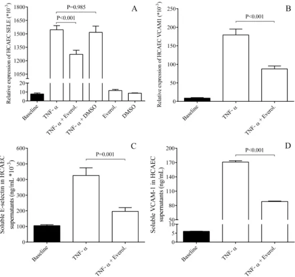
in vitroThere was a higher EC activation level in BMS patients with ISR compared to DES subjects without any complication [6]. Thus, we first investigated whether elevated expression of EC activation dependent adhesion molecules E-selectin and VCAM-1 could be observed in EC cultures under inflammatory conditions with or without everolimusin vitro. Our aim was to study the potential regulatory mechanisms of EC activation that might be caused by distinct coronary stents. E-selectin and VCAM-1 mRNA levels were analyzed in HCAECs and HUVECs after treatment with TNF-αin the presence or absence of everolimus. TNF-αstimu- lation resulted in a robust elevation in both mRNA levels compared to baseline sample. In con- trast, everolimus in the presence of TNF-αsignificantly, however not completely lowered these mRNA levels in HCAECs (P<0.001) (Fig 1A and 1B) and in HUVECs (P<0.001) as well (S1A and S1B Fig). No alteration in these mRNA levels was found by everolimus alone or by vehicle (DMSO) with TNF-αvs. untreated baseline samples.
To provide further evidence about the effect of everolimus on the expression of these adhe- sive receptors, their concentrations were also determined by ELISA in the supernatants of HCAEC samples after treatment by TNF-αin the absence and presence of everolimus. Inflam- mation-raised E-selectin and VCAM-1 concentrations were significantly decreased by everoli- mus (P = 0.001, P<0.001, respectively) (Fig 1C and 1D) in agreement with their altered mRNA levels in ECs above. Hence, thesein vitroresults provide some explanation about the lower level of EC activation with less E-selectin/VCAM-1 in DES individuals.
Everolimus lowered EC inflammation via lowered IL-1
βand IL-6 expression in ECs
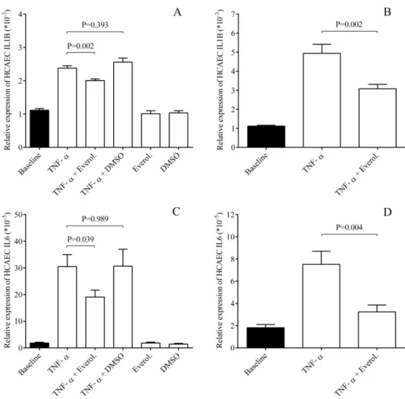
To investigate if everolimus downregulates adhesive molecule expression via modulating a global inflammatory response in ECs, HCAECs were treated with TNF-αthat caused a robust elevation in IL-1βand IL-6 mRNA levels already by 1 hour (Fig 2A and 2C), and further increased by 4 hours (Fig 2B and 2D). In contrast, everolimus moderately but significantly lowered IL-1β(P = 0.002) and IL-6 mRNAs (P = 0.039) already after 1 hour, which were more obvious after 4 hours of treatment (P = 0.002, P = 0.004, respectively). Based on these results, TNF-α-induced EC inflammation could be interrupted by everolimus.
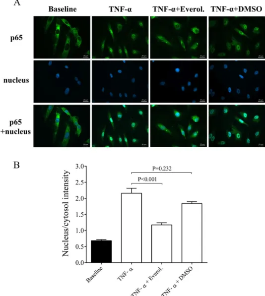
Effect of everolimus on TNF-α-induced NF-κB nuclear translocation in HCAECs
Translocation of the p65 NF-κB subunit into the cell nuclei reliably evaluates the degree of an inflammatory reaction at cellular level [19]. To further analyze the direct effect of everolimus in inflammation-stimulated ECs, early NF-κB p65 nuclear translocation was studied in HCAEC cultures after stimulation with cell culture medium, or TNF-α(100 ng/mL) in the absence or presence of everolimus (0.5μM) or with its solvent for 1 hour. The nucleus/cytosol intensity of p65 staining was studied in these cells. We found that compared to TNF-αstimu- lated cells, everolimus treatment in the presence of TNF-αsignificantly decreased the p65 staining in the cell nucleus (P<0.001). In contrast, TNF-α+DMSO sample did not show alter- ation in p65 staining (Fig 3A and 3B). Accordingly, everolimus was shown to directly influence EC activation via interrupting NF-κB pathway.
Fig 1. Analysis of E-selectin and VCAM-1 expression at mRNA and protein levels in HCAECs after TNF-αstimulation.
HCAEC cells were treated with recombinant TNF-α(100 ng/mL) for 1–24 hours to generate cellular inflammatory conditions.
Elevated E-selectin (SELE) (A) and VCAM-1 mRNA levels (B) induced by TNF-αalready after 1 hour were downregulated by everolimus in HCAECsin vitro. In parallel, soluble E-selectin (C) and VCAM-1 concentrations (D) were measured by ELISA in the supernatants of ECs and were also significantly decreased after 4 and 24 hours, respectively. Mean±SEM, n = 4-8/
group.
https://doi.org/10.1371/journal.pone.0197890.g001
Transcriptional regulation of E-selectin and VCAM-1 expression contributed to everolimus effects
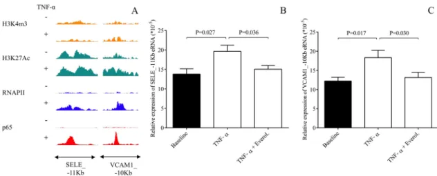
The expression of E-selectin and VCAM-1 are transcriptionally regulated in ECs in an inflam- matory signaling dependent manner, induced by e.g. lipopolysaccharides (LPS) [24]. For fur- ther investigation of potential TNF-αand everolimus-modulated transcriptional regulation of E-selectin and VCAM-1 expression, we here re-analyzed the unstimulated and TNF-α- treated HUVEC-derived publicly available NF-κB transcription factor subunit p65, RNA Poly- merase II (RNAPII), active histone mark H3K27Ac, and active transcription start site mark H3K4m3-specific ChIP-seq data sets [20]. As we expected, TNF-α-induced RNAPII binding was observed at both SELE and VCAM1 gene bodies (S2A and S2B Fig). In addition, two enhancers were identified in the neighboring genomic regions of both genes associating with TNFα-induced p65 and RNAPII binding (Fig 4A,S2A and S2B Fig). Recent studies showed that eRNA expression is a good marker of enhancer activity and is regulated in similar manner
Fig 2. Everolimus decreased EC inflammation via lowered IL-1βand IL-6 expression in ECs. HCAECs were treated with TNF-α(100 ng/mL) with or without everolimus (0.5μM) for 1 and 4 hours, and then IL-1βand IL-6 mRNA levels were quantified by RT-qPCR. This effect was observed already after 1 hour (A, C), while was more pronounced after 4 hours (B, D). Mean±SEM, n = 4-8/group.
https://doi.org/10.1371/journal.pone.0197890.g002
as the neighboring genes in many cell types by different signals [20,25–27]. Therefore, we measured the eRNA expression at one-one selected TNF-α-activated p65 transcription factor- bound enhancers in the neighboring genomic regions of both genes including SELE_-11Kb and VCAM1_-10Kb in unstimulated, TNF-αas well as TNF-αand everolimus-treated HCAECs using RT-qPCR method. TNF-αinduced eRNA expression at both enhancers com- pared to the baseline sample. However, the TNF-α-augmented eRNA expression was signifi- cantly reduced by everolimus treatment (P = 0.036, P = 0.030, respectively) (Fig 4B and 4C).
Overall, we suppose that everolimus inhibits EC activation via altering the TNF-α-induced transcription of EC activation-related genes, such as SELE and VCAM1.
Fig 3. Immunohistochemical staining and analysis of NF-κB activation in TNF-αand everolimus treated endothelial cells. HCAEC cells were treated for 1 hour with MesoEndo Medium (Baseline), 100 ng/ml TNF-αwith or without 0.5μM everolimus or cytokine with the solvent of everolimus (TNF-α+DMSO). Nuclear localization of the NF-κB p65 subunit was monitored by immunostaining. Green: p65 staining; blue: cell nuclei. Scale bar: 20μm (A).
Ratio of the fluorescence intensity of the NF-κB immunostaining in cell nuclei and cytosol was analyzed (B).
Mean±SEM, n = 6-8/group.
https://doi.org/10.1371/journal.pone.0197890.g003
TNF-
αinduced EC inflammation was associated with decreased miR-181b
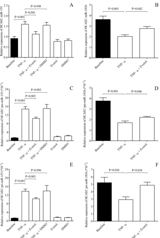
Although E-selectin and VCAM-1 expression were found to be highly regulated at transcrip- tional level in this experimental system, we sought to study the role of post-transcriptional regu- lator of these receptors upon EC inflammation. Since miR-181b modulated VCAM-1 and E- selectin expression in HUVECs amongin vitroconditions [17], we here analyzed the levels of this miRNA in TNF-α-stimulated ECs with or without everolimus as their potential key effec- tor. Both HCAECs and HUVECs were treated by recombinant TNF-αfor 1–4 hours to analyze miR-181b expression along with inflammation-specific miRNAs [25]. As expected, miR-155 and miR-146a as well as the biomarker of EC dysfunction miR-185 [28,29] were elevated by TNF-αcompared to untreated sample in both EC cultures (Fig 5AandS3A and S3B Fig). How- ever, everolimus caused significantly decreased miR-155 and miR-146a levels, with lower miR- 185 expression (data not shown). Importantly, the level of miR-181b was downregulated by the inflammatory stimulus (P<0.001) and the treatment with everolimus restored their expression in both EC cultures (P = 0.042, P = 0.049) (Fig 5BandS3C Fig). As control, we checked that the vehicle (DMSO) with TNF-αand everolimus alone were unable to alter these miRNAs.Precursors of miRNAs were also altered by everolimus in ECs
We subsequently studied whether altered levels of these mature miRNAs above were due to their abnormal transcriptional regulation. Therefore, the levels of pre- and pri-miR-155, and both precursors of miR-181b were quantified by RT-qPCR in HCAECs stimulated with TNF- αwith or without everolimus (Fig 5C–5F). We found that the levels of these miRNA precur- sors were altered in the same manner as seen in mature miRNAs. These findings suggest that miR-155 (C, E) and miR-181b (D, F) expression were modulated at transcription level by TNF-αstimulation and everolimus in ECs.
Endothelial cell miR-181b regulates the SELE and VCAM1 expression
Despite some former available data revealed in HUVECs [17], we wanted to confirm the rela- tionship between miR-181b and SELE and VCAM1 in HCAECs stimulated with TNF-αbyFig 4. Analysis of transcriptional regulation of SELE and VCAM-1 genes in unstimulated and TNF-α-treated HUVECs using publicly available ChIP-seq data sets and the measurement of eRNA expression at two selected enhancers including SELE_-11Kb and VCAM1_-10Kb in HCAECs. The p65, RNAPII, H3K27Ac and H3K4m3-specific ChIP-seq signals at the selected VCAM1 and SELE-associated enhancers were visualized by the Integrative Genomics Viewer (A). Accordingly, HCAECs were then treated with TNF-α(100 ng/mL) with or without everolimus (0.5μM) for 1 hour, and one-one eRNA expression at SELE_-11Kb (B) and VCAM1_-10Kb eRNAs (C) were quantified by RT-qPCR. TNF-α-augmented expression of eRNAs was significantly reduced by everolimus treatment. Mean±SEM, n = 4-8/group.
https://doi.org/10.1371/journal.pone.0197890.g004
Fig 5. Quantification of TNF-αinduced miR-155 and miR-181b levels with the analysis of their precursors in the presence of everolimus upon EC inflammationin vitro. HCAECs were treated by recombinant TNF-αfor 1–4 hours to analyze miR-181b expression along with the inflammation-specific miR-155. First, miR-155 was elevated by TNF-α compared to untreated sample, however, everolimus caused significantly decreased miR-155 levels (A), while miR- 181b was downregulated by the inflammatory stimulus and the treatment with everolimus restored their expression in both EC cultures (B). Levels of pre- and pri-miRNA were altered in the same manner as seen in mature miR-155 (C, E) and miR-181b (D, F), respectively. Mean±SEM, n = 4-8/group.
https://doi.org/10.1371/journal.pone.0197890.g005
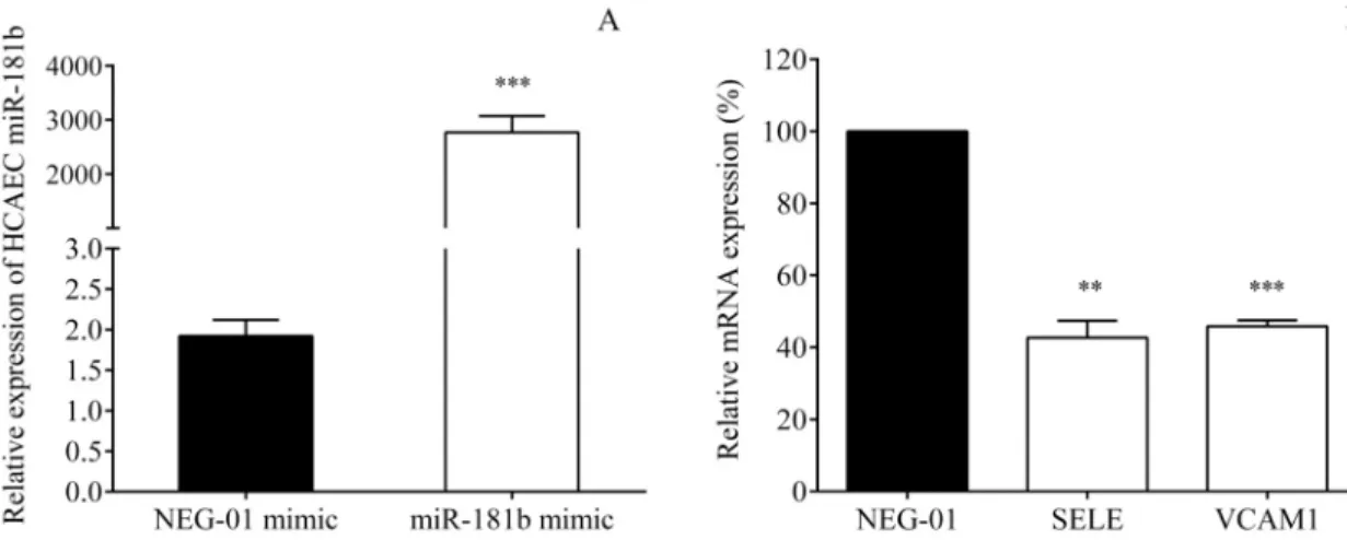
using transfection of specific miR-181b mimic. The overexpression of miR-181b was produced by its specific mimic (Fig 6A), however, the levels of other miRNAs, e.g. miR-155, were not affected (data not shown). As a consequence, SELE (P = 0.006) and VCAM1 mRNA levels (P<0.001) were significantly decreased in the coronary endothelial cells versus control samples transfected with the NEG-01 control mimic (Fig 6B). Based on these results, we confirmed that miR-181b targets E-selectin and VCAM-1 in HCAECs.
Impaired plasma miR-181b correlates with increased plasma levels of related soluble E-selectin and VCAM-1 concentrations
BMS patients with ISR showed higher level of EC activation in contrast to those who received everolimus eluting coronary stent [6]. Former data on soluble E-selectin and VCAM-1 were re-analyzed for this study and depicted inS4A and S4B Fig. There was significantly elevated soluble E-selectin concentrations in BMS subjects with ISR (P = 0.032) (S4A Fig), while VCAM-1 levels were markedly higher (P = 0.160) in comparison with DES cohort without any clinical complication (S4B Fig). Furthermore, to gain more direct evidence about the distinct effect of BMS and DES on vascular inflammation, we determined the level of TNF-αlevels in the plasma samples of our stented patients by ELISA. We found that TNF-αlevels were sig- nificantly lower in those subjects with DES compared to individuals with BMS having ISR (P = 0.049) (S5 Fig). Alterations in these receptor expressions and the level of pro-inflamma- tory cytokine indicated more EC activation when ISR was developed by BMS without the pro- tective effect of everolimus.
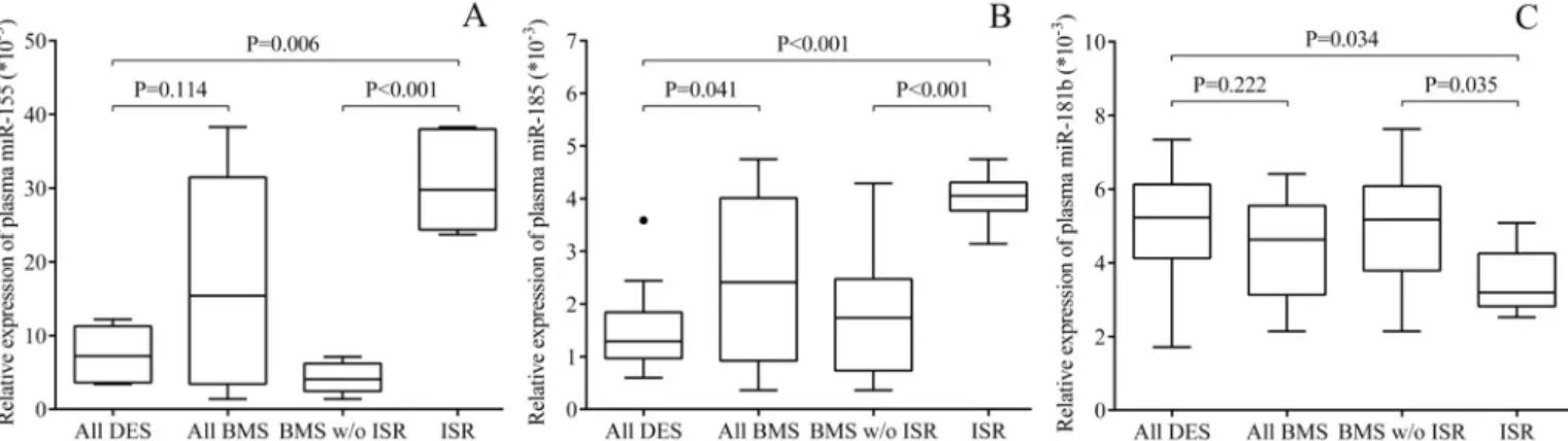
The circulating level of miR-155, miR-185 and miR-181b were quantified by RT-qPCR in the plasma samples of the entire patient population. We sought to investigate if these plasma miRNAs were changed in the same way as seen in ECsin vitro, and to support our findings about abnormal concentrations of soluble adhesive molecules and EC inflammation/dysfunc- tion in case of ISR. First, plasma miR-155 (P = 0.006) and miR-185 (P<0.001) were signifi- cantly upregulated in BMS patients with ISR compared to BMS and DES subjects without any complication (Fig 7A and 7B). These results revealed the presence of EC inflammation and
Fig 6. Overexpression of miR-181b altered the levels of SELE and VCAM1 mRNA in HCAECs. The direct association between miR-181b and SELE/VCAM1 was investigated in HCAECs after stimulation with TNF-αby using transfection of mirVana1miR- 181b mimics (25 pmol) with Lipofectamine RNAiMAX1Transfection Reagent for 24 hours. In parallel, negative control samples were treated with mirVana1miRNA mimic negative control (NEG-01, 25 pmol). After transfection, miR-181b with SELE and VCAM1 mRNAs were quantified by RT-qPCR. Highly increased miR-181b levels (A) resulted in significantly lowered SELE and VCAM1 mRNAs compared to NEG-01 control samples (100%, B).P = 0.006,P<0.001 based on t-test. Mean±SEM, n = 4/
group.
https://doi.org/10.1371/journal.pone.0197890.g006
dysfunction with typical miRNA alterations in those with ISR. Of note, we found some differ- ence between distinct types of stent showing lower inflammation-specific miRNAs in DES subjects. Furthermore, the levels of VCAM-1 and E-selectin repressor miR-181b were signifi- cantly lower in BMS+ISR as compared to other BMS (P = 0.035) or DES implantation without complication (P = 0.034) (Fig 7C). MiR-34a and miR-126 being considered as key miRNAs in vascular inflammation [16,30], were also measured as ‘control miRNAs’ in these plasma speci- mens and showed lower levels in ISR versus DES individuals (P<0.001, P = 0.036, respectively) (S6A and S6B Fig), which were comparable to former results by others [16,30]. These data supported the validity of our patient samples representing pathological vascular conditions after coronary stenting. Finally, correlation tests were performed to study the relationship between plasma VCAM-1 or E-selectin concentrations and miR-181b levels in the pooled patient samples. In accordance with the former data of Sunet al. [17] and the effect of miR- 181b on SELE/VCAM1 we detected (Fig 6), plasma miR-181b expression showed a significant negative correlation with VCAM-1 and E-selectin concentrations (r = -0.441, P = 0.019; r = -0.375, P = 0.049, respectively) (data not shown).
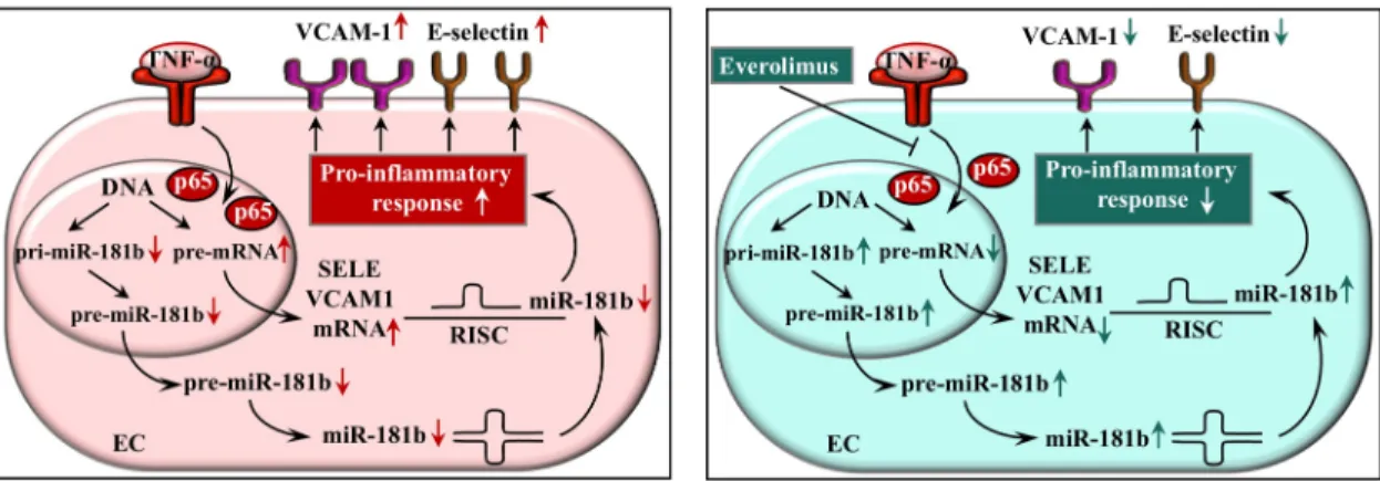
Based on all these data, we propose a model where everolimus acts as a negative regulator of EC activation via inhibiting NF-κB p65 subunit translocation resulting in altered E-selectin and VCAM-1 mRNA levels with their post-transcriptional repressor miR-181b at transcrip- tion level (Fig 8). These cellular events may occur in the early phase of DES implantation showing a beneficial protective effect of everolimus as compared to bare-metal stenting with- out any drug elution.
Discussion
Despite recent development in coronary stenting, ISR associated with VSMC proliferation and EC injury still represent a major clinical issue in those who undergo such intervention [1], and is often associated with enhanced platelet activation with increased amount of microparticles as well [31]. The introduction of DES coated with different drugs, e.g. mTOR inhibitors, has substantially reduced the risk for complications, such as ISR in contrast to BMS in the early phase of stenting [1,2]. Sirolimus during angioplasty prevented VSMC proliferation in an ani- mal model [32], and antagonized VCAM-1 levels induced by TNF-αin cultured HUVECs via inhibiting mTORC2 activity and potentiated ERK1/2 [11]. However, no studies have investi- gated the potential transcriptional and post-transcriptional regulatory mechanisms by which
Fig 7. Quantification of circulating miR-155, miR-185 and miR-181b by RT-qPCR in plasma samples of BMS and DES patients. After total RNA isolation, the expression of circulating miRNAs was quantified in plasma samples by UPL-probe based stem-loop RT-qPCR assay. Plasma miR-155 (A) and miR-185 (B) were significantly upregulated in BMS patients with ISR compared to BMS and DES subjects without any complications, while miR-181b levels (C) were lower in those with ISR versus others without complication. (All DES: n = 21, all BMS: n = 28, ISR: n = 6).
https://doi.org/10.1371/journal.pone.0197890.g007
everolimus can decrease EC activation. Of note, these drugs may result in late in-stent throm- bosis after 1–2 years of intervention [1,33], which may be due to the upregulation of plasmino- gen activation inhitor-1 by ECs [34].
Recently, higher level of EC activation was described with elevated soluble E-selectin and VCAM-1 levels after BMS intervention versus in DES individuals [4–6,8]. Moreover, TNF-α levels were significantly lower in our subjects with DES compared to individuals with BMS having ISR (S5 Fig). These results were in agreement with the paper of McNairet al. [8]. Adhe- sion receptors are expressed on activated ECs stimulated by TNF-αand other inflammatory cytokines, and then involved in leukocyte attachment to ECs [7]. DES displays a beneficial effect to prevent or at least slow down the development of EC activation/dysfunction leading to ISR via locally maintained everolimus concentration of cc. 0.5μM in stented vessels [35].
This anti-proliferative drug has also showed a substantial anti-inflammatory effect in neutro- phils reducing the release of IL-8 and decreasing TNF-α-induced adhesion of neutrophils to ECs [10]. However, no data are available by which mechanisms everolimus can lower EC acti- vation, such as after DES stenting. Hence, in this study, we have systematically investigated the potential transcriptional and post-transcriptional regulatory mechanisms of everolimus.
We first investigated the contribution of transcriptional regulatory mechanisms to the TNF-αand everolimus-dependent regulation of EC activation-linked genes, such as E-selectin and VCAM-1. Cultured HCAECs and HUVECs were challenged with 100 ng/ml concentra- tion of recombinant TNF-α—similarly to Palmieriet al. [36]—to investigate vascular inflam- mation in the presence or absence of everolimus after stenting. Initially, we applied HCAECs as reliable cell culture for the investigation of RNA levels in arterial endothelium [37], while results were also confirmed in HUVECs, which is a fundamental cell culture model to analyze endothelial responses to distinct challenges [18,38].
To analyze the direct effect of everolimus on EC function upon inflammation, we added this drug to the medium of EC cultures in the presence of TNF-α. Everolimus substantially decreased the NF-κB pathway via prevention of p65 translocation into cell nuclei and partially or completely inhibited the TNF-α-dependent effects on E-selectin and VCAM-1 transcription indicating that it effectively antagonized EC activation. Concentrations of VCAM-1 and E- selectin showed similar alterations in the supernatants of ECsin vitroupon inflammation and everolimus treatment. We also assessed very low (0.05μM) and high (5μM) concentration of
Fig 8. Schematic figure about the model to demonstrate the regulatory mechanisms of everolimus on E-selectin (SELE) and VCAM-1 expression. Everolimus decreases EC activation via suppressing the NF-κB pathway with decreased p65 translocation into cell nuclei causing the modulation of E-selectin and VCAM-1 expression as well as miR-181b level at transcriptional and post- transcriptional level, respectively. EC: endothelial cell, TNF-α: tumor necrosis factor alpha, SELE: E-selectin, VCAM-1: vascular cell adhesion molecule-1, RISC: RNA-induced silencing complex.
https://doi.org/10.1371/journal.pone.0197890.g008
everolimus during the setting of these experiments. No difference in RNA levels was observed at low concentration, however, its high concentration resulted in the apoptosis of ECs (data not shown). In agreement with our results, LPS-induced EC activation was also downregulated by rapamycin via mTOR/NF-κB pathway in HUVECs [38]. Our current RT-qPCR-based mea- surements and the re-analysis of publicly available ChIP-seq data sets of HUVECs showed that these genes and SELE_-11Kb and VCAM1_-10Kb eRNAs are activated in ECs by TNF-αcon- firming their TNF-α-dependent transcriptional regulation. Importantly, everolimus decreased the levels of these eRNAs. Others recently described the NF-κB binding site in the -1643 and -1652 regions of miR-99a promoter by ChIP assay, and this miRNA modulated EC inflamma- tion in HUVECs [38].
MiRNAs play a major role in the post-transcriptional regulation of EC activation-depen- dent events in coronary artery disease [12,39]. MiR-223 and miR-141 suppressed ICAM-1 expression in EC culturesin vitro[40,41], while NF-κB mediated inflammation was regulated by elevated expression of miR-146a and miR-155 [25,39]. Moreover, PCI-induced plaque rup- ture was associated with increased miR-155 levels [42]. A number of miRNAs are involved in the development of EC dysfunction, such as upregulated miR-185 in response to high glucose milieu [28], miR-99a in LPS-stimulated [38], and miR-149 in TNF-α-induced EC dysfunction through p38MAPK [36]. These previous data revealed a functional role of these miRNAs in the regulation of various pathological cellular events during vascular inflammation at post- transcriptional level. In our study, TNF-αenhanced miR-146a, miR-155 and miR-185 expres- sion in both EC cultures indicating the cellular inflammatory response and dysfunction as seen earlier [41]. Importantly, we also observed that miR-181b targeted SELE and VCAM1 mRNAs, and TNF-αtranscriptionally repressed miR-181b expression suggesting that TNF-α may enhance E-selectin and VCAM-1 at post-transcriptional level. MiR-181b inhibits impor- tin-α3 expression and NF-κB-responsive VCAM-1 and SELE genes [17]. In other experiments, TNF-αtreatment in HUVECs resulted in decreased miR-141 levels causing enhanced ICAM-1 expression [41], while miR-149 was also decreased due to the same response affecting IL-6 and metalloproteinase-9 expression in EC cultures [38]. Overall, these miRNAs do not only repre- sent a new layer of regulation but may act as new biomarkers in cardiovascular diseases [15].
Here, Pearson’s correlation tests demonstrated a significant reverse correlation between plasma miR-181b levels and plasma VCAM-1 and E-selectin concentrations in our stented patient cohort supporting the relationship between the levels of this post-transcriptional regu- lator and the expression of these adhesive proteins. Based on our data, decreased plasma miR- 181b level may be useful to indicate stent-induced EC activation with enhanced VCAM-1 and E-selectin expression. In apolipoprotein E-deficient/NF-κB-luciferase transgenic mice miR- 181b significantly inhibited atherosclerotic lesion formation, pro-inflammatory gene expres- sion and the influx of lesional macrophages and CD4+ T cells in the vessel wall suggesting the central role of this miRNA in vascular inflammation during atherosclerosis [43].
Our study has some limitations. The number of patients with ISR was relatively small in our former clinical study [6], which was obviously due to the fact that it was a single center study with a limited number of eligible patients per year. Thus, more data are needed to observe the relationship of miR-181b level with soluble E-selectin/VCAM-1 concentrations as a potential biomarker of EC activation and ISR.
We here described for the first time the transcriptional regulatory effects of everolimus on EC inflammation in details. These transcriptional (p65-bound eRNAs SELE_-11Kb and VCAM1_-10Kb) and post-transcriptional regulators (miR-181b) may represent potential ther- apeutic targets upon EC dysfunction. However, further studies are required to prove the func- tional role of these regulatorsin vivo. Similarly, neo-intimal formation and ISR development were effectively modulated by anti-miR-21 [44], or by the overexpression of miR-23b [45]
based on the results of animal models. These data propagate to introduce novel ‘drug’-eluting stents for patients in the near future. As such, a stent system that eluted miR-126 exhibited sig- nificant inhibition of neointimal formation in a rabbit model of restenosis [46].
Conclusions
We provide some pieces of evidence that everolimus acts as a negative regulator of EC activa- tion via suppressed NF-κB pathway with lower p65 translocation into cell nuclei, the modula- tion of the expression of SELE and VCAM-1 and their post-transcriptional repressor miR-181b at transcription level. These data may explain how the level of EC activation can be lowered by everolimus when DES is used for coronary intervention in contrast to BMS implantation.
Supporting information
S1 Table. Sequences of primers for the analysis of mature and precursor miRNAs as well as mRNAs and eRNAs.
(XLSX)
S1 Fig. Measurement of E-selectin and VCAM-1 mRNAs in HUVECs after treatment with TNF-α. HUVECs were treated with recombinant TNF-α(100 ng/mL) for 4 hours to generate cellular inflammatory conditions. Elevated E-selectin (SELE) (A) and VCAM-1 mRNA levels (B) induced by TNF-αwere downregulated by everolimus in HUVECsin vitro. Mean±SEM, n = 4-8/group.
(TIF)
S2 Fig. Analysis of the p65, RNAPII, H3K27Ac and H3K4m3-specific ChIP-seq signals at the genomic loci of VCAM1 and SELE visualized by the Integrative Genomics Viewer. The unstimulated and TNF-α-treated HUVEC-derived publicly available NF-κB transcription fac- tor subunit p65, RNA Polymerase II (RNAPII), active histone mark H3K27Ac, and active tran- scription start site mark H3K4m3-specific ChIP-seq data sets were reanalyzed. The identified TNF-α-activated p65-bound enhancers in the neighboring genomic regions of SELE (A) and VCAM1 (B) genes were indicated by red arrows.
(TIF)
S3 Fig. Quantification of TNF-αinduced miR-146a, miR-155 and miR-181b levels upon inflammation in HUVECsin vitro. HUVECs were treated with TNF-α(100 ng/mL) with or without everolimus (0.5μM) for 1 and 4 hours, and then these miRNA levels were quantified by RT-qPCR. Everolimus caused significantly decreased miR-146a (A) and miR-155 levels (B). The level of miR-181b (C) was downregulated by the inflammatory stimulus and the treatment with everolimus restored their expression in these EC cultures as well. Mean±SEM, n = 4-8/group.
(TIF)
S4 Fig. Re-analysis of soluble E-selectin and VCAM-concentrations in plasma samples of BMS and DES subjects with or without ISR. There were significantly higher E-selectin (A) and markedly elevated VCAM-1 levels in those who had ISR (n = 6) compared to other BMS (n = 22) and DES (n = 21) individuals [6]. Mean±SEM.
(TIF)
S5 Fig. Investigation of TNF-αconcentrations in plasma samples of patients underwent BMS or DES. There were significantly higher plasma levels of TNF-αin those subjects who received BMS and showed ISR (n = 6) compared to individuals with DES (n = 21). Mean±SEM.
(TIF)
S6 Fig. Analysis of plasma miR-34a and miR-126 expression in the presence or absence of ISR in BMS and DES patients. After total RNA isolation, the expression of circulating miR- NAs was quantified in plasma samples by UPL-probe based stem-loop RT-qPCR assay. These miRNAs were significantly lower in those with ISR compared to BMS and DES subjects with- out such complication. Mean±SEM. (All DES: n = 21, all BMS: n = 28, ISR: n = 6).
(TIF)
Author Contributions
Conceptualization: Zsolt Czimmerer, Ja´nos Kappelmayer, Be´la Nagy, Jr.
Data curation: Zsolt Fejes, Zsolt Czimmerer, Tibor Szu¨k, Szila´rd Po´liska, Attila Horva´th, EnikőBalogh, Be´la Nagy, Jr.
Formal analysis: Zsolt Fejes, Zsolt Czimmerer, Szila´rd Po´liska, EnikőBalogh, Vikto´ria Jeney, Ja´nos Kappelmayer, Be´la Nagy, Jr.
Funding acquisition: Istva´n E´des, Ja´nos Kappelmayer.
Investigation: Zsolt Fejes, Zsolt Czimmerer, Szila´rd Po´liska, EnikőBalogh, Vikto´ria Jeney, Be´la Nagy, Jr.
Methodology: Judit Va´radi, Ferenc Fenyvesi, Ja´nos Kappelmayer, Be´la Nagy, Jr.
Project administration: Zsolt Fejes, Tibor Szu¨k.
Resources: Gyo¨rgy Balla, Istva´n E´des.
Software: Attila Horva´th, Judit Va´radi, Ferenc Fenyvesi.
Supervision: Vikto´ria Jeney, Gyo¨rgy Balla, Istva´n E´des, Jo´zsef Balla, Ja´nos Kappelmayer, Be´la Nagy, Jr.
Validation: Zsolt Czimmerer, Tibor Szu¨k, Attila Horva´th, Gyo¨rgy Balla, Be´la Nagy, Jr.
Visualization: Attila Horva´th, Judit Va´radi, Ferenc Fenyvesi.
Writing – original draft: Zsolt Fejes, Be´la Nagy, Jr.
Writing – review & editing: Zsolt Fejes, Zsolt Czimmerer, Jo´zsef Balla, Be´la Nagy, Jr.
References
1. Bavry AA, Bhatt DL. Appropriate use of drug-eluting stents: balancing the reduction in restenosis with the concern of late thrombosis. Lancet 2008; 371: 2134–2143.https://doi.org/10.1016/S0140-6736(08) 60922-8PMID:18572082
2. Costa MA, Simon DI. Molecular basis of restenosis and drug-eluting stents. Circulation 2005; 111:
2257–2573.https://doi.org/10.1161/01.CIR.0000163587.36485.A7PMID:15867193
3. Piatti P, Monti LD. Insulin resistance, hyperleptinemia and endothelial dysfunction in coronary resteno- sis. Curr Opin Pharmacol 2005; 5: 160–164.https://doi.org/10.1016/j.coph.2004.10.004PMID:
15780825
4. Boos CJ, Balakrishnan B, Jessani S, Blann AD, Lip GY. Effects of percutaneous coronary intervention on peripheral venous blood circulating endothelial cells and plasma indices of endothelial damage/dys- function. Chest 2007; 132: 1920–1926.https://doi.org/10.1378/chest.07-1693PMID:18079225 5. Munk PS, Breland UM, Aukrust P, Skadberg O, Ueland T, Larsen AI. Inflammatory response to percuta-
neous coronary intervention in stable coronary artery disease. J Thromb Thrombol 2011; 31: 92–98.
6. Szu¨k T, Fejes Z, Debreceni IB, Kere´nyi A, E´ des I, Kappelmayer J, et al. Integrity(®) bare-metal coronary stent-induced platelet and endothelial cell activation results in a higher risk of restenosis compared to Xience(®) everolimus-eluting stents in stable angina patients. Platelets 2016; 27: 410–419.https://doi.
org/10.3109/09537104.2015.1112368PMID:26765134
7. Carlos TM, Harlan JM. Leukocyte-endothelial adhesion molecules. Blood 1994; 84: 2068–2101. PMID:
7522621
8. McNair ED, Wells CR, Mabood Qureshi A, Basran R, Pearce C, Orvold J, et al. Soluble receptors for advanced glyca- tion end products (sRAGE) as a predictor of restenosis following percutaneous coronary intervention. Clin Cardiol 2010; 33: 678–685.https://doi.org/10.1002/clc.20815PMID:
21089112
9. Panoulas VF, Mastoris I, Konstantinou K, Tespili M, Ielasi A. Everolimus-eluting stent platforms in per- cutaneous coronary intervention: comparative effectiveness and outcomes. Med Devices (Auckl) 2015;
8: 317–329.
10. Vitiello D, Neagoe PE, Sirois MG, White M. Effect of everolimus on the immunomodulation of the human neutrophil inflammatory response and activation. Cell Mol Immunol 2015; 12: 40–52.https://doi.
org/10.1038/cmi.2014.24PMID:24882386
11. Wang C, Qin L, Manes TD, Kirkiles-Smith NC, Tellides G, Pober JS. Rapamycin antagonizes TNF induction of VCAM-1 on endothelial cells by inhibiting mTORC2. J Exp Med 2014; 211: 395–404.
https://doi.org/10.1084/jem.20131125PMID:24516119
12. Zampetaki A, Mayr M. MicroRNAs in vascular and metabolic disease. Circ Res 2012; 110: 508–522.
https://doi.org/10.1161/CIRCRESAHA.111.247445PMID:22302757
13. De Rosa S, Fichtlscherer S, Lehmann R, Assmus B, Dimmeler S, Zeiher AM. Transcoronary concentra- tion gradients of circulating microRNAs. Circulation 2011; 124: 1936–1944.https://doi.org/10.1161/
CIRCULATIONAHA.111.037572PMID:21969012
14. Hulsmans M, Holvoet P. MicroRNA-containing microvesicles regulating inflammation in association with atherosclerotic disease. Cardiovasc Res 2013; 100: 7–18.https://doi.org/10.1093/cvr/cvt161 PMID:23774505
15. He M, Gong Y, Shi J, Pan Z, Zou H, Sun D, et al. Plasma microRNAs as potential noninvasive biomark- ers for in-stent restenosis. PLoS One 2014; 9: e112043.https://doi.org/10.1371/journal.pone.0112043 PMID:25427155
16. Santulli G, Wronska A, Uryu K, Diacovo TG, Gao M, Marx SO, et al. A selective microRNA-based strat- egy inhibits restenosis while preserving endothelial function. J Clin Invest 2014; 124: 4102–4114.
https://doi.org/10.1172/JCI76069PMID:25133430
17. Sun X, Icli B, Wara AK, Belkin N, He S, Kobzik L, et al. MicroRNA-181b regulates NF-κB-mediated vas- cular inflammation. J Clin Invest 2012; 122: 1973–1990.https://doi.org/10.1172/JCI61495PMID:
22622040
18. Jeney V, Balla J, Yachie A, Varga Z, Vercellotti GM, Eaton JW, et al. Pro-oxidant and cytotoxic effects of circulating heme. Blood 2002; 100: 879–887. PMID:12130498
19. Va´radi J, Harazin A, Fenyvesi F, Re´ti-Nagy K, Gogola´k P, Va´mosi G, et al. Alpha-Melanocyte Stimulat- ing Hormone Protects against Cytokine-Induced Barrier Damage in Caco-2 Intestinal Epithelial Mono- layers. PLoS One 2017; 12: e0170537.https://doi.org/10.1371/journal.pone.0170537PMID:28103316 20. Thorvaldsdottir H, Robinson JT, Mesirov JP. Integrative Genomics Viewer (IGV): high-performance
genomics data visualization and exploration. Brief Bioinform 2012; 14: 178–192.https://doi.org/10.
1093/bib/bbs017PMID:22517427
21. Brown JD, Lin CY, Duan Q, Griffin G, Federation AJ, Paranal RM, et al. NF-kappaB directs dynamic super enhancer formation in inflammation and atherogenesis. Mol Cell 2014; 56: 219–231.https://doi.
org/10.1016/j.molcel.2014.08.024PMID:25263595
22. Fejes Z, Po´liska S, Czimmerer Z, Ka´pla´r M, Penyige A, Ga´l Szabo´ G, et al. Hyperglycaemia suppresses microRNA expression in platelets to increase P2RY12 and SELP levels in type 2 diabetes mellitus.
Thromb Haemost 2017; 117: 529–542.https://doi.org/10.1160/TH16-04-0322PMID:27975100 23. Czimmerer Z, Hulvely J, Simandi Z, Varallyay E, Havelda Z, Szabo E, et al. A versatile method to design
stem-loop primer-based quantitative PCR assays for detecting small regulatory RNA molecules. PLoS One 2013; 8: e55168.https://doi.org/10.1371/journal.pone.0055168PMID:23383094
24. Hortelano S, Lo´pez-Fontal R, Trave´s PG, Villa N, Grashoff C, Bosca´ L, et al. ILK mediates LPS-induced vascular adhesion receptor expression and subsequent leucocyte trans-endothelial migration. Cardio- vasc Res 2010; 86: 283–292.https://doi.org/10.1093/cvr/cvq050PMID:20164118
25. Duan Q, Mao X, Xiao Y, Liu Z, Wang Y, Zhou H, et al. Super enhancers at the miR-146a and miR-155 genes contribute to self-regulation of inflammation. Biochim Biophys Acta 2016; 1859: 564–571.
https://doi.org/10.1016/j.bbagrm.2016.02.004PMID:26855180
26. Czimmerer Z, Horvath A, Daniel B, Nagy G, Cuaranta-Monroy I, Kiss M, et al. Dynamic transcriptional control of macrophage miRNA signature via inflammation responsive enhancers revealed using a com- bination of next generation sequencing-based approaches. Biochim Biophys Acta 2018; 1861: 14–28.
https://doi.org/10.1016/j.bbagrm.2017.11.003PMID:29133016
27. Czimmerer Z, Daniel B, Horvath A, Ru¨ckerl D, Nagy G, Kiss M, et al. The Transcription factor STAT6 mediates direct repression of inflammatory enhancers and limits activation of alternatively polarized macrophages. Immunity 2018; 48: 75–90.e6.https://doi.org/10.1016/j.immuni.2017.12.010PMID:
29343442
28. La Sala L, Cattaneo M, De Nigris V, Pujadas G, Testa R, Bonfigli AR, et al. Oscillating glucose induces microRNA-185 and impairs an efficient antioxidant response in human endothelial cells. Cardiovasc Diabetol 2016; 15: 71.https://doi.org/10.1186/s12933-016-0390-9PMID:27137793
29. Hou J, Liu L, Zhu Q, Wu Y, Tian B, Cui L, et al. MicroRNA-185 inhibits angiogenesis in human microvas- cular endothelial cells through targeting stromal interaction molecule 1. Cell Biol Int 2016; 40: 318–328.
https://doi.org/10.1002/cbin.10572PMID:26694763
30. Chen Q, Yang F, Guo M, Wen G, Zhang C, Luong le A, et al. MiRNA-34a reduces neointima formation through inhibiting smooth muscle cell proliferation and migration. J Mol Cell Cardiol 2015; 89(Pt A): 75–
86.https://doi.org/10.1016/j.yjmcc.2015.10.017PMID:26493107
31. Nagy B Jr, Szu¨k T, Debreceni IB, Kappelmayer J. Platelet-derived microparticle levels are signifi- cantly elevated in patients treated by elective stenting compared to subjects with diagnostic catheteri- zation alone. Platelets 2010; 21: 147–151.https://doi.org/10.3109/09537100903477582PMID:
20050761
32. Buerke M, Guckenbiehl M, Schwertz H, Buerke U, Hilker M, Platsch H, et al. Intramural delivery of Siroli- mus prevents vascular remodeling following balloon injury. Biochim Biophys Acta 2007; 1774: 5–15.
https://doi.org/10.1016/j.bbapap.2006.04.018PMID:16920414
33. Joner M, Finn AV, Farb A, Mont EK, Kolodgie FD, Ladich E, et al. Pathology of drug-eluting stents in humans: delayed healing and late thrombotic risk. J Am Coll Cardiol 2006; 48: 193–202.https://doi.org/
10.1016/j.jacc.2006.03.042PMID:16814667
34. Ota H, Eto M, Ako J, Ogawa S, Iijima K, Akishita M, et al. Sirolimus and everolimus induce endothelial cellular senescence via sirtuin 1 down-regulation: therapeutic implication of cilostazol after drug-eluting stent implantation. J Am Coll Cardiol 2009; 53: 2298–2305.https://doi.org/10.1016/j.jacc.2009.01.072 PMID:19520256
35. Abbott Vascular XIENCE®Alpine Everolimus Eluting Coronary Stent Systems. EL2099886, 2015.
https://www.vascular.abbott/us/products/coronary-intervention/xience-alpine-drug-eluting-stent.html 36. Palmieri D, Capponi S, Geroldi A, Mura M, Mandich P, Palombo D. TNFαinduces the expression of
genes associated with endothelial dysfunction through p38MAPK-mediated down-regulation of miR- 149. Biochem Biophys Res Commun 2014; 443: 246–251.https://doi.org/10.1016/j.bbrc.2013.11.092 PMID:24299952
37. Li JB, Wang HY, Yao Y, Sun QF, Liu ZH, Liu SQ, et al. Overexpression of microRNA-138 alleviates human coronary artery endothelial cell injury and inflammatory response by inhibiting the PI3K/Akt/
eNOS pathway. J Cell Mol Med 2017; 21: 1482–1491.https://doi.org/10.1111/jcmm.13074PMID:
28371277
38. Bao MH, Li JM, Luo HQ, Tang L, Lv QL, Li GY, et al. NF-κB-Regulated miR-99a Modulates Endothelial Cell Inflammation. Mediators Inflamm 2016; 2016: 5308170.https://doi.org/10.1155/2016/5308170 PMID:27403035
39. Nazari-Jahantigh M, Wei Y, Schober A. The role of microRNAs in arterial remodelling Thromb Haemost 2012; 107: 611–618.https://doi.org/10.1160/TH11-12-0826PMID:22371089
40. Tabet F, Vickers KC, Cuesta Torres LF, Wiese CB, Shoucri BM, Lambert G, et al. HDL-transferred microRNA-223 regulates ICAM-1 expression in endothelial cells. Nat Commun 2014; 5: 3292.https://
doi.org/10.1038/ncomms4292PMID:24576947
41. Liu RR, Li J, Gong JY, Kuang F, Liu JY, Zhang YS, et al. MicroRNA-141 regulates the expression level of ICAM-1 on endothelium to decrease myocardial ischemia-reperfusion injury. Am J Physiol Heart Circ Physiol 2015; 309:H1303–1313.https://doi.org/10.1152/ajpheart.00290.2015PMID:
26371161
42. Li S, Lee C, Song J, Lu C, Liu J, Cui Y, et al. Circulating microRNAs as potential biomarkers for coronary plaque rupture. Oncotarget 2017; 8: 48145–48156.https://doi.org/10.18632/oncotarget.18308PMID:
28624816
43. Sun X, He S, Wara AKM, Icli B, Shvartz E, Tesmenitsky Y, et al. Systemic delivery of microRNA-181b inhibits nuclear factor-κB activation, vascular inflammation, and atherosclerosis in apolipoprotein E-defi- cient mice. Circ Res 2014; 114:32–40.https://doi.org/10.1161/CIRCRESAHA.113.302089PMID:
24084690
44. Wang D, Deuse T, Stubbendorff M, Chernogubova E, Erben RG, Eken SM, et al. Local MicroRNA Mod- ulation Using a Novel Anti-miR-21-Eluting Stent Effectively Prevents Experimental In-Stent Restenosis.
Arterioscler Thromb Vasc Biol 2015; 35: 1945–1953.https://doi.org/10.1161/ATVBAHA.115.305597 PMID:26183619
45. Gareri C, De Rosa S, Indolfi C. MicroRNAs for Restenosis and Thrombosis After Vascular Injury. Circ Res 2016; 118: 1170–1184.https://doi.org/10.1161/CIRCRESAHA.115.308237PMID:27034278 46. Izuhara M, Kuwabara Y, Saito N, Yamamoto E, Hakuno D, Nakashima Y, et al. Prevention of neointimal
formation using miRNA-126-containing nanoparticle-conjugated stents in a rabbit model. PLoS One 2017; 12: e0172798.https://doi.org/10.1371/journal.pone.0172798PMID:28253326