DETECTION AND MOLECULAR CHARACTERISATION OF EHRLICHIA CANIS IN NATURALLY INFECTED DOGS
IN SOUTH WEST NIGERIA
Olukayode Olugbenga DARAMOLA1, Michael Irewole TAKEET2*, Ibironke Kofoworola OYEWUSI1, Mufutau Atanda OYEKUNLE2 and Adewale Oladele TALABI1 1Department of Veterinary Medicine and Surgery and 2Department of Veterinary Microbiology and Parasitology, College of Veterinary Medicine, Federal University of
Agriculture Abeokuta, Nigeria
(Received 10 August 2017; accepted 6 November 2017)
Canine ehrlichiosis is an important tick-borne rickettsial disease mainly caused by Ehrlichia canis. This study aimed to detect and characterise E. canis in dogs in Abeokuta, Nigeria by microscopy and nested PCR. Blood samples were collected from 205 dogs, thin smears were made, field-stained, and DNA was ex- tracted from the blood samples. A partial region of the 16S rRNA gene was am- plified by polymerase chain reaction (PCR) and sequenced unidirectionally. Ehr- lichial morulae were detected in three dogs (1.5%). The PCR test revealed that 47 dogs (22.9%) were positive for E. canis. The lengths of the sequences obtained range from 374 bp to 376 bp with an average G-C content of 37% and 98–99%
homology with the reference sequences in GenBank. The aligned autochthonous sequences were less polymorphic. The phylogenetic analysis separated sequences reported previously in Nigeria from the autochthonous sequences. The present work shows that the strain of E. canis detected in the study area is genetically dif- ferent from those reported in the northern part of Nigeria and more closely related to sequences from Brazil and India.
Key words: Ehrlichia canis, dog, nested PCR, Abeokuta, 16s rRNA, prevalence
Canine ehrlichiosis is a rickettsial bacteria disease of dogs that has world- wide distribution (Engvall et al., 1996; McBride et al., 1996; Souza et al., 2010;
Fourie et al., 2013; Milanjeet et al., 2014; Cardoso et al., 2016; Cicuttin et al., 2016; Kaewmongkol et al., 2016). Two forms of the disease are known in dogs and humans. In dogs, these include the monocytic form which is caused by Ehr- lichia canis (E. canis) and the granulocytic form that is caused by E. ewingii.
They are transmitted by the brown dog tick (Rhipicephalus sanguineus) and the lone star tick called Amblyomma americanum, respectively. In humans, the mon- ocytic and granulocytic forms are caused by E. chaffeensis and E. ewingii, re-
*Corresponding author; E-mail: takeetmi@funaab.edu.ng; Phone: 0023 (480) 3787-2682
spectively, though Anaplasma phagocytophilum (formerly E. equi) and E. canis have also been reported in human ehrlichiosis (Dumler et al., 2005; Perez et al., 2006).
The clinical manifestation of canine ehrlichiosis varies depending on the immune status, the breeds of dogs infected and the strain of infecting Ehrlichia species. While E. canis is generally believed to cause more severe febrile condi- tion in dogs, E. ewingii infection is less severe (Liddell et al., 2003). Signs of the disease in dogs may include but are not limited to fever, anaemia, haemorrhages, oedema, lymphadenopathy, thrombocytopenia, anorexia and weight loss (Cicut- tin et al., 2016; Lauzi et al., 2016). The organism is characterised by the presence of intracytoplasmic inclusion bodies (morulae) in the monocytes/granulocytes of infected dogs, which can be detected by microscopic examination of peripheral blood (Dagnone et al., 2009). Other techniques such as serology can be used, though with its own limitation to differentiate between previous and ongoing in- fection of dogs (Cicuttin et al., 2016). In recent years, polymerase chain reaction (PCR) has been found to be more sensitive in the detection of Ehrlichia DNA in the blood of infected animals.
The prevalence and molecular characteristics of Ehrlichia canis have been studied extensively around the world (Suksawat et al., 2001; Unver et al., 2001;
Vinasco et al., 2007; Aguiar et al., 2008; Harrus et al., 2011; Nazari et al., 2013;
Milanjeet et al., 2014; Cardoso et al., 2016). One of these studies has revealed that more than one strain may exist in a region (Vinasco et al., 2007). In Nigeria, Kamani et al. (2013) shed light on the molecular characteristics of E. canis de- tected in three northern states of the country. Hence, this study characterised E.
canis detected in naturally infected dogs in the western part of Nigeria and com- pared their sequences with those detected elsewhere in the world and those re- ported by Kamani et al. (2013) in Nigeria.
Materials and methods
Study area and sampled population
The study was carried out in Abeokuta metropolis, Ogun State, Nigeria.
Ogun state is bounded by Lagos State to the south, Oyo and Osun States to the north, Ondo State to the east and the Republic of Benin to the west. A total of 205 dogs consisting local and exotic breeds were randomly sampled mainly from the dogs presented to the Veterinary Teaching Hospital, Federal University of Agriculture, Abeokuta and various veterinary hospitals in the city. The dogs were grouped to young (≤ 1 year) and adult (≥ 1 year) and their sexes were also recorded. The dogs sampled were predominantly exotic breeds, which included Alsatian (102), Rottweiler (18), Boeboel (44), unidentified breed (10) and mon- grel (31). Pregnant dogs and those with a history of recent medication (such as
antibiotic therapy and anti-protozoan therapy) within the last four weeks were excluded from the study.
A blood sample was collected from each dog using cephalic venipuncture into tubes containing ethylenediamine tetraacetic acid (EDTA) as anticoagulant.
The collected blood samples were transported on ice packs to the laboratory.
Thin blood smears were made from each sample collected as well as smears of the buffy coat from the spun capillary tubes, fixed with 98% methanol (Dudal diesel, Madison, USA) and then reverse stained with Field stain A and B (Biolab Diagnostics, USA). The slides were examined under an oil immersion objective of a light microscope (Olympus CX21, China). Sample in which intracytoplas- mic inclusion bodies were demonstrated were adjudged positive for Ehrlichia spp. (Engvall et al., 1996). Blood samples not processed immediately for DNA extraction were stored at –20 °C until use.
DNA extraction and PCR assay for E. canis
Genomic DNA was extracted from whole blood using a commercially available DNA extraction kit (Quick-gDNA MicroPrep, Zymo Research Corpo- ration, Irvine, USA) following the manufacturer’s protocol, and the eluted DNA was stored at –20 °C until use as previously described (Takeet et al., 2013).
Nested PCR that targeted partial regions of the 16S rRNA gene was carried out using the primers listed (Table 1). The primary and the nested reactions were car- ried out using primer sets ECC & ECB and ECAN5 & HE3 (Table 1), respec- tively. The primary reaction primers target 478 bp of Ehrlichia spp. while the nested reaction primers are specific for the amplification of 398 bp of E. canis.
Table 1
The sequences of oligonucleotides used in the nested PCR of Ehrlichia canis detection in naturally infected dogs in Abeokuta, Nigeria
Primer set Primer sequence (5´ – 3´) Reference Band size
ECC AGAACGAACGCTGGCGGCAAGC
ECB CGTATTACCGCGGCT GCTGGCA Murphy et al. (1998) 478
ECAN5 CAATTATTTATAGCCTCTGGCTCTGGCTATAGG
HE3 TATAGGT ACCGTCATTATCTTCCCTAT Milanjeet et al. (2014) 398
Amplification was carried out in 20 µl final volume containing 10 µl of 2 × mastermix (Syd Labs, Inc., USA), 8 µl of nuclease-free water and 0.5 µl (about 10 pmol) each of forward and reverse primer, and 1 µl (about 50 ng) of the genomic DNA in a personal cycler (Biorad, USA). The cycling conditions for the primary PCR reactions were: 94 °C for 3 min initial denaturation, 30 cycles
of 94 °C for 60 s, 59.5 °C for 60 s and 72 °C for 120 s with final extension at 72 °C for 8 min. The nested PCR was carried out also in 20 µl final volume con- taining 10 µl of 2 × mastermix (Syd Labs, Inc., USA), 8 µl of nuclease-free wa- ter and 0.5 µl (about 10 pmol) each of forward and reverse primer, and 1 µl of the primary PCR product as the template. The PCR conditions were: initial dena- turation at 94 °C for 3 min, followed by two amplification steps: the first step consisted of three cycles of 94 °C for 1 min, 55 °C for 2 min, 72 °C for 1.5 min, while the second step consisted of 37 cycles of 94 °C for 1 min, 55 °C for 2 min, 72 °C for 1.5 min with final extension at 72 °C for 8 min. In every amplification, a negative control was included using nuclease-free water instead of DNA.
The PCR products were visualised in 1.5% agarose gel that was stained with ethidium bromide (Amresco, USA) in 1 × TAE buffer (40 mM Tris acetate and 1 mM EDTA) and electrophoresed at 80 V for 60 min along with 10 µl of 100 bp DNA ladder (BioExpress, Kaysville, UT, USA), and then visualised with UV transilluminator (Spectroline, USA). Gel picture was captured using a hand- held camera (Samsung WB50F, China).
Sequence and phylogenetic analyses
To confirm the PCR assay results, four PCR products from the samples that showed expected band size were sequenced directly using Big Dye Termina- tor cycle sequencing kit (Applied Biosystems, Foster City, CA, USA) in the Core Lab of Cornell University Central Laboratory, USA using forward primer ECAN5. Search for homologous sequences in the GenBank was performed using BLASTn (www.ncbi.nlm.nih.gov/BLAST). The obtained sequences were viewed, aligned and compared using BioEdit® (version 7.0.9.0) software. The sequences were also aligned with other published E. canis16S rRNA sequences of dogs from around the world. Phylogenetic analysis of the sequences, including those obtained from GenBank were constructed using Maximum Composite Likeli- hood and Neighbour-Joining methods (Tamura et al., 2004) with a bootstrap con- fidence interval of 1000 replicates in Molecular Evolutionary Genetic Analysis (MEGA 5.0). The sequences obtained from this study have been deposited in GenBank under the accession number KY434110, KY434111, KY434112 and KY434113.
Data analysis
The data were summarised using descriptive statistics. The prevalences of E. canis obtained by PCR and microscopy as well as the prevalences within ages, sexes, and breeds of dogs were compared using Student’s t-test. P < 0.05 was considered statistically significant. The analysis was carried out in SPSS version 19 software.
Ethical considerations
Ethical approval (No. FUNAAB/COLVET/CREC/009/17) was obtained from the Ethical Committee of the College of Veterinary Medicine, Federal Uni- versity of Agriculture Abeokuta, Nigeria, before commencing the project.
Results
Detection of E. canis by microscopy and PCR
Out of the 205 dogs screened by microscopy, ehrlichial morulae were de- tected in the blood (two monocytes and one neutrophil) of three (1.5%) dogs (Fig. 1). Gel electrophoresis of the nested PCR products revealed 47 (22.9%) of the tested dogs to have a band size of about 398 bp which corresponded to the expected fragment size of the amplified partial region of the 16S rRNA gene of E. canis. The three samples that were positive by microscopy were also positive by PCR. Among the screened dogs, there were more females (103) than males (102). The occurrence of E. canis in mongrels (n = 2, 6.5%), was significantly (P < 0.05) lower compared to exotic breeds (n = 45, 25.9%), but the occurrence in female dogs (23.3%) was not significantly different from that in males (22.5%) (X2 = 0.02, P = 0.09). As regards age, the occurrence of E. canis in dogs less than 12 months old (26%) was not significantly different from that in dogs older than one year (21%) (X2 = 0.068, P = 0.4).
Fig. 1a–b. Microscopic screening of Ehrlichia canis in naturally infected dogs using a field-stained blood smear. a: Neutrophil infected by the morulae of E. canis; b: E. canis in an infected
monocyte. Plates (a and b) were re-magnified at × 12 of a Samsung (WB50F) camera
Sequences and phylogenetic analysis
The lengths of the sequences obtained from this study range from 374 to 376 bp with an average G-C content of 37%. Homology search revealed that se-
a b
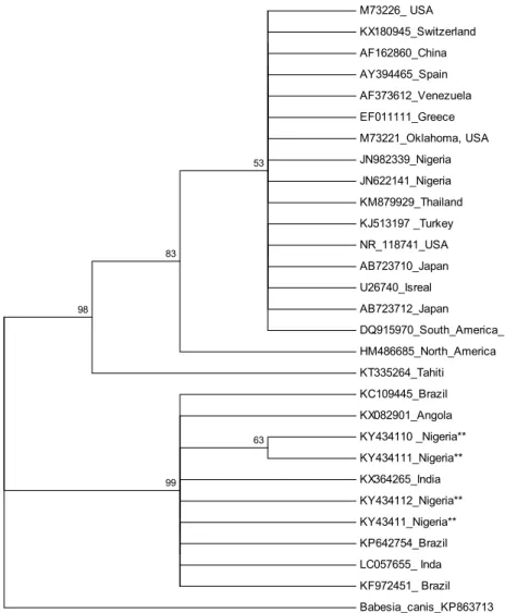
quences obtained from this study had 98–9% homology with the sequences de- posited in GenBank (DQ379966, DQ228505, KP844663, KF878949, KX180945, KX898137, LC057655, M73226, NR118741, JX893522, U54805, KP642754, KJ995842, KJ995837 and KF972451). The aligned autochthonous sequences were less polymorphic with sequences KY434110 showing insertion (C) at point 994 and sequences KY434110 and KY434111 showing alterations (A → C) at point 947. When these sequences were aligned with those previously reported in Nigeria by Kamani et al. (2013), there were considerable variations characterised by complete deletion (at points 926, 962 and 1045), insertion (at points 886, 894, 944, 994 and 1024) and alterations at 912 (T→A), 915 (A→C), 919–921 (AGT→GCT) and 941–942 (TA→AC).
The phylogenetic tree inferred by Maximum Likelihood and Neighbour Joining methods showed the same topologies (Figs 2 and 3). The trees clearly separated the sequences reported previously in Nigeria from the autochthonous sequences into two different clades. The sequences from this study clustered with those from India, Brazil and Angola, while those previously reported in Nigeria clustered among those from the USA, Turkey, Switzerland, Israel, Japan, South America, and Tahiti.
Discussion
This study attempted to shed more light on the occurrence of canine ehr- lichiosis and the molecular characteristics of E. canis detected in naturally in- fected dogs in South West Nigeria by microscopy and by nested PCR that target- ed a partial region of the 16S rRNA gene.
The occurrence of ehrlichial morulae in 1.5% of the dogs sampled and screened by microscopy could not be compared due to the paucity of data and reports on microscopic detection of the parasite in Nigerian dogs. This may be an indication of how difficult microscopic detection of Ehrlichia parasites could be in the blood. The detection of morulae in both monocytes and granulocytes in this study may suggest that both granulocytic and monocytic ehrlichiosis exist among the dog population in Nigeria; hence, there is a need for a wide-scale mo- lecular screening to ascertain which of the Ehrlichia species is/are available in Nigeria as those found in monocytes are generally believed to be either E. canis or E. chaffeensis and those found in granulocytes to be E. ewingii (Blanco and Oteo, 2002; Bowman et al., 2009). Although the infection stage was not classi- fied in the dogs sampled in this study, the three cases in which morulae were de- tected by microscopy may represent acute infection as suggested by other work- ers (Aguero-Rosenfeld et al., 1996; Bakken et al., 1996) who posited that moru- lae are usually detected in blood smears only during acute febrile episodes.
The detection of E. canis DNA in 22.9% of the sampled population of dogs was higher than the prevalence of 12.7% reported by Kamani et al. (2013)
in the northern part of Nigeria and those reported by Nazari et al. (2013) and Lasta et al. (2013) in Malaysia and Brazil, respectively. The higher occurrence reported in this study may partly be associated with the predominance of the tick vector (Rhipicephalus sanguineus) of the parasite in the study area (Oke et al., 2013) and may be due to optimum environmental conditions that favour its breeding. Since we only amplified with E. canis specific primers, there is a need for further studies including other species-specific primers to determine whether mixed infections of these Ehrlichia parasites are possible or exist.
M73226_ USA KX180945_Switzerland AF162860_China AY394465_Spain AF373612_Venezuela EF011111_Greece M73221_Oklahoma, USA JN982339_Nigeria JN622141_Nigeria KM879929_Thailand KJ513197 _Turkey NR_118741_USA AB723710_Japan U26740_Isreal AB723712_Japan
DQ915970_South_America_
HM486685_North_America KT335264_Tahiti KC109445_Brazil KX082901_Angola KY434110 _Nigeria**
KY434111_Nigeria**
KX364265_India KY434112_Nigeria**
KY43411_Nigeria**
KP642754_Brazil LC057655_ Inda KF972451_ Brazil Babesia_canis_KP863713 63
99 98
83
53
Fig. 2. Phylogenetic analysis of 16S rRNA gene partial sequences of E. canis isolates inferred using the Maximum Likelihood method. The percentage (50% and above) of replicate trees in which the associated taxa clustered together in the bootstrap test (1000 replicates) are shown next
to the branches
JN982339_Nigeria AF162860_China KJ513197 _Turkey NR_118741_USA M73221_Oklahoma, USA M73226_ USA JN622141_Nigeria AF373612_Venezuela EF011111_Greece DQ915970_South_America_
AB723712_Japan AB723710_Japan U26740_Isreal
HM486685_North_America AY394465_Spain KX180945_Switzerland KM879929_Thailand KT335264_Tahiti KX082901_Angola KY434112_Nigeria**
KP642754_Brazil KX364265_India KF972451_ Brazil LC057655_ Inda KY434110 _Nigeria**
KY434111_Nigeria**
KY43411_Nigeria**
KC109445_Brazil Babesia_canis_KP863713 50
99 78 100
99 100
56 95
90
53 99
66 63
73
Fig. 3. Phylogenetic analysis of 16S rRNA gene partial sequences of E. canis isolates inferred using the Neighbour-Joining method. The percentage (50% and above) of replicate trees in which
the associated taxa clustered together in the bootstrap test (1000 replicates) are shown next to the branches
The E. canis sequences that were used to construct the phylogenetic trees exhibit diphyletic clades, separating the autochthonous sequences from those previously described in Nigeria. The separation may be an indication that more than one strain of E. canis are in circulation among the dog population in Nige- ria. Clinical manifestation was not reported either in our study or in the previous study on ehrlichial infection of dogs in Nigeria; hence, further investigations on the pathogenicity of these strains in infected dogs are needed. The diphyletic clades exhibited by the autochthonous 16S rRNA partial sequences and those re-
ported previously in Nigeria are not supported by some of the previous works on the phylogenetics of E. canis (Vinasco et al., 2007; Vargas-Hernández et al., 2012). This could be an indication that the 16S rRNA gene of E. canis strains in Nigeria is more polymorphic than those reported elsewhere and this may have potential implications on the clinical outcome and management of canine ehr- lichiosis in Nigeria. Generally, E. canis (Dawson et al., 1993; Unver et al., 2001;
Perez et al., 2006), E. chaffeensis (Dawson et al., 1993; Chen et al., 1997; Rojas et al., 2015) and E. ewingii (Buller et al., 1999; Allen et al., 2014) have been re- ported in human ehrlichiosis around the world but not in Nigeria. This does not necessarily preclude the presence of ehrlichial infection in humans in Nigeria and, as such, the screening for such parasites should be considered in human fe- brile conditions.
In conclusion, this study provides molecular evidence that shows the pres- ence of an E. canis strain in naturally infected dogs in Abeokuta (South West Nigeria), which is genetically different from the strain previously reported in the northern part of Nigeria and more closely related to sequences from Brazil and India.
References
Aguero-Rosenfeld, M. E., Horowitz, H. W. and Wormser, G. P. (1996): Human granulocytic ehr- lichiosis: a case series from a medical center in New York State. Ann. Intern. Med. 125, 904–908.
Aguiar, D. M., Hagiwara, M. K. and Labruna, M. B. (2008): In vitro isolation and molecular char- acterization of an Ehrlichia canis strain from São Paulo, Brazil. Braz. J. Microbiol. 39, 489–493.
Allen, M. B., Pritt, B. S., Sloan, L. M., Paddock, C. D., Musham, C. K., Ramos, J. M., Cetin, N.
and Rosenbaum, E. R. (2014): First reported case of Ehrlichia ewingii involving human bone marrow. J. Clin. Microbiol. 52, 4102–4104.
Bakken, J. S., Krueth, J., Wilson-Nordskog, C., Tilden, R. L., Asanovich, K. and Dumler, J. S.
(1996): Clinical and laboratory characteristics of human granulocytic ehrlichiosis. J. Am.
Med. Assoc. 275, 199–205.
Blanco, J. R. and Oteo, J. A. (2002): Human granulocytic ehrlichiosis in Europe. Clin. Microbiol.
Infect. 8, 763–772.
Bowman, D., Little, S. E., Lorentzen, L., Shields, J., Sullivan, M. P. and Carlin, E. P. (2009): Prev- alence and geographic distribution of Dirofilaria immitis, Borrelia burgdorferi, Ehrlichia canis, and Anaplasma phagocytophilum in dogs in the United States: results of a national clinic-based serologic survey. Vet. Parasitol. 160, 138–148.
Buller, R. S., Arens, M., Hmiel, S. P., Paddock, C. D., Sumner, J. W., Rikihisa, Y., Gaudreault- Keener, M., Manian, F. A., Liddell, A. M., Schmulewitz, N. and Storch, G. A. (1999): Ehr- lichia ewingii, a newly recognized agent of human ehrlichiosis. N. Engl. J. Med. 341, 148–155.
Cardoso, L., Oliveira, A. C., Granada, S., Nachum-Biala, Y., Gilad, M., Lopes, A. P., Sousa, S. R., Vilhena, H. and Baneth, G. (2016): Molecular investigation of tick-borne pathogens in dogs from Luanda, Angola. Parasit. Vectors 9, 252.
Chen, S. M., Yu, X. J., Popov, V. L., Westerman, E. L., Hamilton, F. G. and Walker, D. H. (1997):
Genetic and antigenic diversity of Ehrlichia chaffeensis: comparative analysis of a novel human strain from Oklahoma and previously isolated strains. J. Infect. Dis. 175, 856–863.
Cicuttin, G. L., De Salvo, M. N. and Gury Dohmen, F. E. (2016): Molecular characterization of Ehrlichia canis infecting dogs, Buenos Aires. Ticks Tick Borne Dis. 7, 954–957.
Dagnone, A. S., Souza, A. I. D., André, M. R. and Machado, R. Z. (2009): Molecular diagnosis of Anaplasmataceae organisms in dogs with clinical and microscopical signs of ehrlichiosis.
Rev. Bras. Parasitol. Vet. 18, 20–25.
Dawson, J. E., Candal, F. J., George, V. G. and Ades, E. W. (1993): Human endothelial cells as an alternative to DH82 cells for isolation of Ehrlichia chaffeensis, E. canis, and Rickettsia rickettsii. Pathobiology 61, 293–296.
Dumler, J. S., Choi, K., Garcia-Garcia, J. C., Barat, N. S., Scorpio, D. G., Garyu, J. W., Grab, D. J.
and Bakken, J. S. (2005): Human granulocytic anaplasmosis and Anaplasma phagocytophi- lum. Emerg. Infect. Dis. 11, 1828–1834.
Engvall, E. O., Pettersson, B., Persson, M., Artursson, K. and Johansson, K. E. (1996): A 16S rRNA-based PCR assay for detection and identification of granulocytic Ehrlichia species in dogs, horses, and cattle. J. Clin. Microbiol. 34, 2170–2174.
Fourie, J. J., Stanneck, D., Luus, H. G., Beugnet, F., Wijnveld, M. and Jongejan, F. (2013): Trans- mission of Ehrlichia canis by Rhipicephalus sanguineus ticks feeding on dogs and on arti- ficial membranes. Vet. Parasitol. 197, 595–603.
Harrus, S., Perlman-Avrahami, A., Mumcuoglu, K. Y., Morick, D., Eyal, O. and Baneth, G. (2011):
Molecular detection of Ehrlichia canis, Anaplasma bovis, Anaplasma platys, Candidatus midichloria mitochondrii and Babesia canis vogeli in ticks from Israel. Clin. Microbiol. In- fect. 17, 459–463.
Kaewmongkol, G., Maneesaay, P., Suwanna, N., Tiraphut, B., Krajarngjang, T., Chouybumrung, A., Kaewmongkol, S., Sirinarumitr, T., Jittapalapong, S. and Fenwick, S. (2016): First de- tection of Ehrlichia canis in cerebrospinal fluid from a non-thrombocytopenic dog with meningoencephalitis by broad-range PCR. J. Vet. Intern. Med. 30, 255–259.
Kamani, J., Baneth, G., Mumcuoglu, K. Y., Waziri, N. E., Eyal, O., Guthmann, Y. and Harrus, S.
(2013): Molecular detection and characterization of tick-borne pathogens in dogs and ticks from Nigeria. PLoS Negl. Trop. Dis. 7, e2108.
Lasta, C. S., Santos, A. P., Messick, J. B., Oliveira, S. T., Biondo, A. W., Vieira, R. F. C., Dalmolin, M. L. and González, F. H. D. (2013): Molecular detection of Ehrlichia canis and Anaplasma platys in dogs in Southern Brazil. Rev. Bras. Parasitol. Vet. 22, 360–366.
Lauzi, S., Maia, J. P., Epis, S., Marcos, R., Pereira, C., Luzzago, C., Santos, M., Puente-Payo, P., Giordano, A. and Pajoro, M. (2016): Molecular detection of Anaplasma platys, Ehrlichia canis, Hepatozoon canis and Rickettsia monacensis in dogs from Maio Island of Cape Verde archipelago. Ticks Tick Borne Dis. 7, 964–969.
Liddell, A. M., Stockham, S. L. and Scott, M. A. (2003): Predominance of Ehrlichia ewingii in Missouri dogs. J. Clin. Microbiol. 41, 4617–4622.
McBride, J. W., Corstvet, R. E., Gaunt, S. D., Chinsangaram, J., Akita, G. Y. and Osburn, B. I.
(1996): PCR detection of acute Ehrlichia canis infection in dogs. J. Vet. Diagn. Invest. 8, 441–447.
Milanjeet, H. S., Singh, N., Singh, N., Singh, C. and Rath, S. (2014): Molecular prevalence and risk factors for the occurrence of canine monocytic ehrlichiosis. Vet. Med. 59, 129–136.
Nazari, M., Lim, S. Y., Watanabe, M., Sharma, R. S. K., Cheng, N. A. B. Y. and Watanabe, M.
(2013): Molecular detection of Ehrlichia canis in dogs in Malaysia. PLoS Negl. Trop. Dis.
7, e1982.
Oke, O. A., Idowu, A. B., Ademolu, K. O. and Bamgbose, O. J. (2013): Prevalence of tick infesta- tion on dogs (Canis spp.) presented at the Veterinary Teaching Hospital, Federal Universi- ty of Agriculture Abeokuta, South West Nigeria. Adv. Sci. Tech. 7, 20–24.
Perez, M., Bodor, M., Zhang, C., Xiong, Q. and Rikihisa, Y. (2006): Human infection with Ehrlichia canis accompanied by clinical signs in Venezuela. Ann. N. Y. Acad. Sci. 1078, 110–117.
Rojas, N., Castillo, D. and Marin, P. (2015): Molecular detection of Ehrlichia chaffeensis in Hu- mans, Costa Rica. Emerg. Infect. Dis. 21, 532–534.
Souza, B. M. P. S., Leal, D. C., Barboza, D. C. P. M., Uzêda, R. S., Alcântara, A. C., Ferreira, F., Labruna, B. M., Gondim, L. F. P. and Franke, C. R. (2010): Prevalence of ehrlichial infec- tion among dogs and ticks in Northeastern Brazil. Rev. Bras. Parasitol. Vet. Jaboticabal.
19, 89–93.
Suksawat, J., Xuejie, Y., Hancock, S. I., Hegarty, B. C., Nilkumhang, P. and Breitschwerdt, E. B.
(2001): Serologic and molecular evidence of coinfection with multiple vector-borne patho- gens in dogs from Thailand. J. Vet. Intern. Med. 15, 453–462.
Takeet, M. I., Fagbemi, B. O., Donato, M. D., Yakubu, A., Rodulfo, H. E., Peters, S. O., Wheto, M. and Imumorin, I. G. (2013): Molecular survey of pathogenic trypanosomes in naturally infected Nigerian cattle. Res. Vet. Sci. 94, 555–561.
Tamura, K., Nei, M. and Kumar, S. (2004): Prospects for inferring very large phylogenies by using the neighbor-joining method. Proc. Natl Acad. Sci. U.S.A. 101, 11030–11035.
Unver, A., Perez, M., Orellana, N., Huang, H. and Rikihisa, Y. (2001): Molecular and antigenic comparison of Ehrlichia canis isolates from dogs, ticks, and a human in Venezuela. J. Clin.
Microbiol. 39, 2788–2793.
Vargas-Hernández, G., André, M. R., Faria, J. L. M., Munhoz, T. D., Hernandez-Rodriguez, M., Machado, R. Z. and Tinucci-Costa, M. (2012): Molecular and serological detection of Ehr- lichia canis and Babesia vogeli in dogs in Colombia. Vet. Parasitol. 186, 254–260.
Vinasco, J., Li, O., Alvarado, A., Diaz, D., Hoyos, L., Tabachi, L. and Moro, M. H. (2007): Mo- lecular evidence of a new strain of Ehrlichia canis from South America. J. Clin. Microbiol.
45, 2716–2719.