Szent István University
Postgraduate School of Veterinary Science
Studies of ticks (Acari: Ixodidae) and tick-borne pathogens of dogs in Hungary
PhD dissertation
By
Gábor Földvári
Szent István University
Faculty of Veterinary Science
Postgraduate School of Veterinary Science Budapest, Hungary
Supervisor:
……….
Róbert Farkas, DVM, PhD
Dep. of Parasitology and Zoology, Fac. Vet. Sci., SzIU
Co-supervisors:
Prof. Miklós Rusvai, DVM, PhD
Dep. of Pathology and Forensic Vet. Medicine, Fac. Vet. Sci., SzIU
Prof. Károly Vörös, DVM, PhD, DSc
Dep. and Clinic of Internal Medicine, Fac. Vet. Sci., SzIU
Prepared in eight copies. This is copy number ....
CONTENTS
1. SUMMARY...4
2. INTRODUCTION...5
2.1. Hard tick infestation of dogs...6
2.2. Tick-borne pathogens of dogs...11
2.2.1. Piroplasms...12
2.2.2. Spirochetes...17
3. AIMS...19
4. STUDIES...20
4.1. Hard tick infestation of dogs...20
4.1.1. Materials and methods...20
4.1.1.1. Identification key of tick species infesting dogs in Europe...20
4.1.1.2. Collection of ticks from dogs...21
4.1.1.3. Collection of ticks from field...21
4.1.2. Results...22
4.1.2.1. Identification key of tick species infesting dogs in Europe...22
4.1.2.2. Collection of ticks from dogs...39
4.1.2.3. Collection of ticks from field...42
4.1.3. Discussion...44
4.1.3.1. Identification key of tick species infesting dogs in Europe...44
4.1.3.2. Collection of ticks from dogs and from field...48
4.2. Tick-borne pathogens of dogs...53
4.2.1. Materials and methods...53
4.2.1.1. Detection of small canine piroplasms: case studies...53
4.2.1.2. Molecular examination of babesiae in blood and tick samples...53
4.2.1.3. Molecular examination of spirochetes in blood and tick samples...55
4.2.2. Results...56
4.2.2.1. Detection of small canine piroplasms...56
4.2.2.2. Molecular examination of babesiae in blood and tick samples...58
4.2.2.3. Molecular examination of spirochetes in blood and tick samples...62
4.2.3. Discussion...64
4.2.3.1. Detection of small canine piroplasms...64
4.2.3.2. Molecular examination of babesiae in blood and tick samples...65
4.2.3.3. Molecular examination of spirochetes in blood and tick samples...68
5. NEW RESULTS AND CONCLUSIONS...71
6. REFERENCES...74
7. OWN PUBLICATIONS...87
8. ACKNOWLEDGEMENTS...88
1. SUMMARY
In Europe, the number of reports on canine tick-borne diseases has increased in the past few years. In Hungary, we have had very limited information concerning tick infestation and tick-borne pathogens of dogs. For these reasons, we started to study the tick species and tick-borne pathogens infecting dogs in our country.
Based on morphological studies, a figured practical identification key has been designed for the sixteen hard tick species which have been found on dogs in Europe. The simplicity of this key can help veterinarians and zoologists in tick identification.
In 29 veterinary clinics from six districts of Budapest and 13 counties, 1779 tick specimens were collected from 606 dogs. Most hosts were usually infested with a single female and very few of them had many ticks. The most preferred sites of tick attachment in decreasing order were head, neck and legs. Ixodes ricinus and Dermacentor reticulatus were the most common species. Ixodes canisuga, Haemaphysalis concinna, Ixodes hexagonus, Ixodes acuminatus and Dermacentor marginatus were also found. New data have been provided about the geographical distribution of Dermacentor reticulatus, because the specimens of this species were collected in north-eastern and south-eastern parts of the country too where they had not been found before. Field collections in 31 locations provided new data on the geographical and seasonal occurrence of I. ricinus, D.
reticulatus and other tick species as well.
The occurrence of small canine piroplasms in two dogs was described for the first time in Hungary. These were autochtonous infestations but we need further investigations to know the species, occurrence, vector and origin of this pathogen. The subspecies Babesia canis canis was identified to be the causative agent of babesiosis caused by large Babesia sp. in dogs using molecular biological methods. It was also proven with molecular methods that the geographical distribution of canine babesiosis is larger in the country than it has been previously known. Babesia DNA was detected in free-living and engorged D. reticulatus females for the first time in the country. Presence of B. canis canis in engorged D. reticulatus specimens removed from dogs was also demonstrated with molecular methods.
Molecular evidence was found for the presence of Borrelia sp. in free-living and engorged I.
ricinus females for the first time in Hungary. Three species, B. burgdorferi s.s., B. afzelii and B.
garinii were identified with sequence analysis which are pathogenic to both dogs and humans.
2. INTRODUCTION
Biological complexity of pathogen-tick-host systems is exceptional. More than 3500 years ago, both the Ancient Egyptians and the Ancient Greeks were already aware of ticks and their medical importance (Varma, 1993). Smith and Kilbourne (1893) completed first landmark research proving that Texas fever (caused by Babesia bigemina) was spread from one cow to another by hard ticks (Boophilus annulatus), just as many cattlemen had suspected. Their discovery spurred the search for vectors of malaria-causing Plasmodium and other pathogens worldwide (Pratt and Littig, 1962). Since this first proof of hard tick’s ability to transmit disease agents, scientific attention has continuously been raising. Considering the human and veterinary health importance of ixodid ticks, the scope for exciting novel discoveries is immense. Only during the last 30 years, a considerable increase occurred in the number of publications containing the word “tick” within the ISI Web of Science® database (Fig. 1). Ticks are considered second only to mosquitoes as vectors of human infectious disease agents worldwide but first in Eurasia and North America (Estrada-Peňa and Jongejan, 1999). In Europe during the last two decades there has arisen an increased awareness of ticks and the pathogens they carry (Hillyard, 1996).
0 100 200 300 400 500 600 700 800 900 1000
1975 1977 1979 1981 1983 1985 1987 1989 1991 1993 1995 1997 1999 2001 2003
Figure 1. Number of publications containing the word “tick” between 1975-2004 within the ISI Web of Science® database.
Hard ticks (Acari: Ixodidae) are obligate hematophaguos ectoparasites of a wide variety of terrestrial vertebrates including domestic dogs. Originally evolved as parasites of wild animals, only
a relative minority of the approximately 650 hard tick species, generally those with wide host range can transmit diseases to domesticated animals (Shaw et al., 2001). Because of the wide range of transmitted pathogens, ticks are of considerable medical and veterinary interest worldwide. In Europe, the number of reports of canine tick-borne diseases has increased in the past few years (Shaw et al., 2001; Chandoga et al., 2002; Camacho et al., 2003; Criado-Fornelio et al., 2003a,b).
Emerging tick transmitted canine diseases like babesiosis, anaplasmosis, ehrlichiosis, rickettsiosis, mycoplasmosis, hepatozoonosis and borreliosis have drawn both public and scientific attention to these arthropods (Beugnet, 2002; Kenny et al, 2004a).
Increased mobility of pets and the ability of ticks to find niches in new climatic conditions have resulted in rapid extension of the zoogeographical ranges for many tick species (Glaser and Gothe, 1998; Shaw et al., 2001). The increasing number of ticks has also been associated with growing accessibility of natural environments and an increase in the population of wild host species (deer, small mammals and foxes) that now have a closer association with human activity. For example, Ixodes ricinus has extended its range in Sweden to more northern and western areas since the 1980`s (Talleklint and Jaenson, 1998). Vector of several canine pathogens, Rhipicephalus sanguineus, has also a good adaptive ability and is likely to inhabit new areas throughout Europe (Gothe, 1968; Gothe and Hamel, 1973; Fox and Sykes, 1985; Gothe, 1999). Recently, it has been reported that importation of Dermacentor reticulatus into new regions poses higher risk for canine babesiosis (Zahler and Gothe, 1997 Zahler et al., 2000a).
The importance of ticks is also highlighted by the rapid development of molecular biological methods enabling easy screening of blood samples and ticks for disease agents (Sparagano et al., 1999; Criado-Fornelio et al., 2003b; Monis et al., 2005). There has been an increased awareness of dogs’ ticks because some of them can be dangerous also to humans transmitting zoonotic diseases (Shaw et al., 2001; Beugnet, 2002).
2.1. Hard tick infestation of dogs
Studies examining the hard tick infestation of dogs have been published from the mid 1980s in Europe. Liebisch et al. (1984) recorded the following seven tick species collected from 1624 dogs in Germany (in decreasing order of occurrence): I. ricinus, Ixodes hexagonus, D. reticulatus, R. sanguineus, Ixodes canisuga, Dermacentor marginatus and Haemaphysalis concinna. Beichel et al. (1996) reported the occurrence of only two species, I. ricinus and I. hexagonus on 48 infested
found R. sanguineus, R. turanicus, I. ricinus, R. bursa, I. hexagonus and Haemaphysalis punctata in decreasing order of frequency. Papazahariadou et al. (2003) found only two species, R. sanguineus and R. turanicus on 249 tick-infested animals in northern Greece. Ogden et al. (2000) identified I.
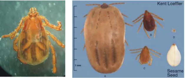
ricinus, I. hexagonus, I. canisuga, H. punctata and D. reticulatus collected from 213 dogs in Great Britain and Ireland. Concerning occurrence, veterinary and zoonotic importance, Ixodes ricinus, Rhipicephalus sanguineus and Dermacentor reticulatus (Fig. 2-4) are the most important species infesting dogs in Europe (Shaw et al., 2001; Beugnet, 2002).
Figure 2. Ixodes ricinus male and unengorged female.
Figure 3. Rhipicephalus sanguineus male, fully engorged female (A), unfed female (B), male (C) and larva (D). Scale is valid only for the right hand picture modified from
http://www.entomology.cornell.edu/MedEnt/TickBioFS/TickBioFS.html.
In Hungary the first comprehensive studies on the occurrence of ixodid tick species were made several decades ago (Kotlán, 1919,1921). Although there is a limited number of publications, in some of them the use of species names (especially in case of D. reticulatus) produced misunderstandings in the later papers. Kotlán (1919) for example used the name D. reticulatus correctly applying the principle of priority which is commonly accepted in zoological nomenclature. Janisch (1959) and Babos (1965) however, considered the name Dermacentor pictus to be valid name for the species with the same morphological characters. Further complications aroused when Kotlán and Kobulej (1972) and Janisch (1986) considered D. reticulatus and D.
marginatus to be synonyms, however, they are morphologically clearly distinct (Arthur, 1960).
Janisch (1959) collected approximately 15000 tick specimens from mammals, birds and from field. He reported that I. ricinus and D. marginatus were the most common species in the country. He had knowledge on the occurrence of D. reticulatus (syn. D. pictus) merely from Fertőd and Tolna which he explained with importation. Babos (1965) included 33 species of five genera into his identification key of hard tick species of Hungary. However, on the basis of current valid species names (Camicas et al., 1998; Horak et al., 2002), these are in fact 24 valid species, 19 of which were registered to occur in the country. Seven of these species, I. ricinus, I. canisuga, H.
concinna, Haemaphysalis inermis, Haemaphysalis parva (syn. Haemaphysalis otophila), D.
reticulatus (syn. D. pictus), and D. marginatus were mentioned to infest dogs in Hungary. Until recently, there has been no information about the temporal and spatial distribution of hard tick species infesting dogs. Farkas and Földvári (2001) examined 160 tick specimens collected from 100 dogs. Four species were found of which I. ricinus and D. reticulatus were the most common. One specimen of D. marginatus and I. hexagonus also occurred. Significant association was found between the presence of clinical signs of canine babesiosis and the infestation of these animals with D. reticulatus (γ=0.53, Px4<< 0.001). The latter species occurred in a greater geographic range than Babos (1965) and Janisch (1959, 1986) previously described.
There have been no other data published on the tick species of dogs, however in a recent study, I. ricinus, H. concinna, D. reticulatus and I. canisuga were found on red foxes in Hungary (Sréter et al., 2003). These carnivores can be considered as relevant to the natural maintenance of tick species that are able to feed on dogs, because the number of foxes is still high (>60000) in the country (Csányi, 2005).
D. reticulatus has been found to be vector of Babesia canis, a tick-borne pathogen of dogs common in Hungary (Janisch, 1986). Because canine babesiosis is a severe and frequent disease in the country (Horváth and Papp, 1996; Csikós et al., 2001), it is crucial to study the geographical and seasonal distribution of this tick species in particular. Previous data on tick infestation of dogs (Farkas and Földvári, 2001), and foxes (Sréter et al., 2005) suggest that the spatial distribution of D.
reticulatus has expanded since the 1950s. According to Meyer-König et al. (2001), this species has extended its distribution from the 31st and 40th northern parallel to the 60th northern parallel. One explanation for this is the adult’s marked ecological plasticity which is also reflected by its occurrence in a variety of ecological zones. Another reason is the nidicolous life of larvae and nymphs which are active during summer only and are particularly protected against extremely unfavourable climatic conditions in the habitats of their hosts, which are burrow-dwelling and ground-living small mammals (Zahler, 1994).
The risk of occurrence of non-indigenous tick species and new tick-borne pathogens (Kálmán et al., 2003; Farkas et al., 2004; Sréter et al., 2004; Sréter-Lancz et al., 2005) has been increasing in Hungary. This may be associated with increased awareness and improved diagnostic methods but also because the number of travelling dogs (with their ticks) has been increasing (Glaser and Gothe, 1998). That is why it is important to monitor both the autochthonous and the imported tick species occurring on dogs and in the field.
Accurate identification is the essential first stage in any study involving ticks. In the case of ticks infesting dogs, it would also be important in the diagnosis of a disease because different species can transmit different pathogens. Morphological features of the adult stages of ticks have traditionally provided the main criteria for distinguishing species (Nuttall and Warburton, 1911, 1915; Arthur, 1960; Babos, 1964; Hillyard, 1996; Estrada-Peňa et al., 2004). However, morphological identification can present difficulties. For example, the large number of species in the keys that do not feed on dogs can be misleading, because most of the identification keys are designed for a particular geographical region and not restricted to host species. For this reason, a non-specialist who tries to identify a tick from a dog has to distinguish it from a number of species that are not feeding on this host. In addition, the tick’s capitulum, which is usually essential for species identification, may become damaged during removal. Furthermore, for nymphal and especially for larval stage, traditional morphological identification is often ambiguous. Such difficulties have made it necessary to find methods of molecular biological identification for ixodid ticks infesting dogs. Hitherto, there have been only a few attempts to discriminate between hard tick species on the basis of their DNA sequences. However, diagnostic methods using restriction enzyme analysis of the second internal transcribed spacer (ITS-2) in the nuclear ribosomal gene have been developed for D. reticulatus by Zahler et al. (1995) and for 17 Ixodes species of the United States by Poucher et al. (1999). The usefulness of molecular methods is more restricted to
Mediterranean region has been published recently by Estrada-Peňa et al. (2004), which includes some but not all dog-infesting species. We have, however, no information on a practical identification key which enables the identification of hard tick species that infest dogs in Europe.
2.2. Tick-borne pathogens of dogs
As hematophaguos arthropods, ticks are well designed to transmit disease agents such as viruses, bacteria, rickettsiae, protozoa, fungi and nematodes (Hillyard, 1996). Transmission of tick- borne pathogens occurs not only from tick to host but both transovarially (i.e. the eggs acquire infection from the egg-laying female) and transstadially (i.e. both larvae and nymphs are able to transmit the pathogen to the next developmental stage) (Beugnet, 2002).
Attaching of ticks firmly to their hosts, facilitates the effective blood feeding, the transmission of pathogens but also the spread of both ticks and microorganisms to different geographical habitats via migrating animals or travelling pets (Kenny et al., 2004b). Although restricted by host movement and climatic factors, many tick species and their pathogens extended their zoogeographical ranges due to the increased mobility of pets (Glaser and Gothe, 1998; Shaw et al., 2001). For example between 1995 and 1998, 36% of cases of monocytic ehrlichiosis reported in Germany occurred in dogs that had travelled for short periods to the Mediterranean area. During the same period, both Ehrlichia canis infection and infestation with R. sanguineus, the vector species traditionally found in Southern Europe, were detected in dogs that have never left Germany (Gothe, 1999).
Tick-borne encephalitis virus (TBEV) (Flaviviridae) is primary pathogenic to humans but also infects dogs (Shaw et al., 2001). Seroprevalence of the virus has been found to be higher in dogs because they come into contact with the infected vector ticks (mainly I. ricinus) more frequently than humans. However, the risk for developing clinical tick-borne encephalitis (TBE) in tick-infested dogs in an endemic area seems to be rather small for unknown reasons. Altogether less than twenty clinical TBE infections of dogs have been reported in Austria, Germany, Sweden and Switzerland (Beugnet, 2002).
Ehrlichia canis is an obligate intracellular Gram negative bacterium infecting white blood cells. It is transmitted by R. sanguineus in Europe and is the causative agent of canine monocytic ehrlichiosis which occurs worldwide and can cause serious symptoms in dogs (Beugnet, 2002).
Anaplasma phagocytophilum (formerly E. phagocytophila, human granulocytic ehrlichiosis agent) is also an obligate intracellular Gram negative bacterium infecting white blood cells. It can be transmitted by I. ricinus in Europe. A. phagocytophilum is the causative agent of canine anaplasmosis (granulocytic ehrlichiosis) which can produce a wide spectrum of clinical
manifestations (Beugnet, 2002).
Hepatozoon canis is an apicomplexan protozoon. Its transmission differs from other tick- borne pathogens because infection of the dog takes place by ingestion of a tick containing the parasite. Its vector species is R. sanguineus. Clinical signs can vary between apparently asymptomatic to severe and life-threatening disease. Infections are reported from Spain, Italy and southern France (Beugnet, 2002).
Beyond these and the two major tick-transmitted infections in Europe (babesiosis and borreliosis) there are some other tick-borne diseases like mycoplasmosis (earlier haemobartonellosis), bartonellosis, tularaemia and, rarely, louping ill (Shaw et al., 2001, Kenny et al., 2004a) as well. Several of the tick-borne infections that affect dogs can cause serious disease in humans, notably borreliosis, anaplasmosis (ehrlichiosis), Mediterranean spotted fever and tick- borne encephalitis. However, the potential zoonotic threat posed by dogs is strongly influenced by the natural cycle of the specific agent with which the dog is infected. Shaw et al. (2001) described three epidemiological scenarios. First, if the transmission of an infectious agent involves ticks with a broad host range (such as I. ricinus), dogs can act directly as sentinels for infection of humans.
Second, by acting as natural hosts for certain nidicolous (endophilic, i.e. its non feeding stages live in the nest of their hosts) ticks (such as R. sanguineus and I. canisuga), dogs significantly increase contact between these species and humans, thereby increasing the risk of transmission (Mumcuoglu et al., 1993). Finally, there is a limited risk of transmission by exposure to infected tick-contents following damage to ticks during grooming of infested animals. This has been reported for H. canis (Beugnet, 2002).
2.2.1. Piroplasms
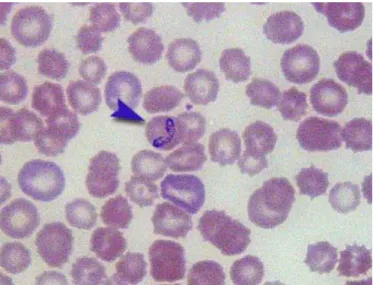
Babesia (Apicomplexa: Piroplasmida) species are tick-transmitted parasites infecting a wide range of wild and domestic vertebrate hosts (Kuttler, 1988). Traditionally, identification of species has been based on host specificity and morphology of the intraerythrocytic piroplasms. Based on these, canine babesiae have been originally recognised to belong to two distinct species, the large pyriform (4-5 µm) Babesia canis (Fig. 5) and the small usually pleomorph (1-2.5 µm) Babesia gibsoni (Fig. 6).
Figure 5. Babesia canis in a Giemsa-stained thin blood smear.
Figure 6. Babesia gibsoni in a Giemsa-stained thin blood smear (from http://www.yamagiku.co.jp/pathology/index.htm).
Canine piroplasms
Large (4-5µm) Small (1-2.5µm)
Babesia gibsoni (Asia) Babesia gibsoni (USA, Spain)
Theileria annae Babesia canis canis
Babesia canis vogeli Babesia canis rossi
Unknown Babesiasp. Theileria equi
Canine piroplasms
Large (4-5µm) Small (1-2.5µm)
Babesia gibsoni (Asia) Babesia gibsoni (USA, Spain)
Theileria annae Babesia canis canis
Babesia canis vogeli Babesia canis rossi
Unknown Babesiasp. Theileria equi
Figure 7. Piroplasms that have been described in dogs.
On the basis of differences in geographical distribution, vector specificity and antigenic properties (Uilenberg et al., 1989; Hauschild et al., 1995), B. canis has been subdivided into three subspecies, namely B. canis canis transmitted by D. reticulatus and R. sanguineus in Europe, B.
canis vogeli transmitted by R. sanguineus in tropical and subtropical regions and B. canis rossi transmitted by Haemaphysalis leachi in South Africa (Fig. 7). These subspecies also differ from each other in pathogenicity. B. canis rossi causes a frequently fatal infection in domestic dogs, even after treatment; B. canis vogeli causes a moderate often clinically unapparent infection; and B. canis canis infections result in a more variable pathogenicity intermediate between B. canis rossi and B.
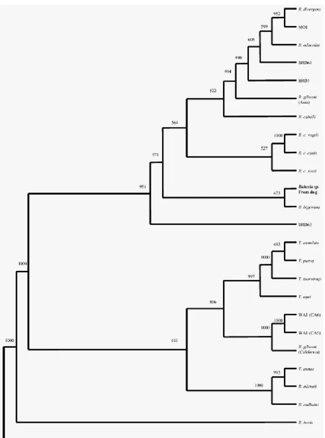
canis vogeli (Uilenberg et al., 1989). Genetic differences between these subspecies have also been proved by Zahler et al. (1998) and Carret et al. (1999). Some authors (Zahler et al., 2004) suggest and some others (Passos et al., 2005) even use the species level for these three subspecies. Baneth et al. (2004) described a fourth subspecies with unknown vector named B. canis presentii which has been detected in cats. Recently, a novel large Babesia sp. has been detected in an infected dog from North America by Birkenheuer et al. (2004) which either represents a new Babesia sp. or is one of the nearly 100 Babesia spp. described for which no genetic data have been reported (Fig 8).
Canine babesiosis caused by B. canis has been reported in several European countries, particularly in the Mediterranean region, where R. sanguineus and D. reticulatus are its vectors.
Cases of autochtonous large Babesia infections have been reported from Belgium, Croatia, France, Germany, Hungary, Italy, Poland, Slovenia, Spain and the Netherlands (Losson et al., 1999;
Beugnet, 2002; Cacció et al., 2002; Duh et al., 2004). Recent studies using molecular methods showed that in France, Slovenia and Spain where these vector species coexist, both B. canis canis and B. canis vogeli could be detected (Cacciò et al., 2002; Criado-Fornelio et al., 2003b; Duh et al., 2004). As a contrast, in Northern Poland where neither D. reticulatus nor R. sanguineus occur, no Babesia sp. has been detected scanning 192 canine blood samples by PCR (Skotorczak et al., 2004).
Figure 8. Phylogenetic tree inferred by distance methods using edited alignment of 18S rDNA sequences. The number on each branch shows the occurrence in 1000 bootstrap replicates
(Birkenheuer et al, 2004).
B. gibsoni occurs in Asia, North America, northern and eastern Africa, Australia and Europe (Birkenheuer et al., 1999; Muhlnickel et al., 2002; Criado-Fornelio et al., 2003a). Recent genetic characterisations demonstrated that small canine piroplasms also represent a greater diversity than previously thought (Zahler et al., 2000b,c; Kjemtrup et al., 2000; Kocan et al., 2001). One of the recently identified small piroplasms, Theileria annae (Zahler et al. 2000b) is phylogenetically closer to B. microti than to B. gibsoni (Fig. 8) and it can be found with a high frequency among Spanish dogs (Camacho et al., 2001). Criado-Fornelio et al. (2003b) described another small piroplasm, namely Theileria equi, from the blood of dogs in Spain using polymerase chain reaction (PCR) and sequencing.
The presence of small babesiae of dogs in Europe was in doubt until the end of the 1980s.
Although some cases have been described recently, knowledge of the veterinary and zoonotic importance of these parasites is still very limited (Casapulla et al., 1988, Zahler et al., 2000b,c;
Suarez et al., 2001, Camacho et al., 2001, 2003; Criado-Fornelio et al., 2003a,b).
Canine babesiosis was first described in Hungary by Wetzl (1905) who used the name Piroplasma canis for the pathogen found in the blood smears of two hunting dogs which visited the county Tolna. The disease was diagnosed again in three dogs from Tolna in the early 1930s (Miklósi, 1931,1932) and in six dogs in the 1970s (Horváth and Papp, 1974) originating from the south-west bank of lake Balaton. Janisch (1986) proved with experimental infections that D.
reticulatus (syn. D. pictus) is the biological vector of B. canis in Hungary. He reported that 70 registered cases had been known until 1986. Since then, it has been a severe and frequent disease in the country (Horváth and Papp, 1996). Csikós et al. (2001) diagnosed babesiosis in 1384 dogs between 1992 and 1999 in Szekszárd and its vicinity. Identification of the pathogen has always been based merely on size and morphology of the intraerythrocytic parasites, and no evidence has been found concerning the subspecies of the large canine Babesia in Hungary. To the best of our knowledge, confirmed cases of B. gibsoni or other small babesiae in dogs have not been reported in Hungary so far.
The detection of both small and large genetically unique canine babesiae that are clinically and morphologically indistinguishable from known piroplasms highlights the need for molecular diagnostics in clinical medicine (Cacciò et al., 2002; Kjemtrup et al., 2000; Zahler et al., 2000b,c).
To date, eight genetically different piroplasms have been described in symptomatic dogs (Fig. 7)
traditional methods allowed (Birkenheuer et al., 2003; Jefferies et al., 2003). Beside PCR-RFLP (PCR and Restriction Fragment Length Polymorphism) (Zahler et al., 1998; Carret et al., 1999) and reverse line blot hybridization (Matjila et al., 2004), sequencing single PCR products remains a reliable and quick diagnostic method (Criado-Fornelio et al., 2003b). For these reasons, considerable changes in the nomenclature, taxonomy and phylogeny of canine piroplasms can be anticipated in the near future.
2.2.2. Spirochetes
Spirochetes are well studied prokaryotes because of their importance in Lyme disease, the most frequent tick-borne human infection in the northern hemisphere (Brouqui et al., 2004). The disease-causing bacteria are flexible, spiral-shaped, Gram-negative spirochetes with internal flagella and belong to Borrelia burgdorferi sensu lato complex (Spirochaetaceae). They have been divided into at least 12 genospecies (Michel et al., 2003; Richter et al., 2004). Six of them: B. burgdorferi sensu stricto, B. afzelii, B. garinii, B. bissettii, B. lusitaniae and B. spielmani, have an unambiguous pathogenic role in human Lyme disease in Central Europe (Strle et al., 1997; Postic et al., 1998;
Collares-Pereira et al., 2004; Richter et al., 2004). Several Ixodes species can transmit these spirochetes to a large number of avian and mammal hosts (Hillyard, 1996). A considerable number of studies examined the occurrence of Lyme disease spirochetes in ticks in Europe. Hubalek and Halouzka (1998) reviewed the literature of European surveys on B. burgdorferi s.l. infection of the most important vector, I. ricinus. They showed that the spirochete was present in all I. ricinus populations in Europe wherever it was examined. There have been surveys in Austria, Belgium, Bulgaria, Croatia, Czech Republic, Denmark, Finland, France, Germany, Great Britain, Ireland, Italy, Lithuania, The Netherlands, Norway, Poland, Russia, Serbia, Slovakia, Slovenia, Spain, Sweden and Switzerland (Hubalek and Halouzka, 1998). Lakos et al. (1991) investigated the presence of the Lyme disease spirochetes in 31 field collected adult I. ricinus ticks in Hungary. Five of them contained Borrelia burgdorferi s.l. Spirochetes were successfully cultivated in four cases, detected by immunofluorescence and dark field microscopy as well in two ticks. Two of the isolated strains were tested by Western blot. These studies were not continued later.
From the epidemiological point of view dogs have been very important since they had been declared an effective factor of spreading human borreliosis (Eng et al., 1988). Borrelia seropositivity is common among dogs because many of them carry a persistent infection for life and only a fraction of infected animals (~5%) enter the disease status (Levy and Magnarelli, 1992;
Beugnet, 2002). Persistent infection even after antibiotic therapy is reportedly common in dogs, because the spirochetes are sequestered in the skin, connective tissue, joint and central nervous
system. Thus, reactivation of infection with recrudescence of disease can occur e.g. in immunocompromised individuals or a co-infection (Shaw et al., 2001). Clinical manifestations of the disease have also been reported in dogs (Greene, 1991). A variety of clinical symptoms (fever, lethargy, lymphadenopathy, kidney disorders, heart block and neuroborreliosis) can be observed (Lissman et al., 1984; Kornblatt et al., 1985). Recurrent polyarthritis is the most frequent manifestation in seropositive animals (Greene, 1991).
Pathogenicity has been firstly proved for B. burgdorferi s.l. in North America (Appel et al., 1993). B. burgdorferi s.s. and B. garinii are found commonly in naturally exposed dogs in Europe and mixed infection with four genospecies (B. burgdorferi s.s., B. afzelii, B. garinii, and B.
valaisiana) may also occur (Hovius et al., 1998). Speck et al. (2001) suggested that there are some differences in clinical symptoms between dogs having borreliosis in Europe and dogs experimentally infected with B. burgdorferi s.s. in the USA.
Canine borreliosis was first described in the USA (Lissman et al., 1984; Kornblatt et al., 1985) and recently in almost all western European countries. B. burgdorferi s.l. infection of dogs have been recorded in France (Doby et al., 1988), Great Britain (May et al., 1991), Belgium (McKenna et al., 1995), Spain (Delgado and Carmenes, 1995), Slovakia (Stefancikova et al., 1996), Germany (Bauerfeind et al., 1998), the Netherlands (Hovius et al., 1999b), Sweden (Egenvall et al., 2000), and Switzerland (Speck et al., 2001). The latter turned out to be a B. afzelii infection in a naturally exposed dog (Speck et al., 2001). Skotorczak and Wodecka (2005) examined blood samples of tick-infected dogs from north-western Poland and revealed the presence of B.
burgdorferi s.s. DNA. We have no literature data concerning canine borreliosis in Hungary, however, veterinary clinics frequently diagnose canine borreliosis based on clinical signs or serological methods (personal communication).
3. AIMS
In Europe, the number of reports on canine tick-borne diseases has increased in the past few years (Shaw et al., 2001; Camacho et al., 2001; Chandoga et al., 2002; Criado-Fornelio et al., 2003a,b; Kenny et al., 2004a). Increased national and international mobility of pets (Glaser and Gothe, 1998), the ability of ticks to find niches in new climatic conditions, growing accessibility of natural environments and an increase in the population of wild host species that now have a closer association with human activity (Csányi, 2005) are reported to be the main reasons (Shaw et al., 2001). In Hungary, we have had very limited information concerning tick infestation and tick-borne pathogens of dogs. For these reasons, we started to study the tick species and tick-borne pathogens infecting dogs in our country. Our main aims were the following:
• To design a practical identification key for the adult tick species occurring on dogs in Europe.
• To examine the tick fauna of dogs and the geographical and seasonal distribution of tick species infesting dogs in Hungary with special emphasis on D. reticulatus.
• To collect data on tick species living in the environments where dogs can be infested with special emphasis on places where canine babesiosis occurs.
• To ascertain whether small babesiae can occur in dogs in Hungary.
• To carry out a molecular survey on B. canis infection of dogs in Hungary in order to clarify the subspecies and obtain detailed information on the geographical and seasonal occurrence of this piroplasm. We also aimed at detecting and identifying Babesia sp. from D. reticulatus specimens fed on dogs and collected from field.
• To detect and identify Borrelia sp. occurring in the blood of dogs and in I. ricinus specimens fed on dogs and collected from field.
4. STUDIES
4.1. Hard tick infestation of dogs
4.1.1. Materials and methods
4.1.1.1. Identification key of tick species infesting dogs in Europe
Species for the identification key were selected on the basis of literature search. The two selecting criteria of tick species for our study were: (1) occurrence in Europe and (2) recorded infestation of dogs. A primary search was carried out in the Nuttall Tick Collection (Department of Entomology, Natural History Museum, London UK) which contains several thousands of specimens that was listed by Keirans (1984) according to host species. This is one of the geographically widest and best quality collections accessible on hard ticks. Faunistic surveys (Liebisch, 1984; Grandes, 1986; Beichel et al., 1996; Papadopoulos et al., 1996; Ogden et al., 2000;
Farkas and Földvári, 2001; Papazahariadou et al., 2003; Földvári and Farkas, 2005a), monographs and reviews (Hillyard, 1996; Camicas et al., 1998; Estrada-Peňa et al., 2004) have also been examined in respect to the two criteria. Cases where tick infestations were only accidental (e.g.
single observation in one report) were not included.
Hard tick specimens used for drawing and examining were unengorged adults kept in 70%
ethanol (except for I. canisuga and H. parva for which only partly engorged females were available) from the tick collection of the Natural History Museum, London, UK. Type specimens were used for identifying generic and specific characters. All morphological features were examined which had been used previously in the description of the earlier publications (Nuttall and Warburton, 1911 and 1915; Arthur, 1960; Babos, 1964; Hillyard, 1996; Walker et al., 2000;
Estrada-Peňa et al., 2004) but only those characters which are relatively easy to distinguish were included into our key. A schematic drawing was prepared by Paul D. Hillyard and Gábor Majoros to show the most relevant morphological signs from dorsal and ventral view of both males and females. Drawings have been also made to each genera and species enabling the recognition of the most important morphological features. Dichotomy was avoided wherever it made the identification easier and only essential characters were mentioned in the keys.
4.1.1.2. Collection of ticks from dogs
After previous consultations, 40 veterinary clinics recruited from a wide geographical area within the country were asked to collect hard ticks from dogs which attended the surgeries in a period of four years. A simple questionnaire was designed to investigate the breed and age of host animal, date of collection and habitat to which the dog had been exposed. Questions on previous acaricidal and/or antibabesial treatment(s) and on symptoms of babesiosis were also asked. Ticks were removed from the dogs during the clinical examination and the questionnaires were completed. Specimens collected from each dog were stored in separate labelled tubes containing 70% ethanol. The samples with the questionnaires were posted to the Department of Parasitology and Zoology, Faculty of Veterinary Science, Szent István University, Budapest.
Counting and identification of species, life stage and sex of ticks were carried out under a stereomicroscope. Standard keys of Babos (1965), Hillyard (1996), Estrada-Peňa et al. (2004) were used for species identification of adults; immature specimens were not identified to species level.
4.1.1.3. Collection of ticks from field
Ticks were collected from the vegetation of 32 different locations including ten (Győr- Moson-Sopron, Vas, Veszprém, Zala, Somogy, Baranya, Pest, Csongrád, Borsod-Abaúj-Zemplén and Heves) counties and six districts of Budapest. Most locations were chosen because frequent infestation of dogs and/or occurrence of canine babesiosis were reported (personal communication).
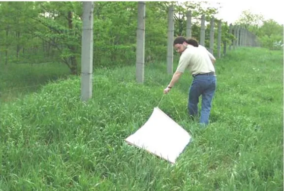
A handle was fixed to a white flannel towel (Fig. 9) in order to use the dragging method for collecting ticks (Hillyard, 1996). Larger towels were used for the undergrowth and smaller, flag-like towels for bushes and shrubs. The coloration of the towel made it easier to find the crawling ticks on it. Field collections were done in April-July and September-November during the main activity peaks of ixodid ticks in Hungary. In order to increase the chance of finding tick specimens, collections were usually carried out in the morning (7-10 a.m.) or in the afternoon (3-7 p.m.) when the relative humidity is usually higher. Dragging lasted for approximately one hour at every field collection sites. Temperature and relative humidity on the ground level was recorded with a Datalogger (Gemini, UK). Characteristic flora and water supply of the area were registered as well.
Time and location of collection were also recorded when tick specimens were found on vegetation or on the clothes of the collector.
Storing and identification of specimens were the same described for ticks originating from dogs.
Figure 9. Collection of ticks from the vegetation with dragging method.
4.1.2. Results
4.1.2.1. Identification key of tick species infesting dogs in Europe
Sixteen hard tick species of five genera were found in literature, which are known to occur regularly on dogs in Europe (Table 1). It is important to note that Dermacentor niveus was excluded. This is regarded as a valid species name by some authors (Horak et al., 2002) and as a synonym of D. marginatus by others (Estrada-Peňa, 1990; Hillyard, personal communication).
Because these two species do not have major morphological difference and occupy the same geographical range (Arthur, 1960), we did not make a differentiation of them. A key was designed to distinguish between stages, sexes and genera. Adults of the 16 species were included in an illustrated identification key below. The adults’ most relevant morphological structures used for identification can be seen in Fig.10.
Table 1. Hard tick species infesting dogs in Europe (Keirans, 1984; Liebisch, 1984; Grandes, 1986;
Beichel et al., 1996; Hillyard, 1996; Papadopoulos et al., 1996; Camicas et al., 1998; Ogden et al., 2000; Papazahariadou et al., 2003; Estrada-Peňa et al., 2004).
Valid name Synonym
Ixodes canisuga (Johnston, 1849) I. vulpinus Schulze, 1937
I. melicola Schulze and Johnston, 1930 I. sciuricola Schulze, 1933
I. barbarossae Schulze, 1937 Ixodes ricinus (Linnaeus, 1758)
Ixodes hexagonus Leach, 1815 I. autumnalis Leach, 1815
I. hexagonus hungaricus Babos, 1964 I. vulpis Pagenstecher, 1861
Haemaphysalis inermis Birula, 1895 Haemaphysalis concinna Koch, 1844
Haemaphysalis punctata Canestrini and Fanzago, 1878 H. cinnabarina punctata Canestrini and Fanzago, 1878 Haemaphysalis parva (Neumann, 1897) H. otophila Schulze, 1918
Rhipicephalus bursa Canestrini and Fanzago, 1878 Rhipicephalus pusillus Gil Collado, 1938
Rhipicephalus turanicus Pomerantsev, 1936 Rhipicephalus sanguineus (Latreille, 1806)
Dermacentor reticulatus (Fabricius, 1794) D. pictus Hermann, 1804 Dermacentor marginatus (Sulzer, 1776) I. hungaricus Karpelles, 1893
D. niveus Neumann, 1897 Hyalomma aegyptium (Linnaeus, 1758)
Hyalomma marginatum marginatum Koch, 1844 H. marginatum Koch, 1844 Hyalomma marginatum rufipes Koch, 1844 H. rufipes Koch, 1844
H. rufipes rufipes Koch, 1944
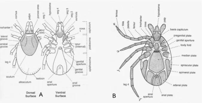
Figure 10. Dorsal and ventral surface of a female (A) and ventral surface of a male (B) hard tick (P.D. Hillyard).
Key to the stages and sexes
1. - Three pairs of legs, spiracles absent.
……….…..larva - Four pairs of legs, spiracles present.
………2 2. - Scutum covers entire dorsum of the body.
………male - Scutum confined to anterior of dorsum.
………3 3. - Genital opening and porose areas absent.
…….………nymph - Porose areas and genital opening present.
………female
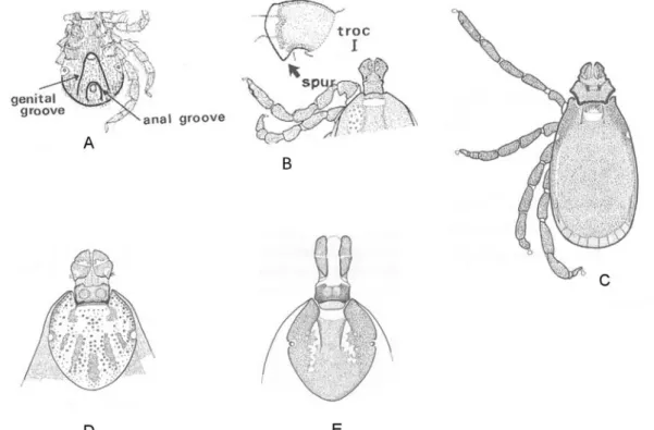
Figure 11. Main characters of Ixodes (A), Haemaphysalis (B), Rhipicephalus (C), Dermacentor (D) and Hyalomma (E) genera (P.D. Hillyard).
Key to the genera
1. - Anal groove circles anus anteriorly. Scutum without eyes or ornamentation; well-marked festoons absent. (Fig. 11A)
………Ixodes
- Anal groove posterior to anus. Scutum with or without eyes and with or without ornamentation; festoons present.
………2
2. - Without eyes or ornamentation. Trochanter I with broad spur. External spur on coxa I absent. (Fig. 11B)
………Haemaphysalis
- Eyes present. Scutum ornate or not. Trochanter I without broad spur. External spur of coxa I present.
………...………3
3. - Scutum inornate. Shape of basis capitulum almost hexagonal in dorsal view. (Fig. 11C) ...Rhipicephalus
- Scutum ornate or not. Shape of basis capitulum subrectangular.
...4
4. - Palps short and wide. Scutum ornate. (Fig. 11D)
...Dermacentor - Palps longer than wide. Scutum inornate. (Fig. 11E)
...Hyalomma
Figure 12. Ixodes canisuga. A. Dorsal view of male; B. Dorsal view of female; C. Dorsal view of female capitulum; D. Ventral view of female basis and coxa I; E. Ventral view of male (P.D.
Hillyard and G. Majoros).
Key to the species of Ixodes females
1. - Article II plus III of palp as long as or longer than width of basis capitulum. Scutum broadly rounded posteriorly or hexagonal. Internal spur of coxa I elongate. Location of genital aperture not between coxae II.
...2
- Article II plus III of palp shorter than width of basis capitulum. Scutum narrowly rounded
genital aperture between coxae IV.
...Ixodes ricinus (Fig. 13 B-D) -Scutum hexagonal. External spurs on coxae I-IV absent. Location of genital aperture between coxae III.
………...Ixodes hexagonus (Fig. 14B-D)
Figure 13. Ixodes ricinus. A. Dorsal view of male; B. Dorsal view of female; C. Dorsal view of female capitulum; D. Ventral view of female basis and coxa I; E. Ventral view of male (P.D.
Hillyard and G. Majoros).
Key to the species of Ixodes males
1. - Internal spur of coxa I prominent. Adanal and epimeral plates clearly shorter than median plate. Median plate as long as or longer than wide.
...2
- Internal spur of coxa I short. Adanal and epimeral plates almost as long as median plate.
Median plate narrow anteriorly but broad posteriorly.
...Ixodes canisuga (Fig. 12 A,E) 2. - Internal spurs on coxae II-IV vestigial. Pregenital plate twice as long as broad. Median
plate much longer than wide.
...Ixodes ricinus (Fig. 13 A,E) - Internal spurs on coxae II-IV absent. Pregenital plate almost hexagonal. Median plate nearly as long as wide ...Ixodes hexagonus (Fig. 14 A,E)
Figure 14. Ixodes hexagonus. A. Dorsal view of male; B. Dorsal view of female; C. Dorsal view of female capitulum; D. Ventral view of female basis and coxa I; E. Ventral view of male (P.D.
Hillyard and G. Majoros).
Key to the species of Haemaphysalis females
1. - Article II of palp projects laterally beyond margin of basis capitulum. Spur on trochanter I prominent in dorsal view……… 2
- Palps do not project laterally beyond basis capitulum. Spur on trochanter I relatively short.
- Cornua present. Scutum almost round. Spur on coxa I prominent.
...Haemaphysalis concinna (Fig. 16 B)
3. - Spur of coxa IV more prominent than that of coxa I
……….Haemaphysalis punctata (Fig. 17 B-E)
- Spur of coxa IV not more prominent than that of coxa I
...Haemaphysalis parva (Fig. 18 B)
Figure 15. Haemaphysalis inermis. A. Dorsal view of male; B. Dorsal view of female capitulum and scutum (P.D. Hillyard).
Figure 16. Haemaphysalis concinna. A. Dorsal view of male capitulum; B. Dorsal view of female capitulum (P.D. Hillyard).
Key to the species of Haemaphysalis males
1. - Article II of palp projects laterally beyond margin of basis capitulum. Basis capitulum 1.5x
...………...2
- Article II of palp without lateral projection. Basis capitulum narrow. Cornua absent. Spurs on all coxae small.
...Haemaphysalis inermis (Fig. 15 A)
- 2. Article III of palp curves inward. Basis capitulum at least 2x broader than long. Cornua prominent. Spur on coxa I prominent.
...Haemaphysalis concinna (Fig. 16 A)
- Article III of palp does not curve inward. Basis capitulum 1.5x broader than long. Cornua blunt. Coxa IV has long, pointed and curved spur.
...Haemaphysalis punctata (Fig. 17 A,C)
- Article III of palp does not curve inward. Basis capitulum 1.5x broader than long. Cornua blunt. Spurs on coxae short and blunt (lacks a long, pointed and curved spur on coxa IV).
...Haemaphysalis parva (Fig. 18 A)
Figure 17. Haemaphysalis punctata. A. Dorsal view of male; B. Dorsal view of female capitulum and scutum; C. Ventral view of male coxa IV; D. Ventral view of female coxa I; E. Dorsal view
of female trochanter I (P.D. Hillyard).
Figure 18. Haemaphysalis parva. A. Ventral view of male; B. Ventral view of female (G.
Majoros).
Key to the species of Rhipicephalus females
1. - Large species (3.5 - 4.0mm). Porose areas large and oval, separated by 1x their height.
Punctation of scutum fine with sparse larger punctation. Genital aperture V-shaped
...………...Rhipicephalus bursa (Fig. 19 D,E)
- Small species (2.2 - 2.4mm). Porose areas small, separated by 2x or more their diameter.
Punctation of scutum fine with sparse larger punctation. Genital aperture U- shaped
….………..Rhipicephalus pusillus (Fig. 20 C-E)
- Medium-sized species (3.0 - 3.8mm). Porose areas small, separated by 1.5 - 2x their diameter. Punctation of scutum fine with sparse larger punctation. Genital aperture U- shaped………...Rhipicephalus sanguineus (Fig. 21 B,C)
- Medium-sized species. Porose areas small, separated by 2x their diameter. Punctation of scutum variable but usually dense and conspicuous. Genital aperture U-shaped
………...Rhipicephalus turanicus (Fig. 22 C-E)
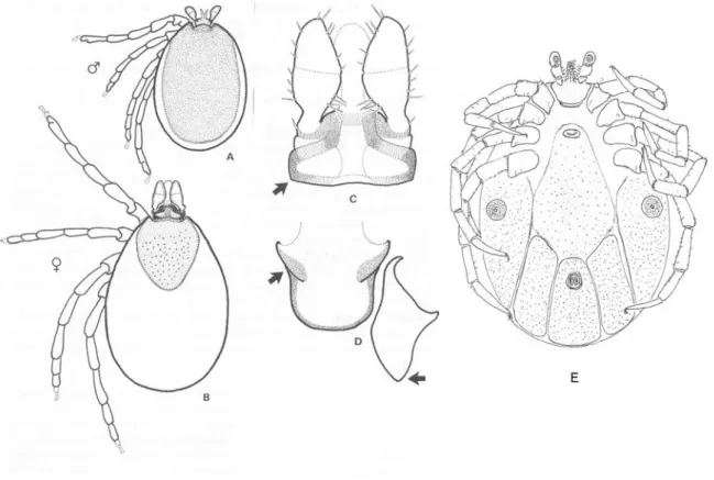
Figure 19. Rhipicephalus bursa. A. Dorsal view of male; B. Ventral view of male; C. Ventral view of male adanal plates; D. Dorsal view of female; E. Ventral view of female (G. Majoros).
Key to the species of Rhipicephalus males
1. - Large species (3.0 - 4.0mm). Punctation of scutum numerous and fine; a few larger punctations in scapular areas. Adanal plates large, subtriangular with broad posterior. Spiracle broad with long, narrow handle-like extension...Rhipicephalus bursa (Fig. 19 A-C)
- Small species (1.8 - 2.2mm). Punctation of scutum fine with larger punctations scattered throughout. Adanal plates narrow, curved inwards posteriorly. Spiracle with short, distinctly curved extension………...Rhipicephalus pusillus (Fig. 20 A,B)
- Medium-sized species (2.7 - 3.3mm). Punctation of scutum usually heavy. Adanal plates vary. Spiracle at least 2x as long as wide………...Rhipicephalus turanicus (Fig. 22 A,B)
- Medium-sized species (2.5 - 3.2mm). Punctation of scutum ranges from fine to large;
usually four more or less regular rows of large punctations visible. Adanal plates elongate triangular with broad posterior. Spiracle shaped like the sole of a slipper………..
...………...Rhipicephalus sanguineus (Fig. 21 A)
Figure 21. Rhipicephalus sanguineus. A. Dorsal view of male; B. Ventral view of female coxa I;
C. Ventral view of female capitulum and scutum (P.D. Hillyard).
Figure 22. Rhipicephalus turanicus. A. Dorsal view of male; B. Ventral view of male; C. Dorsal view of female; D Ventral view of female; E. Ventral view of female genital opening (G.
Majoros).
Key to the species of Dermacentor females and males
1. - Article II of palp with prominent, rear-facing spur. Spur of coxa I not divergent.
... Dermacentor reticulatus (Fig 23)
- Article II of palp without spur. Spur of coxa I clearly divergent.
... Dermacentor marginatus (Fig. 24)
Figure 23. Dermacentor reticulatus. A. Dorsal view of male and left palp; B. Ventral view of female capitulum and scutum; C. Ventral view of female basis and coxa I (P.D. Hillyard).
Figure 24. Dermacentor marginatus. A. Dorsal view of female capitulum and scutum; B. Dorsal view of male capitulum; C. Ventral view of female basis and coxa I (P.D. Hillyard).
Key to the species of Hyalomma females
1. - Scutum virtually round in outline; only a few, scattered, large punctations on scutum. Spur on coxa I wide and widely divergent. Genital aperture broadly oval in outline.
...Hyalomma aegyptium (Fig. 25 B,C)
- Scutum not quite round - outline narrows behind eyes; punctation moderate to numerous, variable in size. Spur of coxa I long and narrow and narrowly divergent. Genital aperture broadly triangular in outline...Hyalomma marginatum marginatum (Fig. 26 C-E)
- Scutum not quite round – outline narrows behind eyes; and entirely covered by large punctations. Spur of coxa I long and narrow and narrowly divergent. Genital aperture broadly oval in outline...Hyalomma marginatum rufipes (Fig. 27 C-E)
Figure 25. Hyalomma aegyptium. A. Dorsal view of male; B. Dorsal view of female capitulum and scutum; C. Ventral view of female basis and coxa I (P.D. Hillyard).
Key to the species of Hyalomma males
1. - Scutum with only a few, scattered large punctations. Spur of coxa I wide and widely
- Surface of scutum smooth with regular, fine punctations and occasional larger punctations.
Spur of coxa I long and narrow and slightly divergent. Adanal plates approx. twice as long as wide and separated by a distance equal to their end width.
...Hyalomma marginatum marginatum (Fig. 26 A,B) - Surface of scutum rugged with many large punctations. Spur of coxa I long and narrow and slightly divergent. Adanal plates approx. twice as long as wide and separated by a distance clearly less than their end width.
...Hyalomma marginatum rufipes (Fig. 27 A,B)
Figure 26. Hyalomma marginatum marginatum. A. Dorsal view of male; B. Ventral view of male; C. Dorsal view of female capitulum and scutum; D Ventral view of female basis and coxa I;
E. Ventral view of female genital opening (P.D. Hillyard and G. Majoros).
Figure 27. Hyalomma marginatum rufipes. A. Dorsal view of male; B. Ventral view of male; C.
Dorsal view of female; D Ventral view of female; E. Ventral view of female genital opening (G.
Majoros).
4.1.2.2. Collection of ticks from dogs
In 29 veterinary clinics from six districts of Budapest and 13 counties 1779 tick specimens were collected from 606 dogs. Infested animals originated from 55 different locations in the country (Fig. 28). Most hosts were usually infested with a single female (Fig. 29) and very few of them had many (up to 78) ticks (Fig. 30). Most of the tick specimens (1666; 93.6%) were adults belonging to six species, the others (113; 6.4%) were nymphs. I. ricinus (872; 52.3%) and D. reticulatus (708;
42.5%) were the most frequently identified species. Forty-six (2.8%) and 33 (2.0%) adults were I.
canisuga and H. concinna, respectively. There were four specimens of I. hexagonus, two of Ixodes acuminatus and only one Dermacentor marginatus. Most of the adults (1268, 76.1%) were semi- engorged or fully engorged females. Specimens of D. marginatus, I. hexagonus, I. acuminatus and I. canisuga were only females.
Figure 28. Locations where ticks were collected from dogs.
Figure 29. Blood-sucking female tick.
Figure 30. Feeding ticks in a dog’s ear.
Single species infestation by either I. ricinus or D. reticulatus occurred on 281 (46.4%) and 217 (35.8%) dogs, respectively. Mixed infestation caused by these two species was detected on 62 dogs (10.2%). D. marginatus and I. hexagonus were found in single infestations, while H. concinna, I. canisuga and nymphs occurred in mixed infestations.
I. ricinus was collected in all locations (Fig. 28). Dogs infested with D. reticulatus were found at 42 out of 55 localities (Fig. 31). Based on the date of tick collection records D. reticulatus and I. ricinus occurred throughout the year (Fig. 32). There was a greater activity peak of these
Figure 31. Locations where Dermacentor reticulatus was found on dogs.
0 50 100 150 200 250 300
1. 2. 3. 4. 5. 6. 7. 8. 9. 10. 11. 12.
D. reticulatus I. ricinus
Figure 32. Seasonal occurrence of D. reticulatus and/or I. ricinus on dogs according to date of collection.
Localization of tick specimens on dogs was recorded in all 180 questionnaires that were returned. The most preferred sites of tick attachment in decreasing order were head, neck and legs.
Living conditions of 103 (57.2%) dogs were given in the questionnaires. Two thirds (68; 66%) of the animals lived in gardens, 23 (22.3%) in flats and there were 12 (11.7%) stray dogs. Walking
forests (15; 17.1%), mixed habitats (15; 17.1%), meadows (12; 13.6%), parks (5; 5.7%), streets (4;
4.5%), or were living in a garden and not walked at all (12; 13.6%). No association was found between living conditions/strolling and the species of ticks that were collected.
Based on clinical signs (e.g. fever, weakness, lethargy, loss of appetite, haemoglobinuria), canine babesiosis was diagnosed by the veterinarians in 113 (18.6%) tick-infested dogs. Thirty-six (31.9%) of these cases were confirmed by the detection of intraerythrocytic Babesia forms in blood smears or amplification of Babesia DNA with PCR. Specimens of D. reticulatus were collected from 90 (79.6%) dogs having clinical signs of canine babesiosis.
4.1.2.3. Collection of ticks from field
In total 421 tick specimens belonging to five species were collected from field. Ticks were found at 31 out of 32 visited sites (with the exception of Veszprém). Fifteen locations were in Budapest and 16 in other parts of the country (Fig. 33). In eight locations (Németbánya, Csévharaszt, Törökbálint, Baktüttös, Gödöllő, Szokolya, Bükk and Pilis) ticks were accidentally found on the body of the collector and not on the towel. Frequency of occurrence was 1-41 specimen/site. Most (315; 74.8%) of the specimens were adults, the others were nymphs (92;
21.9%) and larvae (14; 3.3%). More than half of the collected ticks were females (181; 57.5%).
Figure 33. Locations where ticks were collected from field (* indicates places where D. reticulatus was found and □ where it was not).
counties (with the exception of Borsod-Abaúj-Zemplén and Heves) (Fig. 33). This species occurred in habitats with typically well water supply (pond, lake or channel in the vicinity), dense vegetation (reedy areas; meadows and pastures; river banks; railway embankments; outskirts of deciduous forests or cities; rudimentary habitats along roads and paths) (Fig. 34). The temperature was between 16-30 °C and the relative humidity on the ground usually above 50% where D. reticulatus was collected.
I. ricinus was the second most common species according to the number of collected specimens (135; 42.9%) and the most commonly occurring being present in all districts of Budapest and all counties where collection was carried out (Fig. 33). It was found in the same habitats as D.
reticulatus but also on drier, less humid areas. There were 17 (5.4%) specimens of H. concinna from counties Pest, Somogy, Zala, Veszprém and Vas. Seven specimens of D. marginatus were collected on a sheep run in the vicinity of Nyíregyháza. Two females of H. inermis were found in the mountains Pilis and Börzsöny on the cloths of the collector.
Figure 34. Typical living habitat of D. reticulatus.
4.1.3. Discussion
4.1.3.1. Identification key of tick species infesting dogs in Europe
Sixteen hard tick species were found during our literature search which commonly occur on dogs in Europe. Since migration of birds and international travelling enable ticks to establish in new habitats, it is possible that this list needs to be updated later. We omitted species which were found only accidentally on dogs or if the canine infestation was not autochthonous in Europe.
Our identification key enables easy and appropriate identification of adult tick species known to occur on dogs in Europe. Its main advantage to other keys is that only a limited number of species have to be taken into consideration during the identification. Furthermore, the comparison and distinction of the 16 species can be achieved with fewer morphological characters then in case of the traditional identification keys (Arthur, 1960, Babos, 1965). The short morphological species descriptions and illustrations focused on the adult stages are intended as identification rather than as comprehensive descriptions. With a few exceptions, determination of tick species can be made most confidently on the basis of unengorged females (Hillyard, 1996). However, attention has to be paid, when an engorged female has to be identified. Some characters on these specimens can be modified (e.g. the position of the genital opening) because of the stretching effect of the blood amount in the intestines. Considering these modifications, the key is useful also for engorged ticks. Male specimens often tend to be poor in specific characters though fortunately males are usually found in the company of females. The nymphs however, can be difficult to identify in the absence of adult specimens even with an identification key (Babos, 1965, Hillyard, 1996). When only larvae are available, more serious difficulties arise because these tiny specimens require a relatively advanced level of microscopy. For these reasons, our key is restricted to the adult stage and we suggest to turn to a specialist or to use molecular biological methods if an immature tick specimen needs to be identified to species level.
Based on morphological (Klompen et al., 1997) or molecular biological data sets (Black et al., 1997; Murrell et al., 1999; Dobson and Barker, 1999; Norris et al., 1999), a series of systematic analyses has provided useful information on the relationships of hard tick subfamilies and species.
There are molecular phylogenetic data based on the amplification and analysis of either nuclear (Crampton et al., 1996) or mitochondrial (Black and Piesman, 1994; Caporale et al., 1995; Hubbard et al., 1995; Rich et al., 1995; Mangold et al., 1998) DNA sequences only for some species
biogeography and evolution of hard ticks can be found in reviews from Klompen et al. (2000) and Barker and Murrell (2002). However, there is scant information concerning the molecular analysis of tick species that are usually found on a typical host e.g. dogs. The continuation of our morphological study will be a molecular biological examination of the same species. Since there are already data for 16S mt rDNA sequences in the GenBank® for some of the 16 species, we plan to amplify the missing ones and make a phylogenetic analysis of them.
The simplicity of this key helps in tick identification not only experienced entomologists but also veterinarians who play central role in the diagnosis, treatment and prevention of tick- transmitted diseases of dogs. Several of the tick-borne pathogens can cause serious disease in humans and domesticated animals (Shaw et al., 2001). Because veterinarians play an important role in advising the public as to the zoonotic potential of disease agents transmitted by ticks, they have to be aware of the possible infestation risk exposed by ticks. Therefore, we list the pathogens of humans and dogs which might be carried by these 16 hard tick species (Tables 2-3).
Table 2. Human and/or canine pathogens transmitted by Ixodes and Haemaphysalis species infesting dogs (Gern et al., 1991;Macaigne and Perez-Eid, 1991; Rehacek et al., 1991;Estrada-Peňa
et al., 1995; Hillyard, 1996;Juricova et al., 2002; Spitalska and Kocianova, 2003;Sréter-Lancz et al., 2005; N.a.= no literature data available).
Tick species Pathogen
Ixodes canisuga B. burgdorferi s.l.
Pasteurella pestis Ixodes ricinus louping-ill virus
tick-borne encephalitis virus Rickettsia helvetica
Rickettsia monacensis R. conori
C. burnetii
Anaplasma phagocytophilum B. burgdorferi s.l.
Francisella tularensis Babesia divergens B. microti
Ixodes hexagonus tick-borne encephalitis virus R. conori
B. burgdorferi s.l.
B. microti
Haemaphysalis inermis tick-borne encephalitis virus C. burnetii
Haemaphysalis concinna tick-borne encephalitis virus C. burnetii
F. tularensis Haemaphysalis punctata Bhanja virus
louping-ill virus
Crimean Congo haemorrhagic fever virus tick-borne encephalitis virus
C. burnetii F. tularensis
Listeria monocytogenes