S H O R T R E P O R T Open Access
Molecular detection of vector-borne bacteria in bat ticks (Acari: Ixodidae,
Argasidae) from eight countries of the Old and New Worlds
Sándor Hornok1* , Krisztina Szőke1, Marina L. Meli2, Attila D. Sándor3, Tamás Görföl4, Péter Estók5, Yuanzhi Wang6, Vuong Tan Tu7, Dávid Kováts8, Sándor A. Boldogh9, Alexandra Corduneanu3, Kinga M. Sulyok10,
Miklós Gyuranecz10, JenőKontschán11, Nóra Takács1, Ali Halajian12, Sara Epis13and Regina Hofmann-Lehmann2
Abstract
Background:Despite the increasingly recognized eco-epidemiological significance of bats, data from molecular analyses of vector-borne bacteria in bat ectoparasites are lacking from several regions of the Old and New Worlds.
Methods:During this study, six species of ticks (630 specimens) were collected from bats in Hungary, Romania, Italy, Kenya, South Africa, China, Vietnam and Mexico. DNA was extracted from these ticks and analyzed for vector- borne bacteria with real-time PCRs (screening), as well as conventional PCRs and sequencing (for pathogen identification), based on the amplification of various genetic markers.
Results:In the screening assays,RickettsiaDNA was only detected in bat soft ticks, whereasAnaplasma
phagocytophilumand haemoplasma DNA were present exclusively in hard ticks.BartonellaDNA was significantly more frequently amplified from hard ticks than from soft ticks of bats. In addition toRickettsia helveticadetected by a species-specific PCR, sequencing identified fourRickettsiaspecies in soft ticks, including aRickettsia africae-like genotype (in association with a bat species, which is not known to migrate to Africa), three haemotropic Mycoplasmagenotypes inIxodes simplex, andBartonellagenotypes inI. ariadnaeandI. vespertilionis.
Conclusions:Rickettsiae (from both the spotted fever and theR. felisgroups) appear to be associated with soft rather than hard ticks of bats, as opposed to bartonellae. Two tick-borne zoonotic pathogens (R. helveticaandA.
phagocytophilum) have been detected for the first time in bat ticks. The present findings add Asia (China) to the geographical range ofR. lusitaniae, as well as indicate the occurrence ofR. hoogstraaliiin South Africa. This is also the first molecular evidence for the autochthonous occurrence of aR. africae-like genotype in Europe. Bat
haemoplasmas, which are closely related to haemoplasmas previously identified in bats in Spain and to“Candidatus Mycoplasma haemohominis”, are reported here for the first time from Central Europe and from any bat tick.
Keywords:Chiroptera, Soft tick, Hard tick,Rickettsia, Anaplasma,Bartonella,Haemoplasma
* Correspondence:hornok.sandor@univet.hu
1Department of Parasitology and Zoology, University of Veterinary Medicine, Budapest, Hungary
Full list of author information is available at the end of the article
© The Author(s). 2019Open AccessThis article is distributed under the terms of the Creative Commons Attribution 4.0 International License (http://creativecommons.org/licenses/by/4.0/), which permits unrestricted use, distribution, and reproduction in any medium, provided you give appropriate credit to the original author(s) and the source, provide a link to the Creative Commons license, and indicate if changes were made. The Creative Commons Public Domain Dedication waiver (http://creativecommons.org/publicdomain/zero/1.0/) applies to the data made available in this article, unless otherwise stated.
Background
Bats (order Chiroptera) are the only mammals which ac- tively fly. Among the consequences of this trait, bats show a geographically widespread distribution and may even undergo short to long distance seasonal migration [1].
Additionally, the evolution of flight in bats yielded inad- vertent consequences on their immune functioning, and therefore bats are special in their capacity to act as reser- voir hosts for intracellular pathogens [2]. Bats frequently reach high population densities in or near urban habitats, and their ticks may blood-feed on humans [3, 4], which further increases their veterinary-medical importance.
The presence of DNA from vector-borne bacteria in bat ticks appears to be most extensively studied in Europe. In western Europe,Rickettsiaand Ehrlichiaspe- cies have been molecularly identified in soft ticks (Argas vespertilionis) of bats (in France [5] and the UK [6]).
Another study carried out in central Europe (Poland) failed to detect Borrelia burgdorferi (s.l.), rickettsiae and Anaplasma phagocytophilum in the bat-associated hard tick species,Ixodes vespertilionis[7]. Nonetheless, litera- ture data on molecular analyses of vector-borne bacteria in bat ticks are lacking from several regions of the Old and New Worlds. Therefore, during this study, bat ticks collected in countries representing less-studied regions (eastern and southern Europe, central and southeast Asia, eastern Africa, central America) were screened for the presence of DNA from four important genera of vector-borne bacteria, which include zoonotic species.
Methods
DNA extracts of 307 hard ticks (I. ariadnae: 26 larvae, 14 nymphs, 5 females; I. vespertilionis: 89 larvae, 27
nymphs, 8 females; I. simplex: 79 larvae, 50 nymphs, 9 females) and 323 soft ticks (A. vespertilionis: 321 larvae;
A. transgariepinus: 1 larva; Ornithodoros sp.: 1 larva) were used. The hard ticks (Acari: Ixodidae) were col- lected from 200 individuals of 17 bat species in two countries (Hungary, Romania), whereas soft ticks (Acari:
Argasidae) were removed from 59 individuals of 17 bat species in eight countries (Hungary, Romania, Italy, Kenya, South Africa, China, Vietnam and Mexico) [8,9].
The geographical coordinates and/or locations of collec- tion sites, along with identification of bat and tick spe- cies by expert taxonomists (authoring this study), have already been reported [8, 9]. DNA was extracted indi- vidually from hard ticks, and individually or in pools of 2–3 specimens (if collected from the same host individ- ual) from soft ticks, as reported [8,9].
Bat tick DNA extracts (n= 514) were screened for the presence ofRickettsia helvetica, otherRickettsiaspp., A.
phagocytophilum, haemotropic Mycoplasma spp. and Bartonella spp. with real-time PCRs (Additional file 1:
Table S1). This was followed by conventional PCRs and sequencing of various genetic markers (Additional file2:
Table S2), and phylogenetic analyses (Additional file 3:
Text S1) except forR. helveticaandA. phagocytophilum.
Prevalences were compared with Fisherʼs exact test.
Results and discussion
Rickettsia DNA was only detected in bat soft ticks (all three evaluated species), whereasAnaplasma phagocyto- philum and three haemotropic Mycoplasma genotypes were present exclusively in the hard tick species I.
simplex (Table 1). In addition, Bartonella DNA was
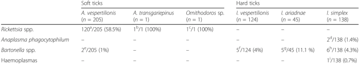
Table 1Prevalence of pathogen DNA in bat ticks according to bat host species and country of origin. The latter are referred to with superscript letters (the cumulative number of bat individuals is equal to or less than the number of positives, because one or more ticks could have been collected from a single bat). After the name of the tick species, the number of analyzed DNA extracts is shown, which corresponds to the number of tick individuals (except forA. vespertilionis, in the case of which pooled samples were also used)
Soft ticks Hard ticks
A. vespertilionis (n= 205)
A. transgariepinus (n= 1)
Ornithodorossp.
(n= 1)
I. vespertilionis (n= 124)
I. ariadnae (n= 45)
I. simplex (n= 138)
Rickettsiaspp. 120a/205 (58.5%) 1b/1 (100%) 1c/1 (100%) – – –
Anaplasma phagocytophilum – – – – – 2d/138 (1.4%)
Bartonellaspp. 2e/205 (1%) – – 5f/124 (4%) 5g/45 (11.1 %) 6h/138 (4.3%)
Haemoplasmas – – – – – 1i/138 (0.7%)
aPipistrellus pipistrellus(Hungary 6×, Italy 1×);Pi. pygmaeus(Hungary 10×);Pi. nathusii(Hungary 1×);Pi. kuhlii(Hungary 1×);Pi. abramus(Vietnam 1×);Pi.cf.
rueppellii(Kenya 1×);Myotis brandtii(Hungary 1×);My. alcathoe(Hungary 2×);My. dasycneme(Hungary 5×);Plecotus auritus(Hungary 1×);Pl. austriacus(Hungary 3×);Nyctalus noctula(Hungary 1×);Eptesicus serotinus(Hungary 1×, Romania 1×);Vespertilio murinus(Hungary 2×, China 1×)
bPi. hesperidus(South Africa 1×)
cBalantiopteryx plicata(Mexico 1×)
dMiniopterus schreibersii(Hungary 1×, Romania 1×)
ePi. pygmaeus(Hungary 2×)
fMy. daubentonii(Romania 2×);My. capaccinii(Romania 1×);Eptesicus serotinus(Romania 1×);Rhinolophus ferrumequinum(Romania 1×)
gMy. alcathoe(Hungary 1×);My. bechsteinii(Hungary 1×);My. daubentonii(Hungary 3×)
hMi. schreibersii(Romania 5×)
iMi. schreibersii(Hungary 1×)
significantly more frequently detected in hard than in soft ticks of bats (Fisherʼs exact test:P= 0.01).
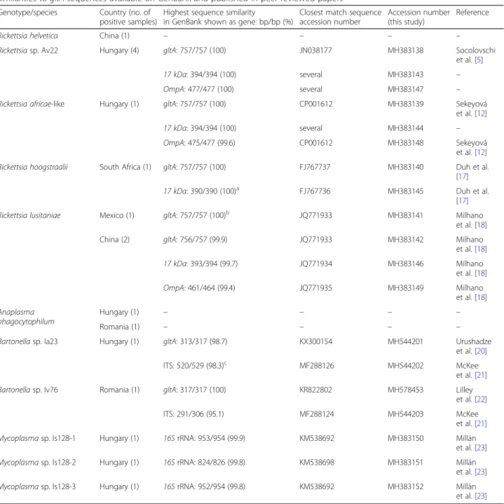
In particular,R. helveticawas identified in one soft tick (A. vespertilionis) from China. This finding is consistent with former reports of R. helvetica in bat fleas [10] and bat faeces [11] in Hungary. Taking into account the bat host-specificity of these PCR-positive ectoparasites, it is possible that bats are susceptible to R. helvetica, al- though based on the very low prevalence this may have low epidemiological significance.
In four samples ofA. vespertilionis from Hungary, the same Rickettsia genotype was identified, which was re- ported from bat soft ticks collected in France (GenBank:
JN038177, see Table2) [12]. More importantly, in oneA.
vespertilionis from Hungary rickettsial DNA was de- tected, which in the amplified part of thegltA gene had 99.9–100% sequence identity (depending on the nucleo- tide at position 679: C or T) to sequences of R. africae from Ethiopia (GenBank: CP001612) and from migratory bird fleas reported in neighboring Slovakia (GenBank:
HM538186) [13]. Two other markers were also success- fully amplified from this sample: the 17 kDa gene se- quence was identical with that of several Rickettsia species, whereas the OmpA sequence showed 2 bp dif- ferences from that ofR. africae(Table2).
Interestingly, theOmpAsequence from thisA. vesperti- lioniswas identical with that of the Rickettsiastrain “At- lantic rainforest” (GenBank: MF536975 [14]) and Rickettsia sp. “Atlantic rainforest Aa46” (GenBank:
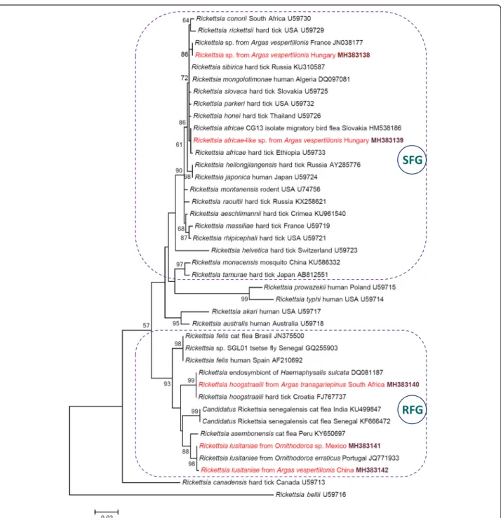
KY113110 [15]), which represent a genetic variant of the human pathogenR. parkeri[14,15] detected so far only in the New World. Nevertheless, we consider the species de- tected inA. vespertilionis to belong toR. africaebecause of the following four reasons: (i) thegltAgene is a reliable genetic marker for species identification and phylogenetic comparison of rickettsiae [13,16]; (ii)R. africaewas iden- tified based on this gene in previous studies (e.g. [13]); (iii) thegltA phylogenetic analysis confirmed that the rickett- sial genotype fromA. vespertilionis collected in Hungary clustered withR. africae, but apart fromR. parkeri(Fig.1);
and (iv) the OmpA gene of the type strain ofR. parkeri (GenBank: U43802) was only 98.3% (469/477 bp) identical with theOmpAsequence obtained here.
The soft tick containing the R. africae-like DNA was collected from Myotis dasycneme, which occurs north of the Mediterranean Basin and is a facultative, middle distance migrant bat species, not known to move between Europe and Africa [1]. Therefore, this result implies the autochthonous occurrence of a R.
africae-like genotype in Europe. In the phylogenetic analysis, this genotype was clearly separated (with moderate, 72% bootstrap support value) from the Rickettsia sp. from A. vespertilionis reported in France (Fig. 1).
In addition,R. hoogstraaliiwas identified in a soft tick from South Africa (Table 2). This rickettsia has only been reported from Europe and North America [17], therefore its occurrence in Africa is new. Similarly, R.
lusitaniae was formerly only reported in Europe (Portugal) [18] and Central America (Mexico) [19], the latter being confirmed in the present study (Table 2).
However, a gltAgenotype highly similar toR. lusitaniae (1 bp difference from JQ771933, i.e. 99.9% identity) was also shown here, for the first time, to occur in Asia (China) (Table 2). The level of OmpA sequence diver- gence of this Chinese isolate (MH383149) was the same (3 bp) from R. lusitaniae in Portugal (JQ771935) and fromR. lusitaniaein Mexico (GenBank: KX377432).
In summary, bat soft ticks contained the DNA of three Rickettsia species from the spotted fever group (SFG), and two further ones from the Rickettsia felis group (RFG) (Fig.1).
Anaplasma phagocytophilum DNA was detected here in the hard tick species,I. simplex, in both Hungary and Romania. Previously, Anaplasma sp. DNA was also shown to be present in bat feces in Hungary (GenBank:
KP862895). This low prevalence in bat ticks, suggests that bats may be susceptible to this pathogen, but most likely play a subordinate (if any) role in the epidemiology of granulocytic anaplasmosis in the evaluated region.
Bartonellae associated with bat ectoparasites, including ticks, have been reported for the first time in Hungary [10]. Based on high Ct values of the majority of bartonella-positive samples here, sequencing was only possible from two hard ticks (oneI. ariadnaeand oneI.
vespertilionis; Table 2). Based on two genetic markers (gltA and ITS), Bartonella sp. “Ia23” from I. ariadnae was relatively (Table2: 98.2–98.7%) similar toBartonella sp. isolates detected in bats (My. emarginatus) in Georgia, Caucasus [20,21]. InI. vespertilionis, known to feed on humans [3],Bartonella sp.“Iv76”was shown to be present (Table2). ThegltAsequence of this genotype was 100% (317/317 bp) identical to “Candidatus Bartonella hemsundetiensis”, reported from Finland [22]
(GenBank: KR822802, Table2), but only 99.7% (316/317 bp) identical to Bartonella sp. isolates (GenBank:
KX300127, KX300131, KX300136) detected in bats (My.
blythii) in Georgia, Caucasus [20]. The ITS sequence of Bartonella sp.“Iv76” was 95.1% (291/306 bp) and 93.8%
(287/306 bp) identical to Bartonella sp. isolates (Gen- Bank: MF288124 and KX420717, respectively) from bats (My. blythiiand My. emarginatus, respectively) sampled in Georgia, Caucasus [21]. The ftsZ sequence similarity of Bartonella sp. “Iv76” (GenBank: MH544204) to bat-associated bartonellae available on GenBank from Georgia [20] was below 85.5% (data not shown).
In Europe, molecular evidence on the occurrence of bat haemoplasmas has hitherto been reported from western
countries, i.e. Spain [23] and the Netherlands [11]. Based on blood and fecal samples, respectively, these studies suggested infections of bats with the relevant agents. Hae- moplasmas are regarded as predominantly vector-borne [24]. However, bat-associated haemoplasmas have not hitherto been identified in blood-sucking arthropods.
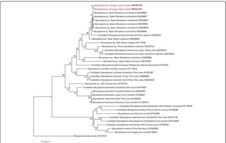
Here, three haemotropic Mycoplasma genotypes have
been detected in a tick specimen (I. simplex), collected in Hungary (Table2).Ixodes simplexis specialized to its host, Miniopterus schreibersii[25], from which bat species hae- moplasma genotypes having 99.8–99.9% 16SrRNA gene similarity to those from I. simplex collected in Hungary (Table 2) have been reported in Spain [23]. Importantly, these bat-associated haemoplasmas are phylogenetically Table 2Results of molecular analyses and sequence comparisons. Species names of rickettsiae are based on highest sequence similarities togltAsequences available on GenBank and published in peer-reviewed papers
Genotype/species Country (no. of positive samples)
Highest sequence similarity
in GenBank shown as gene: bp/bp (%)
Closest match sequence accession number
Accession number (this study)
Reference
Rickettsia helvetica China (1) – – – –
Rickettsiasp. Av22 Hungary (4) gltA: 757/757 (100) JN038177 MH383138 Socolovschi
et al. [5]
17 kDa: 394/394 (100) several MH383143 –
OmpA: 477/477 (100) several MH383147 –
Rickettsia africae-like Hungary (1) gltA: 757/757 (100) CP001612 MH383139 Sekeyová
et al. [12]
17 kDa: 394/394 (100) several MH383144 –
OmpA: 475/477 (99.6) CP001612 MH383148 Sekeyová
et al. [12]
Rickettsia hoogstraalii South Africa (1) gltA: 757/757 (100) FJ767737 MH383140 Duh et al.
[17]
17 kDa: 390/390 (100)a FJ767736 MH383145 Duh et al.
[17]
Rickettsia lusitaniae Mexico (1) gltA: 757/757 (100)b JQ771933 MH383141 Milhano
et al. [18]
China (2) gltA: 756/757 (99.9) JQ771933 MH383142 Milhano
et al. [18]
17 kDa: 393/394 (99.7) JQ771934 MH383146 Milhano
et al. [18]
OmpA: 461/464 (99.4) JQ771935 MH383149 Milhano
et al. [18]
Anaplasma phagocytophilum
Hungary (1) – – – –
Romania (1) – – – –
Bartonellasp. Ia23 Hungary (1) gltA: 313/317 (98.7) KX300154 MH544201 Urushadze
et al. [20]
ITS: 520/529 (98.3)c MF288126 MH544202 McKee
et al. [21]
Bartonellasp. Iv76 Romania (1) gltA: 317/317 (100) KR822802 MH578453 Lilley
et al. [22]
ITS: 291/306 (95.1) MF288124 MH544203 McKee
et al. [21]
Mycoplasmasp. Is128-1 Hungary (1) 16SrRNA: 953/954 (99.9) KM538692 MH383150 Millán
et al. [23]
Mycoplasmasp. Is128-2 Hungary (1) 16SrRNA: 824/826 (99.8) KM538698 MH383151 Millán
et al. [23]
Mycoplasmasp. Is128-3 Hungary (1) 16SrRNA: 952/954 (99.8) KM538692 MH383152 Millán
et al. [23]
Rickettsia helveticaandAnaplasma phagocytophilumwere detected by using species-specific primers (Additional file1: Table S1) and sequencing was not possible due to high Ct values
aAmplification ofOmpAgene was not successful
bAmplifications of17 kDaandOmpAgenes were not successful
cAmplification of theftsZgene was not successful
close to“CandidatusMycoplasma haemohominis”, as re- ported [23] and as also shown here (Fig.2).
Conclusions
Rickettsiae (from both the spotted fever and the R. felis groups) appear to be associated with soft rather than hard ticks of bats, as opposed to bartonellae. Although with low prevalence, two tick-borne zoonotic pathogens
(R. helvetica and A. phagocytophilum) have been detected for the first time in bat ticks. The present find- ings add Asia (China) to the geographical range of R.
lusitaniae, as well as indicate the occurrence ofR. hoog- straalii in South Africa. This is also the first molecular evidence of a R. africae-like genotype in Europe, in association with a bat host species that is not known to migrate to Africa. Bat haemoplasmas, which are
Fig. 1Maximum-likelihood tree of spotted fever group (SFG: encircled with dashed line),Rickettsia felisgroup (RFG: encircled with dashed line) and other rickettsiae based on thegltAgene. Sequences from this study are highlighted with red color and bold accession numbers. Branch lengths represent the number of substitutions per site inferred according to the scale shown
phylogenetically close to“Ca.M. haemohominis”, are re- ported here for the first time from central Europe and from any bat tick.
Additional files
Additional file 1:Table S1.Technical data for real-time PCRs used for screening. (DOCX 18 kb)
Additional file 2:Table S2.Technical data for conventional PCRs used for sequencing. (DOCX 21 kb)
Additional file 3:Text S1.Methods. (DOCX 20 kb)
Abbreviations
Ct:Threshold cycle; ftsZ: Cell division protein; gltA: Citrate synthase; ITS: 16S- 23S rRNA intergenic spacer region; OmpA: Outer membrane protein-A
Acknowledgements
Part of the molecular work was performed using the logistics of the Center for Clinical Studies, Vetsuisse Faculty, Zurich, Switzerland. The authors thank the Wildlife Recovery Center Valpredina (Italy) for their collaboration.
Funding
Molecular work in Hungary was supported by NKFIH 115854. This research was also supported by the 12190-4/2017/FEKUTSTRAT grant of the Hungar- ian Ministry of Human Capacities. ADS was supported by the János Bolyai Re- search Scholarship of the Hungarian Academy of Science.
Availability of data and materials
The sequences obtained and/or analyzed during the present study are deposited in the GenBank database under the accession numbers MH383138-MH383152, MH544201-MH544204 and MH578453. All other rele- vant data are included in the article.
Authors’contributions
SH designed the Hungarian part of the study, participated in DNA extraction, supervised molecular phylogenetic analyses and wrote the manuscript. ADS, TG, PE, YW, VTT, DK, SAB, AC, AH and SE provided important samples and contributed to the study design and the manuscript. KS extracted most of the DNA. MLM, KMS, MG, NT and JK performed molecular and phylogenetic analyses. RHL designed the Swiss part of the study and significantly contributed to the manuscript. All authors read and approved the final manuscript.
Ethics approval
Permissions for bat capture were provided by the National Inspectorate for Environment and Nature in Hungary (no. 14/2138-7/2011), the Vietnam Administration of Forestry of the Vietnamese Ministry of Agriculture and Rural Development (no. 1206/TCLN-BTTN), the School of Medicine at Shihezi University in China (no. AECSU2015-01), the Underground Heritage Commission in Romania (no. 305/2015), the Kenya Wildlife Service (no. KWS/BRM/5001) and the Secretary of the Environment and Natural Resources in Mexico (no.
SEMARNAT-08-049). Permission for bat capture was not needed in Italy, where six bat ticks were collected from bats rescued and hospitalized at the Wildlife Recovery Center. Permissions for bat hospitalization at the Wildlife Recovery Center in Italy were authorized with D.G.R. n. 5485 of 13.07.2001. The bat band- ing license numbers are TMF-14/32/2010 (DK), 59/2003 (PE), TMF-493/3/2005 (TG), TMF-513/1/2004 (SAB) and 305/2015 (ADS). Bats were released after re- moval of ticks.
Fig. 2Maximum-likelihood tree of haemotropicMycoplasmaspp. based on the16SrRNA gene. Sequences from this study are highlighted with red color and bold accession numbers. After the country name, the isolation source is indicated with genus and species name. Branch lengths represent the number of substitutions per site inferred according to the scale shown
Consent for publication Not applicable.
Competing interests
The authors declare that they have no competing interests.
Publisher’s Note
Springer Nature remains neutral with regard to jurisdictional claims in published maps and institutional affiliations.
Author details
1Department of Parasitology and Zoology, University of Veterinary Medicine, Budapest, Hungary.2Clinical Laboratory, Department of Clinical Diagnostics and Services, and Center for Clinical Studies, Vetsuisse Faculty, University of Zurich, Zurich, Switzerland.3Department of Parasitology and Parasitic Diseases, University of Agricultural Sciences and Veterinary Medicine, Cluj-Napoca, Romania.4Department of Zoology, Hungarian Natural History Museum, Budapest, Hungary.5Department of Zoology, Eszterházy Károly University, Eger, Hungary.6Department of Pathogenic Biology, School of Medicine, Shihezi University, Shihezi, China.7Institute of Ecology and Biological Resources, Vietnam Academy of Science and Technology, Hanoi, Vietnam.8Hungarian Biodiversity Society, Budapest, Hungary.9Directorate, Aggtelek National Park, Jósvafő, Hungary.10Institute for Veterinary Medical Research, Centre for Agricultural Research, Hungarian Academy of Sciences, Budapest, Hungary.11Plant Protection Institute, Centre for Agricultural Research, Hungarian Academy of Sciences, Budapest, Hungary.12Department of Biodiversity, School of Molecular and Life Sciences, Faculty of Science and Agriculture, University of Limpopo, Sovenga, South Africa.13Department of Biosciences and Pediatric Clinical Research Center“Romeo and Enrica Invernizzi”, University of Milan, Milan, Italy.
Received: 27 July 2018 Accepted: 7 January 2019
References
1. Hutterer R, Ivanova T, Meyer-Cords C, Rodrigues L. Bat migrations in Europe.
A review of banding data and literature. Naturschutz und Biologische Viefalt 28. Bonn: Federal Agency for Nature Conservation; 2005. p. 162.
2. Brook CE, Dobson AP. Bats as‘special’reservoirs for emerging zoonotic pathogens. Trends Microbiol. 2015;23:172–80.
3. Piksa K, Nowak-Chmura M, Siuda K. First case of human infestation by the tickIxodes vespertilionis(Acari: Ixodidae). Int J Acarol. 2013;38:1–2.
4. Jaenson TG, Tälleklint L, Lundqvist L, Olsen B, Chirico J, Mejlon H.
Geographical distribution, host associations, and vector roles of ticks (Acari:
Ixodidae, Argasidae) in Sweden. J Med Entomol. 1994;31:240–56.
5. Socolovschi C, Kernif T, Raoult D, Parola P.Borrelia,Rickettsia, andEhrlichia species in bat ticks, France, 2010. Emerg Infect Dis. 2012;18:1966–75.
6. Lv J, Fernández de Marco MDM, Goharriz H, Phipps LP, McElhinney LM, Hernández-Triana LM, et al. Detection of tick-borne bacteria and babesia with zoonotic potential inArgas(Carios)vespertilionis(Latreille, 1802) ticks from British bats. Sci Rep. 2018;8:1865.
7. Piksa K, Stańczak J, Biernat B, Górz A, Nowak-Chmura M, Siuda K. Detection ofBorrelia burgdorferi sensu latoand spotted fever group rickettsiae in hard ticks (Acari, Ixodidae) parasitizing bats in Poland. Parasitol Res. 2016;115:
1727–31.
8. Hornok S, Szőke K, Kováts D, Estók P, Görföl T, Boldogh SA, et al. DNA of piroplasms of ruminants and dogs in ixodid bat ticks. PLoS One. 2016;11:
e0167735.
9. Hornok S, Szőke K, Görföl T, Földvári G, Tu VT, Takács N, et al. Molecular investigations of the bat tickArgas vespertilionis(Ixodida: Argasidae) and Babesia vesperuginis(Apicomplexa: Piroplasmida) reflect“bat connection” between central Europe and central Asia. Exp Appl Acarol. 2017;72:69–77.
10. Hornok S, Kovács R, Meli ML, Kontschán J, Gönczi E, Gyuranecz M, et al. First detection of bartonellae in a broad range of bat ectoparasites. Vet Microbiol. 2012;159:541–3.
11. Hornok S, Szőke K, Estók P, Krawczyk A, Haarsma AJ, Kováts D, et al.
Assessing bat droppings and predatory bird pellets for vector-borne bacteria: molecular evidence of bat-associatedNeorickettsiasp. in Europe.
Antonie Van Leeuwenhoek. 2018;111:1707–17.
12. Sekeyová Z, Mediannikov O, Roux V, Subramanian G, Spitalská E, Kristofík J, et al. Identification ofRickettsia africaeandWolbachiasp. inCeratophyllus
gareifleas from passerine birds migrated from Africa. Vector Borne Zoonotic Dis. 2012;12:539–43.
13. Roux V, Rydkina E, Eremeeva M, Raoult D. Citrate synthase gene
comparison, a new tool for phylogenetic analysis, and its application for the rickettsiae. Int J Syst Bacteriol. 1997;47:252–61.
14. Acosta ICL, Luz HR, Faccini-Martínez ÁA, Muñoz-Leal S, Cerutti JC, Labruna MB. First molecular detection ofRickettsiasp. strain Atlantic rainforest in Amblyomma ovaleticks from Espírito Santo State, Brazil. Rev Bras Parasitol Vet. 2018;27:420–2.
15. Paddock CD, Allerdice MEJ, Karpathy SE, Nicholson WL, Levin ML, Smith TC, et al. Unique strain ofRickettsia parkeriassociated with the hard tick Dermacentor parumapertusNeumann in the western United States. Appl Environ Microbiol. 2017;83:e03463–16.
16. Portillo A, de Sousa R, Santibáñez S, Duarte A, Edouard S, Fonseca IP, et al.
Guidelines for the detection ofRickettsiaspp. Vector Borne Zoonotic Dis.
2017;17:23–32.
17. Duh D, Punda-Polic V, Avsic-Zupanc T, Bouyer D, Walker DH, Popov VL, et al.
Rickettsia hoogstraaliisp. nov., isolated from hard- and soft-bodied ticks. Int J Syst Evol Microbiol. 2010;60:977–84.
18. Milhano N, Palma M, Marcili A, Núncio MS, de Carvalho IL, de Sousa R.
Rickettsia lusitaniaesp. nov. isolated from the soft tickOrnithodoros erraticus (Acarina: Argasidae). Comp Immunol Microbiol Infect Dis. 2014;37:189–93.
19. Sánchez-Montes S, Guzmán-Cornejo C, Martínez-Nájera Y, Becker I, Venzal JM, Labruna MB.Rickettsia lusitaniaeassociated withOrnithodoros yumatensis(Acari: Argasidae) from two caves in Yucatan, Mexico. Ticks Tick Borne Dis. 2016;7:1097–101.
20. Urushadze L, Bai Y, Osikowicz L, McKee C, Sidamonidze K, Putkaradze D, et al. Prevalence, diversity, and host associations ofBartonellastrains in bats from Georgia (Caucasus). PLoS Negl Trop Dis. 2017;11:e0005428.
21. McKee CD, Kosoy MY, Bai Y, Osikowicz LM, Franka R, Gilbert AT, et al.
Diversity and phylogenetic relationships amongBartonellastrains from Thai bats. PLoS One. 2017;12:e0181696.
22. Lilley TM, Veikkolainen V, Pulliainen AT. Molecular detection ofCandidatus Bartonella hemsundetiensis in bats. Vector Borne Zoonotic Dis. 2015;15:706–8.
23. Millán J, López-Roig M, Delicado V, Serra-Cobo J, Esperón F. Widespread infection with hemotropic mycoplasmas in bats in Spain, including a hemoplasma closely related to“CandidatusMycoplasma hemohominis”. Comp Immunol Microbiol Infect Dis. 2015;39:9–12.
24. Neimark H, Johansson KE, Rikihisa Y, Tully JG. Proposal to transfer some members of the generaHaemobartonellaandEperythrozoonto the genus Mycoplasmawith descriptions of“CandidatusMycoplasma haemofelis”,
“CandidatusMycoplasma haemomuris”,“CandidatusMycoplasma haemosuis”and“CandidatusMycoplasma wenyonii”. Int J Syst Evol Microbiol. 2001;51:891–9.
25. Hornok S.Ixodes simplexNeumann, 1906. In: Estrada-Peña A, Mihalca AD, Petney TN, editors. Ticks of Europe and North Africa: A Guide to Species Identification. Cham: Springer International Publishing. 2017. p. 103–7.