1. Introduction
The application of biopharmaceuticals for oral ad- ministration is a topic of great interest in the phar- maceutical industry due to the inherent advantages of oral delivery [1]. Biopharmaceutical formulations are more stable in the solid state than in liquid form;
in addition, it has many advantages like storage at ambient temperature, longer shelf-life, easier prod- uct shipping and the possibility of controlled drug delivery [2]. Currently, the pharmaceutical industry is typically applying freeze drying and spray drying
processes in order to obtain solid biopharmaceuticals, however, both technologies have disadvantages [3, 4].
Freeze drying is a time-consuming batch technology, has high energy consumption, and the freezing can cause degradation of the biomolecules. On the con- trary, spray drying can be operated continuously, is more economical, but can induce inactivation of heat-sensitive molecules, due to the high drying tem- perature. Electrospinning (ES), which is originally a fiber drawing technology, can be a promising alter- native for continuous drying technologies, providing
Electrospinning scale-up and formulation development of PVA nanofibers aiming oral delivery of biopharmaceuticals
E. Hirsch1, P. Vass1, B. Démuth1, Zs. Pethő1, E. Bitay2, S. K. Andersen3, T. Vigh3, G. Verreck3, K. Molnár4,5, Zs. K. Nagy1, Gy. Marosi1*
1Department of Organic Chemistry and Technology, Faculty of Chemical Technology and Biotechnology, Budapest University of Technology and Economics, H-1111, Budapest, Műegyetem rakpart 3, Hungary
2Faculty of Technical and Human Sciences, Sapientia Hungarian University of Transylvania, 540485 Târgu-Mureş, Op. 9., Cp. 4., Romania
3Oral Solids Development, Janssen R&D, B-2340 Beerse, Turnhoutseweg 30, Belgium
4Department of Polymer Engineering, Faculty of Mechanical Engineering, Budapest University of Technology and Economics, H-1111, Budapest, Műegyetem rakpart 3, Hungary
5MTA–BME Research Group for Composite Science and Technology, Műegyetem rakpart 3, H-1111 Budapest, Hungary
Received 5 December 2018; accepted in revised form 4 February 2019
Abstract.Electrospinning is a promising drying technology providing a rapid and gentle drying at ambient temperature, thus electrospinning of polyvinyl alcohol aqueous solutions was investigated for the solid formulation of biopharmaceuticals.
The commonly used single-needle electrospinning does not have adequate productivity to satisfy the industrial requirements, therefore our aim was to study the scale-up of the technology by using high-speed electrospinning. High molecular weight polyethylene oxide as a secondary polymer was applied to enhance the fiber formation of polyvinyl alcohol. While polyvinyl alcohol-polyethylene oxide formulations resulted in adequate fiber formation it was not possible to process them further as the friability of the fibers was too low. In order to increase the friability, the effect of adding various sugars (mannitol, glucose, lactose, saccharose, and trehalose) was investigated. The results showed that mannitol was the best friability en- hancing excipient because of its crystallinity and low moisture content in the fibrous sample. In contrast, glucose, lactose, saccharose, and trehalose were amorphous with higher moisture content and fibers containing these were grindable only after post-drying.
Keywords:nanomaterials, electrospinning, scale-up, biopharmaceuticals, oral formulation https://doi.org/10.3144/expresspolymlett.2019.50
*Corresponding author, e-mail:gmarosi@mail.bme.hu
© BME-PT
a rapid and gentle drying at ambient temperature [5–
7]. Thus, ES technology can be applied for the solid formulation of various biopharmaceuticals such as enzymes, peptides, proteins, monoclonal antibodies, oligonucleotides and live cells [8–11]. As both freeze and spray drying can induce degradation of biophar- maceuticals, they are typically formulated with var- ious excipients to preserve the activity, amongst which the most commonly used excipients are sug- ars (like mannitol, saccharose, and trehalose being the typical choices) [5, 12, 13]. The exact protection mechanism is not known, but there are two hypothe- ses (namely the vitrification and water replacement hypotheses), that explain the protective effect of sug- ars on the active pharmaceutical ingredient (API) stability [14, 15].
Considering the sensitivity of biopharmaceuticals the use of aqueous solutions during electrospinning is strongly preferred. This preference makes the de- velopment more challenging, from the productivity point of view. Owing to the high boiling point of aqueous solutions compared to the volatile organic solvents, the productivity of the method is reduced.
Using the conventional laboratory-scale ES equip- ment, single-needle electrospinning (SNES) with aqueous solutions, only 0.5–1.0 ml/h feeding rate was achieved up to now [16–18].
SNES equipment does not have adequate productiv- ity, thus in order to satisfy the industrial requirements, the scale-up of ES is necessary. Different methods have been created to scale up the process and there are equipments commercially available on the mar- ket [19]. The most commonly used technique is multi- needle ES, which has a well-known drawback that is related to the alteration of the electric field profile induced by the presence of other electrospinning jets nearby [20, 21]. The other technique used for the scale-up of the electrospinning process is the needle- less ‘free-surface’ ES [22]. From the free-surface of the polymer solution, several jets can be created, how- ever evaporation of the solvent results in the change of solution composition and viscosity. High-speed electrospinning (HSES) technique seems to be a prom- ising way to meet the pharmaceutical requirements of the scale-up [23]. The fiber formation process, ac- celerated by electrostatic and centrifugal forces, al- lows mass production of nanofibers, while the con- centration of the constituents is maintained at a constant level.
Another great challenge besides increasing the pro- ductivity is to achieve appropriate downstream processability to enable tableting [24, 25]. In the case of water-based ES, the residual water content of the dried samples has great importance, because it has a high impact on the friability due to the water plasti- cizing effect [26]. In this respect, the grindability of the fibers and the properties (e.g. flowability) of the ground fibers are also critical. Furthermore, the water content could affect the API chemical and microbio- logical stability during long-term storage [27, 28].
The present research aimed to investigate the advan- tages of HSES method during the scale-up of water- based ES for the solid formulation of biopharmaceu- ticals. Polyvinyl alcohol (PVA) was chosen as a basic polymer matrix for the aqueous electrospin- ning experiments as it shows good compatibility with biomolecules [29–31]. The development of a formulation consisting of polymers and excipients was carried out to achieve adequate fiber formation with high productivity and good downstream process- ability.
2. Experimental 2.1. Materials
Polyvinyl alcohol (PVA, Mw: 130 000 g/mol) pur- chased from Sigma-Aldrich (Merck, Darmstadt, Germany) and polyethylene oxide (PEO, Mw: 2 000 000 g/mol) supplied by Colorcon (Dartford, England) were used as polymer matrices. Mannitol (Mannogem EZ, SPI Pharma, Wilmington, USA), glucose (glucose-monohydrate, Hungrana, Szabad- egyháza, Hungary), lactose (lactose-monohydrate, Meggle Pharma, Wasserburg, Germany), saccharose (Reanal, Budapest, Hungary) and trehalose (trehalose dihydrate, Sigma-Aldrich, St. Louis, USA) were used as additives to enhance the grindability. Dis- tilled water was used to prepare the polymer solu- tions for electrospinning.
2.2. Viscosity measurement
The viscosity of the solutions was determined using an AR 2000 rotational rheometer (TA Instruments, New Castle, USA) in parallel plate configuration.
The upper moving plate of 40 mm diameter and the lower Peltier plate, which set the temperature of the solutions to 25 °C, were made of stainless steel. The viscosities were measured at linearly increasing shear rate from 20 to 60 1/s. There were no practically
relevant changes in the measured viscosities as a function of shear rate.
2.3. Electrospinning technologies
2.3.1. Single-droplet electrospinning (SDES) SDES technology was used for the screening of polymer concentration required for obtaining fibers.
One droplet of the polymer solution was placed on top of a metal rod (d= 5 mm) and high voltage (20 kV DC) was applied provided by an NT-35 direct cur- rent power supplier (MA2000, Unitronik Ltd, Nagy - kanizsa, Hungary). The fibers were collected on a grounded aluminum foil immobilized to the collector plate. The metal tip and the collector were vertically at a distance of 15 cm (see Figure 1a).
2.3.2. Single-needle electrospinning (SNES) Lab-scale electrospinning device, SNES was used with a 0.5 mm inner diameter nozzle which was con- nected with high voltage supply and the polymer so- lution was dosed by a SEP-10S Plus type syringe pump (Aitecs, Vilnius, Lithuania) (see Figure 1b). The electrical potential applied on the nozzle, the collec- tor distance, and the feeding was 20–30 kV, 15–30 cm, and 0.5–1 ml/h respectively, furthermore it was ad- justed for each solution to obtain adequate fiber for- mation. All of the experiments were carried out at room temperature.
2.3.3. High-speed electrospinning (HSES) The scaled-up electrospinning experiments were per- formed using a high-speed electrostatic spinning setup consisting of a round-shaped, stainless steel atomizer connected to a high-speed motor [23]. The high ro- tation speed of the atomizer and the multiple holes in it (number of holes: 8, diameter of the holes: 330 µm, diameter of the atomizer 34 mm) allowed higher pro- ductivity. The polymer solution was fed with a syringe pump (SEP-10S Plus, Aitecs, Vilnius, Lithuania) at
20–80 ml/h. The rotational speed of the atomizer (2000–9000 rpm), and the voltage applied (0–40 kV) were tested during the experiments. A drying air flow (2 bar) and the electrostatic forces directed the fibers to the grounded metal collector covered with aluminum foil, which was at a fixed distance (35 cm) from the spinneret in a vertical arrangement (see Fig- ure 1c ). The experiments were performed at room temperature.
2.4. Post drying
Post drying was carried out in a vacuum oven (Va- ciotem-T, JP Selecta, Barcelona) at 25 °C and 0.05 bar.
The fibers were dried for 48 hours.
2.5. Grinding
The nanofibers were ground using different technolo- gies in order to evaluate the friability of the fibers.
A sieve with 0.8 mm grid, a cutting mill (IKA A11., IKA-WERKE GmbH & Co. KG, Staufen, Germany) at 28 000 rpm, and a hammer mill (IKA MF10, IKA- WERKE GmbH & Co. KG, Staufen, Germany) with 1 mm grid and 3000 rpm were used in the experi- ments.
2.6. Scanning electron microscopy (SEM) Morphology of the samples was examined by a JEOL 6380LVa (JEOL, Tokyo, Japan) type scanning elec- tron microscope in high vacuum after electrospinning and grinding. Samples were fixed by conductive dou- ble-sided carbon adhesive tape and coated with gold using ion sputter (JEOL 1200, JEOL, Tokyo, Japan).
The applied accelerating voltage was 15 kV and the working distance was between 10–30 mm.
2.7. Water content measurement
The water content of the samples was measured right after the electrospinning process and after the post- drying using a moisture balance (Sartorius MA40,
Figure 1.Schematic representation of SDES equipment (a) used for polymer screening, SNES equipment (b) used for the lab-scale experiment, and a scaled-up electrospinning set-up, HSES equipment (c).
Sartorius, Göttingen, Germany). The water content was determined based on the weight loss of approx- imately 0.1 g sample after 10 min at 105 °C.
2.8. Particle size distribution measurement The particle size distribution was measured with a Mastersizer 2000 (Malvern Instruments Ltd., Malvern, UK) laser diffraction-based particle size analyzer.
2.9. Raman spectroscopy
A Kaiser RamanRxn2® Hybrid in-situ analyzer (Kaiser Optical Systems, Ann Arbor, USA) connected with PhAT (Pharmaceutical Area Testing) probe was applied to acquire the spectra of the fibrous samples for the characterization of the crystallinity of the com- pounds. The samples were illuminated with an Invic- tus laser diode (400 mW, 785 nm). Reflection mode was used for measuring reference samples and fibers.
The diameter of the laser spot size was extended to 6 mm and the nominal focus length was 250 mm. The examined spectral range was between 200 and 1890 cm–1. During the measurements, the resolution was 4 cm–1, which provided 1691 variables during data processing. An acquisition time of 60 seconds gave adequate Raman intensity and signal-to-noise ratio. The wavenumbers between 1532–300 cm–1was included for analysis, because there were no charac- teristic peaks in this region. The spectra were baseline corrected (Automatic Whittaker Filter) and normal- ized (area = 1) using MATLAB software.
2.10. X-ray diffraction (XRD)
X-ray diffraction patterns were recorded with a PAN- alytical X’pert Pro MDP X-ray diffractometer (Alme- lo, The Netherlands) using Cu-Ka radiation (1.506 Å) and Ni filter. The applied voltage was 40 kV, while the current was 30 mA. The reference samples and the fibrous samples were analyzed between 2θ an- gles of 4 and 42°.
3. Results and discussion
The morphology of the fibers produced by electro- spinning (ES) can be affected by solution parame- ters such as composition and viscosity (i.e.through component concentrations and component type), and process parameters such as applied voltage, feeding rate, and the collector distance. The influ- ence of the polymer concentration on morphology and fiber diameter was studied using single-droplet
electrospinning (SDES, Figure 1a). The electrospin- ning process using aqueous polyvinyl alcohol (PVA) solutions was transferred to the single-needle elec- trospinning (SNES, Figure 1b) device and the feasi- bility of scale-up was evaluated using high-speed electrospinning (HSES, Figure 1c). The SDES, SNES, and HSES manufacturing techniques were compared with respect to fiber morphology and productivity.
The solution composition was modified with differ- ent excipients to obtain grindable fibers.
The aim of the study was to produce fibers with the smallest possible fiber diameter, high productivity, and good grindability. Finally, the formulations were characterized based on water content, crystallinity, and physical stability.
3.1. Screening of fiber formation of aqueous PVA solution
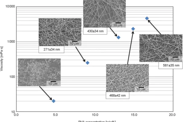
For the fast evaluation of fiber formation, SDES tech- nology was used, requiring only a small amount (ap- proximately 200 µl) of polymer solution. With in- creasing polymer concentration the viscosity of the solutions increased as well, which has a large effect on product morphology [32]. Several polymer solu- tions with different PVA concentration (4.8, 9.1, 13.0, 14.9, 16.7%, throughout the article all concen- trations are given as w/w%) were prepared and elec- trospun. Electrospinning of polymer solutions with too low concentration results in the formation of par- ticles. By increasing the concentration, shape of the particles changes to fibers. At an optimal concentra- tion range, stable and reproducible fiber production can be attained. Fiber diameter can be increased using higher viscosity solutions; however, using too high polymer concentrations, the highly viscous so- lutions can cause problems during feeding as they cannot be pumped.
The results of SDES shown in Figure 2 were evalu- ated based on fiber formation and fiber diameter of the formulations. Fibers were not formed at 4.8%
due to the viscosity being too low, while as soon as the viscosity was increased to ca. 250 mPa·s fiber for- mation occurred. For all PVA concentrations higher than or equal to 9.1% fibers were formed with aver- age diameters in the submicron range. As expected fiber diameter increased with increasing concentra- tion due to the higher viscosity. The solution con- taining 16.7% PVA was too viscous for further ex- periments using a syringe pump.
3.2. Comparison of electrospinning methods SDES, SNES, and HSES were compared with re- spect to applicability for water-based electrospin- ning. For the comparison, solutions containing 9.1 13.0, 14.9% PVA were prepared as these concentra- tions were known to form fibers and were feasible for production. The methods were evaluated based on fiber morphology, and productivity and the results are shown in Table 1.
The fiber diameters and morphology obtained using SDES were similar to SNES and HSES, which also shows the feasibility of SDES technology for screen- ing. Using HSES, the fiber diameters were slightly smaller for 9.1 and 13.0% PVA concentrations and bigger for the highest concentration; however, there was an increase in the standard deviation. The appar- ent smaller fiber diameters and wider fiber distribu- tions are likely caused by the rotating spinneret i.e.
both electrostatic and centrifugal forces for the at- omization (Table 1).
Using SNES the maximal feeding rate for the water- based systems proved to be 0.5 ml/h, which is very low even in laboratory scale (maximum 75 mg/h).
In comparison, using HSES 40 ml/h (maximum 6 g/h) feeding rate could be achieved, however, the experiment resulted not only in fiber production but droplets were also detected on the collector (Fig- ure 3) indicating that either process conditions or so- lution composition should be optimized to further improve fiber formation, and this will be discussed in the next section.
3.3. Optimization of solution formulation When HSES was used for the formulation of PVA aqueous solution, besides fiber formation droplets were also detected on the collector, owing to the high Figure 2.The viscosity of the PVA aqueous solutions and the obtained fiber morphology using SDES. Scanning electron microscope (SEM) images of the fibers were taken and the average fiber diameter of each formulation was calcu- lated from 10 measurements.
Table 1.Average fiber diameter and standard deviation (SD) (calculated from 10 measurements) of the formulations obtained from 9.1, 13.0 and 14.9% PVA aqueous solution using SDES, SNES, and HSES electrospinning methods.
ES method
9.1% PVA 13.0% PVA 14.9% PVA
Productivity [g/h]
Fiber diameter [nm]
SD [nm]
Fiber diameter
[nm] SD Fiber diameter
[nm]
SD [nm]
SDES 271 34 430 34 468 042 –
SNES 279 41 427 43 445 080 0.045–0.075
HSES 232 41 398 61 477 109 3.6–6.0
breakage ratio of the jet to droplets at the nozzle. In the PVA aqueous solution the entanglement of the polymer chains is low due to the hydrogen bonds be- tween the polymer chains and the water molecules [33]; however, if the entanglement is sufficiently high, then fiber formation takes place [34, 35]. Using higher PVA concentration (13.0 and 14.9%) with higher viscosity was not suitable to increase the en- tanglement and resulted in droplets and fibers on the collector.
3.3.1. Role of PEO
Another strategy for reducing the droplet formation was to add high molecular weight polyethylene oxide (PEO) in low concentration to the PVA solution. PEO is a really good fiber forming polymer, thus it can help the fiber formation of various blends [36, 37].
PEO can interact with the PVA molecules by compet- ing with water molecules and combined with the larg- er molecule size results in an increase of the entan- glement.
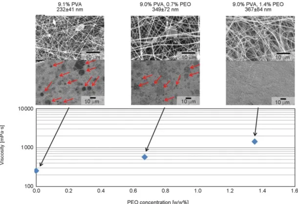
Different concentrations of PEO (0.5, 0.7, 0.9, 1.4%) were added to the PVA solution. By adding 1.4%
PEO, the fiber formation was adequate and there were no droplets on the collector while adding 0.9% or less PEO, several droplets could be found on the col- lector. The droplet formation is shown in Figure 3 as
a function of PEO concentration. The PEO contain- ing formulations had larger fiber diameters, which slightly increased with the PEO concentration but only within the range of standard deviation.
The viscosity of the solution containing 9.0% PVA and 1.4% PEO was 1420 mPa·s which is comparable to the 13.0% PVA solution’s viscosity (1320 mPa·s), however, in the presence of 1.4% PEO, the droplet formation could be avoided. The entanglement of the polymer chains in the PVA solution was increased by the addition of PEO, which resulted in adequate fiber formation (no droplets), but with an increase in fiber diameter of ~50% (to 367 from 232 nm). While the fiber quality and process productivity met the de- sired targets (wrt. morphology and production rate), the fibrous mat was sticky and the grinding was not possible, which is an important attribute in down- stream processing. Consequently, while the use of PEO as a secondary polymer is a feasible solution to prevent droplet formation during water-based ES, further optimization is needed in order to improve the grindability.
3.3.2. Grindability optimization
Processability of the fibers (e.g. milling, powder properties, etc.) is a critical aspect for the develop- ment of solid pharmaceutical products. The produced
Figure 3.SEM images of electrospun fibers from PVA and PEO containing solution using HSES. Droplet formation, vis- cosity, and fiber diameter as a function of PEO concentration. Droplets are indicated with arrows.
fibers are collected in a form of a fibrous mat, which cannot be directly used for the production of a tablet, hence the fiber mat needs to be grounded to a pow- der before further processing. Therefore, the opti- mization of the polymer matrix was carried out con- sidering the grindability of the fibers. The composi- tion of the electrospinning solution was modified in order to adjust both the grindability and spinnability using HSES, which was applied in the subsequent experiments.
From biopharmaceutical formulations of freeze and spray drying it is a well-known strategy to use sug- ars to protect the molecules during drying and for
processing [13], therefore it was decided to investi- gate the impact of the following 5 sugars: glucose, lactose, mannitol, saccharose, and trehalose. The electrospinnability and grindability were evaluated in parallel, with the function of PEO concentration.
The aim of the optimization was to obtain grindable fibers with the diameter in the submicron range using a robust electrospinning process without droplet for- mation. The minimal sugar concentration needed to obtain grindable fibers was determined by adding different amount of mannitol to the PVA-PEO solu- tion. The use of twice as much mannitol as polymer resulted in fibers with adequate grindability.
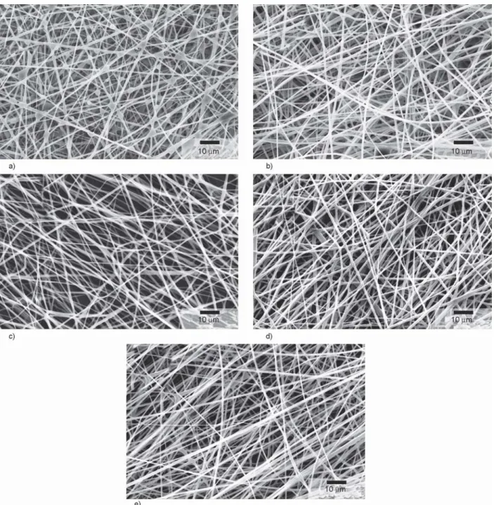
Figure 4.SEM images of PVA-PEO fibers containing a) glucose, b) lactose, c) mannitol, d) saccharose, and e) trehalose pro- duced with HSES equipment.
To prevent the droplet formation during ES, different concentrations (0.4, 0.6, 0.8, 1.1%) of PEO were added to the PVA solution containing mannitol as a grinding aid. High concentration of PEO can increase the fiber diameter and decrease the grindability of the fibers, thus the minimal concentration needed to be found. The composition containing 0.4% PEO re- sulted in droplets on the collector. Fiber formation was adequate and there was no droplet formation from the aqueous solution containing 7.7% PVA, 0.6% PEO and 15.3% mannitol. However, the fiber diameter increased (856±214 nm) with the addition of mannitol but it was necessary as it was needed to modify the formulation to achieve good grindability.
The use of 0.8 and 1.1% PEO increased the fiber di- ameter (to 750 and 1120 nm) further, which was not preferred.
Besides mannitol, glucose, lactose, saccharose, and trehalose were used to enhance the grindability of PVA-PEO fibers. The solutions prepared for electro- spinning contained 7.7% PVA, 0.6% PEO, and 15.3%
sugar based on the mannitol optimization. Using each grindability aid the fiber formation was ade- quate and there was no droplet formation, the fibers are shown in Figure 4. The average diameter of the fibers using mannitol, glucose, lactose, saccharose, and trehalose was 856, 908, 895, 912, and 969 nm respectively with similar standard deviation (Table 2).
The increased total concentration of the polymer so- lution (10.5 to 23.6%) can be the reason for the in- creased fiber diameter compared to the formulations without sugars. The viscosity of the solutions con- taining different sugar-type excipients was between 644 and 713 mPa·s (Table 2) which was lower com- pared to the solution containing only PVA and PEO (viscosity 1420 mPa·s). The sugar molecules make secondary interaction with the water molecules re- moving them from the PVA hydrogen bonds and cre- ating an emulsion-like structure. The disruption of
the hydration layer of the PVA resulted in higher en- tanglement of the polymer chains. The minimal PEO concentration needed to prevent droplet formation also decreased in the presence of sugars. The high entanglement of the polymer molecules made the fiber formation possible at lower viscosity.
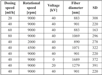
3.4. Screening of process parameters
Besides solution composition, process parameters have a high impact on fiber formation and morphol- ogy [7]. Thus, screening of the feeding rate, the ro- tational speed of the atomizer and the applied volt- age was carried out using HSES with a solution containing 7.7% PVA, 0.6% PEO and 15.3% man- nitol (Table 3). Rotational speed and feeding rate had no significant effect on the fiber diameter and fiber formation in the examined range; however, the in- crease of the feeding rate resulted in droplet forma- tion. When using 60 ml/h or higher feeding rates, droplet and fiber formation could be observed during the ES process. High voltage of 0, 20, and 40 kV was applied to the atomizer. With the decrease of voltage, there was an increase in fiber diameter, which is ex- pected as fiber formation and size is controlled by the electrostatic force [V] and centrifugal force [rpm].
The optimal parameters were: 9000 rpm, 40 kV, and the feeding rate could be raised up to 40 ml/h in HSES experiments, which is an 80 times higher pro- duction rate compared to SNES.
3.5. Characterization
3.5.1 Effect of water content on grindability The grindability of the fibers is an important aspect of the downstream proceassability to obtain a final
Table 2.Viscosity of 7.7% (1.00 g) PVA, 0.6% (0.08 g) PEO, and 15.3% (2.00 g) sugar-containing aqueous solutions (10 ml) and the average fiber diameter ob- tained using HSES.
Sugar excipient
Viscosity [mPa·s]
Fiber diameter [nm]
SD [nm]
Mannitol 683 856 214
Glucose 713 908 319
Saccharose 644 912 180
Lactose 704 895 294
Trehalose 675 969 318
Table 3.Screening of process parameters during high-speed electrospinning using a solution containing 7.7%
PVA, 0.6% PEO and 15.3% mannitol.
Dosing speed [ml/h]
Rotational speed [rpm]
Voltage [kV]
Fiber diameter
[nm]
SD
20 9000 40 883 308
40 9000 40 901 220
60 9000 40 883 163
80 9000 40 1069 296
40 2000 40 890 278
40 4500 40 1071 322
40 9000 40 901 220
40 9000 0 1689 372
40 9000 20 1279 391
40 9000 40 901 220
dosage form e.g. tablet. The grindability of the PVA- PEO-sugar formulations was tested using a sieve, a cutting mill, and a hammer mill. Comparing the dif- ferent grinding technologies, the hammer mill was chosen for further examination because it can be in- tegrated in a continuous technology line. The ground materials obtained using the hammer mill were char- acterized and SEM images were taken before and after milling to investigate milling impact on fiber characteristics.
The fibrous mats recovered from HSES were typi- cally 2 cm×5 cm. The milling converted the mats to
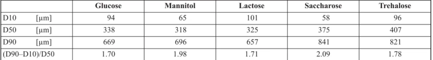
micron-sized particles as seen in Figure 5 composed of submicron fibers (Figure 5d). The morphology was not affected by the grinding. The fibers were easily milled and non-sticking to the mill. The particle size distribution of the milled powders is shown in Table 4, and it can be observed that the average par- ticle size after milling was similar for the formula- tions with glucose, mannitol, and lactose, but slightly higher for the formulations with saccharose and tre- halose. All five produced powder had low-density (e.g. 0.0385 g/cm3for mannitol) with poor flowabil- ity. From the particle size, good flowability would
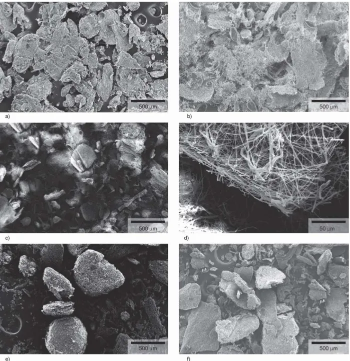
Figure 5.SEM images of ground PVA-PEO-Sugar fibers containing a) glucose, b) lactose, c)–d) mannitol, e) saccharose, and f) trehalose. Electrospinning was accomplished using HSES and the obtained nanofibrous mats were ground using a hammer mill.
be expected, however, due to the fibrous structure, the particles had poor flowability. While the five formulations (cf. Table 2) have similar properties fur- ther characterization of the produced powders is nec- essary in order to understand the impact of milling.
The formulation containing mannitol was grindable right after the electrospinning process; however, the samples containing glucose, lactose, saccharose, and trehalose were too sticky to be ground. It was assumed that this was caused by the high water content of the powder, with water acting as a plasticizer and this was confirmed by water content measurements with the results shown in Table 5. In order to see if a grind- able product could be obtained; all formulations were post dried to achieve lower water content. After post- drying, all sugar containing formulations were grind- able and thereby suitable for downstream process- ing (Table 5). The water content influenced the grindability of the samples significantly, due to its plasticizing effect, and by lowering the water con- tent the fibers were friable which enhanced the grindability.
3.5.2. Physical state of the fibers
Pharmaceutically used sugars have different water sorption capability depending on their physical state (crystalline or amorphous) [38]. Crystalline sugars interact with water based on surface adsorption;
meanwhile, amorphous materials absorb a large number of water molecules into the bulk structure.
The moisture content, hence also the downstream processability of the samples, is influenced by not only the drying conditions but also the physical state of the substances [39].
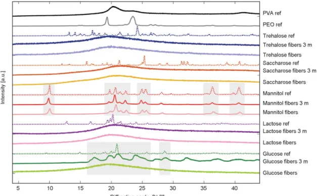
Thus, the physical state of the polymers and excipi- ents were examined using X-ray diffraction (XRD) and Raman spectrometry. The polymers (PVA and PEO) proved to be amorphous in each formulation and were stable over the examined 3 months (Fig- ure 6). Mannitol was crystalline and the δ polymorph was present in the fibers, which was proven by the characteristic peaks of the δ polymorph shown on the Raman and XRD spectra (Figure 6, 7).
On the contrary, the glucose, lactose, saccharose, and trehalose containing formulations showed peak widen- ing and shifting due to their amorphous structure.
Over the 3 months storage at low humidity condi- tions, the samples containing lactose, saccharose, and trehalose preserved the amorphous state, while glu- cose crystallized over the storage time (Figure 6, 7).
Amorphous solids are in not-equilibrium state, in some cases crystalline transition takes place. Further characterization will be needed to investigate the in- fluencing factors of the phenomenon.
Crystallinity, such as in the case of mannitol, can be advantageous to achieve enhanced grindability, due to the low water content and the friable nature of the sample. On the other hand, glucose, lactose, saccha- rose, and trehalose containing formulations were amorphous and had higher water content. The amor- phous formulations were also friable after post-dry- ing and the samples were grindable only at low water content. It can be concluded that the influencing fac- tors of the grindability of fibers were not only the fiber formation and drying conditions but also the water content and the physical state of the excipients.
While post drying was necessary for glucose, lactose, saccharose, and trehalose containing formulations, Table 4.Particle size distribution of ground PVA-PEO-Sugar fibers. D-values (D10, D50, D90) were used to describe particle
size distributions, which are the intercepts for the particle size of the 10, 50 and 90% of the cumulative mass.
Table 5.Water content (H2O%) and grindability of the fibrous mat containing different sugars.
Glucose Mannitol Lactose Saccharose Trehalose
D10 [µm] 94 65 101 58 96
D50 [µm] 338 318 325 375 407
D90 [µm] 669 696 657 841 821
(D90–D10)/D50 1.70 1.98 1.71 2.09 1.78
Glucose Mannitol Lactose Saccharose Trehalose
After HSES H2O [%] 13.3 5.5 11.2 9.4 14.0
Grindable? NO YES NO NO NO
After post-drying H2O [%] 7.1 3.7 4.7 5.7 7.8
Grindable? YES YES YES YES YES
it's possible that it can be avoided by further opti- mizing the ES conditions.
4. Conclusions
The capability of fiber formation from polyvinyl al- cohol (PVA) aqueous solutions was examined using
single-droplet electrospinning (SDES), single-needle electrospinning (SNES), and high-speed electrospin- ning (HSES) technologies. The scale-up and the op- timization of the PVA system were carried out to achieve adequate fiber formation with high produc- tivity and good downstream processability.
Figure 6.XRD graph of references and PVA-PEO-sugar fibers right after electrospinning and post-drying. XRD measure- ments were done after 3 months storage in low humidity conditions to study the stability of the components (curves indicated with 3 m).
Figure 7.Raman spectra of references and PVA-PEO-sugar fibers right after electrospinning and post-drying, and after 3 months (3 m) storage in low humidity conditions.
SDES was found to be a feasible method for the fast screening of polymer concentration needed for fiber formation and the fiber diameter obtained using SDES was similar to SNES and HSES. The feeding rate during the electrospinning process could be raised from 0.5 to 40 ml/h using HSES equipment, which is an 80 times higher production rate com- pared to SNES. When HSES was used for the formu- lation of PVA aqueous solutions, fiber formation was achieved, but drying was incomplete as droplets were observed on the collector. The composition of the so- lution was modified to improve fiber formation using high molecular weight polyethylene oxide (PEO) in low concentration as a secondary polymer. While PVA-PEO formulations resulted in adequate fiber formation and productivity, it was not possible to process them further as the friability of the fibers was too low i.e.too sticky. The grindability of the fibers was enhanced by adding glucose, mannitol, lactose, saccharose, or trehalose to the polymer solution.
The PVA-PEO-sugar formulations resulted in grind- able fibers in the submicron range though fiber di- ameter was increased compared to formulations with- out sugars. PVA and PEO polymers, as well as glu- cose, lactose, saccharose, and trehalose were amor- phous and these fibrous samples were grindable only after a post-drying step due to the high moisture con- tent and low friability of these samples. The water content influenced the grindability of the formulations significantly, due to its plasticizing effect, and by lowering the water content the fibers were friable which enhanced the grindability.
The use of mannitol was the most applicable as there was no need for post drying to achieve a grindable product after the electrospinning process. The low water content and the crystalline structure of manni- tol in the fibrous sample is possibly the reason for the better grindability. It can be concluded that the in- fluencing factors of the grindability of fibers were not only the fiber formation and drying conditions but also the formulation composition, final water content and the physical state of the excipients.
In this work, potential matrixes for the formulation of biopharmaceuticals were developed and charac- terized. The scale-up of the water-based electrospin- ning was accomplished using HSES technique, which shows a possible way for the solid formula- tion of highly sensitive biopharmaceuticals.
Acknowledgements
The research reported in this paper was supported by OTKA grants K-112644, PD-128241, KH-124541, and Ph.D. schol- arship from Gedeon Richter’s Talentum Foundation. Support of grant BME FIKP-BIO by EMMI is kindly acknowledged.
References
[1] Moroz E., Matoori S., Leroux J-C.: Oral delivery of macromolecular drugs: Where we are after almost 100 years of attempts. Advanced Drug Delivery Re- views, 101, 108–121 (2016).
https://doi.org/10.1016/j.addr.2016.01.010
[2] Maa Y-F., Prestrelski J. S.: Biopharmaceutical powders particle formation and formulation considerations. Cur- rent Pharmaceutical Biotechnology, 1, 283–302 (2000).
https://doi.org/10.2174/1389201003378898
[3] Vass P., Démuth B., Hirsch E., Nagy B., Andersen S. K., Vigh T., Verreck T., Csontos I., Nagy Zs. K., Marosi Gy.:
Drying technology strategies for colon-targeted oral de- livery of biopharmaceuticals. Journal of Controlled Re- lease, 296, 162–178 (2019).
https://doi.org/10.1016/j.jconrel.2019.01.023
[4] Langford A., Bhatnagar B., Walters R., Tchessalov S., Ohtake S.: Drying of biopharmaceuticals: Recent de- velopments, new technologies and future direction.
Japan Journal of Food Engineering, 19, 15–24 (2018).
https://doi.org/10.11301/jsfe.18514
[5] Broeckx G., Vandenheuvel D., Claes J. J. I., Lebeer S., Kiekens F.: Drying techniques of probiotic bacteria as an important step towards the development of novel pharmabiotics. International Journal of Pharmaceutics, 505, 303–318 (2016).
https://doi.org/10.1016/j.ijpharm.2016.04.002
[6] Huang S., Vignolles M-L., Chen X. D., Le Loir Y., Jan G., Schuck P., Jeantet R.: Spray drying of probiotics and other food-grade bacteria: A review. Trends in Food Science and Technology, 63, 1–17 (2017).
https://doi.org/10.1016/j.tifs.2017.02.007
[7] Drosou C. G., Krokida M. K., Biliaderis C. G.: Encap- sulation of bioactive compounds through electrospin- ning/electrospraying and spray drying: A comparative assessment of food-related applications. Drying Tech- nology, 35, 139–162 (2017).
https://doi.org/10.1080/07373937.2016.1162797
[8] Hu X., Liu S., Zhou G., Huang Y., Xie Z., Jing X.: Elec- trospinning of polymeric nanofibers for drug delivery applications. Journal of Controlled Release, 185, 12–
21 (2014).
https://doi.org/10.1016/j.jconrel.2014.04.018
[9] Jiang H., Wang L., Zhu K.: Coaxial electrospinning for encapsulation and controlled release of fragile water- soluble bioactive agents. Journal of Controlled Release, 193, 296–303 (2014).
https://doi.org/10.1016/j.jconrel.2014.04.025
[10] Salalha W., Kuhn J., Dror Y., Zussman E.: Encapsula- tion of bacteria and viruses in electrospun nanofibres.
Nanotechnology, 17, 4675–4681 (2006).
https://doi.org/10.1088/0957-4484/17/18/025
[11] Sanders E. H., Kloefkorn R., Bowlin G. L., Simpson D.
G., Wnek G. E.: Two-phase electrospinning from a sin- gle electrified jet: Microencapsulation of aqueous reser- voirs in poly(ethylene-co-vinyl acetate) fibers. Macro- molecules, 11, 3803–3805 (2003).
https://doi.org/10.1021/ma021771l
[12] Angkawinitwong U., Sharma G., Khaw P. T., Brocchini S., Williams G. R.: Solid-state protein formulations.
Therapeutic Delivery, 6, 59–82 (2015).
https://doi.org/10.4155/tde.14.98
[13] Emami F., Vatanara A., Park E. J., Na D. H.: Drying tech- nologies for the stability and bioavailability of biophar- maceuticals. Pharmaceutics, 10, 131/1–131/22 (2018).
https://doi.org/10.3390/pharmaceutics10030131
[14] Mensink M. A., Frijlink H. W., van der Voort Maarschalk K. V. D., Hinrichs W. L. J.: How sugars protect proteins in the solid state and during drying (review): Mecha- nisms of stabilization in relation to stress conditions.
European Journal of Pharmaceutics and Biopharmaceu- tics, 114, 288–295 (2017).
https://doi.org/10.1016/j.ejpb.2017.01.024
[15] Grasmeijer N., Stankovic M., de Waard H., Frijlink H.
W., Hinrichs W. L. J.: Unraveling protein stabilization mechanisms: Vitrification and water replacement in a glass transition temperature controlled system. Biochim- ica et Biophysica Acta, 1834, 763–769 (2013).
https://doi.org/10.1016/j.bbapap.2013.01.020
[16] Wágner I., Nagy Zs. K., Vass P., Fehér Cs., Barta Zs., Vigh T., Sóti P. L., Harasztos A. H., Pataki H., Balogh A., Verreck G., van Assche I., Marosi Gy.: Stable formula- tion of protein-type drug in electrospun polymeric fiber followed by tableting and scaling-up experiments. Poly- mers for Advanced Technologies, 26, 1461–1467 (2015).
https://doi.org/10.1002/pat.3569
[17] Chew S. Y., Wen J., Yim E. K. F., Leong K. W.: Sus- tained release of proteins from electrospun biodegrad- able fibers. Biomacromolecules, 6, 2017–2024 (2005).
https://doi.org/10.1021/bm0501149
[18] Sipos E., Szabó Z. I., Rédai E., Szabó P., Sebe I., Zelkó R.: Preparation and characterization of nanofibrous sheets for enhanced oral dissolution of nebivolol hy- drochloride. Journal of Pharmaceutical and Biomedical Analysis, 129, 224–228 (2016).
https://doi.org/10.1016/j.jpba.2016.07.004
[19] Persano L., Camposeo A., Tekmen C., Pisignano D.: In- dustrial upscaling of electrospinning and applications of polymer nanofibers: A review. Macromolecular Ma- terials and Engineering, 298, 504–520 (2013).
https://doi.org/10.1002/mame.201200290
[20] Theron S. A., Yarin A. L., Zussman E., Kroll E.: Mul- tiple jets in electrospinning: Experiment and modeling.
Polymer, 46, 2889–2899 (2005).
https://doi.org/10.1016/j.polymer.2005.01.054
[21] Zhou F-L., Gong R-H., Porat I.: Mass production of nanofibre assemblies by electrostatic spinning. Polymer International, 58, 331–342 (2009).
https://doi.org/10.1002/pi.2521
[22] Yu M., Dong R-H., Yan X., Yu G-F., You M-H., Ning X., Long Y-Z.: Recent advances in needleless electro- spinning of ultrathin fibers: From academia to industrial production. Macromolecular Materials and Engineer- ing, 302, 1700002/1–1700002/19 (2017).
https://doi.org/10.1002/mame.201700002
[23] Nagy Zs. K., Balogh A., Démuth B., Pataki H., Vigh T., Szabó B., Molnár K., Schmidt B. T., Horák P., Marosi Gy., Verreck G., van Assche I., Brewster M. E.: High speed electrospinning for scaled-up production of amor- phous solid dispersion of itraconazole. International Journal of Pharmaceutics, 480, 137–142 (2015).
https://doi.org/10.1016/j.ijpharm.2015.01.025
[24] Démuth B., Nagy Zs. K., Balogh A., Vigh T., Marosi Gy., Verreck G., van Assche I., Brewster M. E.: Downstream processing of polymer-based amorphous solid disper- sions to generate tablet formulations. International Jour- nal of Pharmaceutics, 486, 268–286 (2015).
https://doi.org/10.1016/j.ijpharm.2015.03.053
[25] Szabó E., Démuth B., Nagy B., Molnár K., Farkas A., Szabó B., Balogh A., Hirsch E., Nagy B., Marosi Gy., Nagy Zs. K.: Scaled-up preparation of drug-loaded elec- trospun polymer fibres and investigation of their con- tinuous processing to tablet form. Express Polymer Let- ters, 12, 436–451 (2018).
https://doi.org/10.3144/expresspolymlett.2018.37
[26] Blasi P., D’Souza S. S., Selmin F., DeLuca P. P.: Plas- ticizing effect of water on poly(lactide-co-glycolide).
Journal of Controlled Release, 108, 1–9 (2005).
https://doi.org/10.1016/j.jconrel.2005.07.009
[27] Breen E. D., Curley J. G., Overcashier D. E., Hsu C. C., Shire S. J.: Effect of moisture on the stability of a lyo - philized humanized monoclonal antibody formulation.
Pharmaceutical Research, 18, 1345–1353 (2001).
https://doi.org/10.1023/A:1013054431517
[28] Kaialy W., Khan U., Mawlud S.: Influence of mannitol concentration on the physicochemical, mechanical and pharmaceutical properties of lyophilised mannitol. In- ternational Journal of Pharmaceutics, 510, 73–85 (2016).
https://doi.org/10.1016/j.ijpharm.2016.05.052
[29] Nagy Zs. K., Wagner I., Suhajda Á., Tobak T., Harasz- tos A. H., Vigh T., Sóti P. L., Pataki H., Molnár K., Marosi Gy.: Nanofibrous solid dosage form of living bacteria prepared by electrospinning. Express Polymer Letters, 8, 352–361 (2014).
https://doi.org/10.3144/expresspolymlett.2014.39
[30] Zeng J., Aigner A., Czubayko F., Kissel T., Wendorff J.
H., Greiner A.: Poly(vinyl alcohol) nanofibers by elec- trospinning as a protein delivery system and the retar- dation of enzyme release by additional polymer coat- ings. Biomacromolecules, 6, 1484–1488 (2005).
https://doi.org/10.1021/bm0492576
[31] Vajdai A., Szabó B., Süvegh K., Zelkó R., Újhelyi G.:
Tracking of the viability of Stenotrophomonas mal- tophiliabacteria population in polyvinylalcohol nano - fiber webs by positron annihilation lifetime spectro - scopy. International Journal of Pharmaceutics, 429, 135–
137 (2012).
https://doi.org/10.1016/j.ijpharm.2012.03.018
[32] Stubbe B., Li Y., Vergaelen M., van Vlierberghe S., Dubruel P., de Clerck K., Hoogenboom R.: Aqueous electrospinning of poly(2-ethyl-2-oxazoline): Mapping the parameter space. European Polymer Journal, 88, 724–732 (2017).
https://doi.org/10.1016/j.eurpolymj.2016.09.014
[33] Briscoe B., Luckham P., Zhu S.: The effects of hydro- gen bonding upon the viscosity of aqueous poly(vinyl alcohol) solutions. Polymer, 41, 3851–3860 (2000).
https://doi.org/10.1016/S0032-3861(99)00550-9
[34] Tao J., Shivkumar S.: Molecular weight dependent struc- tural regimes during the electrospinning of PVA. Ma- terials Letters, 61, 2325–2328 (2007).
https://doi.org/10.1016/j.matlet.2006.09.004
[35] Nagy Zs. K., Nyúl K., Wagner I., Molnár K., Marosi Gy.:
Electrospun water soluble polymer mat for ultrafast re- lease of Donepezil HCl. Express Polymer Letters, 4, 763–772 (2010).
https://doi.org/10.3144/expresspolymlett.2010.92
[36] Balogh A., Farkas B., Verreck G., Mensch J., Borbás E., Nagy B., Marosi Gy., Nagy Zs. K.: AC and DC elec- trospinning of hydroxypropylmethylcellulose with poly - ethylene oxides as secondary polymer for improved drug dissolution. International Journal of Pharmaceu- tics, 505, 159–166 (2016).
https://doi.org/10.1016/j.ijpharm.2016.03.024
[37] Borbás E., Balogh A., Bocz K., Müller J., Kiserdei É., Vigh T., Sinkó B., Marosi A., Halász A., Dohányos Z., Szente L., Balogh G. T., Nagy Zs. K.: In vitrodissolu- tion–permeation evaluation of an electrospun cyclodex- trin-based formulation of aripiprazole using μFlux™.
International Journal of Pharmaceutics, 491, 180–189 (2015).
https://doi.org/10.1016/j.ijpharm.2015.06.019
[38] Hancock B. B., Shamblin S. L.: Water vapour sorption by pharmaceutical sugars. Pharmaceutical Science and Technology Today, 1, 345–351 (1998).
https://doi.org/10.1016/S1461-5347(98)00088-1
[39] Szakonyi G., Zelkó R.: The effect of water on the solid state characteristics of pharmaceutical excipients: Mo- lecular mechanisms, measurement techniques, and quality aspects of final dosage form. International Jour- nal of Pharmaceutical Investigation, 2, 18–25 (2012).
https://doi.org/10.4103/2230-973X.96922