University of Szeged Faculty of Pharmacy
Department of Pharmaceutical Technology and Regulatory Affairs
Summary of Ph.D. thesis
APPLICATION OF CYCLODEXTRINS AND MUCOADHESIVE PRESERVATIVE SYSTEM IN OPHTHALMIC FORMULATIONS
Tivadar Bíró Pharmacist
Supervisor:
Dr. habil. Zoltán Aigner Ph.D.
SZEGED
2021
University of Szeged
Doctoral School of Pharmaceutical Sciences Educational Program: Pharmaceutical Technology
Head: Prof. Dr. Ildikó Csóka Ph.D.
Institute of Pharmaceutical Technology and Regulatory Affairs Supervisor: Dr. habil. Zoltán Aigner Ph.D.
Tivadar Bíró
APPLICATION OF CYCLODEXTRINS AND MUCOADHESIVE PRESERVATIVE SYSTEM IN OPHTHALMIC FORMULATIONS
Complex Examination Committee:
Head: Prof. Dr. Piroska Szabó-Révész D.Sc., Institute of Pharmaceutical Technology and Regulatory Affairs, University of Szeged
Members: Prof. Dr. Ildikó Bácskay Ph.D., Department of Pharmaceutical Technology, University of Debrecen
Dr. habil. Géza Regdon Jr. Ph.D., Institute of Pharmaceutical Technology and Regulatory Affairs, University of Szeged
Reviewer Committee:
Head: Prof. Dr. Loránd Kiss D.Sc, Institute of Pharmaceutical Chemistry, University of Szeged
Reviewers: Prof. Dr. Ildikó Bácskay Ph.D., Department of Pharmaceutical Technology, University of Debrecen
Dr. Lajos Szente D.Sc., Cyclolab Ltd., Budapest
Members: Dr. habil Andrea Vasas Ph.D., Department of Pharmacognosy, University of Szeged
Dr. habil. Gerda Szakonyi Ph.D., Institute of Pharmaceutical Analysis, University of Szeged
SZEGED 2021
1. INTRODUCTION
Ocular drug delivery provides a challenging opportunity to develop optimal formulations with proper therapeutic effect and acceptable patient compliance, because it is restricted by many factors, like complex anatomical structure, defensive reflex mechanisms, rapid drainage and applicability issues. The main goal is to meet the requirements of patient-based therapy and technological formulation aspects.
Eyes are one of the most important organs of the human body. In the case of any dysfunction of vision, serious drawback can be appeared in daily activities. Patient compliance is a key factor, thus finding the optimal administration route which is self- applicable by the patients, and optimal formulation with accomplished therapeutic effect and zero irritation is the mission what researchers are trying to complete. The special environment of the eye makes the formulation optimization difficult. Several methods are developed for enhanced ocular drug delivery. Mainly topical eye drop solutions are in the focus of research laboratories. Besides the advantages of these formulations (self-applicable, non-invasive, convenient, economical), many difficulties are known, which need to be overcome: short retention time, low drug absorption, low bioavailability and problematic microbiological stability in multi-dose products.
To increase the efficiency of the ocular delivery of the drug, the enhancement of water solubility and the contact time of the drug on the surface of the cornea are necessary. Addition of solubility enhancer cyclodextrin (CD) derivatives and mucoadhesive polymers, the permeability of active ingredients is improved, and the retention time is increased in the ocular surface. Therefore, preferable efficacy and bioavailability can be achieved. Antimicrobial stability of topical ophthalmic formulations is especially important. According to previous studies, the mostly used preservative, benzalkonium-chloride (BK) is irritant and toxic on corneal epithelial cells; therefore, novel non-toxic, antimicrobial agents are required.
2. AIMS
Formulation optimization is a major challenge in the field of ocular drug delivery. The aim of this work was the development of innovative eye drop formulation containing prednisolone (PR) in water-soluble CD complex with acceptable physiological, rheological and mucoadhesive parameters, adequate microbiological stability, optimal toxicity and drug permeability using CD inclusion complex and preservative, mucoadhesive zinc-hyaluronate (ZnHA) – zinc-gluconate (ZnGlu) system.
The main steps of the project were as follows:
I. Formation and optimization of CD inclusion complex with the chosen active pharmaceutical ingredient (API), PR tested by phase solubility test and membrane diffusion study;
II. Setting the physiological parameters using pH, surface tension and osmolality measurements;
III. Investigation of the viscosity and mucoadhesive properties;
IV. Testing the microbiological stability according to the standards of European Pharmacopoeia (EP);
V. In vitro cytotoxicity studies on human corneal epithelial cell line (HCE-T);
VI. Permeability tests on in vitro HCE-T and ex vivo porcine cornea models.
3. MATERIALS AND METHODS
3.1. Materials
PR was purchased from Henan Lihua Pharmaceutical Company (Henan, China).
hydroxypropyl-β-CD (HPBCD) was obtained from Wacker-Chemie GmbH (Munich, Germany), hydroxypropyl-γ-CD (HPGCD) was kindly donated by Cyclolab Ltd. (Budapest, Hungary), ZnHA and ZnGlu from Gedeon Richter Plc. (Budapest, Hungary). BK, NaCl, boric acid, borax (for borate buffer) and dimethyl sulfoxide (DMSO) were obtained from Molar Chemical Ltd. (Halásztelek, Hungary). Mucin (porcine gastric mucin type II) was purchased from Sigma Aldrich (Saint Louis, Missouri, USA). Ringer-buffer was used as medium during the experiments. The pH of Ringer buffer was set to 7.4.
3.2. Methods
3.2.1. Characterization of PR-CD inclusion complex
3.2.1.1. Phase solubility test
The phase solubility of PR was measured by adding it in excess amount to HPBCD- and HPGCD-containing solutions (purified water was used as solvent) with different concentrations (0-150 mM) and allowing it to be intermixed for 48 hours. Thereafter the solutions were filtered with a 0.45 µm membrane filter (Millex-HV Syringe Driven Filter Unit, 0.45 µm, EMD Millipore, Billerica, MA, USA) and analysed with UV spectrophotometry (wavelength: 248 nm, Unicam UV/Vis Spectrometer, ATI Unicam, Cambridge, UK). The type of diagrams and the ratio of complexes were determined and the apparent stability constants of complexes (Ks) were calculated.
3.2.2. Preparation of products
0.1% PR was used in the formulations as API. Defined amounts of HPGCD or HPBCD were dissolved in borate buffer (prepared by water for injection, filtered on 0.22 µm membrane filter). After addition of PR, products were sonicated for 10 minutes, until total dissolution of API. Then 0.5% ZnHA and 0.5% ZnGlu was added to the system. Osmolality was set with NaCl to about 300 mOsm/kg, the pH was about 6.20 in every formulation. Every eye drop was prepared in aseptic environment. The containers were stored in fridge for at least 24 hours for completely wetting of polymer.
3.2.3. Study of diffusion through dialysis membrane
The amount of CD for the optimal penetration of API was determined by drug diffusion monitoring. Zellutrans/Roth cellulose dialysis membrane tube (10 mm wide, 6.4 mm diameter, MWCO: 12 000-14 000 D) was used for the experiment. The membrane pouches were closed with Spectra/Por Closures. The sample (2.00 mL) was injected into the pouches and put into 25 mL of borate buffer containing aqueous acceptor phase (pH = 7.4) tempered at 35 °C. At various time intervals (15, 30, 60, 120, 180 and 240 min) 1.00 mL of sample was removed from the acceptor phase and refilled with the buffered solution. Four samples were measured parallel at the same time. The PR content was analysed with UV spectrophotometry.
3.2.4. Viscosity
A Physica MCR 101 rheometer with cone-plate measuring device (Anton Paar, Graz, Austria, CP25-1, cone angle 0.997°, 25 mm diameter) was used for the measurement. The formulations were investigated at 25 °C; the shear rate was increased from 0.1 to 100 1/s, the means of the data at 100 1/s shear rate were calculated at the evaluation. The viscosity values were illustrated as a function of the concentration of CD derivatives.
3.2.5. Surface tension
The surface tension of the samples was measured with OCA 20 contact angle system (Dataphysics Instruments GmbH, Filderstadt, Germany) by analysing the shape of pendant drop. The values of surface tension were determined with SCA 20/22 software module using the Young-Laplace equation.
3.2.6. Efficacy of antimicrobial preservation
The applicability of ZnHA – ZnGlu as ophthalmic preservative system was investigated versus BK, because the other components in the formulation could affect its antimicrobial effect. The antimicrobial effectiveness of the ophthalmic samples was determined according to the standards of the EP. ZnHA – ZnGlu and BK as preservatives were tested on control strains, Staphylococcus aureus (ATCC 6538), Pseudomonas aeruginosa (ATCC 9027) and Candida albicans (ATCC 10231). Inoculum suspensions of the microorganisms were prepared by using a sterile suspending fluid containing 9 g/L NaCl. The number of colony- forming units (CFU) was determined with plate count. Preserved samples were inoculated with the suspensions of bacteria and fungus by adding 106 CFU per millilitre. According to the standard method, three parallel samples were removed at zero hours and at appropriate intervals (6 hours, 24 hours, 7 days, 14 days, 28 days), and plated to Sabouraud-dextrose fluid agar (fungus) or tryptic soy fluid agar (bacteria). Bacteria-containing samples were incubated
at 30°C – 35°C for 24 hours and fungus-containing samples at 20°C – 25°C for 48 hours. The reduction of these values was converted to log10 and compared with requirements A and B of the EP (EP-A, EP-B). The aim was to determine whether the preservative effect of ZnHA meets the requirements of EP in the presence of CD derivatives.
3.2.7. Investigation of mucoadhesion
The mucoadhesion of CD-containing eye drops was determined by the tensile test method. Samples prepared with and without ZnHA–ZnGlu. The purpose was to determine the effect of ZnHA on mucoadhesion and to establish if the presence of the type of CD has an effect in mucoadhesion. The measurement was performed with a TA.XT Plus Texture analyzer (ENCO, Spinea, Italy) instrument equipped with a 1 kg load cell and a cylinder probe with a diameter of 1 cm. The sample (20 μL) was attached to the cylinder probe and placed in contact with a filter paper disc wetted with 50 μL of an 8% w/w mucin dispersion or simulated lachrymal fluid (blank, pH=7.4). The mucin dispersion was made with simulated lachrymal fluid. 2500 mN preload was used for 3 minutes. The cylinder probe was moved upward to separate the sample from the substrate at a prefixed speed of 2.5 mm/min.
3.2.8. Preparation of human corneal epithelial cell line (HCE-T) model
Human corneal epithelial cells (HCE-T; RCB 2280; RIKEN BRC, Tsukuba, Japan) were immortalized by transfection with a recombinant SV40-adenovirus vector. The cells were grown in Dulbecco’s Modified Eagle’s Medium/F-12 (Gibco, Life Technologies, Carlsbad, California, USA) supplemented with 10% fetal bovine serum (Gibco, Life Technologies, Carlsbad, California, USA), 0.5% DMSO, 5 µg/mL recombinant human insulin and 10 ng/mL recombinant human epidermal growth factor (EGF) in a humidified incubator with 5% CO2
at 37 °C. All plastic surfaces were coated with 0.05% rat tail collagen dispersed in sterile distilled water before cell seeding in culture dishes. The culture medium was changed every second day. HCE-T cells were cultured first in liquid-liquid condition for 5-8 days. To create the air-liquid condition the medium from the upper compartment was removed and only 1 mL of medium was added to the lower compartment to keep the liquid level at the appropriate height for the next 5-8 days. To measure transepithelial electrical resistance (TEER), cells were fed with 500 µL medium in the upper compartment every second day.
3.2.9. Cell viability measurement by impedance
The final concentration of the PR in the formulations for cell culture experiments was 100 µg/mL. The formulations were diluted in Ringer buffer (pH = 7.4). We tested the following samples (Table 1.):
Table 1. Composition of investigated samples by toxicity and permeability studies. Defined amounts were completely dissolved or suspended in Ringer buffer.
For cell viability measurements For permeability assay
I. PR/HPBCD/ZnHA/ZnGlu F1 PR
II. PR/HPGCD/ZnHA/ZnGlu F2 PR/HPBCD
III. PR/HPBCD/BK F3 PR/HPGCD
IV. PR/HPGCD/BK F4 PR/HPBCD/ZnHA/ZnGlu
V. ZnHA/ZnGlu F5 PR/HPGCD/ZnHA/ZnGlu
VI. BK
Impedance was measured at 10 kHz by an RTCA SP instrument (ACEA Biosciences, San Diego, CA, USA). For background measurements 50 μL of cell culture medium was added to the wells, then cells were seeded at a density of 5×10³ cells/well to 96-well plate with gold electrodes (E-plate 96, ACEA Biosciences) coated with collagen. Cells were cultured for 4-5 days in CO2 incubator at 37 °C and monitored every 10 minutes until the end of experiments.
Cells were treated at the beginning of the plateau phase of growth. The treatment solutions were dissolved in Ringer buffer. Triton X-100 (TX-100) detergent (1 mg/mL) was used as a reference compound to induce cell toxicity. Cell index was defined as Rn-Rb at each time point of measurement, where Rn is the cell-electrode impedance of the well when it contains cells and Rb is the background impedance of the well with the medium alone.
3.2.10. Immunohistochemistry
To evaluate morphological changes in HCE-T cells caused by the different formulations, cell viability assay was followed by immunostaining for junctional proteins zonula occludens protein-1 (ZO-1), occludin, β-catenin and E-cadherin. Cells were grown on glass coverslips (Menzel-Glaser, Braunschweig, Germany) at a density of 4×10⁴ cells/coverslips and treated with different formulations containing PR for 30 minutes. After the treatment coverslips were washed with phosphate buffer (PBS) and the cells were fixed with 3% paraformaldehyde solution for 15 minutes at room temperature. The cells were permeabilized by 0.2% TX-100 solution for 10 minutes and the nonspecific binding sites were blocked with 3% bovine serum albumin in PBS. Primary antibodies rabbit anti-ZO-1 (AB_138452, 1:400; Life Technologies, Carlsbad, CA, USA), rabbit anti-β-catenin (AB_476831, 1:400), rabbit anti-occludin (AB_2533977, 1:100; Life Technologies, Carlsbad, CA, USA) and mouse anti-E-cadherin (AB_397580, 1:400; Life Technologies, Carlsbad, CA, USA) were applied as overnight treatment. Incubation with secondary antibodies Alexa Fluor-488-labeled anti-mouse (AB_2534088, 1:400; Life Technologies, Invitrogen, USA) and anti-rabbit IgG Cy3 conjugated (AB_258792, 1:400) lasted for 1 hour. Hoechst dye 33342 was used to stain cell nuclei. After mounting the samples (Fluoromount-G; Southern Biotech, Birmingham, USA)
staining was visualized by a Visitron spinning disk confocal system (Visitron Systems GmbH, Germany).
3.2.11. Permeability study on HCE-T cell culture model
TEER was measured to check the barrier integrity by an EVOM volt-ohmmeter (World Precision Instruments, Sarasota, FL, USA) combined with STX-2 electrodes, and was expressed relative to the surface area of the monolayers as Ω × cm2. TEER of cell-free inserts was subtracted from the measured data. HCE-T cells were seeded at a density of 105 cells onto Transwell inserts (polycarbonate membrane, 0.4 µm pore size, 1.12 cm2 surface area;
3401, Corning Life Sciences, Tewksbury, Massachusetts, USA) and cultured for 5-8 days at liquid-liquid and for 5-8 days at air-liquid interface. The culture medium was changed and TEER was checked every second day. For the permeability experiments the inserts were transferred to 12-well plates containing 1.5 mL Ringer buffer in the acceptor (lower/basal) compartments. In the donor (upper/apical) compartments 0.5 mL buffer was pipetted containing different formulations (F1-F5) of PR for 30 minutes. Plates were kept on a horizontal shaker (120 rpm) during the assay. Samples from both compartments were collected and the PR concentration was detected by high-performance liquid chromatography (HPLC). Briefly, cleared volume was calculated from the concentration difference of the tracer in the acceptor compartment (Δ[C]A) after 30 minutes and donor compartments at 0 hour ([C]D), the volume of the acceptor compartment (VA; 1.5 mL) and the surface area available for permeability (A; 1.1 cm2).
3.2.12. Ex vivo permeability assay
Fresh porcine eyes were collected from Large White Pigs (weight 90-115 kg, male and female, 6-7 months) from local slaughterhouse. Enucleated eyeballs were washed by isotonic saline solution (NaCl, 0.9%; B. Braun, Melsungen, Germany) and stored in Ringer buffer in a container held on ice until application. Transport and excision were performed within 2 hours after death of animals. Excised corneas were placed into vertical diffusion chambers (Standard Vertical Ussing/Diffusion Chambers, made of acrylic with 0.64 cm2 diffusion surface, Harvard Apparatus, Holliston, MA, USA). Epithelial side of the cornea was faced to donor phase of the system. Donor and acceptor phase volume were equally 3.5 mL. After 30 min incubation of corneas (Ringer buffer, 37 °C), the donor compartment was removed, and 3.5 mL sample was injected. 500 µL samples were collected from acceptor compartment at defined time intervals and refilled with 500 µL Ringer-buffer at each sample collection.
Continuous oxygenation was ensured during the experiment for mixing of the system and
mimicking physiological circumstances for the tissue. 6 parallels were measured for each type of formulation (F1-F5). PR content of samples was analyzed by HPLC. Apparent permeability was calculated at each sample.
3.2.13. TEER measurement in ex vivo model
During the ex vivo permeability assay, the integrity of porcine corneas was monitored by TEER measurement. Ag/AgCl electrodes were used connected with Millicell® ERS-2 Epithelial Volt-Ohm Meter (EMD Millipore Corporation, Billerica, MA, USA). The resistance was measured at 0 min, 15 min, 150 min and 300 min in each cornea containing vertical diffusion chambers. Blank resistance was measured and subtracted to obtain exactly the TEER of ex vivo cornea-based model.
3.2.14. Quantification by High Performance Liquid Chromatography
The quantitative measurement of PR was performed by HPLC using Agilent Infinity 1260 (Agilent, Santa Clara, CA, USA). Phenomenex Gemini NX C18 column (150x4.6 mm, 5 µm) was used with the official method of European Pharmacopoeia. Highly purified and filtered water was in channel A, HPLC grade acetonitrile/HPLC grade methanol 50/50 V/V% in channel B, 1 mL/min flow rate, 25 °C temperature. Gradient elution was used for the separation. 20 µL volume of samples was injected and analyzed on 254.4 nm wavelength.
3.2.15. Statistical analysis
All data presented are means ± SD. The values were compared using the one-way ANOVA followed by Dunett’s test by GraphPad Prism 5.0 software (GraphPad Software Inc., San Diego, USA). Changes were considered statistically significant at P < 0.05.
4. RESULTS AND DISCUSSION
4.1. Characterization of PR-cyclodextrin inclusion complex 4.1.1. Phase solubility test
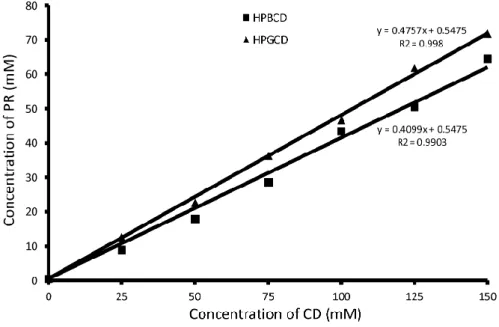
The phase solubility of PR in HPBCD and HPGCD containing aqueous solutions (0-150 mM) is shown in Figure 1.
Fig. 1. Phase solubility diagrams of PR in aqueous HPBCD (■) and HPGCD (▲) solutions at 25 °C
The solubility of PR was increased linearly by increasing the concentration of HPBCD or HPGCD. The diagrams are Higuchi AL type for both cyclodextrins, therefore the formation of 1:1 complex can be assumed. In case of HPBCD, the apparent stability constant of the complex is 1286.4 M-1, the constant of the PR-HPGCD complex was measured to be 1778.5 M-1. It is stated that PR has greater affinity for complex-formation with HPGCD.
4.2. Study of diffusion through dialysis membrane
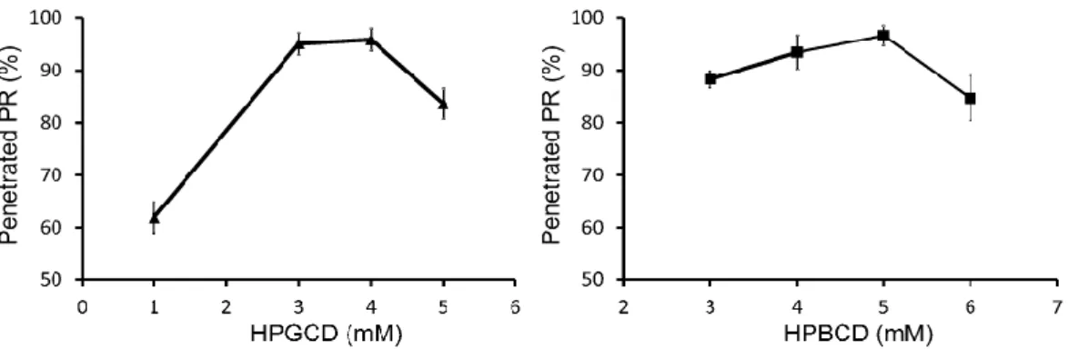
The concentrations of CD derivatives for the penetration of PR were optimized. The penetrated PR as a function of the concentration of CD is shown in Figure 2.
Fig. 2. PR penetration through semi-permeable membrane as a function of the concentration of HPGCD and HPBCD after 240 min
The results show that 4 mM HPGCD and 5 mM HPBCD induce the highest diffusion of PR through the dialysis membrane. Under the optimal CD concentration, a part of free, hydrophobic drug remained in the pouches. Above the optimal CD level, the excess amount of CD keeps the free PR in complex, therefore less amount of free API is detectable in the acceptor phase. Thereafter, mucoadhesive ZnHA–ZnGlu additives were added to the eye drops, which ensure antimicrobial, preservative effect in the formulations. It was found that the application of biopolymer has no effect on the diffusion of PR.
4.3. Viscosity
The viscosity of ZnHA and PR-CD complex containing ophthalmic formulations was measured. According to previous reports, the viscosity should be under 30 mPa s. Above this level blurred vision and discomfort appear, which result the faster elimination due to reflex mechanisms of the eye. The results show that the viscosity of our formulations is appropriate, in the range of 9.2-24.2 mPa s.
4.4. Surface tension
No significant difference was found between the values. The surface tensions of the eye drops are about 60 mN/m, which is higher than the surface tension of lacrimal fluid.
Ophthalmic products were investigated by Han K. et al., and the range of the surface tension values was between 34.3 and 70.9 mN/m. According to this study, the surface tension of formulations meets the requirements for ophthalmic products.
4.5. Efficacy of antimicrobial preservation
In Sample I. (with HPBCD) and II. (with HPGCD) ZnHA–ZnGlu were used as preservative compounds.
The logarithmic decrease of Staphylococcus aureus was 1.5 after 6 hours, 2 after 24 hours, and no bacteria were detected in the samples after 7 days. In case of Pseudomonas aeruginosa, the logarithmic decrease is higher at the earlier period, but the bacterium appeared in every sample. The logarithmic decrease of Candida albicans was 1 after 6 hours, and no fungi were detected later. In summary, the preservative effect of ZnHA–ZnGlu containing samples meets the EP-B criteria.
The microbiological stability of Sample III. (with HPBCD) and IV. (with HPGCD), which contained BK as preservative agent, was tested. The logarithmic decrease of Staphylococcus aureus was 1 after 6 hours, 2 after 24 hours, no bacteria were detected later in Sample III. For Pseudomonas aeruginosa the logarithmic decrease was 2 and no change was detected later. In Candida albicans containing samples, no fungi were found after 7 days.
In Sample IV., for Staphylococcus aureus and Candida albicans, the number of CFU was zero after 6 hours. In case of Pseudomonas aeruginosa the antimicrobial effect was lower due to the known resistance of the bacterium against BK. The microbiological stability of Samples III-IV. meets the requirements of the EP.
4.6. Investigation of mucoadhesion
There is a large difference in the measured force between the blank and the mucin dispersion. All the samples show mucoadhesivity, samples prepared with ZnHA have significantly higher values than samples prepared without it. This proves the importance of the presence of ZnHA because the interpenetration between the ZnHA chains and mucin can be assumed. These samples can have better mucoadhesive properties and cause decreased administration frequency and a lower active ingredient concentration. The type of CD does not play an important role in mucoadhesion because there is no significant difference between the samples prepared with HPBCD and HPGCD.
4.7. Cell viability assay
Impedance measurement showed significant cell damage after treatment with all three formulations containing BK (III, IV, VI). PR containing formulations with HPBCD, HPGCD, ZnHA and ZnGlu did not show any cytotoxic effect. Normalized cell index was significantly higher in ZnHAZnGlu containing sample (V), than in formulation with BK (VI). As a comparison, maximal toxicity was detected in cells treated with the reference damaging agent Triton X-100 detergent (Fig. 3).
Figure 3. Cell viability of HCE-T corneal epithelial cells after 1-hour treatment with formulations measured by impedance. Values are presented as means ± SD, n = 6-12. Statistical analysis: ANOVA
followed by Dunett’s test. (*p<0.05; ***p<0.001 compared to control), Triton X-100 (Tx-100).
4.8. Immunohistochemistry
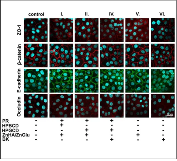
The cornea epithelial cells formed tight paracellular barrier visualized by the localization of the junctional proteins ZO-1, β-catenin, E-cadherin and occludin. The cells were tightly apposed, and all junctional proteins were localized at the intercellular connections forming pericellular belts in the control groups (Fig. 4). Morphological change can be observed in the case of BK containing sample (VI) by the localization of E-cadherin protein. In the case of target eye drops formulated with ZnHA–ZnGlu, no morphological change and no toxic effect were detected on the cell culture. It can be stated that ZnHA–ZnGlu combination is non-toxic alternative preservative system, which is tolerable on HCE-T cells in the applied concentration. The results of BK containing samples confirmed the toxic and expected irritative effect on the eye surface.
Figure 4. Effects of PR and pharmaceutical excipients containing formulations on junctional morphology of HCE-T corneal epithelial cells. Immunostaining for zonula occludens-1 (ZO-1), occludin tight junction proteins and β-catenin, E-cadherin adherens junction proteins after 1-hour
treatment.
4.9. Permeability study on HCE-T cell culture model
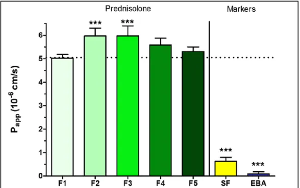
After 30-minute treatment only dissolved PR-CD complex containing formulation 2 and formulation 3 showed significantly higher Papp values (F2: 5.97 × 10⁻⁶ cm/s, F3: 5.97 × 10⁻⁶ cm/s) compared with PR suspension (F1: 5.02 × 10⁻⁶ cm/s). Papp values were minimally lower in ZnHA/ZnGlu containing formulations (F4: 5.59 × 10⁻⁶ cm/s, F5: 5.31 × 10⁻⁶ cm/s). The model showed low Papp values for the two hydrophilic paracellular marker molecules indicating a good barrier. PR-CD complex containing solutions and PR-CD-ZnHA–ZnGlu containing target formulations were studied compared with sample which contains the same amount of PR in suspension form.
Figure 5. Permeability of PR (100 µg/mL in each formulations) (F1-F5) across HCE-T epithelial cell layers (30-minute assay). Values for paracellular permeability markers fluorescein (SF) and Evans blue labeled albumin (EBA) are also shown. Statistical analysis: ANOVA followed by Dunett’s test.
***p<0.001. Values are presented as means ± SD, n = 4. p<0.001 compared to F1.
The permeability was significantly higher in samples containing the dissolved PR-CD complex compared with the suspension. Because the concentration of dissolved API is higher in PR-CD solutions, and higher towards the corneal epithelial cell layer, faster drug permeation is expected through the epithelial layer. In the target formulations with biopolymer, the permeability is minimally lower, due to the diffusion restrictive effect of the polymer structure (Fig. 5.).
4.10. Ex vivo permeability assay
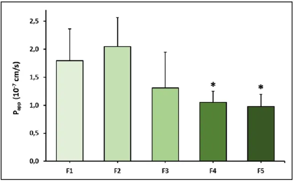
Permeability of PR was tested on ex vivo porcine cornea model in the case of previously mentioned formulations (F1-F5) (Fig. 6.). In the case of PR-HPBCD (F2), permeability was higher (2.05 × 10⁻⁷ cm/s) compared with PR containing suspension (F1, 1.79 × 10⁻⁷ cm/s) Permeability in PR-HPGCD (F3) complex containing samples was lower (1.31 × 10⁻⁷ cm/s).
Considering the relatively high standard deviations in F1-F3, no significant difference can be stated between their Papp values. In the presence of ZnHA/ZnGlu (F4-F5) significantly lower permeability values were measured (F4: 1.05 × 10⁻⁷ cm/s, F5: 0.98 × 10⁻⁷ cm/s). The monitored TEER values were in the range of 1052-2818 Ω × cm2. According to the results, no significant difference was found between the eye drop suspension and solutions.
Permeability values were significantly lower in ZnHA–ZnGlu containing formulations in
comparison with the suspension form. Considering the continuous mixing, vertical position and complexity of porcine cornea, the optimal attributes are not observed in this model in solution-based samples. The permeability is lower in target formulations because polymer structure affects the drug diffusion negatively, however, the retention time is increased on the eye surface due to the increased viscosity and mucoadhesion. In the case of ex vivo study the whole porcine cornea is used formed by lipophilic and hydrophilic layers. Therefore, difference shall be observed in the permeability on in vitro HCE-T and ex vivo porcine cornea models. The formulations can be optimal in vivo, because less irritation and lachrymal secretion are expected due to the not irritative solution form of eye drops.
Figure 6. Permeability of PR in different formulations (F1-F5) across in ex vivo porcine cornea model. Statistical analysis: ANOVA followed by Dunett’s test. *p<0.05 compared to F1. Values are
presented as means ± SD, n = 6.
As the eye drop contacts for longer time, reflex lachrymal secretion and eye blinking are limited, prolonged drug absorption is expected after administration, which results in less frequent application. We assume that the permeability of PR is optimal in the target formulations, which may result in enhanced therapeutic effect.
.
5. SUMMARY AND CONCLUSIONS
In this work PR-containing aqueous solutions were formulated by CD inclusion complex and addition of mucoadhesive, antimicrobial ZnHA and ZnGlu. The following results have been achieved:
Aqueous solutions of PR were formulated by inclusion complexation with HPBCD and HPGCD.
The investigation of the diffusion of PR through the dialysis membrane revealed that 5 mM of HPBCD, and 4 mM of HPGCD caused the highest penetration of API in in vitro circumstances. The addition of ZnHA and ZnGlu had no effect on the penetration properties.
The measurement of viscosity showed that the 5 mM HPBCD- and 4 mM HPGCD- containing products had the lowest viscosity values, meeting the requirements of the EP.
It was found that the concentration of CDs has no effect on the surface tension of the eye drops, and these values are optimal compared with previously investigated ophthalmic products.
The mucoadhesive properties of ZnHA-containing formulations were proved with the tensile test, resulting in a higher retention time of the eye drop on the surface. The type of CD derivative has no influence on mucoadhesivity.
During the preservative effectiveness test, the applicability of ZnHA–ZnGlu combination was proven as an antimicrobial, preservative system. The microbiological stability of ZnHA-containing products meets the requirements of the EP in case of S. aureus, P. aeruginosa and C. albicans.
The formulations are non-toxic in vitro according to the immunohistochemistry and impedance measurement on HCE-T model
Eye drops had optimal permeability according to the results of in vitro HCE-T and ex vivo porcine cornea models.
These novel compositions are promising for overcoming the challenges of ocular drug delivery by optimal permeability, ensured microbiological stability, minimal irritation, and acceptable patient compliance.
LIST OF ORIGINAL PUBLICATIONS
I. Bíró, T., Horvát, G., Budai-Szűcs, M., Csányi, E., Urbán, E., Facskó, A., Szabó-Révész, P., Csóka, I., Aigner, Z., Development of prednisolone-containing eye drop formulations by cyclodextrin complexation and antimicrobial, mucoadhesive biopolymer. Drug Design, Development and Therapy (2018) 12, 2529–2537.
DOI: 10.2147/DDDT.S165693
Q1 IF (2018): 3.208 Citation: 8
II. Bíró T., Aigner Z., Current Approaches to Use Cyclodextrins and Mucoadhesive Polymers in Ocular Drug Delivery – A Mini-Review. Scientia Pharmaceutica (2019) 87, 15.
DOI: 10.3390/scipharm87030015
Q2 IF (2019): 0 Citation: 4
III. Bíró T., Bocsik A., Jurišić Dukovski B., Gróf I., Lovrić J., Csóka I., Deli M.A., Aigner Z.
New approach in ocular drug delivery: in vitro and ex vivo investigation of cyclodextrin containing, mucoadhesive eye drop formulations. Drug Design, Development and Therapy (2021) 15, 351-360.
DOI: 10.2147/DDDT.S264745
Q1 IF (2020): 3.216 Citation: 0
PRESENTATIONS RELATED TO THE THESIS
1. Tivadar Bíró, Zoltán Aigner: Megnövelt biohasznosulású szemészeti készítmény formulálása és vizsgálata.
Local Scientific Student’s Association, p. 170, Szeged, Hungary, 2016.
2. Tivadar Bíró, Zoltán Aigner: Megnövelt biohasznosulású szemészeti készítmény formulálása és vizsgálata.
XII. Ottó Clauder Memorial Competition, p. 13, Budapest, Hungary, 2016.
3. Tivadar Bíró, Zoltán Aigner: Megnövelt biohasznosulású szemészeti készítmény formulálása és vizsgálata.
National Conference of Scientific Student’s Association, p. 443, Pécs, Hungary, 2017.
4. Tivadar Bíró, Zoltán Aigner: Megnövelt biohasznosulású gyulladáscsökkentő szemcsepp formulálása.
Hungarian Chemical Society, Crystallization and Drug Formulation Department 10th Conference, p. 29, Balatonszemes, Hungary, 2017.
5. Tivadar Bíró, Gabriella Horvát, Mária Budai-Szűcs, Erzsébet Csányi, Edit Urbán, Andrea Facskó, Piroska Révész- Szabó, Ildikó Csóka, Zoltán Aigner:
Characterization of prednisolone containing eye drop formulations by mucoadhesive–
preservative system and cyclodextrin inclusion complex formation.
7th BBBB International Conference on Pharmaceutical, Sciences, Acta Pharmaceutica Hungarica 87 (3-4) p. 178, Balatonfüred, Hungary, 2017.
6. Tivadar Bíró, Gabriella Horvát, Alexandra Bocsik, Ilona Gróf, Edit Urbán, Mária A. Deli, Erzsébet Csányi, Piroska Szabó-Révész Ildikó Csóka, Zoltán Aigner: Formulation of steroid containing eye drops with cyclodextrin derivatives and mucoadhesive preservative system 12th Central European Symposium on Pharmaceutical Technology and Regulatory Affairs, Acta Pharmaceutica Hungarica 88 (3) p. 139-140, Szeged, Hungary, 2018.
7. Tivadar Bíró, Zoltán Aigner: Ciklodextrinek és mukoadhezív, antimikróbás biopolimer alkalmazása megnövelt hatékonyságú szemészeti készítmények formulálása céljából
XIII. Ottó Clauder Memorial Competition, p. 18, Budapest, Hungary, 2018.
8. Tivadar Bíró, Zoltán Aigner: Novel ophthalmic formulations to increase the efficacy and stability
I. Symposium of Young Researchers on Pharmaceutical Technology, Biotechnology and Regulatory Science, p. 18, Szeged, Hungary, 2019.
9. Tivadar Bíró, Zoltán Aigner: In vitro and ex vivo investigation of steroid containing ocular drug delivery systems
II. Symposium of Young Researchers on Pharmaceutical Technology, Biotechnology and Regulatory Science, p. 6, Szeged, Hungary, 2020.
10. Tivadar Bíró, Alexandra Bocsik, Ilona Gróf, Bisera Jurišić Dukovski, Jasmina Lovrić, Mária A. Deli, Zoltán Aigner: Innovatív szemcseppek toxicitásának és hatóanyag-permeabilitásának vizsgálata in vitro és ex vivo modellek alkalmazásával
Congressus Pharmaceuticus Hungaricus XVI; Acta Pharmaceutica Hungarica 90. p. 86, Debrecen, Hungary, 2020.