hjic.mk.uni-pannon.hu DOI: 10.33927/hjic-2020-25
THE EFFECT OF pH ON BIOSURFACTANT PRODUCTION BY BACILLUS SUBTILIS DSM10
RÉKACZINKÓCZKY1 ANDÁRONNÉMETH*1
1Department of Applied Biotechnology and Food Science, Budapest University of Technology and Economics, M ˝uegyetem rkp. 3, Budapest, 1111, HUNGARY
The genusBacillushas long been known for its ability to produce many industrially useful products. These bacteria mostly produce extracellular products like organic acids, enzymes and biosurfactants. In this paper, the production of surfactin using theBacillus subtilisstrain DSM10 is investigated. Biosurfactant was produced in a lab-scale 1-liter fermenter. pH control using different bases (NH4OH and NaOH) was compared to observe whether the amount of produced biosurfac- tant or the quality of the product was influenced. The formation of the product was followed by measuring the surface tension, and the product formed was analyzed by reversed-phase chromatography. The investigation of the effect of pH control showed that it can be omitted during the fermentation of the biosurfactant. The highest concentration of surfactin (5g/L) was achieved without pH control in contrast with when the pH was kept constant (pH = 7).
Keywords: biosurfactant, surfactin, fermentation, purification, Bacillus subtilis
1. Introduction
In recent years, the microbial production of tensio-active molecules with various properties, e.g. emulsifying, wet- ting, foaming, detergency, solubilizing and dispersing, has been gaining interest [1]. Biosurfactants are am- phiphilic compounds produced by a variety of microbial communities. Natural surfactants are of great importance in the pharmaceutical, cosmetic, agricultural and food in- dustries due to their beneficial properties including low toxicity, biodegradability, high selectivity and activity un- der extreme environmental conditions [2,3]. The market of bio-based surfactants is predicted to be worth $5.52 billion by 2022 [4]. This is unsurprising given our high degree of dependency on kinds of hygiene products, the majority of which include surfactants or emulsifiers.
Biosurfactants are classified based on their chemical structure into the following groups: glycolipids, lipopep- tides, fatty acids and lipids, as well as polymeric and particulate biosurfactants [3,5]. One of the most effec- tive biosurfactants is surfactin. This lipopeptide-type sur- factant contains a cyclic peptide linked to a fatty acid chain (Fig. 1) [6]. Bóka et al. reported that the molec- ular weights of surfactins range from 993 Da to 1049 Da [7]. Several gram-positive Bacillusspecies naturally produce surfactins, which help the bacteria to stabilize their cell membranes and adhere to a surface [8,9]. The biosynthesis of surfactin occurs through different mecha- nisms: the conversion of glucose or glycerol as a substrate
*Correspondence:naron@f-labor.mkt.bme.hu
to glucose 6-phosphate through the glycolytic pathway, providing the main precursor of carbohydrates located in the hydrophilic part and the oxidation of glucose to pyruvate then to acetyl-CoA, which serves as a precur- sor for the synthesis of lipids and amino acids (Asp, Glu, Leu, Val). However, if the substrate is a hydrocarbon, the metabolism is shifted towards the lipolytic pathway (β- oxidation into acetyl-CoA) and gluconeogenesis (acetyl- CoA involved in the synthesis of the precursor glucose 6-phosphate) [1].
The effectiveness of surfactants is defined by their ability to reduce the surface tension (ST), defined as the cohesive force between molecules which is proportional to the concentration of surfactant in the solution [2]. Their efficiency is measured by the critical micelle concentra- tion (CMC) [1,11]. Above the CMC, surfactants form mi- cellar structures, but below it, the aggregates dissociate into monomers. Lipopeptides fromB. subtilisare partic- ularly compelling because their surface activity has been reported to be strong [6,12,13]. Powerful surfactants can decrease the surface tension of water (72mN/m at20◦C) to less than30mN/m [14]. The emulsifying capacity can be monitored by calculating the emulsification index (EI,
%) and emulsion stability. As the pH decreases, surfactin becomes less soluble in water because the carboxyl group is protonated [12]. Under neutral or basic conditions, the carboxyl group is in the ionic form, thus its solubility and emulsification capability increases [15]. Moro et al. eval- uated the influence of the pH on the stability of the surfac- tants produced by species ofB. subtilis,B. gibsoniiandB.
Figure 1: Chemical structure of surfactin: peptide loop of amino acids: five L-amino acids (Val, Asp, Leu, Glu and Leu) two D-amino acids (Leu and Leu), and aα,β- hydroxy C13-C15 fatty acid chain [10]
amyloliquefaciens[9]. All isolates exhibited surface ten- sions below30mN/m. In strongly acidic conditions, the emulsifying activity significantly decreased for bothB.
subtilis ODW02andB. subtilis ODW15. As the pH was increased from7to12, the stability of the surfactant pro- duced byB. subtilis ODW02decreased even further, but the one byB. subtilis ODW15remained stable. In the case ofB. amyloliquefaciens MO13, a significant increase was observed both under acidic and basic conditions. This dif- ferent pH-responsive behavior makes surfactin applicable in a variety of industrial fields.
The choice of fermentation cultivation media (i.e. of average composition, carbon source, nitrogen source and trace elements) as well as fermentation strategies (i.e.
temperature, pH, aeration and agitation) also need to be considered in relation to the type of applications. Based on a literature review, the best combination among the fermentation conditions was1.5vvm at300rpm, result- ing in a maximum yield of surfactin of6.45g/L [16].B.
subtilis ATCC 21332was grown on an iron-enriched min- imal salt (MSI) medium including glucose (40g/L). In a recent study, Yang et al. used nanoparticles (NPs) to im- prove the total yield of surfactin.5g/L of Fe NPs were added to the fermentation medium ofB. amyloliquefa- ciens MT45, which increased the titer of surfactin from 5.94to9.18g/L. Modifying the biosynthesis of surfactin with metabolic engineering tools can further increase production and titers of surfactin inB. subtilis, as demon- strated by Wu et al. (12.8g/L) [17]. Few studies have fo- cused on individual characteristics and relative amounts of surfactin variants in the extracts of lipopeptides. Many surfactin variants exist with various lengths of fatty acid chains and different amino acid sequences [5]. Akpa et al. analyzed the replacement of L-glutamic acid with four other amino acids, namely L-leucine, L-valine, L- isoleucine and L-threonine, in the culture medium. The presence of Thr was found to be favorable for the syn- thesis of longer (C15-C16) fatty acid chains inB. subtilis S499[18]. Supplementation with Mn2+, Cu2+and Ni2+
ions can also promote the production of novel variants of surfactin with different components of fatty acids (C16- C18) and amino acids (central aspartic acid methyl ester residue instead of aspartate) [19]. This approach can be useful for studying specific biological activities.
The objectives of our study were to explore the effect of pH control on the production of biosurfactants (i.e. on titers and productivity) by theB. subtilis strain DSM10 and evaluate the properties of biosurfactants, i.e. type, surface tension reduction and emulsifying activity.
2. Materials and methods 2.1 Cultivation conditions
Bacillus subtilis DSM10 (NCAIM B.02624T) was used for biosurfactant fermentation in this study. Cultivation was performed at37◦Cin250ml Erlenmeyer flasks con- taining 100 ml of an inorganic medium based on the composition used by Joshi et al. (2013): 34 g glucose, 1.0g NH4NO3,6.0 g KH2PO4,2.7g Na2HPO4,0.1 g MgSO4•7H2O,1.2·10−3g CaCl2•2H2O,1.65·10−3g FeSO4•7H2O,1.5·10−3g MnSO4•4H2O and2.2·10−3 g Na-EDTA [20].
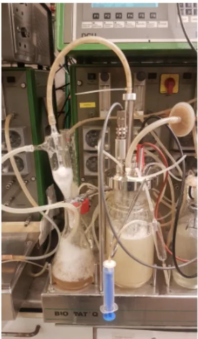
The experiments were carried out in a 1 L bench- top bioreactor, with a working volume of 0.8 L (Bio- stat Q fermenter, B. Braun Biotech International, Ger- many) and a 10% v/v inoculum. For biosurfactant pro- duction, the temperature was adjusted to 37◦Cwith an agitation speed of300rpm and an aeration rate of0.25 vvm. The pH was controlled by 25% H2SO4 and two different bases, namely 25% NH4OH and 25% NaOH.
Biosurfactant fermentation without external pH control served as a controlled experiment. A cyclone separator for reducing foam was connected to the outlet airstream of the fermenter (Fig. 2). The foam could overflow from the fermenter via the air outlet, through the cyclone sep- arator to the collector flask.
2.2 Analysis of biomass
Bacterial growth was monitored by measuring the optical density of the fermentation broth at600nm using a Phar- macia LKB Ultrospec Plus spectrophotometer in compar- ison with that of the centrifuged supernatant of the sam- ple.
The biomass concentration (g cell dry weight/L) was determined by using a calibration curve (R2= 1):
Biomass [g/L] = 0.4283·OD600+ 1.4568 (1) The sampled broth was centrifuged at6,000rpm for15 mins. The cell pellets were collected and dried at105◦C to constant weight by a Sartorius MA35 moisture ana- lyzer to measure the cell dry weight.
Figure 2:Fermentation setup with foam-separating glass cyclone
2.3 Analysis of glucose consumption
Glucose consumption was determined using the Waters Breeze 2 HPLC System. The mobile phase was 5 mM H2SO4and the rate of elution was0.5mL/min. A BIO- RAD Aminex HPX-87H (300 ×7.8 mm, 9 µm) col- umn (65◦C) was applied with a Refractive Index detector (40◦C).
The glucose concentration was calculated from the peak area by the following calibration curve equation (R2= 1):
Glucose [g/L] = 4·10−6·PeakArea + 0.0147 (2)
2.4 Analysis of biosurfactants
Surface tension measurement
The surface activities of biosurfactants produced by the bacterial strains were determined by measuring the sur- face tension of the samples of cell-free broth using the stalagmometric method with a Traube Stalagmometer (2.5 mL, Wilmad-LabGlass LG-5050-102 Stalagmome- ter Tube for samples of low viscosity) at room temper- ature (25◦C). To increase the accuracy of the surface tension measurements, the averages of triplicates were used in this report. The surface tension can be determined based on the number of drops that fall per unit volume, the density of the sample and the surface tension of a liq- uid reference, e.g. deionized water.
The actual number of drops was calculated using N =N0+x−y
c (3)
whereN denotes the number of drops of the sample cal- culated to the nearest tenth of a drop;N0represents an in- teger of drops counted between capillary-scale readingsx
andy;xandystand for capillary-scale readings based on the maximum data point as0and the minimum data point as40;xandy refer to the distances in millimeters from the beginning of each scale; andc is the capillary-scale calibration in millimeters per drop.
The surface tension (ST in mN/m) was calculated ac- cording to
ST =STw·Nw·D N·Dw
(4) where STwdenotes the surface tension of water at25◦C (72mN/m);Nwrepresents the number of water drops (20 drops);D stands for the density of the sample in g/mL;
Nrefers to the number of drops of sample, andDwis the density of water at25◦C.
Emulsifying activity
The emulsifying activity was determined by the addition of 2 mL of sunflower oil to the same volume of cell- free sample or surfactin solution in a test tube, which was vortex-mixed vigorously for2mins. [21]. The tubes were incubated at25◦Cand the emulsification index (EI) de- termined after24hours according to:
EIt= He
Ht
100 (5)
whereHe andHt are the height of emulsion and total height of the liquid in the tube, respectively.
To study the emulsion stability, the same protocol was used. The emulsification index (EI, %) was determined after 1h and the EI measured after24h (EI24,%), the tubes were incubated at25◦C. The emulsion stability was expressed as a function of the changes in EI over the24 h.
High-performance liquid chromatography (HPLC) The surfactin concentration was measured by HPLC us- ing a Waters Alliance 2695 Separations Module, which is a high-performance liquid chromatographic system equipped with a Waters 2996 photodiode array detector, at205nm and a Symmetry C18 Column (4.6×150mm, 5µm - Waters, Ireland). The mobile phase consisted of 20% v/v trifluoroacetic acid (TFA) (3.8 mM) and 80%
v/v acetonitrile. The elution rate was1mL/min at25◦C and the sample volume was10µL. The purified surfactin was identified by using commercially available surfactin (Wako Chemicals) as the authentic compound [22].
2.5 Isolation of the biosurfactant
The method for purifying the biosurfactant was adapted from the one outlined by Joshi et al. (2008) [23]. The cell-free broth was obtained by centrifuging the fermen- tation broth at4,000 rpm for 20mins. at 4◦C using a Janetzki MLW K23D centrifuge. The cell-free broth was used for further purification steps. The biosurfactant was recovered from the supernatant by acid precipitation: the
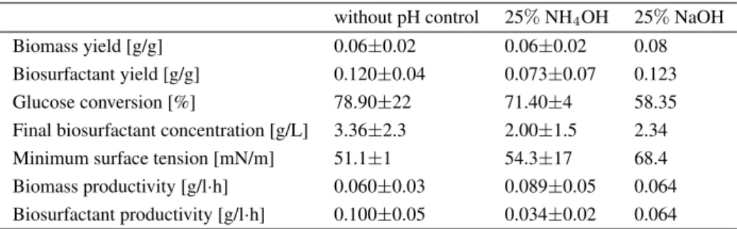
Table 1:Summary of the results of the fermentations
without pH control 25%NH4OH 25%NaOH
Biomass yield [g/g] 0.06±0.02 0.06±0.02 0.08
Biosurfactant yield [g/g] 0.120±0.04 0.073±0.07 0.123
Glucose conversion [%] 78.90±22 71.40±4 58.35
Final biosurfactant concentration [g/L] 3.36±2.3 2.00±1.5 2.34
Minimum surface tension [mN/m] 51.1±1 54.3±17 68.4
Biomass productivity [g/l·h] 0.060±0.03 0.089±0.05 0.064 Biosurfactant productivity [g/l·h] 0.100±0.05 0.034±0.02 0.064
pH was adjusted to2.0 using6N HCl and kept at4◦C overnight. The precipitate was collected by centrifuga- tion at4,000rpm for20mins. at4◦C, then resuspended in distilled water. The pH was adjusted to7.0using6N NaOH and the solution lyophilized by a Christ Alpha 2-4 LSC freeze dryer. The concentration of biosurfactant was determined gravimetrically from the resulting yellowish white powder. The concentration of biosurfactant was determined gravimetrically from the lyophilized powder.
The identity of the purified biosurfactant was checked by HPLC.
2.6 Calculation of fermentation parameters
To compare the results of the fermentation, the following parameters were determined.Substrate (glucose) conversion was calculated accord- ing to:
∆S% = S0−Sf
S0 (6)
whereS0andSfdenote the initial substrate and final glu- cose concentrations, respectively.
The biomass yield on glucose (Yx
S, g/g) was defined by:
Yx
s = xf−x0 S0−Sf
(7) wherexfandx0are the final and initial biomass concen- trations, respectively.
The biosurfactant yield on glucose (YP
s, g/g) was de- fined by:
YP
s = Pf−P0
Sf−S0 (8)
wherePf and P0 are the final and initial biosurfactant concentrations, respectively.
The volumetric productivities Jx= xmax
txmax
(9) and
JP = Pmax tPmax
(10) (g/l·h) were calculated as the quotients of the maximum biomass concentration (xmax, g/l) or the maximum bio- surfactant concentration (Pmax, g/l) and the fermentation time (txmax ortPmax, h) when the maximum concentra- tion was achieved, respectively.
3. Results and Discussion
To evaluate the effect of pH on surfactin production, a se- ries of batch fermentations were performed either with or without pH control. The biosurfactant solution was analysed quantitatively and qualitatively using the HPLC method reported by Mubarak et al. [24].
An overview of the calculated parameters of the batch runs can be seen in Table 1. In the absence of pH con- trol, the maximum biomass concentration achieved was 4.00g/L after35h (Fig. 3). Without pH control, the in- creased acidity of the medium inhibited further growth at pH 4.4. The maximum biosurfactant concentration was 4.99g/L, which resulted in the surface tension decreas- ing to50.1 mN/m. In pH-controlled fermentations, the biomass yields (0.06and0.08g/g - pH adjusted with25%
NH4OH and25%NaOH to7.0, respectively) were sim- ilar to that in the absence of pH control (0.06g/g) (Ta- ble 1), while the production of biosurfactants was unable, with a few exceptions, to reduce the surface tension sig- nificantly (66.5mN/m with25%NH4OH,Table 1;Figs.
4 and5). This may account for the presence of residual glucose concentrations of8to12g/L (Figs. 4and5). The maximum surfactant concentrations were3.50and2.34 g/L by adjusting the pH using NH4OH and NaOH, re- spectively. Although these results are similar to the av- erage yield of surfactants (3.36 g/L) in the absence of pH control, a significant drop in productivity of approxi-
Figure 3:Fermentation of Surfactin - without external pH control
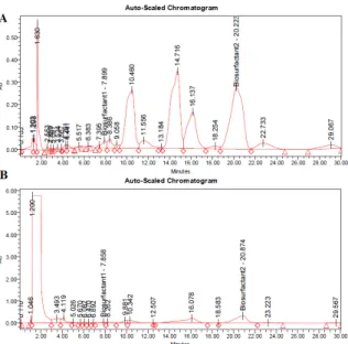
Figure 4:A) HPL chromatogram of a1.25g/L surfactin standard, B) HPLC chromatogram of the isolated biosur- factant fraction from the foam out sample
Figure 5: Fermentation of Surfactin - pH controlled by 25% NH4OH.
mately50%was observed (Table 1).
The highest value of the emulsifying activity (EI24) was in excess of70%at the end of the exponential phase of the growth curve (at35h,Fig. 3). The EI24values ob- tained from samples extracted from pH-controlled exper- iments increased from45to55%(Figs. 5and6, respec- tively). These inconsistencies can be explained in part by the fact that differentBacillusspecies and strains are ca- pable of producing numerous surfactin variants with dis- tinct properties. It is highly likely that the production of surfactants is independent of cell growth as EI24 de- creased and ST increased during the stationary phase per- haps as a result of degradation by enzymatic hydrolysis or uptake under substrate-limiting conditions [25,26].
Since the spectrum of lipopeptide-type biosurfactants is broad, the profiles of the extracts obtained after the purification process were compared to the surfactin stan- dard.Fig. 4presents two representative chromatograms:
Figure 6:Fermentation of Surfactin - pH controlled by 25%NaOH
(A) surfactin standard and (B) purified culture broth of B. subtilis DSM10. Comparatively speaking, the sam- ples from our study exhibited similar peaks (number of peaks, retention time). The intense peak at the beginning of the chromatogram indicates the presence of some non- retained impurities, namely contaminants and inorganic salts such as NH4NO3, which are often co-extracted with the targeted biosurfactant, that may need to be sepa- rated. Overall, based on the separation of peaks and re- tention time, the isolated biosurfactant was identified as surfactin.
The surfactin titer ofB. subtilis DSM10was compared with the results from relevant studies (Table 2). Even though the fermentation strategies differ to some extent, the surfactin concentration from our work compares well with the values previously reported in the literature.
4. Conclusion
The aim of this study was to assess the biosurfactant- producing capability ofBacillus subtilis DSM10and es- tablish an economically feasible fermentative process.
This paper investigated the effect of pH control on the amount of biosurfactant production. The maximum amount of biosurfactant (approximately 5 g/L) was re- covered from fermentation experiments in the absence of pH control at37◦C. Furthermore, a preliminary char- acterization of the surface-active compounds produced during fermentation was conducted. HPLC analysis con- firmed the presence of surfactin in the purified product.
Table 2:Surfactin production byBacillusspecies Strain Surfactin titer [g/L] Ref.
B. subtilis ATCC 21332 6.45 [16]
B. subtilis 2.00 [27]
B. subtilis SPB1 4.92 [28]
B. subtilis DSM10T 3.99 [29]
B. subtilis #573 4.80 [6]
B. subtilis CN2 7.15 [30]
B. subtilis DSM10 4.99 this work
The minimum surface tension was50mN/m. The emul- sifying activity achieved using sunflower oil was approx- imately70%. These results represent an initial step to- wards large-scale production of this biosurfactant. From a technical and economic standpoint, the fermentative pro- cess of surfactin carried out in the absence of pH control in a mineral salt medium using glucose as the sole car- bon source seems to be an effective strategy for pilot- and industrial-scale production.
Acknowledgment
The research was supported by the Gedeon Richter’s Talentum Foundation, founded by Gedeon Richter Plc.
(Gedeon Richter Ph.D. fellowship). The research re- ported in this paper has been supported by the Na- tional Research, Development and Innovation Fund TUDFO/51757/2019-ITM, Thematic Excellence Pro- gram.
REFERENCES
[1] Santos, D. K. F.; Rufino, R. D.; Luna, J. M.; San- tos, V. A.; Sarubbo, L. A.: Biosurfactants: Multi- functional biomolecules of the 21st century.Int. J.
Mol. Sci.2016,17(3), 1–31DOI: 10.3390/ijms17030401
[2] Satpute, S. K.; Banpurkar, A. G.; Dhakephalkar, P.
K.; Banat, I. M.; Chopade, B. A.: Methods for in- vestigating biosurfactants and bioemulsifiers: A re- view. Crit. Rev. Biotechnol. 2010,30(2), 127–144
DOI: 10.3109/07388550903427280
[3] Fenibo, E. O.; Douglas, S. I.; Stanley, H.
O.: A review on microbial surfactants: Produc- tion, classifications, properties and characteriza- tion. J. Adv. Microbiol. 2019, 18(3), 1–22 DOI:
10.9734/jamb/2019/v18i330170
[4] Markets and Markets. Natural Surfactants Market (Bio-based Surfactants) https:
//www.marketsandmarkets.com/search.asp?
search=surfactants
[5] Sajna, K. V.; Höfer, R.; Sukumaran, R. K.; Got- tumukkala, L. D.; Pandey, A.: White biotechnol- ogy in biosurfactants. in: Industrial biorefineries and white biotechnology; Elsevier B.V., 2015; pp.
499–521DOI: 10.1016/B978-0-444-63453-5.00016-1
[6] Gudiña, E. J.; Fernandes, E. C.; Rodrigues, A. I.;
Teixeira, J. A.; Rodrigues, L. R.: Biosurfactant pro- duction byBacillus subtilisusing corn steep liquor as culture medium.Front. Microbiol.2015,6, 1–7
DOI: 10.3389/fmicb.2015.00059
[7] Bóka, B.; Manczinger, L.; Kecskeméti, A.; Chan- drasekaran, M.; Kadaikunnan, S.; Alharbi, N. S.;
Vágvölgyi, Cs.; Szekeres, A.: Ion trap mass spec- trometry of surfactins produced byBacillus subtilis SZMC6179J reveals novel fragmentation features of cyclic lipopeptides.Rapid Commun. Mass Spec- trom.2016,30(13), 1581–1590DOI: 10.1002/rcm.7592
[8] From, C.; Hormazabal, V.; Hardy, S. P.; Granum, P.
E.: Cytotoxicity inBacillus mojavensisis abolished following loss of surfactin synthesis: Implications for assessment of toxicity and food poisoning po- tential.Int. J. Food Microbiol.2007,117(1), 43–49
DOI: 10.1016/j.ijfoodmicro.2007.01.013
[9] Moro, G. V.; Almeida, R. T. R.; Napp, A. P.;
Porto, C.; Pilau, E. J.; Lüdtke, D. S.; Moro, A.
V.; Vainstein, M. H. Identification and ultra-high- performance liquid chromatography coupled with high-resolution mass spectrometry characterization of biosurfactants, including a new surfactin, iso- lated from oil-contaminated environments.Microb.
Biotechnol.2018,11(4), 759–769DOI: 10.1111/1751- 7915.13276
[10] Surfactin - Wikimedia Commons. https:
//commons.wikimedia.org/wiki/File:
Surfactin.png
[11] De, S.; Malik, S.; Ghosh, A.; Saha, R.; Saha, B.: A review on natural surfactants.RSC Adv.2015,5(81), 65757–65767DOI: 10.1039/c5ra11101c
[12] Nitschke, M.; Pastore, G. M.: Production and properties of a surfactant obtained from Bacil- lus subtilis grown on cassava wastewater.
Bioresour. Technol. 2006, 97(2), 336–341 DOI:
10.1016/j.biortech.2005.02.044
[13] Fonseca, R. R.; Silva, A. J. R.; De França, F. P.;
Cardoso, V. L.; Sérvulo, E. F. C.: Optimizing car- bon/nitrogen ratio for biosurfactant production by a Bacillus subtilisstrain. Appl. Biochem. Biotechnol.
2007,137(1), 471–486DOI: 10.1007/s12010-007-9073-z
[14] Seydlová, G.; Svobodová, J.: Review of surfactin chemical properties and the potential biomedical applications.Cent. Eur. J. Med.2008,3(2), 123–133
DOI: 10.2478/s11536-008-0002-5
[15] Long, X.; He, N.; He, Y.; Jiang, J.; Wu, T.: Bio- surfactant surfactin with pH-regulated emulsifica- tion activity for efficient oil separation when used as emulsifier.Bioresour. Technol.2017,241, 200–
206DOI: 10.1016/j.biortech.2017.05.120
[16] Yeh, M.-S.; Wei, Y.-H.; Chang, J.-S.: Bioreactor de- sign for enhanced carrier-assisted surfactin produc- tion withBacillus subtilis.Process Biochem.2006, 41(8), 1799–1805DOI: 10.1016/j.procbio.2006.03.027
[17] Wu, Q.; Zhi, Y.; Xu, Y.: Systematically engineering the biosynthesis of a green biosurfactant surfactin byBacillus subtilis168.Metab. Eng.2019,52, 87–
97DOI: 10.1016/j.ymben.2018.11.004
[18] Akpa, E.; Jacques, P.; Wathelet, B.; Paquot, M.;
Fuchs, R.; Budzikiewicz, H.; Thonart, P.: Influence of culture conditions on lipopeptide production by Bacillus subtilis.Appl. Biochem. Biotechnol. - Part A Enzym. Eng. Biotechnol.2001,91, 551–561DOI:
10.1385/ABAB:91-93:1-9:551
[19] Bartal, A.; Vigneshwari, A.; Bóka, B.; Vörös, M.; Takács, I.; Kredics, L.; Manczinger, L.;
Varga, M.; Vágvölgyi, Cs.; Szekeres, A.: Effects
of different cultivation parameters on the pro- duction of surfactin variants by a Bacillus sub- tilis strain. Molecules, 2018, 23(10), 2675 DOI:
10.3390/molecules23102675
[20] Joshi, S. J.; Geetha, S. J.; Yadav, S.; De- sai, A. J.: Optimization of bench-scale produc- tion of biosurfactant by Bacillus licheniformis R2. APCBEE Procedia 2013, 5, 232–236 DOI:
10.1016/j.apcbee.2013.05.040
[21] Vaz, D. A.; Gudiña, E. J.; Alameda, E. J.; Teix- eira, J. A.; Rodrigues, L. R.: Performance of a biosurfactant produced by a Bacillus subtilis strain isolated from crude oil samples as com- pared to commercial chemical surfactants. Col- loids Surf. B: Biointerfaces2012,89, 167–174DOI:
10.1016/j.colsurfb.2011.09.009
[22] Freitas de Oliveira, D. W.; Lima França, Í. W.;
Nogueira Félix, A. K.; Lima Martins, J. J.; Apare- cida Giro, M. E.; Melo, V. M. M.; Gonçalves, L.
R. B.: Kinetic study of biosurfactant production by Bacillus subtilis LAMI005 grown in clarified cashew apple juice.Colloids Surf. B: Biointerfaces 2013,101, 34–43DOI: 10.1016/j.colsurfb.2012.06.011
[23] Joshi, S.; Bharucha, C.; Desai, A. J.: Produc- tion of biosurfactant and antifungal compound by fermented food isolate Bacillus subtilis 20B.
Bioresour. Technol.2008,99(11), 4603–4608 DOI:
10.1016/j.biortech.2007.07.030
[24] Mubarak, M. Q. E.; Hassan, A. R.; Hamid, A. A.;
Khalil, S.; Isa, M. H. M.: A simple and effective isocratic HPLC method for fast identification and quantification of surfactin.Sains Malaysiana2015, 44(1), 115–120DOI: 10.17576/jsm-2015-4401-16
[25] Mukherjee, S.; Das, P.; Sivapathasekaran, C.; Sen, R.: Antimicrobial biosurfactants from marineBacil- lus circulans: Extracellular synthesis and purifica- tion. Lett. Appl. Microbiol. 2009, 48(3), 281–288
DOI: 10.1111/j.1472-765X.2008.02485.x
[26] Rodríguez, N.; Salgado, J. M.; Cortés, S.;
Domínguez, J. M.: Alternatives for biosurfac- tants and bacteriocins extraction fromLactococcus lactis cultures produced under different pH condi- tions.Lett. Appl. Microbiol. 2010,51(2), 226–233
DOI: 10.1111/j.1472-765X.2010.02882.x
[27] Amani, H.; Mehrnia, M. R.; Sarrafzadeh, M. H.;
Haghighi, M.; Soudi, M. R.: Scale up and applica- tion of biosurfactant from Bacillus subtilis in en- hanced oil recovery. Appl. Biochem. Biotechnol.
2010,162(2), 510–523DOI: 10.1007/s12010-009-8889-0
[28] Ghribi, D.; Ellouze-Chaabouni, S.: Enhancement of Bacillus subtilislipopeptide biosurfactants produc- tion through optimization of medium composition and adequate control of aeration. Biotechnol. Res.
Int.2011, 1–6DOI: 10.4061/2011/653654
[29] Willenbacher, J.; Rau, J. T.; Rogalla, J.; Syldatk, C.;
Hausmann, R.: Foam-free production of surfactin via anaerobic fermentation ofBacillus subtilisDSM 10T.AMB Express2015,5(1)DOI: 10.1186/s13568-015- 0107-6
[30] Bezza, F. A.; Chirwa, E. M. N.: Production and applications of lipopeptide biosurfactant for biore- mediation and oil recovery by Bacillus subtilis CN2. Biochem. Eng. J. 2015, 101, 168–178 DOI:
10.1016/j.bej.2015.05.007
![Figure 1: Chemical structure of surfactin: peptide loop of amino acids: five L-amino acids (Val, Asp, Leu, Glu and Leu) two D-amino acids (Leu and Leu), and a α, β-hydroxy C13-C15 fatty acid chain [10]](https://thumb-eu.123doks.com/thumbv2/9dokorg/777653.35437/2.892.83.417.90.308/figure-chemical-structure-surfactin-peptide-amino-acids-hydroxy.webp)