Proceedings 2018, 2, 1069; doi:10.3390/proceedings2131069 www.mdpi.com/journal/proceedings
Proceedings
Hierarchically Combined Periodic SERS Active 3D Micro- and Nanostructures for High Sensitive
Molecular Analysis
†István Rigó 1, Miklós Veres 1, Orsolya Hakkel 2 and Péter Fürjes 2,*
1 Institute for Solid State Physics and Optics, Wigner Research Centre for Physics, HAS, H-1121 Budapest, Hungary; rigo.istvan@wigner.mta.hu (I.R.); veres.miklos@wigner.mta.hu (M.V.)
2 MEMS Laboratory, Institute of Technical Physics and Materials Science, Centre for Energy Research, HAS, H-1121 Budapest, Hungary; hakkel.orsolya@energia.mta.hu
* Correspondence: furjes@mfa.kfki.hu (P.F.); Tel.: +36-1-392-2698
† Presented at the Eurosensors 2018 Conference, Graz, Austria, 9–12 September 2018.
Published: 14 December 2018
Abstract: To increase the local field intensity of Raman scattering, gold nanospheres were entrapped in gold coated periodic inverse pyramid structures, being SERS substrates by themselves. The applicability of this complex structure for sensitive molecule detection was proved by comparison of the detected Raman signals with and without particle entrapment. Moreover its relevance in molecular diagnostic was also proposed considering the specific surface functionalisation of the gold nanoparticles.
Keywords: SERS; micromachining; 3D micro- and nanostructure; molecular analysis
1. Introduction
Raman spectroscopy is finding many applications in biology, life sciences and other areas.
Raman scattering is inherently weak, but its sensitivity can be improved by implementing surface- enhanced Raman scattering (SERS). Surface enhanced Raman scattering (SERS) evolves in the vicinity of nanostructured metallic surfaces or nanostructures achieving several orders of magnitude enhancement in the Raman signal and extremely improved sensitivity reaching the attomolar (10–18 M) concentration ranges [1]. This highly sensitive detection performance of SERS was utilized for analysing molecules located in the few nanometer distance or immobilised on the surface of nanoparticles trapped in a specially designed microstructure.
2. Experimental
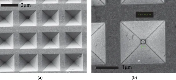
According to our approach special size fitted SERS active substrate was prepared by micromachining techniques in silicon wafer to be applicable for particle entrapment [2,3]. Periodic array of inverse pyramid structures were manufactured by anisotropic silicon etching and covered by gold. Gold nanospheres were trapped in the pyramidal cavities subsequently (Figure 1).
Proceedings 2018, 2, 1069 2 of 4
(a) (b)
Figure 1. Scanning electron microscopic images of the periodic array of gold coated inverse pyramids (a), and the entrapped gold nanoparticle (b).
SERS performance of the hierarchically combined structures was analyzed and compared by using a highly diluted benzophenone solution dripped onto different surfaces. The SERS spectra were recorded on a Renishaw 1000 micro-Raman spectrometer using 785 nm excitation. The resulted Raman spectra are demonstrated in Figure 2 in case of flat Au layer for reference (curve a), array of gold coated inverse pyramids (curve b), array of gold coated inverse pyramids with entrapped nanoparticles (curve c). The selectivity and sensitivity of the proposed combined 3D structure and molecule detection method were characterized by octadecanethiol surface functionalization. The recorder spectrum was also added in Figure 2 (curve d).
Figure 2. Reference normal Raman (a) and surface enhanced Raman spectra of benzophenone recorded on array of gold coated inverse pyramids (b), array of gold coated inverse pyramids with entrapped nanoparticles (c) and the latter with octadecanethiol surface functionalization (d).
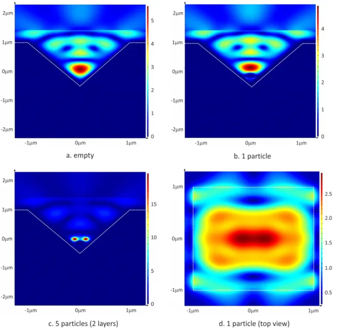
To understand the analytical performance the near-field intensity distributions of inverse pyramid arrays were studied by FDTD simulations using the Lumerical FDTD Solutions v.8.15.736 software. Silicon was used as substrate material of the inverse pyramids with 200 nm gold coating, and 1 or 5 periodically ordered gold nanospheres (Ø 250 nm) were placed into the pyramids in 1 and 2 layers, respectively. Near-field profiles were demonstrated in Figure 3.
The significant localised intensity enhancements in specific hotspots of the combined structure were clearly demonstrated by the simulation results.
Proceedings 2018, 2, 1069 3 of 4
Figure 3. Relative near-field intensity distributions (a.u.) of inverse pyramids being empty (a) and filled with 1 (b) or 5 (c) gold nanoparticles on the monitor cross-section perpendicular to the polarization plane, and from above in case of 1 entrapped particle (d).
3. Results and Conclusions
Special hierarchically combined 3D SERS active structures were designed and fabricated by micromachining technologies. The proposed sensitivity enhancement of Raman spectroscopy was proved by FDTD simulations and experimentally also. The analytical performance of the developed SERS structure was compared to plain and conventional periodic gold surfaces in case of different model molecules. The significant sensitivity and selectivity of the applied SERS structures and spectroscopic method were demonstrated and their applicability in high performance molecular analysis was proposed.
Author Contributions: O.H. and P.F. constructed and developed the 3D SERS microstructures, I.R. and M.V.
designed and implemented the Raman spectroscopic analysis.
Acknowledgments: The technical support of the experts Magda Erős, Margit Payer and Levente Illés in micro-, nanofabrication is highly appreciated.
Conflicts of Interest: The authors declare no conflict of interest.
Proceedings 2018, 2, 1069 4 of 4
References
1. Ryder, A.G. Surface enhanced Raman scattering for narcotic detection and applications to chemical biology. Curr. Opin. Chem. Biol. 2005, 9, 489–493.
2. Rigó, I.; Veres, M.; Fürjes, P. SERS active periodic 3D structure for trapping and high sensitive molecular analysis of particles or cells. Proceedings 2017, 1, 560.
3. Rigó, I.; Veres, M.; Himics, L.; Tóth, S.; Czitrovszky, A.; Nagy, A.; Fürjes, P. Comparative analysis of SERS substrates of different morphology. Procedia Eng. 2016, 168, 371–374.
© 2018 by the authors. Licensee MDPI, Basel, Switzerland. This article is an open access article distributed under the terms and conditions of the Creative Commons Attribution (CC BY) license (http://creativecommons.org/licenses/by/4.0/).