Peripheral cannabinoid-1 receptor blockade restores hypothalamic leptin signaling
Joseph Tam1,*, Gerg}o Szanda2,5, Adi Drori1, Ziyi Liu2, Resat Cinar2, Yoshihiro Kashiwaya3, Marc L. Reitman4, George Kunos2,**
ABSTRACT
Objective: In visceral obesity, an overactive endocannabinoid/CB1receptor (CB1R) system promotes increased caloric intake and decreases energy expenditure, which are mitigated by global or peripheral CB1R blockade. In mice with diet-induced obesity (DIO), inhibition of food intake by the peripherally restricted CB1R antagonist JD5037 could be attributed to endogenous leptin due to the rapid reversal of hyperleptinemia that maintains leptin resistance, but the signaling pathway engaged by leptin has remained to be determined.
Methods: We analyzed the hypothalamic circuitry targeted by leptin following chronic treatment of DIO mice with JD5037.
Results: Leptin treatment or an increase in endogenous leptin following fasting/refeeding induced STAT3 phosphorylation in neurons in the arcuate nucleus (ARC) in lean and JD5037-treated DIO mice, but not in vehicle-treated DIO animals. Co-localization of pSTAT3 in leptin-treated mice was significantly less common with NPYþthan with POMCþARC neurons. The hypophagic effect of JD5037 was absent in melanocortin-4 receptor (MC4R) deficient obese mice or DIO mice treated with a MC4R antagonist, but was maintained in NPY/mice kept on a high-fat diet.
Conclusions: Peripheral CB1R blockade in DIO restores sensitivity to endogenous leptin, which elicits hypophagia via the re-activation of melanocortin signaling in the ARC.
Ó2017 The Authors. Published by Elsevier GmbH. This is an open access article under the CC BY-NC-ND license (http://creativecommons.org/licenses/by-nc-nd/4.0/).
Keywords Peripheral CB1 blockade; Leptin resistance; POMC; NPY; Diet-induced obesity
1. INTRODUCTION
Visceral obesity and its metabolic complications, commonly called the metabolic syndrome, represent a growing public health concern worldwide[1]. Accumulating evidence supports the pathogenic role of an overactive endocannabinoid/CB1receptor (CB1R) system in obesity/
metabolic syndrome [2e4]. Indeed, the CB1R antagonist/inverse agonist rimonabant was effective in reducing body weight in obese/
overweight people and also improved the associated insulin resis- tance, fatty liver, and dyslipidemia[5], but the therapeutic development of this class of compounds was halted due to neuropsychiatric side effects mediated by blockade of CB1R in the central nervous system [6]. More recent evidence indicates that activation of CB1R in periph- eral tissues, including adipose tissue[7], liver[8], skeletal muscle[9], the endocrine pancreas[10], and proinflammatory macrophages[11], contributes to visceral adiposity and its metabolic complications, and its selective blockade by CB1R antagonists/inverse agonists with limited brain penetrance can improve the obese phenotype in animal
models of diet-induced metabolic syndrome without eliciting behaviors attributable to blockade of CB1R in the CNS[11e13].
In a previous study, we reported that the peripherally restricted CB1R inverse agonist JD5037 was as effective as its brain penetrant parent compound SLV319 (ibipinabant) in normalizing all of the metabolic consequences of a high-fat diet (HFD), including the normalization of body weight as well as causing transient but pronounced hypophagia [13]. The latter effect was surprising in view of the dominant role of central neural circuits in the control of food intake. To resolve this paradox, we posited that the hypophagic effect of chronic blockade of peripheral CB1R is mediated by endogenous leptin, as a result of the rapid reversal of the hyperleptinemia and associated leptin resistance of diet-induced obese (DIO) mice[13]. Reversal of the hyperleptinemia, in turn, could be attributed to reduced leptin production due to direct inhibition of CB1R in adipocytes and sympathetic nerve endings in adipose tissue, as well as increased leptin clearance in the kidney, due to inhibition of CB1R in renal proximal tubular cells[13].
1Obesity and Metabolism Laboratory, The Institute for Drug Research, School of Pharmacy, Faculty of Medicine, The Hebrew University of Jerusalem, Jerusalem 9112001, Israel2Laboratory of Physiologic Studies, National Institute on Alcohol Abuse and Alcoholism, USA3Laboratory of Metabolic Control, National Institute on Alcohol Abuse and Alcoholism, USA4Diabetes, Endocrinology and Obesity Branch, National Institute of Diabetes and Digestive and Kidney Diseases, National Institutes of Health, Bethesda, MD 20892, USA
5 Current address: Department of Physiology, Faculty of Medicine, Semmelweis University, Budapest, Hungary.
*Corresponding author. Obesity and Metabolism Laboratory, The Institute for Drug Research, School of Pharmacy, Faculty of Medicine, POB 12065, Jerusalem 91120, Israel.
Fax:þ972 2 675 7015. E-mail:yossit@ekmd.huji.ac.il(J. Tam).
**Corresponding author. Laboratory of Physiologic Studies, National Institute on Alcohol Abuse and Alcoholism, National Institutes of Health, Bethesda, MD 20892, USA.
Fax:þ1 301 480 0257. E-mail:george.kunos@nih.gov(G. Kunos).
Received May 5, 2017Revision received June 14, 2017Accepted June 16, 2017Available online 24 June 2017 http://dx.doi.org/10.1016/j.molmet.2017.06.010
Original Article
The ability of peripherally generated leptin to reach its hypothalamic receptors reconciles the paradox of modulating a centrally regulated function using a drug restricted to the periphery. The question remains:
which neural circuits are activated by endogenous leptin, once the leptin resistance in DIO mice is reversed by treatment with a periph- erally restricted CB1R inverse agonist. Generally, leptin coordinates the activity of the appetitive neural circuitry primarily via promoting the activity of pro-opiomelanocortin (POMC)/cocaine- and amphetamine- regulated transcript (CART) neurons [14e16] and subsequent anorexigenic signaling bya-melanocyte-stimulating hormone (a-MSH) via the melanocortin 4 receptor (MC4R) [17] and simultaneously inhibiting the activity of orexigenic neuropeptide Y (NPY)/agouti-related peptide (AgRP) neurons in the arcuate nucleus (ARC)[18,19]. There- fore, we examined the relative role of these two pathways in the anorexigenic effect of endogenous leptin in DIO mice chronically treated with the peripheral CB1R antagonist JD5037. The results indicate that resensitization of DIO mice to leptin is reflected in increased leptin-induce phosphorylation of STAT3 in both POMC and NPY neurons, with the former playing a key role in the anorexigenic and weight reducing actions of endogenous leptin, as reflected in the absence of these effects in MC4R knockout mice or DIO mice treated with an MC4R antagonist.
2. MATERIALS AND METHODS
2.1. Animals and experimental protocol
The experimental protocol was approved by the Institutional Animal Care and Use Committees of the NIAAA and Hebrew University of Jerusalem. Male 6-week old NPY/ mice (129S-Npytm1Rpa/J) and their littermate controls were obtained from the Jackson Laboratory.
Adult, male, genetically obese MC4R/mice (MC4Rtm1Lowl/J) were generated by heterozygote mating using wild-type littermates as controls. Mice were maintained under a 12 h light/dark cycle and fed ad libitum. To generate DIO, C57Bl6/J and NPY/mice were fed a HFD (Research Diet, D12492; 60% calories from fat, 20% from protein, and 20% from carbohydrates), with age-matched lean controls receiving a standard laboratory diet (STD, NIH-31 rodent diet) for 12e 14 weeks. To achieve normoleptinemia in DIO mice, we adapted a protocol described by Knight et al.[20]. Briefly, leptin-deficientob/ob mice were implanted with an osmotic minipump (model 2001D, Alzet Osmotic Pumps; Durect, Cupertino, CA) delivering leptin dissolved in phosphate-buffered saline (PBS) at a rate of 150 ng/h for 12 weeks, during which time they were fed a HFD to induce DIO. Control groups of wild-type mice on HFD andob/obmice on STD were also implanted with minipumps delivering PBS at the same rate. Pumps were replaced every 28 days. HFD-induced obese C57Bl/6J, NPY/ and normo- leptinemicob/obmice, and genetically obese MC4R/mice on STD were treated daily with JD5037 (3 mg/kg/day, po) or vehicle (Veh; 4%
DMSOþ1% Tween80 in normal saline) for 7 days. Body weight and food intake were monitored daily. Mice were euthanized by cervical dislocation under anesthesia, and their brains and trunk blood were collected for further analyses.
2.2. Leptin sensitivity
Leptin sensitivity was assessed in lean and DIO mice and DIO mice treated daily with JD5037 (3 mg/kg po.) for 7 days followed by twice daily treatment with leptin (3 mg/kg, ip) or vehicle for an additional 4 days. One hour after the last dose of leptin or vehicle, mice were anesthetized, perfused via the left ventricle with 5 mL of 0.9% saline for 1 min followed by 60e80 mL of cold 0.1 M phosphate buffer (pH 7.4) containing 4% paraformaldehyde for 15 min at room temperature.
Then, the perfused brains were removed, post-fixed in the same fixative for 6 h at 4C and further processed for immunohistochemical analyses.
2.3. Immunohistochemistry
After fixation, the brains were cryoprotected with 0.1 M phosphate buffer (pH 7.4) containing 20% sucrose for 72 h and then rapidly frozen in isopentane pre-cooled to70C with dry ice. Serial coronal sections (30mm) were cut using a cryostat through the brain region containing the ARC. After inactivating the endogenous peroxidase activity with 0.6% hydrogen peroxidase (SigmaeAldrich, St. Louis, MO), sections were incubated separately with avidin and biotin solutions (Vector Lab, Burlingame, CA) for blocking nonspecific binding of endogenous biotin, biotin-binding protein, and lectins. Then, the sections were incubated free-floating in 0.01 M PBS (pH 7.4) containing 2% normal donkey serum (Jackson ImmunoResearch Labs, West Grove, PA), 0.3% Triton X-100 (SigmaeAldrich, St. Louis, MO) and rabbit anti-pSTAT3 antibody (1:1,500; Cell Signaling, Beverly, MA) or rabbit anti-c-Fos antibody (1:10,000; Santa Cruz Biotechnology, CA) for 43 h at 4 C. The immunoreaction product was visualized using the Vectastain elite ABC kit (Vector Lab., Burlingame, CA) and 30,30-diaminobenzidine (Sigmae Aldrich, St. Louis, MO) as a chromogen. After thorough washes, sec- tions were mounted on gelatin-coated slides. Following dehydration in ethanol, sections were cleared in xylene and coverslipped in PermountÒ(Fisher Scientific, Fair Lawn, NJ). Staining for pSTAT3 and c-Fos were visualized using a brightfield light source and captured with a digital camera mounted on an Olympus BX41 microscope.
Sections earmarked for double labeling with pSTAT3þPOMC or pSTAT3þNPY were processed according to the indirect immunofluo- rescence method of Coons[21]. Briefly, following washes in PBS, the sections were incubated free-floating in PBS containing Triton X-100, blocking serums and two primary antibodies: rabbit anti-pSTAT3 (1:500; Cell Signaling, Beverly, CA) and chicken anti-POMC (1:1,000;
Abcam, Cambridge, MA) or rabbit anti-pSTAT3 (1:500; Cell Signaling) and chicken anti-NPY (1:2,000; Novus Biologicals) for 43 h at 4C.
Then, the sections were incubated at room temperature in PBS con- taining Triton X-100, blocking serum, and donkey anti-Rabbit Alexa Fluor 594 (1:250; Invitrogen) for 1 h, and then Triton X-100, blocking serum, and goat anti-chicken Alexa Fluor 488 (1:250; Invitrogen) for another hour. After thorough washes in PBS, all sections were mounted on gelatin-coated microscope slides, coverslipped with Vectashield (Vector Lab.), and analyzed using a Zeiss LSM700 confocal microscope.
The localization of the immunoreactive signals was identified using Hof’s mouse brain atlas. Six corresponding sections in the ARC, organized in a consecutive rostral to caudal sequence from0.94 mm to2.92 mm relative to the bregma, were counted for pSTAT3 and c- Fos positive cells and analyzed for co-localization of pSTAST3 with POMC or NPY positive cells. Cell counts from 4 mice per group were obtained from both sides of the brain in each section. pSTAT3-, c-Fos-, POMC-, and NPY-positive cells as well as double labeled cells were scored in each ARC section from vehicle and leptin-treated animals.
The percentage of double-positive pSTAT3þPOMC or pSTAT3þNPY cells was also determined by cell counting.
2.4. Blood biochemistry
Serum leptin levels were measured by an ELISA kit (Millipore, Billerica, MA, and R&D Systems, Minneapolis, MN).
2.5. Chronic infusion of SHU-9119
To assess whether hypothalamic MC4R mediates the response to peripheral CB1R antagonism in DIO mice, vehicle (saline) or the MC4R
antagonist SHU-9119 (Tocris; 24 nmol/day) was delivered over 7 days by intracerebroventricular (icv) infusion using osmotic minipumps. The effects of simultaneous 7-day treatment with daily oral doses of vehicle or JD5037 (3 mg/kg, po.) on body weight, food intake, and serum leptin levels were determined as described above.
2.6. Fasting/refeeding paradigm
To determine whether peripheral CB1R blockade resensitizes endog- enous leptin action, we used a fasting/refeeding paradigm. Briefly, male, 6e7 week old C57Bl/6J mice were kept on STD or HFD for 5 weeks. During the last week of feeding, the mice were treated with either vehicle or JD5037 (3 mg/kg, po.) for 1 week. Following the last dose, the mice were fasted for 16 h and then food was introduced for an additional 4 h before sacrifice. The hypothalamus and trunk blood were collected from each mouse, and the phosphorylation of STAT3 was determined by western blotting, while serum leptin levels were measured by an ELISA kit (Millipore, Billerica, MA).
2.7. Endocannabinoid measurements
Mice were euthanized and the brain and hypothalamus were rapidly dissected and kept at80C until processed. Arachidonoyl ethano- lamide (AEA) and 2-arachidonoylglycerol (2-AG) in brain cortex and hypothalamus were extracted, purified, and quantified by the stable isotope dilution LC-MS/MS method described previously[13].
2.8. Materials
Rimonabant was obtained from the National Institute of Drug Abuse Drug Supply Program. JD5037, kindly provided by Jenrin Discovery, Inc.[22]. For oral administration by gavage, rimonabant and JD5037 were dissolved in 4% DMSOþ1% Tween-80 in saline. SHU-9119 was purchased from Tocris and was dissolved in saline to be administered by an Alzet osmotic minipump (Durect, Cupertino, CA) at the rate of 24 nmol/day as described previously[23,24].
2.9. Statistics
Data are expressed as meanSEM. Unpaired two-tailed Student’s t- test was used to determine differences between vehicle- and drug- treated groups. Results in multiple groups were compared by one- or two-way ANOVA followed by a Bonferroni or Sidak’s multiple comparisons test, as appropriate (GraphPad Prism v6 for Windows).
Significance was set atP<0.05.
3. RESULTS
3.1. Hyperleptinemia is required for the hypophagic and weight reducing effects of peripheral CB1R blockade
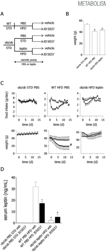
We have previously demonstrated that the weight reducing and hypophagic effects of JD5037 in DIO mice develop rapidly within 7 days of treatment and are associated with a similar rapid reversal of hyperleptinemia and leptin resistance[13]. Here we sought to further test whether hyperleptinemia is, in fact, a prerequisite of the efficacy of peripheral CB1R blockade. To this end, we tested the effects of JD5037 in obese mice with substantially different leptin status using an approach based on that developed by Knight et al.[20], as detailed in the Methods section and illustrated inFigure 1A and B.
Treatment with a daily oral dose of JD5037 (3 mg/kg) for 14 days failed to significantly affect body weight, food intake, and plasma leptin levels in obese but normoleptinemic animals (ob/obmice on HFD chronically infused with leptin) and was similarly ineffective in aleptinemicob/ob mice. On the other hand, and in accordance with our previousfindings [13], JD5037 treatment alleviated hyperleptinemia, caused transient
Figure 1: Effects of peripheral CB1R blockade on food intake and body weight in obese mice with different leptin status. (A)Experimental protocol yielding obese mice with aleptinemia (ob/obmice on standard diet [STD] and chronically infused with PBS), normoleptinemia (ob/obmice on high-fat diet [HFD] infused with leptin), or hyperleptinemia (wild-type mice on HFD, infused with PBS). Horizontal line indicates the period of leptin or PBS infusion.(B)Mean body weights at the end of the pretreatment period. n¼10, 13, and 11 forob/obSTD,ob/obHFDþleptin and WT HFDþPBS groups, respectively. *P<0.0001 as compared toob/obSTDþPBS group.(C)Daily food intake and body weight of vehicle-treated (open symbols) and JD5037-treated animals (filled symbols), n¼6 in all 6 groups.#P<0.001 for the effect of JD5037 vs vehicle treatment.(D)Serum leptin concentrations on day 14 of JD5037 or vehicle treatment, n¼4e6 mice per group. Leptin was undetectable in sera ofob/obmice infused with PBS.#P<0.0001 relative to vehicle-treated WT mice.
hypophagia, and significantly reduced body weight in wild-type DIO mice (Figure 1C and D). Furthermore, the weight-reducing effect of JD5037 in the last group could be counteracted by daily treatment with a pharmacologic dose of leptin which also reversed the JD5037- induced decrease in plasma leptin (Supplementary Fig. 1). Together, these findings support the notion that the anorectic and weight reducing effects of peripheral CB1R blockade in DIO are contingent on hyperleptinemia and its drug-induced reversal.
3.2. Peripheral CB1R blockade restores hypothalamic leptin sensitivity
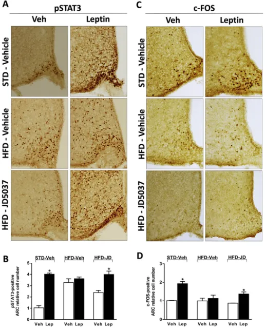
Next, we analyzed leptin-induced STAT3 phosphorylation in the diencephalon, a marker of leptin signaling, to test whether peripheral CB1R blockade restores leptin sensitivity in the mediobasal hypothal- amus. As illustrated inFigure 2A, B, leptin treatment robustly increased STAT3 phosphorylation in the ARC of lean control mice with a few
responsive cells also present in the adjacent ventromedial hypothal- amus (VMH). Although the number of pSTAT3þcells in the VMH was increased in vehicle-treated DIO mice compared to lean control mice, leptin treatment was without any effect on hypothalamic pSTAT3 in DIO mice, similar tofindings by others[20,25]. In contrast, in DIO mice treated with JD5037 for 7 days, leptin treatment robustly increased STAT3 phosphorylation, similarly to its effect in lean controls. To determine the effects of leptin treatment, HFD status and peripheral CB1R blockade on neuronal activity in the ARC, we examined the expression of c-Fos protein in consecutive brain sections used to evaluate pSTAT3 expression. We found that the leptin-induced in- crease in c-Fos expression in the ARC of lean mice was completely absent in vehicle-treated DIO mice but was partially restored in JD5037-treated DIO mice (Figure 2C, D). Thus, peripheral CB1R blockade restores hypothalamic leptin-induced STAT3 phosphorylation and neural activation in DIO mice.
Figure 2: Peripheral CB1R blockade restores hypothalamic leptin-induced STAT3 phosphorylation in DIO mice.Effect of leptin (3 mg/kg, ip twice daily for 4 days) on tyrosine705-phosphorylated STAT3-positive (pSTAT3þ) neurons(A)or c-Fos expression(C)in hypothalamic coronal sections obtained from lean and DIO C57Bl/6J mice treated for 7 days with either vehicle or JD5037 (3 mg/kg, po). Quantification of the number of pSTAT3þcells(B)or c-Fosþcells(D)in the ARC. Data are meanSEM from 4 animals in each group. *P<0.05 relative to the corresponding control group treated with vehicle (Veh).
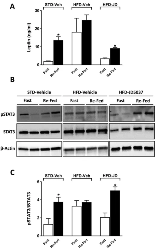
Analyses of leptin sensitivity typically involve the use of pharmaco- logical doses of leptin even in lean mice. We therefore tested whether changes in the phosphorylation status of STAT3 are also detectable in response to changes in the plasma levels of endogenous leptin within its physiological range. Leptin plasma levels drop during fasting and
increase upon refeeding [26]. We therefore tested the effects of overnight fasting followed by brief refeeding on plasma leptin levels and pSTAT3/STAT3 status in the mediobasal hypothalamus. As shown inFigure 3, plasma leptin level at the end of the fasting period was low in lean mice, was about ten times higher in vehicle-treated DIO mice,
Figure 3: Peripheral CB1R blockade restores sensitivity to endogenous leptin.Wild-type C57Bl/6J mice on STD or HFD were treated orally for one week with 3 mg/kg/day JD5037 or vehicle. After the last dose the mice were fasted for 16 h, then provided access to food for 4 h, after which they were sacrificed and brain tissue and plasma were collected for quantifying plasma leptin(A), and hypothalamic pSTAT3/STAT3(BandC), as detailed in Methods. Quantitation of pSTAT3 in panel B was done by densitometry, and values are expressed as the ratio of pSTAT3/STAT3. Data are meanSEM from 5 animals in each group. *P<0.05 relative to the corresponding value in the fasted group.
Figure 4: Peripheral CB1R blockade increases leptin-induced tyrosine705-STAT3 phosphorylation in NPY-positive neurons.Representative photomicrographs showing tyrosine705-pSTAT3þneurons (red) and NPYþneurons (green) in the hypothalamic ARC 45 min after the last injection of leptin (3 mg/kg, ip. twice daily for 4 days) or vehicle(A).
Insets (AeF): Examples of NPYþneurons coexpressing pSTAT3. Quantitation of the percentage of pSTAT3þNPY neurons in coronal brain sections(B). pSTAT3þNPY neurons were counted from four brain sections from each group. *P<0.05 relative to the corresponding control group treated with Veh.#P<0.05 relative to the leptin-treated mice in the STD- Veh group.
and returned to near control levels in DIO mice treated for one week with daily doses of 3 mg/kg JD5037. Refeeding elicited significant, several-fold increases in plasma leptin in both lean mice and DIO mice treated with JD5037, but no significant further increase was evident in vehicle-treated DIO mice. STAT3 phosphorylation quantified using Western blots showed parallel changes, being significantly increased upon refeeding in lean and in DIO mice treated with JD5037 but not in vehicle-treated DIO animals. This indicates that JD5037 treatment of obese mice reverses their resistance to endogenous leptin.
3.3. Inhibition of NPY signaling by leptin is not involved in the weight reducing and hypophagic effects of peripheral CB1R blockade in DIO mice
In the above experiments, JD5037 treatment restored leptin responsiveness in the ARC of DIO mice and normalized their body weight. These effects of leptin can be mediated either by inhibition of orexigenic NPY/AgRP neurons or activation of anorexigenic POMC/
CART neurons in the ARC[27,28]. Wefirst explored the possibility that the restored effects of leptin that result from peripheral CB1R Figure 5: Peripheral CB1R blockade is equieffective in wild-type and NPYL/Lmice on HFD. (A)Body weight and food-intake of WT and NPY/mice kept on STD or HFD and treated with JD-5037 or vehicle. *P<0.01, JD5037 vs vehicle, n¼5 in all groups except for STD groups (n¼4) and NPY/HFD JD-5037 treated (n¼4).(B)Serum leptin concentrations on day 7 of JD5037 treatment. *P<0.01, vs STD;#P<0.01 and###P<0.001 vs HFD vehicle group. Numbers of observations were as inA.
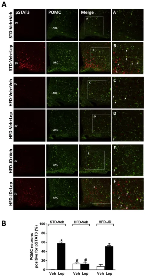
Figure 6: Peripheral CB1R blockade increases leptin-induced tyrosine705-STAT3 phosphorylation in POMC-positive neurons.Representative photomicrographs showing tyrosine705pSTAT3þneurons (red) and POMC-positive neurons (green) in the hypothalamic ARC 45 min after the last injection of leptin (3 mg/kg, ip twice daily for 4 days) or vehicle (A). Insets (AeF): examples of POMCþneurons co-expressing pSTAT3. Quantitation of the percentage of pSTAT3þPOMC neurons in coronal brain sections(B). pSTAT3þPOMC neurons were counted from four brain sections from each group. *P<0.05 relative to the corresponding control group treated with Veh.#P<0.05 relative to the vehicleþleptin- treated mice in the STD-Veh group.
blockade involve the NPY/AgRP pathway. As shown inFigure 4, leptin induced detectable STAT3 phosphorylation in about one third of NPY- positive neurons in lean mice, as assessed by double immunohisto- chemistry and confocal microscopy. HFD suppressed leptin-activated STAT3 phosphorylation in NPY-positive neurons, and this suppression was only partially reversed by chronic treatment with JD5037 (3 mg/
kg, po) for 7 days (Figure 4A, B).
To further test the functional role of altered NPY signaling in mediating the weight reducing and hypophagic effects of JD5037, we treated HFD-induced obese NPY/ mice and their HFD-fed wild-type littermates with JD5037 (3 mg/kg, po) for 7 days and monitored their body weight, food intake, and serum leptin levels.
Compared to wild-type mice on STD, both wild-type and NPY/ mice on HFD displayed similar significant increases in body weight (Figure 5A), visceral adiposity (Supplementary Fig. 2B), and serum leptin levels (Figure 5B). JD5037 caused robust decreases in body weight, food intake, and serum leptin levels in NPY/ animals, which were similar or, in the case of serum leptin, even slightly greater than its parallel effects in wild-type DIO littermates (Figure 5A,B, and Supplementary Fig. 2). These findings argue against the involvement of NPY in the weight reducing effect of JD5037, although they do not rule out the possible involvement of AgRP.
3.4. Peripheral CB1R blockade restores hypothalamic leptin sensitivity via POMC signaling
Since leptin activates the anorexigenic POMC/CART neurons in the ARC [14,16], we next explored whether peripheral CB1R blockade affects this neuronal pathway. Consistent with previous reports [28], leptin administration stimulated STAT3 phosphorylation in POMC-positive neurons of lean mice, as assessed by double immunohistochemistry and confocal microscopy (Figure 6). The percentage of POMCþ/ pSTAT3þneurons in the ARC (57.43.3%) was significantly higher than the percentage of NPYþ/pSTAT3þ neurons (see Figure 4, 35.96.8%,P<0.01). Leptin-stimulated STAT3 phosphorylation in these neurons was completely abrogated in DIO mice, although basal pSTAT3 levels were significantly increased. Leptin-induced STAT3 phosphorylation was fully restored in DIO mice treated with JD5037 (3 mg/kg, po.) for 7 days (Figure 6) prior to leptin treatment, suggesting that peripheral CB1R blockade enhances hypothalamic leptin signaling via the activation of anorexigenic POMC neurons.
The anorexigenic effect of leptin is believed to be mediated bya-MSH acting on MC4R[29]. To explore the functional role of POMC-positive neurons in mediating the hypophagic effect of JD5037, we treated STD-fed obese MC4R/mice and their HFD-fed littermate controls with JD5037 (3 mg/kg, po.) for 7 days and monitored their body weight, food intake, and serum leptin levels. As shown inFigure 7, the
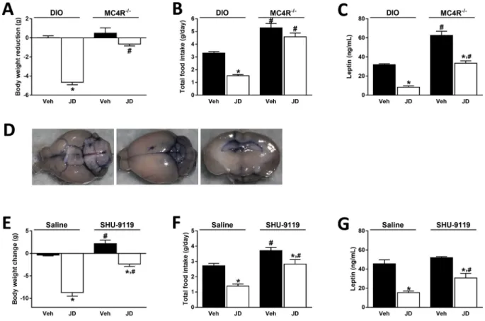
Figure 7: MC4R deactivation abolishes the hypophagic and weight reducing effects of peripheral CB1R blockade.JD5037 (3 mg/kg, po, for 7 days) reduced body weight (A)and food intake(B)in DIO mice but not in MC4R/animals. Absolute body weights of DIO mice are: 42.30.7 g (Veh) vs 36.40.9 g (JD5037), and of MC4R/mice:
43.21.5 (Veh) vs 42.31.8 g (JD5037). Serum leptin levels were similarly reduced by JD5037 in both mouse strains, although the net values were much greater in MC4R/ mice(C). Treatment of DIO mice with SHU-9119 (24 nmol/day, icv via osmotic minipump, for 7 days) attenuated the reductions in body weight, food intake and serum leptin levels induced by JD5037 (3 mg/kg/day, po, for 7 days) (EeG). Absolute body weights in saline-infused DIO mice are 50.01.6 g (Veh) vs 41.90.8 g (JD5037), and in SHU-9119- infuced DIO mice are 53.80.9 g (Veh) vs 49.51.2 g (JD5037).D, representative pictures of a cannulated brain demonstrating the location of the cannula and the injection site of SHU-9119 into the 3rd ventricle (blue staining). Data are obtained from 4 to 5 animals in each group. *P<0.05 relative to the corresponding obese group treated with Veh;
#P<0.05 relative to the corresponding DIO group treated with either Veh or JD5037.
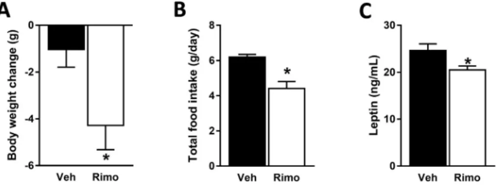
weight reducing and hypophagic effects of JD5037 observed in DIO mice were absent in MC4R/mice (Figure 7A, B). Although JD5037 treatment reduced serum leptin levels in both mouse strains, the absolute levels remained significantly higher in MC4R/compared to DIO mice (Figure 7C). To further test the role of MC4R in mediating the increased hypothalamic signaling of leptin in DIO mice treated with JD5037, we continuously infused via a minipump the potent MC4R antagonist, SHU-9119 (24 nmol/day, icv) to HFD-fed animals that were simultaneously treated with daily oral doses of JD5037 (3 mg/kg) or vehicle for 7 days. The correct location of the icv infusion of SHU-9119 into the 3rd ventricle was verified postmortem by the infusion of to- luidine blue (Figure 7D). In the presence of SHU-9119, the effects of JD5037 on body weight, food intake, and serum leptin levels were significantly attenuated (Figure 7EeG). Unlike these results, treatment of MC4R/ mice with the globally acting CB1R inverse agonist rimonabant resulted in reductions in body weight, food intake and serum leptin levels (Figure 8AeC).
3.5. Effects of high-fat diet and peripheral CB1R blockade on hypothalamic endocannabinoids
Our laboratory has earlier documented an inverse relationship between leptin and hypothalamic endocannabinoids levels and a role in appetite regulation[30]. We therefore measured the tissue levels of AEA and 2- AG in the cortex and hypothalami of lean and DIO mice as well as DIO mice chronically treated with JD5037 (3 mg/kg, po.). As seen in Supplementary Figure 3, keeping mice on HFD resulted in a significant increase in hypothalamic 2-AG and a smaller, not statistically signifi- cant increase in AEA levels. In contrast, treatment of DIO mice with JD5037 significantly reduced AEA, but not 2-AG levels. These changes were specific for the hypothalamus, as brain endocannabinoid levels remained unchanged by either diet or drug treatment.
4. DISCUSSION
Rimonabant, thefirst-in-class, globally acting CB1R antagonist/inverse agonist is an anti-obesity agent that not only reduced body weight but also improved all the associated metabolic abnormalities[5]. Although neuropsychiatric side-effects led to its withdrawal from the market, this was not unexpected in view of the known obligatory role of endocannabinoids and CB1R in the mesolimbic dopaminergic reward pathway in mediating both natural and drug reward[31]. Subsequent evidence that non-brain-penetrant CB1R antagonists are, as or nearly as, effective as rimonabant in improving metabolic end points could be attributed to the presence of CB1R at low yet functionally relevant levels
in tissues involved in metabolic control and their upregulation in obesity. On the other hand, the efficacy of peripheral CB1R blockade in reducing food intake in DIO mice was surprising in view of the dominant role of a central neural circuitry in appetite control. The absence of a similar effect in mice with defective leptin signaling or in wild-type DIO mice treated with a leptin antagonist supported the hypothesis that the hypophagic effect of peripheral CB1R blockade is mediated by endogenous leptin, due to the rapid reversal of hyper- leptinemia and the associated leptin-resistant state[13]. This notion is further supported by the presentfindings that the ability of peripheral CB1R blockade to reduce food intake and body weight is abrogated by clamping serum leptin levels of DIO mice at the normal physiologic range (Figure 1), or by the infusion of exogenous leptin to counter the leptin lowering effect of JD5037 in hyperleptinemic DIO mice (Supplementary Figure 1). These observations are also in agreement with the concept of Knight et al. [20] that hyperleptinemia is both necessary and sufficient for maintaining leptin resistance in DIO.
The presentfindings further indicate that once the leptin resistance of DIO mice is reversed by peripheral CB1R blockade, endogenous leptin acts in the hypothalamus primarily via activating POMC neurons resulting in MC4R activation, as indicated both by increased STAT3 phosphorylation in POMC neurons and a parallel increase in c-Fos expression in the ARC. The dominant role of melanocortin signaling is further supported by the failure of peripheral CB1R blockade to reduce food intake and body weight in obese MC4R/mice and its reduced efficacy in DIO mice simultaneously exposed to a MC4R antagonist. In contrast, the hypophagic effect of JD5037 is fully maintained, if not increased, in HFD-fed NPY/mice compared to their HFD-fed wild- type littermates, which is similar to the maintained hypophagic effect of rimonabant in overnight fasted NPY/mice[30]. The preferential activation of POMC neurons is also indicated by the significantly greater co-localization of pSTAT3 with POMC than with NPY expressing arcuate neurons following leptin challenge of JD5037-treated DIO mice. Nevertheless, the co-expression of pSTAT3 with some NPY neurons could reflect their inhibition by leptin that would result in reduced AgRP expression and, as a consequence, disinhibition of melanocortin signaling. This mechanism would be retained in NPY/ mice as gene deletion of NPY does not affect the AgRP content of these neurons[32].
Refeeding after fasting was associated with a significant increase in serum leptin and a parallel increase in hypothalamic pSTAT3 in lean and JD5037-treated DIO mice, but not in vehicle-treated DIO mice.
This is compatible with the role of pSTAT3 signaling in the metabolic effects of endogenous leptin, as proposed previously[33,34]. The HFD-
Figure 8: MC4R deactivation does not abolish the hypophagic effect of global CB1R blockade.Rimonabant (3 mg/kg, po., for 7 days) reduced body weight(A), food intake (B)and serum leptin levels(C)in MC4R/mice. Absolute body weights are 52.52.9 g (Veh) vs 49.62.3 g (rimonabant). Data are meanSEM from 4 to 5 animals in each group. *P<0.05 relative to the corresponding control group treated with Veh.
induced increase in the baseline level of hypothalamic pSTAT3 (Figure 2) is also compatible with recent evidence that ARC neurons in DIO mice remain sensitive to endogenous leptin in leptin-resistant states, elicited by high-fat diets [35,36], or chronic central infusion of leptin [37]. However, ourfindings also indicate that at the high circulating levels of leptin associated with DIO, leptin receptors are maximally activated, so that any further increase in leptin either from endogenous or exogenous sources results in no change in either pSTAT3 or food intake. The apparent dissociation between functional leptin resistance and continued leptin signaling in ARC neurons in DIO may be related to the control of endogenous reactive oxygen species in the hypothalamus[38].
Although the role of the LepR/pSTAT3/aMSH/MC4R pathway in the acute metabolic effects of leptin is well established[17,33,39], genetic inactivation of STAT3 in POMC neurons reduced, but did not abolish, the hypophagic effect of leptin[40], whereas STAT3 inactivation in AgRP/NPY neurons also attenuated leptin-induced hypophagia [41].
This suggests that both POMC and AgRP/NPY neurons could be targets of the acute metabolic effects of leptin. Although STAT3 phosphory- lation following chronic peripheral CB1R blockade could be detected in a small proportion NPYþneurons (Figure 4), genetic deletion of NPY did not affect the acute metabolic response of DIO mice to leptin (Figure 5).
Nevertheless, thesefindings do not rule out the role of the AgRP/NPY pathway in the hypophagic effects of leptin under different conditions.
Indeed, recentfindings indicate that AgRP neurons drive consumma- tory feeding but are dispensable for reward-driven feeding of palatable foods, such as a HFD used in the present study[42].
It was reported that young pre-obese MC4R/mice retain sensitivity to exogenous leptin [29] and to leptin receptor antagonist [35], whereas older animals with manifest obesity do not respond to such treatments. This implies that there is an age and/or obesity-dependent switch towards the POMC pathway to convey the central effects of leptin on energy metabolism[35]. Whether such a shift is predomi- nantly age or obesity related remains to be determined, but it is noteworthy that the age-related decrease in central leptin sensitivity was found to be promoted by concurrent obesity[43]. Nevertheless, the abovefindings are in good agreement with the present observa- tions that once peripheral CB1R blockade resensitizes the mediobasal hypothalamus to leptin, the hormone signals primarily via the mela- nocortin system to reduce food intake and body weight in obese animals.
Earlier findings indicate that both peripheral and central CB1R are involved in the orexigenic effects of endocannabinoids. Peripherally restricted CB1R antagonists inhibit food intake in HFD-fed mice [12,13,44]in a leptin-dependent manner[13], but failed to inhibit the hunger-induced increase in food intake in STD-fed lean mice, which could still be inhibited by rimonabant in a leptin-independent manner [13]. Similarly, JD5037 treatment failed to reduce food intake and body weight in MC4R/mice, whereas rimonabant retained its efficacy. It could be argued that intact leptin signaling via MC4R mediates the hypophagic effect of peripheral CB1R blockade, but not the effect of rimonabant, which, in these animals, is likely due to blockade of CB1R in the CNS. Indeed, pharmacological blockade of MC4R was reported to cause a delayed increase in hypothalamic endocannabinoid levels [45], and a possible similar increase in the hypothalami of MC4R/ mice may contribute to their hyperphagia via increased CB1R activa- tion, which would be antagonized by rimonabant.
Paradoxically, altered central CB1R activity may also contribute to the hypophagic effect of peripheral CB1R blockade. Our laboratory has earlier reported that leptin-deficientob/obmice have elevated 2-AG levels in the hypothalamus, whereas leptin treatment of these
animals preferentially reduces hypothalamic AEA [30]. Interestingly, the same pattern was evident in the present study, in which HFD feeding increased hypothalamic 2-AG (in agreement with thefindings of others[46,47]), and JD5037 treatment decreased hypothalamic AEA levels. This is compatible with the hypothesis that the hypophagic effect of JD5037 is mediated by endogenous leptin [13], in part through reducing central AEA/CB1R signaling.
Hyperleptinemia is thought to be responsible for maintaining a leptin resistant state in obesity[20], and we have earlier demonstrated that peripheral CB1R blockade reverses the hyperleptinemia of DIO mice by antagonizing leptin production in adipose tissue and promoting leptin clearance by the kidney[13]. Because circulating leptin can reach the mediobasal hypothalamus [48], normalization of plasma leptin may directly lead to resensitization of hypothalamic leptin receptors, resulting in increased signaling by endogenous leptin. Alternatively, circulating leptin may regulate the sensitivity of hypothalamic leptin receptors indirectly, through vagal afferent neurons. Leptin receptors are expressed in vagal afferents innervating the stomach and duo- denum, and their activation was reported to promote satiety[49,50], whereas their genetic deletion led to hyperphagia and obesity[51].
Others found that catabolic changes in visceral fat depots due to overexpression of uncoupling protein-1 (UCP1) lead to leptin- dependent hypophagia, which can be abolished by vagal deaf- ferentation[52]. Increased energy expenditure by CB1R blockade also involves increased UCP1 expression in white adipocytes [53], and vagal deafferentation was found to abolish the hypophagic effect of rimonabant[54]. Taken together, thesefindings could suggest that an interaction between endocannabinoids and leptin at the level of vagal sensory afferents plays a role in the resensitization of hypothalamic leptin signaling by peripheral CB1R blockade. Studies are in progress to explore this intriguing possibility.
AUTHOR CONTRIBUTIONS
JT, AD, GS, ZL, RC, and YK conducted the experiments and analyzed the data. MLR provided material and analyzed the data. JT and GK designed and supervised the experiments and wrote the manuscript.
ACKNOWLEDGMENTS
This work was supported by intramural funds from the National Institute on Alcohol Abuse and Alcoholism, NIH to G.K., and by the Israel Science Foundation (ISF) research grant to J.T. (Grant #617/14). G. S. is supported by a János Bólyai Research Scholarship of the Hungarian Academy of Sciences.
CONFLICT OF INTEREST
None declared.
APPENDIX A. SUPPLEMENTARY DATA
Supplementary data related to this article can be found athttp://dx.doi.org/10.1016/j.
molmet.2017.06.010.
REFERENCES
[1] Kopelman, P.G., 2000. Obesity as a medical problem. Nature 404:635e643.
[2] Kunos, G., Osei-Hyiaman, D., Liu, J., Godlewski, G., Batkai, S., 2008. Endo- cannabinoids and the control of energy homeostasis. Journal of Biological Chemistry 283:33021e33025.
[3] Mazier, W., Saucisse, N., Gatta-Cherifi, B., Cota, D., 2015. The endocanna- binoid system: pivotal orchestrator of obesity and metabolic disease. Trends in Endocrinology and Metabolism 26:524e537.
[4] Silvestri, C., Di Marzo, V., 2013. The endocannabinoid system in energy ho- meostasis and the etiopathology of metabolic disorders. Cell Metabolism 17:
475e490.
[5] Despres, J.P., Golay, A., Sjostrom, L., Rimonabant in Obesity-Lipids Study, G, 2005. Effects of rimonabant on metabolic risk factors in overweight patients with dyslipidemia. The New England Journal of Medicine 353:2121e2134.
[6] Moreira, F.A., Grieb, M., Lutz, B., 2009. Central side-effects of therapies based on CB1 cannabinoid receptor agonists and antagonists: focus on anxiety and depression. Best Practice & Research. Clinical Endocrinology & Metabolism 23:
133e144.
[7] Cota, D., Marsicano, G., Tschop, M., Grubler, Y., Flachskamm, C., Schubert, M., et al., 2003. The endogenous cannabinoid system affects energy balance via central orexigenic drive and peripheral lipogenesis. The Journal of Clinical Investigation 112:423e431.
[8] Osei-Hyiaman, D., DePetrillo, M., Pacher, P., Liu, J., Radaeva, S., Batkai, S., et al., 2005. Endocannabinoid activation at hepatic CB1 receptors stimulates fatty acid synthesis and contributes to diet-induced obesity. The Journal of Clinical Investigation 115:1298e1305.
[9] Eckardt, K., Sell, H., Taube, A., Koenen, M., Platzbecker, B., Cramer, A., et al., 2009. Cannabinoid type 1 receptors in human skeletal muscle cells participate in the negative crosstalk between fat and muscle. Diabetologia 52:664e674.
[10] Nakata, M., Yada, T., 2008. Cannabinoids inhibit insulin secretion and cyto- solic Ca(2þ) oscillation in islet beta-cells via CB1 receptors. Regulatory Peptides 145:49e53.
[11] Jourdan, T., Godlewski, G., Cinar, R., Bertola, A., Szanda, G., Liu, J., et al., 2013. Activation of the Nlrp3 inflammasome in infiltrating macrophages by endocannabinoids mediates beta cell loss in type 2 diabetes. Nature Medicine 19:1132e1140.
[12] Tam, J., Vemuri, V.K., Liu, J., Batkai, S., Mukhopadhyay, B., Godlewski, G., et al., 2010. Peripheral CB1 cannabinoid receptor blockade improves car- diometabolic risk in mouse models of obesity. The Journal of Clinical Inves- tigation 120:2953e2966.
[13] Tam, J., Cinar, R., Liu, J., Godlewski, G., Wesley, D., Jourdan, T., et al., 2012.
Peripheral cannabinoid-1 receptor inverse agonism reduces obesity by reversing leptin resistance. Cell Metabolism 16:167e179.
[14] Cheung, C.C., Clifton, D.K., Steiner, R.A., 1997. Proopiomelanocortin neu- rons are direct targets for leptin in the hypothalamus. Endocrinology 138:
4489e4492.
[15] Elias, C.F., Lee, C., Kelly, J., Aschkenasi, C., Ahima, R.S., Couceyro, P.R., et al., 1998. Leptin activates hypothalamic CART neurons projecting to the spinal cord. Neuron 21:1375e1385.
[16] Schwartz, M.W., Seeley, R.J., Campfield, L.A., Burn, P., Baskin, D.G., 1996.
Identification of targets of leptin action in rat hypothalamus. The Journal of Clinical Investigation 98:1101e1106.
[17] Seeley, R.J., Yagaloff, K.A., Fisher, S.L., Burn, P., Thiele, T.E., van Dijk, G., et al., 1997. Melanocortin receptors in leptin effects. Nature 390:349.
[18] Schwartz, M.W., Peskind, E., Raskind, M., Boyko, E.J., Porte Jr., D., 1996.
Cerebrospinalfluid leptin levels: relationship to plasma levels and to adiposity in humans. Nature Medicine 2:589e593.
[19] Elias, C.F., Aschkenasi, C., Lee, C., Kelly, J., Ahima, R.S., Bjorbaek, C., et al., 1999. Leptin differentially regulates NPY and POMC neurons projecting to the lateral hypothalamic area. Neuron 23:775e786.
[20] Knight, Z.A., Hannan, K.S., Greenberg, M.L., Friedman, J.M., 2010. Hyper- leptinemia is required for the development of leptin resistance. PLoS One 5:
e11376.
[21] Coons, A.H., 1958. Fluorescent antibody methods. General Cytochemical Methods 1:399e422.
[22] Chorvat, R.J., Berbaum, J., Seriacki, K., McElroy, J.F., 2012. JD-5006 and JD- 5037: peripherally restricted (PR) cannabinoid-1 receptor blockers related to SLV-319 (Ibipinabant) as metabolic disorder therapeutics devoid of CNS lia- bilities. Bioorganic & Medicinal Chemistry Letters 22:6173e6180.
[23] Nogueiras, R., Wiedmer, P., Perez-Tilve, D., Veyrat-Durebex, C., Keogh, J.M., Sutton, G.M., et al., 2007. The central melanocortin system directly controls peripheral lipid metabolism. The Journal of Clinical Inves- tigation 117:3475e3488.
[24] Wiedmer, P., Chaudhary, N., Rath, M., Yi, C.X., Ananthakrishnan, G., Nogueiras, R., et al., 2012. The HPA axis modulates the CNS melanocortin control of liver triacylglyceride metabolism. Physiology & Behavior 105:
791e799.
[25] Cristino, L., Busetto, G., Imperatore, R., Ferrandino, I., Palomba, L., Silvestri, C., et al., 2013. Obesity-driven synaptic remodeling affects endo- cannabinoid control of orexinergic neurons. Proceedings of National Academy of Sciences of the United States of America 110:E2229eE2238.
[26] Weigle, D.S., Duell, P.B., Connor, W.E., Steiner, R.A., Soules, M.R., Kuijper, J.L., 1997. Effect of fasting, refeeding, and dietary fat restriction on plasma leptin levels. The Journal of Clinical Endocrinology & Metabolism 82:
561e565.
[27] Sohn, J.W., Elmquist, J.K., Williams, K.W., 2013. Neuronal circuits that regulate feeding behavior and metabolism. Trends in Neuroscience 36:
504e512.
[28] Munzberg, H., Huo, L., Nillni, E.A., Hollenberg, A.N., Bjorbaek, C., 2003. Role of signal transducer and activator of transcription 3 in regulation of hypo- thalamic proopiomelanocortin gene expression by leptin. Endocrinology 144:
2121e2131.
[29] Marsh, D.J., Hollopeter, G., Huszar, D., Laufer, R., Yagaloff, K.A., Fisher, S.L., et al., 1999. Response of melanocortin-4 receptor-deficient mice to anorectic and orexigenic peptides. Nature Genetics 21:119e122.
[30] Di Marzo, V., Goparaju, S.K., Wang, L., Liu, J., Batkai, S., Jarai, Z., et al., 2001.
Leptin-regulated endocannabinoids are involved in maintaining food intake.
Nature 410:822e825.
[31] Solinas, M., Goldberg, S.R., Piomelli, D., 2008. The endocannabinoid system in brain reward processes. British Journal of Pharmacology 154:369e383.
[32] Patel, H.R., Qi, Y., Hawkins, E.J., Hileman, S.M., Elmquist, J.K., Imai, Y., et al., 2006. Neuropeptide Y deficiency attenuates responses to fasting and high-fat diet in obesity-prone mice. Diabetes 55:3091e3098.
[33] Bates, S.H., Stearns, W.H., Dundon, T.A., Schubert, M., Tso, A.W., Wang, Y., et al., 2003. STAT3 signalling is required for leptin regulation of energy bal- ance but not reproduction. Nature 421:856e859.
[34] Buettner, C., Pocai, A., Muse, E.D., Etgen, A.M., Myers Jr., M.G., Rossetti, L., 2006. Critical role of STAT3 in leptin’s metabolic actions. Cell Metabolism 4:
49e60.
[35] Ottaway, N., Mahbod, P., Rivero, B., Norman, L.A., Gertler, A., D’Alessio, D.A., et al., 2015. Diet-induced obese mice retain endogenous leptin action. Cell Metabolism 21:877e882.
[36] Horvath, T.L., Sarman, B., Garcia-Caceres, C., Enriori, P.J., Sotonyi, P., Shanabrough, M., et al., 2010. Synaptic input organization of the melanocortin system predicts diet-induced hypothalamic reactive gliosis and obesity. Pro- ceedings of National Academy of Sciences of the United States of America 107:14875e14880.
[37] Pal, R., Sahu, A., 2003. Leptin signaling in the hypothalamus during chronic central leptin infusion. Endocrinology 144:3789e3798.
[38] Diano, S., Liu, Z.W., Jeong, J.K., Dietrich, M.O., Ruan, H.B., Kim, E., et al., 2011. Peroxisome proliferation-associated control of reactive oxygen species sets melanocortin tone and feeding in diet-induced obesity. Nature Medicine 17:1121e1127.
[39] Meek, T.H., Matsen, M.E., Damian, V., Cubelo, A., Chua Jr., S.C., Morton, G.J., 2014. Role of melanocortin signaling in neuroendocrine and
metabolic actions of leptin in male rats with uncontrolled diabetes. Endo- crinology 155:4157e4167.
[40] Xu, A.W., Ste-Marie, L., Kaelin, C.B., Barsh, G.S., 2007. Inactivation of signal transducer and activator of transcription 3 in proopiomelanocortin (Pomc) neurons causes decreased pomc expression, mild obesity, and defects in compensatory refeeding. Endocrinology 148:72e80.
[41] Gong, L., Yao, F., Hockman, K., Heng, H.H., Morton, G.J., Takeda, K., et al., 2008. Signal transducer and activator of transcription-3 is required in hypo- thalamic agouti-related protein/neuropeptide Y neurons for normal energy homeostasis. Endocrinology 149:3346e3354.
[42] Denis, R.G., Joly-Amado, A., Webber, E., Langlet, F., Schaeffer, M., Padilla, S.L., et al., 2015. Palatability can drive feeding independent of AgRP neurons. Cell Metabolism 22:646e657.
[43] Rostas, I., Tenk, J., Miko, A., Furedi, N., Soos, S., Solymar, M., et al., 2016.
Age-related changes in acute central leptin effects on energy balance are promoted by obesity. Experimental Gerontology 85:118e127.
[44] Argueta, D.A., DiPatrizio, N.V., 2017. Peripheral endocannabinoid signaling controls hyperphagia in western diet-induced obesity. Physiology & Behavior 171:32e39.
[45] Matias, I., Vergoni, A.V., Petrosino, S., Ottani, A., Pocai, A., Bertolini, A., et al., 2008. Regulation of hypothalamic endocannabinoid levels by neuropeptides and hormones involved in food intake and metabolism: insulin and melano- cortins. Neuropharmacology 54:206e212.
[46] Morello, G., Imperatore, R., Palomba, L., Finelli, C., Labruna, G., Pasanisi, F., et al., 2016. Orexin-A represses satiety-inducing POMC neurons and con- tributes to obesity via stimulation of endocannabinoid signaling. Proceedings of National Academy of Sciences of the United States of America 113:
4759e4764.
[47] Knani, I., Earley, B.J., Udi, S., Nemirovski, A., Hadar, R., Gammal, A., et al., 2016. Targeting the endocannabinoid/CB1 receptor system for treating obesity in Prader-Willi syndrome. Molecular Metabolism 5:1187e1199.
[48] Faouzi, M., Leshan, R., Bjornholm, M., Hennessey, T., Jones, J., Munzberg, H., 2007. Differential accessibility of circulating leptin to individual hypothalamic sites. Endocrinology 148:5414e5423.
[49] Peters, J.H., Ritter, R.C., Simasko, S.M., 2006. Leptin and CCK selectively activate vagal afferent neurons innervating the stomach and duodenum.
American Journal of Physiology: Regulatory, Integrative and Comparative Physiology 290:R1544eR1549.
[50] Wang, Y.H., Tache, Y., Sheibel, A.B., Go, V.L., Wei, J.Y., 1997. Two types of leptin-responsive gastric vagal afferent terminals: an in vitro single-unit study in rats. American Journal of Physiology 273:R833eR837.
[51] de Lartigue, G., Ronveaux, C.C., Raybould, H.E., 2014. Deletion of leptin signaling in vagal afferent neurons results in hyperphagia and obesity. Mo- lecular Metabolism 3:595e607.
[52] Yamada, T., Katagiri, H., Ishigaki, Y., Ogihara, T., Imai, J., Uno, K., et al., 2006.
Signals from intra-abdominal fat modulate insulin and leptin sensitivity through different mechanisms: neuronal involvement in food-intake regulation. Cell Metabolism 3:223e229.
[53] Perwitz, N., Wenzel, J., Wagner, I., Buning, J., Drenckhan, M., Zarse, K., et al., 2010. Cannabinoid type 1 receptor blockade induces transdifferentiation to- wards a brown fat phenotype in white adipocytes. Diabetes, Obesity &
Metabolism 12:158e166.
[54] Gomez, R., Navarro, M., Ferrer, B., Trigo, J.M., Bilbao, A., Del Arco, I., et al., 2002. A peripheral mechanism for CB1 cannabinoid receptor-dependent modulation of feeding. The Journal of Neurosciences 22:9612e9617.