Preparation and evaluation of metoprolol tartrate patches containing different polymer components
Ph.D. thesis
József Papp
Semmelweis University
Doctoral School of Pharmaceutical Sciences
Supervisor: Dr. Sylvia Marton, Ph.D.
Consulent: Dr. Romána Zelkó, D.Sc.
Reviewers: Dr. Judit Balogh, Ph.D.
Dr. Miklós Vecsernyés, Ph.D.
President of the Theoretical Exam Committe: Dr. György Bagdy, D.Sc.
Members of the Theoretical Exam Committe: Dr. Kornélia Tekes, Ph.D.
Dr. Erzsébet Csányi, Ph.D.
Budapest
2012
1
CONTENT
1. ABBREVIATIONS
42. INTRODUCTION
63. LITERATURE REVIEW
83.1. Therapeutic Systems
83.2. Transdermal Therapeutic Systems
93.3. Skin as a barrier and transfer to systemic blood circulation
103.4. Physicochemical properties of drugs with skin permeation
133.5. Enhancing the permeation into the skin
143.5.1. Application of physical activation 15
3.5.1.1. Iontophoresis 15
3.5.1.2. Electroporation 16
3.5.1.3. Phonophoresis 16
3.5.1.4. Thermoresponsive systems 16
3.5.2. Application of prodrugs 17
3.5.3. Application of enhancers 17
3.6. Classification of TTS
193.6.1. Membrane permeation controlled TTS 19
3.6.2. Adhesive dispersion controlled TTS 20
3.6.3. Matrix diffusion controlled TTS 21
3.6.4. Microreservoir controlled TTS 22
3.7. Kinetic consideration of TTS
233.7.1. Release kinetics from matrix devices 24
3.7.2. Release kinetics from gelled liquid reservoir systems 26 3.7.3. Analysis of the release profiles according to different kinetic models 26
4. OBJECTIVES
285. EXPERIMENTAL PART
295.1. Materials and methods
292
5.1.1. Materials 29
5.1.1.1. Application of metoprolol tartrate 29
5.1.1.2. Application of acrylates 31
5.1.1.3. Application of cellulose ether polymers 32
5.1.1.3.1. Methylcellulose 32
5.1.1.3.2. Hypromellose 34
5.1.2. Methods 36
5.1.2.1. Method for preparation of patches 36
5.1.2.2. Organoleptic examination of different compositions for preparing
applicable patches 36
5.1.2.3. Viscosity measurements 36
5.1.2.4. Optimization of composition according to the drug liberation
examinations 37
5.1.2.5. Analysis of the release profiles according to different kinetic models 38
5.1.2.6. FT-IR examinations 38
5.1.2.7. Non-invasive stability screening of patches with
ATR-FTIR examinations 38
5.1.2.8. Positron annihilation lifetime spectroscopy (PALS) 38
5.2. Results
415.2.1. Preformulation examinations 41
5.2.1.1. Preparation of Eudragit films for further formulation of matrices
containing metoprolol tartrate 41
5.2.1.2. Optimization of formulations using different Metolose types 43
5.2.1.3. Viscosity aspects for the preformulations 44
5.2.2. Results of the analysis of the TTS systems 45
5.2.2.1. Drug liberation examinations 45
5.2.2.2. Storage of patches 47
5.2.2.3. FT-IR examinations 49
5.2.2.4. Positron annihilation lifetime spectroscopy (PALS) 52
5.3. Discussions
535.3.1. Formulation considerations of applicable patches 53 5.3.2. Kinetic considerations of the release profiles 54
3
5.3.3. Non-invasive stability screening of patches 55 5.3.4. Relationship between FT-IR spectra and kinetic data of
patches 55
5.3.5. Aspects of the positron annihilation lifetime
spectroscopy (PALS) results 57
6. NEW SCIENTIFIC RESULTS AND CONCLUSIONS
617. SUMMARY
638. ÖSSZEFOGLALÓ
649. REFERENCES
6510. PUBLICATIONS AND LECTURES
7911. ACKNOWLEDGEMENTS
804
1. ABBREVIATIONS
A ~ cross-sectional area Ai ~ initial drug concentration Ai ~ relative intensities
ATR-FTIR ~ Attenuated total reflectance Fourier transform infrared spectroscopy AUC ~ area under curve
CS ~ saturation solubility CNS ~ central nervous system D ~ diffusion coefficient
Da ~ Dalton
dC ~ concentration gradient dt ~ time of diffusion dx ~ thickness of a layer
dw ~ quantity of the dissolved substance F ~ fraction released from matrix devices FT-IR ~ Fourier transform infrared spectroscopy G ~ rotation speed
h ~ hour
h( ) ~ lifetime distribution function
iv ~ intravenous
JPE ~ Japanese Pharmaceutical Excipients k ~ rate constant of the drug release L ~ thickness of matrix film
Mt ~ amount of the drug released at time t
M∞ ~ total amount of the released drug at infinite time
n ~ number of samples
o-Ps ~ ortho-positronium atom
PALS ~ positron annihilation lifetime spectroscopy PSA ~ pressure sensitive adhesives
Ph. Eur. ~ European Pharmacopoeia
Ps ~ positronium
R ~ radius of the free volume hole R2 ~ correlation coefficient
RSD ~ relative standard deviation S(t) ~ lifetime spectrum
t0 ~ lag-time (min) of the dissolution TTS ~ transdermal therapeutic system
USP/NF ~ United States Pharmacopeia – National Formulary
% w/w ~ percentage weight in weight
I ~ separate lifetimes
β ~ shape parameter of the curve
R ~ constant for the positronium lifetime
ε ~ porosity
Τ ~ tortuosity
τ63.2 ~ time (min) when 63.2% of M∞ has been dissolved
5 ~ positronium lifetime
2 ~ medium long lifetime τ3 ~ positron lifetime spectra
6
2. INTRODUCTION
Transdermal therapeutic systems (TTSs) allow delivery of contained drug into the systemic circulation via permeation through skin layers at a controlled rate. These systems are easy to apply and remove as and when desired. This approach of drug delivery is more pertinent in case of chronic disorders, such as hypertension, which require long-term dosing to maintain therapeutic drug concentration. Transdermal delivery of cardiovascular drugs offers several advantages, and transdermal forms of nitroglycerin and clonidine have been marketed. Transdermal delivery of one of the common class of cardiovascular drugs, – blockers –, has also been investigated and can offer benefits. Metoprolol tartrate is a selective hydrophilic ß-blocking agent for the treatment of mild and moderate hypertension and also for long term management of angina pectoris. Metoprolol tartrate with hydrophilic character is 95% absorbed and has a bioavailability of 40- to 50% in oral dosage forms. Peak plasma concentrations are achieved after 2–3 hours, and half life of the molecule is 3 to 7 hours. This makes frequent dosing necessary to maintain the therapeutic blood levels of the drug for long- term treatment. The n-octanol/water partition coefficient is 0.98 at pH 7.4. Therefore, metoprolol is an ideal drug candidate for transdermal drug delivery, which makes a once-a-day dose treatment possible, thus improving the patient compliance.
Matrix type TTS could be an appropriate vehicle for sustain-release of metoprolol tartrate. The application of different polymer matrices enables the control of the rate and the amount of the released drug. Acrylic polymers (Eudragit) and cellulose ether polymers (Metolose) are often used in pharmaceutical preparations with different purposes, such as binding-, coating- and sealing tablets, and also for retarding the drug release from tablets. Acrylic polymers have good skin compatibility, and are able to form films. Metolose polymers can also be characterized with good skin compatibility and film forming ability. Applying the casting method they can be ideally combined with each other for film forming. Choose of different types of these excipients enables the influence of the drug release rate- and amount from the vehicles containing metoprolol tartrate.
In order to reveal information about the structure of polymer films containing drugs are mainly NMR, X-ray diffraction and thermoanalytical methods applied. The use of
7
atomic force microscopy makes possible to get much more detailed information about their 3D surface properties. A new promising method for the analysis of polymer films is the application of positron annihilation lifetime spectroscopy (PALS), which gives valuable data about the supramolecular characteristics of patches with the determination of the sizes and distributions of free volume holes of polymers.
8
3. LITERATURE REVIEW
3.1. THERAPEUTIC SYSTEMS
The classical dosage forms are usually 2 dimensional including dose in volume. The next generations of drug products are 3 dimensional forms, where the third dimension is time.
The 3 dimensional preparations include:
- prolonged action preparations - controlled release dosage forms - targeted delivery systems - pulsatile release.
Among these categories the controlled release dosage forms contain the therapeutic systems. The most popular representatives of this class are the transdermal therapeutic systems (TTS).
The applied drug can be given by the use of therapeutic systems during a predetermined time with steady-state delivery rate. These systems do not always contain the usual (oral) dosage, which can lower the onset of side-effects. They are also able to sustain constant therapeutic steady-state blood level, because the eliminated drug is supplied by these systems to the body and / or to the determined organ.
With the recognition of the advantages of these systems, they are widely used in therapies of several diseases.
They can be categorized as follows [1]:
I. Therapeutic systems with systemic effect - parenteral systems
- transdermal therapeutic systems - oral therapeutic systems
- oral osmotic therapeutic systems
9 - pulsating therapeutic system - rectal therapeutic systems
II. Therapeutic systems with local effect
- therapeutic systems for intrauterine application - ophthalmic therapeutic systems.
3.2. TRANSDERMAL THERAPEUTIC SYSTEMS
The drug content of transdermal therapeutic systems (TTS) is able to penetrate through the different layers of the skin to reach the systemic circulation and / or the organ, where the drug is ready to give its therapeutic effect.
There are many advantages of these systems [2-5]:
- the drug avoids the liver, which results in delayed metabolization of the drug - it does not oppress the liver and the GI tract
- it enhances the biological half time of the drug (through avoiding the liver) - it results in higher bioavailability of the drug
- toxicity and side effects are lowered
- it enables an application form without pain - they ensure precise dosage
- smaller doses can also give the same therapeutic effect
- there can be also drugs applied, which could be used only via iv. injection or infusion because of the metabolization
- they lower the risk of microbiological contamination
- the effect of food and drinks does not affect the liberation of the drug - drugs with short biological half time can be also applied
- enhance the patient compliance
- the eventually negative effect of the drug can be terminated with the removal of the patches.
10
Most of the dermal preparations are creams and ointments, which do not enable precise dosage of the drug, because the thickness of the applied preparation cannot be exactly determined, and it is fast unavoidable to wash off some part of the system [6]. To reach the required local therapeutic effect they need to be applied not only once a day.
These clinical facts lead to the development of the transdermal therapeutic systems (TTS), which are able to ensure systemic effect with predetermined time with steady- state delivery rate.
There are also disadvantages of TTS [7-10]:
- at the first time of application, a certain time is needed to reach the therapeutic blood level
- drugs, the application of which leaded to skin irritation are not applicable in TTS
- chemical modifiers in patches can also lead to skin irritation
- extended application at the same surface of the skin can cause allergy symptoms - a limited range of drugs can be applied in TTS because of the slight penetration
into the skin structure.
3.3. SKIN AS A BARRIER AND TRANSFER TO SYSTEMIC BLOOD CIRCULATION
Skin, as the biggest organ, plays a very important role in the protection of the body through chemical- and physical protection. The three main layers of the skin are:
- epidermis - dermis - subcutis.
The main barriers to transdermal delivery are the stratum corneum and the stratum lucidum in the epidermis layer, respectively. The stratum corneum is a horny layer of the skin; the thickness of this layer is between 10 -15 µm and consists of 10-25 layers. It is a parallel array of thin plates, which are stratified on each other and consist of mainly
11
proteins [11, 12]. There can be found intercellular lamellar lipids between these thin layers. They are residue of the membrane surrounding each epidermal cell and they come into existence when the epidermal cells are embodied. This lipid contains ceramides, free sterols, free acids, sterol esters, triglycerides, etc. These lipids are able to organize themselves into membrane bilayers, although they do not contain any phospholipids [13]. All the components of the interstitial lipids contribute to the barrier function of the stratum corneum. Figure 1 illustrates the different layers of the skin.
Figure 1 – The different layers of the skin [14]
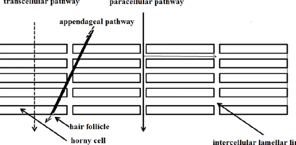
The transepidermal delivery of the drugs has three major pathways in the stratum corneum [15, 16]
- paracellular - transcellular - appendageal.
12
The paracellular pathway involves the lamellar lipids into the transport of drugs, which are essentially responsible for the sticking of the epidermal cells. During the transcellular transportation the drugs pass through the cells of the stratum corneum.
Most of the substances diffuse through this layer via the paracellular lipoidal pathway [17]. Nonetheless, paracellular and transcellular routes can play a role in combination during the transportation of drugs. The appendage route composed of hair follicles, sebaceous glands and sweat glands is considered to be substantially less important for drug transport, as this route accounts for less than 0.1 percent of the total surface area of the skin. This route may be of some importance for large ionic molecules. Drugs which diffuse through the sebaceous glands come first into the dermis and then into the subcutis [18, 19]. Figure 2 shows the routes of transdermal delivery.
Figure 2 – Delivery pathways of transdermal delivery
The other important barrier for many drugs is the stratum lucidum, which consist of 1 or 2 cell lines. It is an electronegative barrier and the electronegative drugs are not able to cross the stratum lucidum because of this negative shift. The positively charged drugs are pulled by this layer. Iontophoretic treatment can extinguish the effect of this barrier [20, 21]. The drugs can pass through the dermis and the subcutis easier.
13
At the surface of the skin there can be found an acid protecting layer, through which all the hydrophilic and also the hydrophobic drugs need to pass [22].
3.4. PHYSICOCHEMICAL PROPERTIES OF DRUGS WITH SKIN PERMEATION
The lipids in the epidermis play a very important role in the penetration of drugs into the skin. The protein parts of the epidermis can bind water and they become hydrated.
This hydrated state helps also in the better penetration of drugs. With this hydration the dermis behaves as a protein gel regarding the penetration of the drugs. Whether the drug can penetrate into the skin and reach the systemic blood circulation and / or the specific organ depends also on the structure of the drug. Most drugs are not suitable for transdermal delivery for one or more reasons. The desirable physicochemical, pharmacokinetic and biopharmaceutical characteristics of a candidate drug are:
- low daily dose (less than 20 mg/day) - short half-life (10 hours or less)
- low molecular weight (less than 400 Da) - low melting point (less than 200°C)
- high lipid solubility (octanol/water partition coefficient – /logP/ between -1.0 and 4)
- high skin permeability (permeability coefficient greater than 0.5 x 10⁻³ cm/h) - non-irritating and non-sensitizing to skin.
Low oral bioavailability and low therapeutic index, which requires tight control of plasma levels, are not required characteristics of drugs for transdermal delivery, but they confirm the application of a candidate drug in TTS [23- 28].
Generally only a few of the candidates could fulfill all these requirements: the high oral daily dose, large patch size, skin irritation or sensitization forms the main barriers for
14
the transdermal application. An acceptable balance between these requirements must be established for the drug delivery in TTS.
Drugs can have high skin permeability with the following characteristics:
- high dipole moments
- able to form hydrogen binding - are amphiphilic
- have low molecular weight.
If the drugs are applied on the skin they can
- treat local diseases or
- they can form reservoirs glued to the cells of the stratum corneum or of the dermis or
- they can penetrate into the deeper regions of the skin or - they are metabolized by the enzymes of the skin or - they can penetrate into the microcircular system.
3.5. ENHANCING THE PERMEATION INTO THE SKIN
Transdermal systems are applied in not such a wide range in the practice because of the poor permeation of the drugs into the skin. There are several possibilities to improve the permeation of drugs into the skin:
Physical methods
- scraping of the stratum corneum
- hydration of the stratum corneum [29, 30]
- iontophoresis [31]
- electroporation
15 - application of ultrasound energy on the skin - application of thermal energy.
Chemical methods:
- synthesis of lipophilic drugs - delipidation of the stratum corneum
- application of enhancers to help the penetration of drugs into the skin [32-34].
Biochemical methods:
- synthesis of prodrugs which are able for bioconversion in the skin [35, 36]
- application of enhancers can reduce the residence time in the skin, and thus decrease the cutaneous metabolism of drugs.
3.5.1. Application of physical activation
3.5.1.1. Iontophoresis
Iontophoresis involves application of a low intensity electric current to facilitate the permeation of a drug into the skin. Iontophoresis can increase the rate of transdermal penetration of hydrophilic compounds [37, 38].
Peptides could not be applied in TTS because of their high molecular weight, instability, low biological half-life and mainly polar characteristics [39]. The application of iontophoresis helps to transport protein molecules into the skin (transport of insulin) [40].
16 3.5.1.2. Electroporation
Electroporation is a method of application of short, high voltage electrical pulses to the skin. These electrical pulses form transient aqueous pores in the stratum corneum through which the drug molecules pass with electrophorese and / or electroosmose. This method allows a much more effective diffusion of drugs, respectively in the case of proteins [41-43]. The disadvantage of this system is the nerve irritation caused by the application of electrical pulses. The application of 2 phase continuous current can minimize the irritation and the lesion of the nerve endings. Electroporation can be achieved also with patches of which structure is much more difficult and therefore the production is more expensive.
3.5.1.3. Phonophoresis
Phonophoresis (or sonophoresis) uses ultrasound energy in order to enhance the skin penetration of active substances [44-46]. When skin is exposed to ultrasound, the waves propagate to a certain level and cause several effects that assist skin penetration. One of these effects is the cavitation, which means formation and subsequent collapse of gas bubbles in a liquid. This cavitation leads to formation of holes in the corneocytes, enlargement of intercellular spaces, and perturbation of stratum corneum lipids. Another effect is the heating of skin due to the energy loss of the propagating ultrasound wave (scattering and absorption effects). The resulting temperature elevation will increase the fluidity of the stratum corneum lipids and increase the diffusivity of molecules through the skin barrier. These main effects can be assisted by acoustic microstreaming caused by the acoustic shear stress which is due to unequal distribution of pressure forces.
3.5.1.4. Thermoresponsive systems
A new direction for pharmaceutical and medical research have been made with formulations based on stimuli-responsive polymers, because temporal drug release may be required, when the severity of disease symptoms fluctuate with time. If discontinuous changes in drug release rates are required in response to a small change
17
of temperature, thermoresponsive polymers may be used to develop hydrogels which react with their physical characteristics on the changes of their environment [47-49].
Thermoresponsive transdermal therapeutic systems consist of thermoresponsive agents (polymer, liquid crystals, liposomes), which suffer phase transition according to the change of temperature regulating drug delivery. This phase transition may be soluble- insoluble state variation, sol-gel transition, liquid crystal phase transitions, crystalline- amorphous phase-oscillation [50-52], etc.
3.5.2. Application of prodrugs
Prodrugs are inactive forms of drugs, which are transferred in the body with hydrolytic- and enzymatic bioconversion into the active forms of drugs. The application of prodrugs has an important role in the case of drugs, which could not penetrate in the original form into the skin [53, 54].
At the application of TTS the drugs pass the different layers of the skin, where they can be modified by the metabolic enzymes (such as o-methyltransferase, aryl-hydrocarbon- hydrolase) [55]. The inactive prodrugs are metabolized with these enzymes into the active drugs. This bioconversion can be applied for example in the case of the less permeable estrogen. These esterificated estrogens (estradiol acetate, estradiol diacetate, estradiol valerate) can penetrate into the skin much more intensively and they are fast metabolized by the esterase enzyme in the skin and form the active estradiol [56-58].
3.5.3. Application of enhancers
Increasing drug penetration through the skin needs penetration enhancers which modify the lipid organization in the stratum corneum. Depending on their molecular structure they can act in the following ways:
- intercalating into the sublattice, which leads to the modification of the sublattice
18 - disturbing of the lamellar packing
- forming separate small domains in the lateral packing in the lamellae
- forming enhancer-rich large separate domains in the intercellular regions [59, 60].
The structure of skin is modified as a result at the application of enhancers:
- they can change the hydration of the skin thanks to their higher water content in comparison with the skin. In this case the evaporation of the water in the TTS can lead to the hydration of the skin.
- lipophilic enhancers form a thin layer on the surface of the skin and lower the evaporation of the endogenous water from the skin (W/O emulsions) [61].
The transepidermal transportation of drugs can be positively affected by the hydration of the different layers of the skin. The transfollicular pathway can lead to a problem that the drug and the enhancer can be separated from each other in the sebaceous glands.
The keratolytic agents can loosen up the structure of the stratum corneum.
The surface active ingredients can solubilize the fat layer, which can quicken the transport during the lipid layer. These enhancers can be saturated and unsaturated fatty acids, from which the dodecyl chain length or the unsaturated C18 chain length is the most favorable for increasing drug transport across the skin [62, 63]. The fatty acids are applied with hydrocortisone or indomethacin [64, 65].
There are also other enhancers such as dimethyl sulfoxide [66, 67].
There is no general preference for any particular penetration enhancer or family of enhancers, because their effect depends on the interactions with the applied drug. The different types of enhancers can be varied with each other to goal a better absorption into the skin.
19
3.6. CLASSIFICATION OF TTS
In the general characterization of TTS are the following components:
- backing films - drug reservoir
- rate-controlling membrane
- pressure sensitive adhesives (PSA)
- protective pressure sensitive adhesive release liner.
In many cases one component can fulfill the role of the other components.
The TTSs can be classified on the base of the drug-release mechanism:
- membrane permeation controlled patch - adhesive dispersion controlled patch - matrix diffusion controlled patch - microreservoir controlled patch.
The TTSs are characterized by steady-state delivery rate, the whole dose and the complete area of drug release.
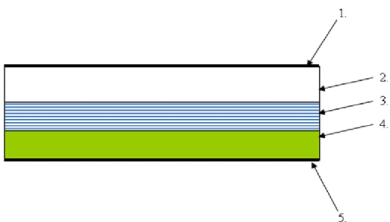
3.6.1. Membrane permeation controlled TTS
The release-rate from the reservoir is controlled by a membrane which is permeable for the drug. This membrane can be porous or not porous polymer membrane. The outer surface of the membrane is thin, compatible with the drug and hypoallergenic. The pressure sensitive adhesive ensures the cohesion between the skin and the TTS (Figure 3). The release-rate of the drug is influenced by the structure and the composition of the
20
reservoir, by the composition of the rate-controlling membrane and by the thickness, the structure and the permeability coefficient of the adhesive.
1. impermeable backing liner; 2. drug reservoir; 3. rate-controlling membrane;
4. pressure sensitive adhesive; 5. release liner
Figure 3 – Structure of membrane permeation controlled patch
The main advantage of this TTS is the nearly constant drug release-rate and the disadvantage is the intensive growth of drug release (dose dumping), if the membrane is damaged [68-70].
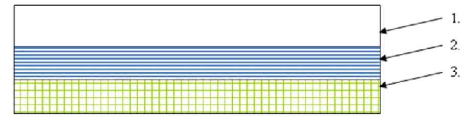
3.6.2. Adhesive dispersion controlled TTS
In this simplified membrane permeation controlled patch the drug enhancer adhesive matrix polymer contains the drug (it is dispersed in polyisobutylene, polyacrylate polymers). This polymer matrix is clamped to the impermeable backing liner in one or more layers [71, 72]. The disadvantage of this type of TTS is that the drug release-rate is getting smaller during the time of application because of the expansion of the polymer layer (the drug needs to diffuse through this layer). This release-rate can be enhanced with the application of more polymer layers, which have more drug content [73]. Figure 4 illustrates the structure of adhesive dispersion controlled patches.
21
1. impermeable backing layer; 2. drug in adhesive; 3. release liner
Figure 4 – Structure of adhesive dispersion controlled patch
3.6.3. Matrix diffusion controlled TTS
The matrix diffusion controlled patches (Figure 5) have more simple structure in comparison with the other type of TTS’s: they do not contain any rate-controlling membrane [74, 75].
1. impermeable backing layer; 2. absorbent pad; 3. occlusive baseplate;
4. drug reservoir; 5. adhesive; 6. release liner
Figure 5 – Matrix diffusion controlled patch
Drug reservoir is prepared by dissolving the drug and polymer in a common water- based solvent. The insoluble drug should be homogenously dispersed in hydrophilic or
22
lipophilic polymer. The required quantity of plasticizer (triethylcitrate, polyethylene glycol, propylene glycol and permeation enhancer) is then added and mixed properly.
The medicated polymer is then moulded into rings with defined surface area and controlled thickness over the teflon cover on horizontal surface followed by solvent evaporation at an elevated temperature. The formed film is then separated from the rings, which is then mounted onto an occlusive base plate in a compartment fabricated from a drug impermeable backing. Adhesive polymer is then spread along the circumference of the film [76]. Commonly used polymers for matrix are cross linked polyethylene glycol, polyacrylates, ethylcellulose, polyvinylpyrrolidone and hydroxypropyl methylcellulose. In an ideal case, the swelling of the polymer, which contains the drug, can be influenced that way, that the fluxation of water into the polymer and the diffusion of the drug occur at steady-state rate. Advantages of matrix patches include absence of dose dumping, direct exposure of polymeric matrix to the skin and no interference of adhesive.
3.6.4. Microreservoir controlled TTS
The drug is suspended in an aqueous solution of water miscible drug solubilizer e.g.
polyethylene glycol. The drug suspension is homogenously dispersed by a high shear mechanical force in lipophilic polymer, forming thousands of microscopic drug reservoirs (10-200 μm). The dispersion is quickly stabilized by cross-linking the polymer chains in-situ, which produces a medicated polymer disc of a specific area and fixed thickness. Occlusive base plate mounted between the medicated disc and adhesive form backing prevent the loss of drug through the backing membrane [77]. This system is exemplified by development of Nitrodisc®. Figure 6 presents the structure of microreservoir controlled patch.
23
Figure 6 – Microreservoir controlled patch
3.7. KINETIC CONSIDERATION OF TTS
Based on observations of experimental data, it may be stated that the differences in the therapeutic effect of different drug products (which contain the same drug constituents) are most frequently caused by differences in the rates at which the active ingredients are liberated from the given dosage form [78].
The process of diffusion has been described quantitatively by Fick, who adapted equations of thermal conductivity applied earlier by Fourier to diffusion. The definition of the law is as follows: the quantity of the dissolved substance (dw) which diffuses at a constant temperature through a cross-sectional area (A) within a time (dt), through a layer of thickness (dx) which is perpendicular to surface A on the effect of a concentration gradient (dC):
(1)
where D is the diffusion coefficient (the quantity of dissolved substance which diffuses through a 1 cm2 cross-sectional area per unit time).
1. impermeable backing layer; 2. drug in adhesive; 3. release liner 4. drug reservoir
dx DA dC dt
dw
24
For the characterization of different dosage forms are applied different kinetic models, which are based on the equation of Fick.
The TTS can be grouped in 2 major types respectively the release kinetics of drugs:
- adhesive matrix systems - gelled liquid reservoir systems.
3.7.1. Release kinetics from matrix devices
In dissolved systems, an adhesive matrix system contains drug dissolved at or below the saturation solubility of the drug in the adhesive. Diffusion of drug occurs from the surface of patch into water or skin. In ideal case, the fraction (F) released from a matrix system is described as follows [79]:
(2)
where:
Mt= amount of drug released into a sink at time t M∞= amount released on depletion of the device D= diffusion coefficient
L= thickness of matrix film n= integer
This equation (2) is more complex in nature, so for simplicity it can be distinguished an early-time and a late portion and they can characterized with different equations [80, 81].
2 2 2
2 2 t
) 1 n 2 (
L 4
t ) 1 n 2 ( D exp 8 1
F
M
M
25
The drug can be found in dispersed-type systems as finely divided particles uniformly dispersed in the polymer matrix. Therefore, the initial drug concentration (Ai) is greater than the saturation solubility (CS). The drug release kinetics from a dispersed-type matrix system is described as follows [82]:
(3)
where:
CS= saturation solubility Ai= initial drug concentration
Once the dispersed drug is exhausted from the matrix, the drug release kinetics essentially follows the kinetics of drugs from dissolved systems.
The porous matrix system has continuous macroscopic channels. The release kinetics is mainly dependent on the transport of drug through these tortuous channels. The following equation (4) describes the release kinetics of a drug from these porous matrix systems [83]:
(4)
where:
ε
= porosity Τ= tortuosityThe porosity can be determined from the volume fraction of the transdermal device that is permeated by solvent. Tortuosity is a measure of the diffusion length, which the solute would traverse through the channel in excess of linear path [84, 85].
In the absence of porosity and tortuosity in the matrix device, equations 3 and 4 are the same.
5 , s 0 i s i
t ) C A 2 ( L DC A F 2
5 , 0 s t
( 2 A C
s) C t
M D
26
3.7.2. Release kinetics from gelled liquid reservoir systems
Reservoir patches contain the drug mainly in solution in appropriate vesicles. The viscosity of this solution is increased with an appropriate gelling or thickening agent to make the fabrication of the reservoir patches easier. The release kinetics of the drug through the gel and the rate-controlling membrane of a liquid reservoir patch follow Fick’s law of diffusion.
3.7.3. Analysis of the release profiles according to different kinetic models
In the in vitro evaluation of patches are often applied the following models, which are more simplified as the ones described above and are often used in the kinetic characterization of transdermal devices.
Zero-order model
The drug release from the dosage form follows a steady-state release running at a constant rate [86, 87]:
M
t/M
∞= kt
(5)where
Mt= the amount of the drug released at time t,
M∞= the total amount of the released drug at infinite time, k= the zero order rate constant of the drug release.
This model can be applied for drug dissolution from pharmaceutical dosage forms that do not disaggregate and release the drug slowly (assuming that area does not change and no equilibrium conditions are obtained).
27 First-order model
The drug activity within the reservoir is assumed to decline exponentially and the release rate is proportional to the residual activity:
M
t/M = 1– exp(–kt)
(6)where
k= first order rate constant of the drug release.
Weibull model
A general empirical equation described by Weibull was adapted to the dissolution / release process. This equation can be successfully applied to almost all kinds of dissolution curves and is commonly used in kinetic studies.
To characterize the dissolution profile of patches the Weibull distribution was applied in the following form [88]:
(7)
where
Mt=the dissolution (%) at time t (min), M∞=the dissolution (%) at infinite time, t0=the lag-time (min) of the dissolution, β=shape parameter of the curve,
τ63.2=time (min) when 63.2% of M∞ has been dissolved.
2 . 63
0 t
t exp t
1 M M
28
4. OBJECTIVES
In the literature overview of the thesis, I summarized the references which are connected with the transdermal delivery and their applications in the pharmaceutical practice. I gave an overview about transdermal delivery devices, especially focused on the matrix type ones. I represented the different polymers which were used to form matrices and enable the drug release from the obtained transdermal systems. The kinetic aspect of the controlled drug release in transdermal therapeutic systems is also presented regarding to their drug depot characteristics.
The objectives of the experimental part of my thesis were:
- to formulate patches containing standard amount of metoprolol tartrate and different amounts of polymers, which are official in the European Pharmacopoeia,
- to characterize in vitro the metoprolol tartrate release from the prepared patches,
- to analyze the relationship between the drug release and the composition of the patches regarding to the different polymer ratios,
- to evaluate the applicability and reliability of different kinetic models to describe the drug release from the patches,
- to give a relationship between the drug release and the ability of the polymer matrix to form secondary structures,
- to evaluate in process the patches with appropriate polymer ratio for the desirable drug release by non-invasive FT-IR spectroscopy.
29
5. EXPERIMENTAL PART
5.1. Materials and methods
5.1.1. Materials
Metoprolol tartrate of Ph. Eur. was selected as a highly water-soluble model drug.
The following excipients were applied for the preformulation examinations and for the preparation of the patches:
- Eudragit RL 30 D (USP/NF, JPE, Ph. Eur.; Evonik Röhm GmbH, Darmstadt, Germany)
- Eudragit RS 30 D (USP/NF, JPE, Ph. Eur.; Evonik Röhm GmbH, Darmstadt, Germany)
- Eudragit NE 30 D (USP/NF, JPE, Ph. Eur.; Evonik Röhm GmbH, Darmstadt, Germany)
- Triethylcitrate (Hungaropharma, Budapest, Hungary)
- Polyethylene glycol (PEG400) (Hungaropharma, Budapest, Hungary) - Sucrose stearate S1570 (Mitsubishi Chemical Co., Japan)
- Sucrose laurate L1695 (Mitsubishi Chemical Co., Japan)
- Metolose 90 SH 100.000 SR (USP, Ph. Eur.; Shin-Etsu Chemical Company, Tokyo, Japan)
- Metolose SM 4000 (USP, Ph. Eur.; Shin-Etsu Chemical Company, Tokyo, Japan)
5.1.1.1. Application of metoprolol tartrate
Metoprolol tartrate is a selective hydrophilic ß-blocking agent for the treatment of mild and moderate hypertension and also for long term management of angina pectoris.
Metoprolol tartrate is 95% absorbed and has a bioavailability of 40- to 50%. In the blood circulating system it is in the first step 12% protein bound, then rapidly enters the
30
CNS and has moderate lipid solubility. The metabolism of this drug is hepatically (primarly by CYP2D6). The metabolization occurs also mainly in the liver.
Approximately 95% of the drug is excreted renally and less than 5% of the drug is excreted unchanged in urine.
Reduction of exercise induced tachycardia is proportional to the logarithm of the plasma concentration over the range of 20–100 μg/L and effects are detected at 50–100 μg/L.
Peak plasma concentrations are achieved after 2–3 hours. Half life of the molecule is 3 to 7 hours. The plasma half-life is about 4 hours [89, 90], which makes frequent dosing necessary to maintain the therapeutic blood levels of the drug for long-term treatment.
The n-octanol/water partition coefficient is 0.98 at pH 7.4. Therefore, metoprolol is an ideal drug candidate for transdermal drug delivery. The drug has previously been investigated for its skin permeation potential with menthol as a penetration enhancer [91], as a part of liposomal formulations [8] and also as an active ingredient of transdermal matrix films [92]. Figure 7 shows the structure of metoprolol tartrate.
Figure 7 – Chemical structure of metoprolol tartrate [93]
31 5.1.1.2. Application of acrylates
Acrylic polymers have been known for a long time. They have favorable biocompatibility, good skin adhesion property and good compatibility with a wide range of drugs and excipients.
Polyacrylates are saturated hydrocarbon polymers (Figure 8). Therefore they are highly resistant to oxidation and do not require the addition of stabilizers, the application of which could lead to skin irritation. These polymers are inherently tacky, and therefore, in general they do not need low molecular weight tackifiers and plasticizers to provide the stickiness and softness.
where R1 is H-, CH3-, R2 is H-, CH3-, CH3CH2-, CH2CH2N(CH3)2-, R3 is CH3-, R4 is CH3-, C4H9-
Figure 8 – Chemical structure of acrylic polymers [94]
Poly acrylic esters are produced by copolymerization of acrylic esters, acrylic acid and other functional monomers. Commonly used modifying monomers include vinyl acetate, methyl acrylate, methyl and ethyl methacrylate, acrylic and methacrylic acid, acrylnitrile and certain amine-functional monomers. These modifying monomers can be used to change the solubility and / or permeability of the acrylic polymers. Water- soluble or hydrophilic monomers such as vinylpyrrolidone, 2-hydroxyethyl acrylate and 2-ethyl acrylate have been used to increase the hydrophilicity of the polymers [94].
32
5.1.1.3. Application of cellulose ether polymers
Cellulose ethers are derivates of cellulose. They are formed through partial or total substitution of the hydroxyl groups in the so called etherification. There are different forms of cellulose ethers such as carboxymethylcellulose, methylcellulose, ethylcellulose and hydroxymethyl-, hydroxyethyl- and hydroxypropylcellulose, etc. The physical and chemical properties of cellulose ethers depend on the way and the grade of substitution. Table 1 summarizes the data of the cellulose types which are applied in the preparation of patches.
Table 1 – Characteristics of the applied Metolose types [95]
Metolose types Methoxyl Hydroxypropoxyl Viscosity*
content (%) content (%) (cP)
SM 4000 27.5-31.5 – 3000-5600
90 SH 100.000 SR 19.0-24.0 4.0-12.0 75.000-140.000
*: USP viscosity
5.1.1.3.1. Methylcellulose
Methylcellulose is formed from long-chain substituted cellulose in which approximately 27–32% of the hydroxyl groups are in the form of the methyl ether (Figure 9). The different types of methylcellulose have degrees of polymerization in the range 50–1000, with molecular weights in the range 10.000–220.000 Da. The degree of substitution of methylcellulose is defined as the average number of methoxy (CH3O) groups attached to each of the anhydroglucose units along the chain. The degree of substitution also affects the physical properties of methylcellulose, such as its solubility and the viscosity level of solutions, which can be achieved with the application of methylcellulose.
33
Figure 9 – Chemical structure of methylcellulose [94]
Methylcellulose is widely used in oral and topical pharmaceutical formulations. The following application forms can occur in tablet formulations:
- binding agents (low- or medium-viscosity grades of methylcellulose).
Methylcellulose is added either as a dry powder or in solution [96], - disintegrants (high viscosity grades of methylcellulose) [97], - to produce sustained-release preparations,
- to spray-coat (highly substituted low-viscosity grades of methylcellulose) tablet cores to mask unpleasant taste or to modify the release of drug by controlling the physical characteristics of the granules [98],
- to seal tablet cores prior to sugar coating.
In liquid formulations the following application fields are involved:
- emulsify different types of oils (low-viscosity grades of methylcellulose),
- suspend or thicken orally administered liquids (low-viscosity grades of methylcellulose) [99]. Methylcellulose delays the settling of suspensions and increases the contact time of drugs, such as antacids, etc.
In topically applied creams and gels are applied high-viscosity grades of methylcellulose as a thickening agent. In ophthalmic formulations has been also used
34
high-viscosity grade of methylcelluloses [100]. Methylcellulose types can be also found in injectable formulations.
Therapeutically, methylcellulose is used as a bulk laxative; it has also been used to aid appetite control in the management of obesity. Practically insoluble in acetone, methanol, chloroform, ethanol (95%), ether, saturated salt solutions, toluene, and hot water. It is soluble in glacial acetic acid and in a mixture of equal volumes of ethanol and chloroform. In cold water, methylcellulose swells and disperses slowly to form a clear to opalescent, viscous, colloidal dispersion.
5.1.1.3.2. Hypromellose
Hypromellose is a partly O-methylated and O-(2-hydroxypropylated) cellulose (Figure 10). The different types of hypromellose vary in viscosity and extent of substitution.
Grades may be distinguished by appending a number indicative of the apparent viscosity, in mPas, of a 2% w/w aqueous solution at 20°C.
Molecular weight is approximately 10.000–1.500.000.
where R is H-, CH3- or CH3CH(OH)CH2-
Figure 10 – Chemical structure of hypromellose [94]
These excipients are applied in oral, ophthalmic, nasal, and topical pharmaceutical formulations. In tablet formulations hypromellose is primarily used:
- tablet binder excipient [101], - film-coating agent [102],
35
- to form matrix for use in extended release tablet formulations [103, 104], - to retard the release of drugs from a matrix (at levels of 10-80% w/w), - to film-coat tablets (at levels of 2-20% w/w).
Hypromellose is also an ideal candidate as a suspending and thickening agent in topical formulations. It is a much better candidate for ophthalmic formulations because of the clarity of solutions made with hypromellose compared with the solutions made with methylcellulose. It forms fewer undissolved fibers. Therefore at low concentration level (0.45–1.0% w/w) hypromellose may be added as a thickening agent for eye drops and artificial tear solutions. It is also used commercially in liquid nasal formulations at a concentration of 0.1% [105]
In topical gels and ointments hypromellose can be applied as an emulsifier, suspending agent, and stabilizing agent. As a protective colloid it can prevent droplets and particles from coalescing or agglomerating. It is also widely used in cosmetics and food products.
Hypromellose powder is a stable material, although it is hygroscopic after drying.
Solutions are stable at pH 3–11. A reversible sol–gel transformation upon heating and cooling can be observed with hypromellose. The gelation temperature is 50–90°C, depending upon the grade and concentration of this material. Viscosity of the solution decreases as temperature is increased below the gelation temperature, while beyond the gelation temperature viscosity increases as temperature is increased. Aqueous solutions of hypromellose are quite enzyme-resistant, thus provides good viscosity stability during long-term storage [94].
36
5.1.2. Methods
5.1.2.1. Method for preparation of patches
In the first step, 2/3 part of water was heated to 70 ºC. Metoprolol tartrate, the different types of Metolose of various proportions and the different additives were dissolved homogeneously in the hot water. The remaining 1/3 part of the water, stored at 5 ºC, was added after homogenizing. This mixture was stirred until dissolving of the components and afterwards it was cooled. At room temperature (25 ºC) the different acrylic polymer solutions were added to the system applying a low stirring rate to avoid forming of air bubbles.
This preparation method enabled that metoprolol tartrate is completely dissolved before embedded in the matrix and is fully dispersed in the polymer system in the course of the drying process. In the cooling step of the preparation of the patches, there is an increase in the viscosity of the solution containing Metolose, which makes more difficult to mix it with the polyacrylate solution thoroughly.
The homogenous mixture was filled into a gum ring of a constant diameter (54 mm).
Each sample contained 7.5 g of this mixture. The metoprolol tartrate concentration of the mixture was 1.11% w/w in each sample. The drying of the samples was performed at room temperature for a 3 day period. The further investigations were done after this 3 day period. First the thickness of patches was checked and in each formulation was found similar.
5.1.2.2. Organoleptic examination of different compositions for preparing applicable patches
For the determination of the incompatibility between the model drug and the polymer matrix forming components I prepared patches and after 3 days of storage in exsiccator with organoleptic observation I chose the macroscopically homogeneous candidates for further examinations.
5.1.2.3. Viscosity measurements
The prepared gel formulations at room temperature were filled into the cylinder of the viscosimeter (HAAKE VT550 Rheometer, Haake GmbH, Karlsruhe, Germany). The
37
measurements were done at 25°C. For the evaluation the balance viscosity values of each formulation (n=3) were taken. The viscosity – time curves were recorded with the following parameters: disc SV2, G= 50 1/min, measurement time was 15 min.
5.1.2.4. Optimization of composition according to the drug liberation examinations
This test was performed by Hanson SR8-Plus (Hanson Research, Chatsorth, USA) according to Ph. Eur. regulation – Paddle over disk. TTS samples after 3 days of storage were placed into a disk apparatus. Then they were immersed into the temperature- controlled 400 ml acceptor medium (pH = 6.00 buffer solution). The acceptor medium was kept at 32 ±1°C and mixed at the rate of 25 rpm with rotating pad. Samples were taken at predetermined time points with AutoPlus Maximizer system and an Auto Plus MultiFill collector (Hanson Research, Chatsorth, USA). The sample volume was 10 ml, which was replaced each time with the equivalent of dissolution medium. The active content of the samples was determined with an Auto Plus On-Line UV/VIS Autosamples spectrophotometer at 274 nm on the basis of a calibration curve recorded earlier. The examination cell is shown in Figure 11.
Figure 11 – Drug liberation examination cell [93]
38
5.1.2.5. Analysis of the release profiles according to different kinetic models
The following mathematical models were evaluated considering the drug release profiles of the patches. The non-linear parameter estimation of the release models applied for matrices was made with the Solver function of the computer package Microsoft Excel 5.0.
In the evaluation of the matrices we applied the following models:
zero-order model first-order model Weibull distribution.
5.1.2.6. FT-IR examinations
FT-IR spectra of the cast film patches were obtained using a JASCO FT/IR-4200 spectrometer in 4000-400 cm-1 wavenumber range. 32 scans were performed at a resolution of 4 cm−1. The system was operated in the transmission mode. Spectra Analysis software was applied for the determination of the peak area within the wavenumber range of 1757.7-2811.8 cm-1.
5.1.2.7. Non-invasive stability screening of patches with ATR-FTIR examinations
The prepared matrices with and without metoprolol tartrate were stored 1 month at 40±2°C and 75±5% relative humidity in open container.
The ATR-FTIR spectra of the stored patches with and without metoprolol tartrate samples were scanned over wavenumber range of 4000–600 cm−1 using Able Jasco FT- IR 4200 type A spectrometer with ATR Pro470H single reflection ATR accessory. 32 scans were performed at a resolution of 4 cm−1.
5.1.2.8. Positron annihilation lifetime spectroscopy (PALS)
Positron annihilation lifetime spectroscopy (PALS) is a unique method since it is exceptionally sensitive to the free volume. It is frequently used to determine the size distribution of free volume holes in polymers. All of these measurements are based on
39
the interaction of the free volume holes and the so called ortho-positronium atom.
When positrons are injected to an amorphous molecular material they form three different states in the simplest case. A part of them forms positronium (Ps) atoms with the electrons of the material. This exotic atom, which is the bound state of an electron and a positron, has two ground states depending on the relative orientation of the spins of the constructing particles. The para-Ps, in which the spins are anti-parallel, has a very short intrinsic lifetime (125 ps). The usual reactivity of Ps atoms is not very high and in most of the cases the two particles bound in para-Ps (the positron and the electron) annihilate with each other by the intrinsic lifetime ( 1). The other ground state of positronium is the ortho-Ps (o-Ps), in which the spins are parallel. This Ps state has a very long lifetime in a vacuum (141 ns) and, even at very low reactivity, it interacts with the electrons of the surrounding material considerably. Usually the positron of the ortho-Ps does not annihilate with its own electron but rather with an electron of the material. This interaction decreases the intrinsic lifetime significantly and usually a much shorter lifetime is observed (1-10 ns). Even so, this is the longest lifetime component of positron lifetime spectra ( 3).
The third state of positrons in materials is usually due to positrons not able to form Ps atoms. These positrons diffuse almost freely in the material but they should also annihilate sooner or later with surrounding electrons, i.e., with the electrons of polymeric chains in our case. This provides a medium long lifetime ( 2) that characterizes the average electron density of the material. This lifetime component usually reflects tiny changes of the electron structure of a material very sensitively. In amorphous materials as polymers the size of free volume holes is not uniformly distributed. Consequently, the simple assumption on the number and the nature of positron states is not necessarily correct. In these materials, instead of one well defined o-Ps state, a number of similar states occur depending on the size of the free volume around the positronium. So the lifetime spectrum, S(t) is not a simple sum of exponential curves and instead of:
i
i
i
t
A t
S exp( )
(8)it can be described by a continuous distribution of lifetimes:
40
0
d ) exp(
)
( t
h t
S
(9)where i-s are the assumed separate lifetimes, Ai-s are their relative intensities, while the function h( ) is the lifetime distribution function which is proportional with the size distribution of free volume holes in the material.
In polymers the formed o-Ps atoms tend to be trapped in free volume holes and, as mentioned above, their annihilation is not governed by their intrinsic lifetime but by the electron density in the holes. Their lifetime is associated with the size of the free volume around them:
1
2 1 1
2
2
1R R R
R R R
s i n
(10)where is the positronium lifetime, R is the radius of the free volume hole, and R is a constant. Consequently, on the basis of o-Ps lifetime distributions we gain a detailed picture on the size distribution of free volume holes [106, 107].
The positron source applied for the measurements was made of carrier free 22NaCl of the activity of 4x10⁵ Bq. The active sodium chloride was sealed between two very thin kapton foils. The source was then placed between two pieces of the sample treated identically before. Positron lifetime spectra were recorded by a conventional fast-fast coincidence system. The system was constructed from standard ORTEC electronic units, while the detectors from BaF2 scintillator crystals and XP2020Q photomultipliers.
The time resolution of the system was about 200 picoseconds. The spectra were evaluated into three lifetime components. The two shorter lifetime components (165ps, 460ps) are mixtures of different positron and positronium states. As they are too complicated to interpret physically, they were ignored in the evaluations. The longest living positron state is due to ortho-positronium. The lifetime of this positronium state is, in the case of polymers, connected with the size of free volume holes [108]. The longer this lifetime, the larger the holes are.
41
5.2. RESULTS
5.2.1. Preformulation examinations
5.2.1.1. Preparation of Eudragit films for further formulation of matrices containing metoprolol tartrate
The aim of the preformulation examinations was to create matrix type patches with the combination of polyacrylate polymer and different additives to embed the model drug (metoprolol tartrate). For this reason there were investigated several types of polyacrylate polymer solutions in combination with metoprolol tartrate for compatibility. For these examinations I prepared different samples with Eudragit RL 30 D, Eudragit RS 30 D and Eudragit NE 30 D with different additive such as triethyl- citrate, PEG400, Sucrose stearate S1570, Sucrose laurate L1695. Acrylates are suitable to formulate oral dosage forms which are able to release their content in different parts of the GI tract. According to their functional groups they can influence at which pH values they swell and get permeable. There are also several types which enable the applied drugs to be released with time control under independent pH values [109].
There can be found several examples in the literature for the application of Eudragit polymers to formulate transdermal matrices: mainly the following two types are mentioned as a part of matrix formulations: Eudragit RS and Eudragit RL [110-114].
These two types have trimethyl-ammonioethylmethacrylate as functional group which enables high permeability and pH independent swelling. The application of Eudragit NE 30 D (aqueous dispersion of a neutral copolymer based on ethyl acrylate and methyl methacrylate) in matrix type patches can be also advantageous, because of its time- controlled drug release properties [69, 115, 116]. For the formulation of matrices these 3 types were chosen.
The aim of the first formulations in the developing of matrices was to obtain structures which can be manufactured with film casting method resulting in patches which are flexible and applicable on the skin. Eudragit RS and Eudragit RL were applied for preparing matrices alone and also with different plasticizers. The summary of formulations can be seen in Table 2.
![Figure 1 – The different layers of the skin [14]](https://thumb-eu.123doks.com/thumbv2/9dokorg/1377812.113375/12.892.205.766.421.853/figure-different-layers-skin.webp)
![Figure 7 – Chemical structure of metoprolol tartrate [93]](https://thumb-eu.123doks.com/thumbv2/9dokorg/1377812.113375/31.892.224.672.612.839/figure-chemical-structure-metoprolol-tartrate.webp)
![Figure 8 – Chemical structure of acrylic polymers [94]](https://thumb-eu.123doks.com/thumbv2/9dokorg/1377812.113375/32.892.237.659.478.685/figure-chemical-structure-acrylic-polymers.webp)
![Table 1 – Characteristics of the applied Metolose types [95]](https://thumb-eu.123doks.com/thumbv2/9dokorg/1377812.113375/33.892.126.767.502.622/table-characteristics-applied-metolose-types.webp)
![Figure 9 – Chemical structure of methylcellulose [94]](https://thumb-eu.123doks.com/thumbv2/9dokorg/1377812.113375/34.892.293.566.126.360/figure-chemical-structure-methylcellulose.webp)
![Figure 10 – Chemical structure of hypromellose [94]](https://thumb-eu.123doks.com/thumbv2/9dokorg/1377812.113375/35.892.298.598.634.854/figure-chemical-structure-hypromellose.webp)
![Figure 11 – Drug liberation examination cell [93]](https://thumb-eu.123doks.com/thumbv2/9dokorg/1377812.113375/38.892.256.631.683.1041/figure-drug-liberation-examination-cell.webp)
