1
Intranasal Nanoparticulate Systems as Alternative Route of Drug
Areen Alshweiata,b, Rita Ambrusa and IIdikó Csókaa
aFaculty of Pharmacy, Institute of Pharmaceutical Technology and Regulatory Affairs, University of Szeged, Szeged, Hungary.
bFaculty of Pharmaceutical Science, The Hashemite University, Zarqa, Jordan.
Corresponding authors Areen Alshweiat
Faculty of Pharmacy, Institute of Pharmaceutical Technology and Regulatory Affairs, University of Szeged H- 6720, Eötvös u. 6. Szeged, Hungary. Tel+36702227752, Fax +366254557
Email areen.alshweiat@hu.edu.jo
Abstract: There is always a need for alternative and efficient methods of drug delivery. The nasal cavity can be considered as a non-invasive and efficient route of administration. It has been used for local, systemic, brain targeting, and vaccination delivery. Although many intranasal products are currently available on the market, the majority is used for local delivery with fewer products available for the other targets. As nanotechnology utilization in drug delivery has rapidly spread out, the nasal delivery has become attractive as a promising approach. Nanoparticulate systems facilitate drug transportation across the mucosal barrier, protect the drug from nasal enzyme degradation, enhance the delivery of vaccines to the lymphoid tissue of the nasal cavity with an adjuvant activity, and offer a way for peptide delivery into the brain and the systemic circulation, in addition to their potential for brain tumor treatment. This review article aims at discussing the potential benefit of the intranasal nanoparticulate systems, including nanosuspensions, lipid and surfactant, and polymer-based nanoparticles as regards productive intranasal delivery. The aim of this review is to focus on the topicalities of nanotechnology applications for intranasal delivery of local, systemic, brain, and vaccination purposes during the last decade, referring to the factors affecting delivery, regulatory aspects, and patient expectations.
This review further identifies the benefits of applying the Quality by Design approaches (QbD) in product development.
According to the reported studies on nanotechnology-based intranasal delivery, potential attention has been focused on brain targeting and vaccine delivery with promising outcomes. Despite the significant research effort in this field, nanoparticle-based products for intranasal delivery are not available. Thus, further efforts are required to promote the introduction of intranasal nanoparticulate products that can meet the requirements of regulatory affairs with high patient acceptance.
Keywords: Intranasal, nanoparticulate system, drug delivery, Quality by Design, regulatory, patients’ expectations.
1. INTRODUCTION
Clear evidence of the nasal cavity as an effective route of administration has attracted research groups to concentrate on exploiting this region as an alternative means for systemic and brain delivery of drugs and vaccines to overcome the inconvenience of already available routes.
Over the past decades, nanotechnology has gained an advanced position in drug delivery approaches. A nanoparticulate system holds a great value over the manipulative characteristics of the applied therapeutics, such as solubility, permeability, and half- life. These features allow the extended use of nanoparticulates for cancer targeting and controlled release purposes. Many parenteral, oral, and topical nanoparticulate therapeutics are available on the market and clinical trial stages [1–8].
The term nanotechnology is also widely used and defined as the control and manipulation of matter at the nano-scale (10-100 nm). However, the particles within the size range of 1-1000 nm are considered as nanoparticles in practice. Nanoparticles are regarded as special due to the fact that particles on the nanometer scale have unique optical, electronic, and structural/functional properties distinctive from the normal size. Moreover, higher permeability, a large surface to volume ratio, and higher mucoadhesion can be achieved as a consequence of nanosizing [9–13].
Nanosystems form a special group regarding their regulatory acceptance. Related guidelines and relevant chapters of the European Medicines Agency (EMA) and the Food and Drug Administration (FDA) must be applied during all manufacturing stages from material selection and formulation to the final production. Furthermore, the FDA has emphasized the application of the Quality by Design (QbD) methodology, which can be especially useful for novel, high risk dosage forms and administration routes. The adoption of the International Council on Harmonization (ICH) guidelines for pharmaceutical development-Q8, risk management-Q9 and quality system-Q10 provides great potential for careful planning during the formulation and development even in the early phase of the research [14–20].
A high number of successful applications of nanoparticulate systems in drug delivery motivated to apply this technology in the case of the intranasal route; in order to improve drug delivery and to overcome the limits of this administration route. A combination of novel nanotechnology developments together with increased knowledge on intranasal delivery can efficiently lead to substantial advances in drug delivery with enhanced bioavailability and patient acceptance.
2. NASAL CAVITY 2.1 Nasal Anatomy
As known from anatomy studies, the human nasal cavity (Fig 1) is composed of two symmetrical chambers (nostrils) separated by the median septum, the area inside each chamber is divided into the nasal vestibule area and the main nasal cavity containing the respiratory and olfactory regions. The total surface area and volume of the nasal cavity are 150 cm2 and 15 ml, respectively [21,22].
The nasal respiratory area is the largest part of the nasal cavity, it is confined between the septal and lateral walls and it contains the superior, middle, and inferior turbinates forming the slit-like area that is responsible for the humidification and temperature regulation of the inspired air [23].
The uppermost region of the nasal cavity is the olfactory region, which is responsible for the sense of smell. This area comprises 10% of the total intranasal cavity and the olfactory information is sent from the olfactory bulb via the olfactory neuron into the piriform cortex, amygdala, and entorhinal cortex; where this can promote direct brain transport [24,25].
The cell lining type varies along the nasal cavity; the vestibules are covered by non-ciliated squamous and transitional epithelium with poor blood perfusion, whereas the respiratory region is covered by epithelium consisting of ciliated, pseudostratified, and columnar epithelium cells with a rich blood supply from the underlying lamina propria. The presence of microvilli along with columnar cells intensifies the surface area available for drug absorption, as each cell covered with 300 microvilli, and their fine projections (cilia) are fundamental to mucus transport into the nasopharynx. The topographical and physiological features of the respiratory region are responsible for being the main region for permeation. Similar to the respiratory area, the olfactory region is covered by pseudostratified epithelium with a specialized refractory receptor for smell perception. Prior to transfer, the olfactory component must be dissolved in the serous fluid that is produced and secreted by Bowman's gland [26–29].
3
Besides the significance of the respiratory region for systemic absorption, it plays a crucial role in direct drug delivery into the brain through the trigeminal region. The olfactory and trigeminal structures act as the only available apertures of the central nervous system (CNS) entry [30].
The anatomical aspect plays a crucial role in nasal delivery. To get the benefit of the high surface area of the nasal cavity and its consequences on higher absorption, the formulation must be spread over a large mucosal area. The place of distribution inside the cavity is essential for the activity; for example, for local delivery, systemic delivery, and vaccines, broad distribution is required whilst in brain targeting the drug must be delivered into the upper parts of the nose containing the olfactory region in addition to covering the trigeminal nerve, which may have a contribution in targeting. Such factors must be considered in selecting the dosage form and designing the delivery devices to ensure the proposed deposition and coverage of the formulation to get the intended response [31,32].
Fig. (1). Anatomical structure of the human nasal cavity.
2.2 Nasal Cavity as Drug Route of Administration
The distinction of the intranasal route is ascribed mainly to the anatomical and physiological characteristics of the nasal cavity.
The nasal cavity offers a number of advantages, such as: avoiding first-pass metabolism, high surface area, high permeation, high vascularization, and having a nose to brain direct pathway as well as circumventing the blood brain barrier. Thus, the nasal route has the potential for the delivery of drugs that suffer from extensive first-pass metabolism, poor solubility, and degradation in the gastrointestinal tract. It is also an attractive site for vaccine and peptide delivery that have been parenterally administered so far. The intranasal route is a non-invasive, non-sterile, and easily administered method that can enhance patients' compliance [33–39].
On the other hand, many limitations could adversely affect nasal delivery, these include: mucociliary clearance, restricted volume of nasal administration (max. 200 ul), presence of enzymes and efflux transporters, pathological and environmental factors that affect intranasal blood supply. Moreover, the narrow nasal valve represents a potential obstacle to an efficient drug delivery [40–43].
2.3 Emerging Intranasal Application from Local to Systemic, Brain, and Vaccine Delivery
The intranasal delivery of drugs has been initially utilized for the local treatment of topical conditions. Various marketed drugs have been used to treat congestion, nasal allergies, infections, and nasal polyps. Decongestants, steroids, and antihistamines are the most common drugs that are nasally applied for their local action [44–48].
As a consequence of the previously mentioned advantages, the nasal cavity has evolved from local administration into a route for systemic, brain targeting, and vaccine delivery. This extension has opened up the possibilities for all drug delivery purposes, including cancer treatment [49–53].
Intranasal products with systemic effects are commercially available for certain drugs such as zolmitriptan, sumatriptan, ergotamine, butorphanol tartrate, and fentanyl as well as peptides such as calcitonin, desmopressin, buserelin, and nafarelin [54–61]. Other drugs have been nasally introduced for the treatment of urgent conditions such as migraine, seizures, opioid overdose, and pain breakthrough in cancer [62–67].
Maximum 2% of drugs are capable of reaching the brain after systemic administration due to the presence of the protective brain capillary endothelium. The exploitation of the intranasal route to target the CNS is an attractive approach to circumvent the blood brain barrier (BBB) and deliver the drug directly through the cribriform plate, olfactory and trigeminal regions.
Alzheimer’s disease, depression, migraine, schizophrenia, HIV consequences, and multiple sclerosis are all CNS diseases that systemic administration has failed to treat. The availability of an effective delivery rather than the drugs was the missing part for achieving considerable therapeutic outcomes [68–70].
The intranasal cavity offers easy administration for vaccines, inducing both mucosal and systemic immunity. The importance of this site has evolved from the nature of infections itself since the majority of viral and bacterial infections start from the mucosal tissues. Both innate and adaptive immune responses can be directly initiated after the delivery of the antigen via nasal- associated lymphoid tissues (NALT) through the distinctive M cells into the antigen sampling cells, dendritic cells, B-cells, and T-cells, being responsible for the humoral immune responses mediated by secretory IgA antibodies [71–74]. The pharmaceutical aspects of intranasal vaccination have been thoroughly discussed by Sharma et al. [75].
3. NANOTECHNOLOGY AS A FURTHER ASPECT OF THE INTRANASAL DELIVERY OF DRUGS 3.1 Rationale for using Nanotechnology for Intranasal Delivery
Within the last decades, tremendous efforts have focused on intranasal delivery and its potential for different applications beyond its local importance; to achieve systemic delivery and brain targeting in addition to mucosal and systemic vaccination.
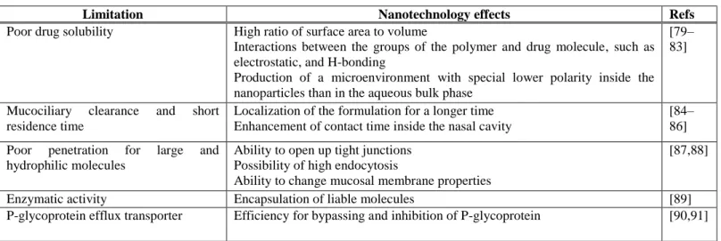
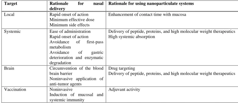
Hypothetically, the ideal route is available. Nevertheless, many limitations can hamper its efficiency. The combination of nanotechnology as the drug preparation method and intranasal delivery as the route is supposed to provide an effective delivery system. Nanotechnology offers the criteria for achieving high solubility and dissolution rates, which are the key factors for drug absorption and activity. Furthermore, this technology can protect the drugs from nasal enzyme activity, counteract the mucociliary action to increase contact time and promote permeation. Table 1 lists the nanotechnology effects on the major nasal delivery limitations and Table 2 identifies the rationale for using the nasal passage as a route for administration and nanoparticles as a technology for various targets. Many risks can also be increased, such as toxicity or even the inhalation of the nano-scale particles. Thus the identification of the risks associated with the intranasal delivery of nanoparticles must be cautiously evaluated [76–78]
Table 1. Nanotechnology solutions for intranasal delivery limitations.
Limitation Nanotechnology effects Refs
Poor drug solubility High ratio of surface area to volume
Interactions between the groups of the polymer and drug molecule, such as electrostatic, and H-bonding
Production of a microenvironment with special lower polarity inside the nanoparticles than in the aqueous bulk phase
[79–
83]
Mucociliary clearance and short residence time
Localization of the formulation for a longer time Enhancement of contact time inside the nasal cavity
[84–
86]
Poor penetration for large and hydrophilic molecules
Ability to open up tight junctions Possibility of high endocytosis
Ability to change mucosal membrane properties
[87,88]
Enzymatic activity Encapsulation of liable molecules [89]
P-glycoprotein efflux transporter Efficiency for bypassing and inhibition of P-glycoprotein [90,91]
5
Table 2. Rationale for the use of the nasal route and nanoparticulate systems with various targets.
Target Rationale for nasal delivery
Rationale for using nanoparticulate systems
Local Rapid onset of action
Minimum effective dose Minimum side effects
Enhancement of contact time with mucosa
Systemic Ease of administration Rapid onset of action Avoidance of first-pass metabolism
Avoidance of gastric deterioration and enzymatic degradation
Delivery of peptide, proteins, and high molecular weight therapeutics High systemic absorption
Brain Circumvention of the blood brain barrier
Noninvasive application of anti-tumor agents
Drug targeting
Delivery of peptide, proteins, and high molecular weight therapeutics
Vaccination Noninvasive
Induction of mucosal and systemic immunity
Adjuvant activity
3.2 Pharmaceutical Factors of the Influence of Nanoparticles on Intranasal Delivery
The pharmacokinetics and bioavailability of the applied drug (vaccine) are ruled by different factors related to the properties of both the active pharmaceutical agent (API) and the formulations. These factors determine the mechanism of absorption through the nasal mucosa [92].
Nanomaterials possess distinctive physicochemical properties compared to their conventional counterparts, and these properties provide nanoparticles with beneficial characteristics. The physicochemical characteristics of nanoparticles which most influence their administration through the nasal route include size, shape, chemical composition, physiochemical stability, crystal structure/polymorphism, surface area, surface charge, and surface energy in addition to drug loading and drug entrapment efficiency.
Particle size is a critical evaluation parameter to assess the desired properties of nanoparticles due to its consequences on surface area and viscosity, and thus drug dissolution, release, absorption, and stability [93]. Due to their small size, nanoparticles are usually used as a drug carrier via passive transport, active transport, and endocytosis [94,95]. However, the mechanism by which nanoparticles enhance drug transport is not fully described. Some studies considered that nanocarriers interact with the mucus layer and release the drug in the mucus or at the mucus–epithelial cell interface, while other studies implied that the drug-loaded nanoparticles themselves cross the mucosal barrier. Both cases involve the uptake of nanoparticles into the respiratory or olfactory epithelium and then drug payload diffused into the systemic circulation or to the CNS. Surface charge also plays an important role in the interactions of nanoparticles with biological systems. For example, positively charged nanoparticles have been designed to improve nasal adhesion with the nasal mucosa via the electrostatic interaction with the sialic groups of mucin [96]. Furthermore, it has also been observed that the surface charge of nanoparticles alters blood-brain barrier integrity and transmembrane permeability [95].
There is no clear trend found which is concerned with the influence of nanoparticle size on drug uptake into the tissue [97].
Therefore, the effect of these important factors on drug permeability has been discussed in many studies. Some authors studied the in vitro transport across nasal epithelium, ex vivo across nasal mucosa or in vivo with animal models. Brooking et al [98]
studied the transport of 12’1-radiolabelled latex nanoparticles by using a range of particle sizes and surface coatings across rat nasal mucosa. Among 20, 100, 500, and 1000 nm of non-modified nanoparticles, the 20 nm sized particles showed the highest
extent in the systemic circulation. The 20 nm sized nanoparticles showed 2-fold higher blood concentration than the 100 nm sized particles, while 500 and 1000 nm sized particles showed similar lower levels of uptake; half of these seen for the 100 nm sized particles. The surface modification of 100 nm sized particles changed the surface charge. This change had a significant effect on the uptake of the particles into the systemic circulation. Coating the particles with poloxamine 908 (-14 mV zeta potential) resulted in a significant reduction in uptake compared with the uncoated particles (-49 mV zeta potential). However, coating of the polystyrene particles with Poly-I-lysine (PLL) (25 kDa) and PLL (I28 kDa) with zeta potentials +33 and +19 mV, respectively, did not significantly change the levels of particles transported into the blood stream as compared to the uncoated particles despite these former particles had a positive surface charge. It is worth noting that these results contradicted what has been previously proved, namely that PLL is able to open the tight junction and increase the transport across the nasal membrane into the blood stream. It was expected that PLL-coated nanoparticles would give greater transport across the nasal membrane [99]. On the other hand, 100 nm chitosan modified particles resulted in a significant increase in the transport of particles into the blood stream due to its mucoadhesive effects and ability to open the tight junction.
Gartziandia et al [100] studied the transport of polymeric and lipid-based nanoparticles with the same surface charge (-23 mV) across olfactory monolayers in rats. 100 nm sized nanostructured lipid carriers (NLCs) penetrated to a higher extent compared to the 220 poly(lactic-co-glycolic acid) (PLGA) nanoparticles. Moreover, the positively charged chitosan-coated NLCs increased the transcellular transport by almost threefold compared to the uncoated NLCs. Mistry et al [101] studied the transport of different-sized fluorescent carboxylated polystyrene nanoparticles across excised porcine olfactory epithelium mounted in a vertical Franz Diffusion Cell. 20, 100, and 200 nm of the non-modified nanoparticles (surface charge -42 mV) were compared to Polysorbate 80-modified polystyrene nanoparticles (-21 mV) and chitosan-modified nanoparticles (+42 mV surface charge). Polysorbate 80-coated (PEGylated) particles penetrated deeper in the tissue compared to the uncoated and chitosan-coated nanoparticles.
A study by Ahmad et al [102] discussed the effects of nanoemulsion (NE) particle size on the permanence of the NE within the nasal cavity. NEs with droplet size of 80, 200, 500, and 900 nm were compared. The NEs were prepared from Labrafac®WL1349/Labrafac® CC and Soluto®. The results showed that the smaller the droplet size, the higher permanence within the nasal mucosa. The study also confirmed the translocation of 100 nm in the nasal mucosa and along the trigeminal nerve to the olfactory bulb. However, large nanodroplets (900 nm) were not transported to the olfactory bulb.
The shape of nanoparticles influences their stability, absorption, and cellular uptake. The spherical shape is the most stable thermodynamically. However, these effects are cross-linked with particle size and surface charge. Gratton et al [103]
designed a series of particles with different sizes and shapes to study the interdependent effect of size and shape on their internalization by human cervical carcinoma epithelial (HeLa) cells. Nanoparticles and microparticles were prepared by using particle replication with non-wetting templates method. The particles were made of cationic, cross-linked poly(ethylene glycol) hydrogels. Three series were produced as follows: cubic-shaped particles (2, 3, and 5 μm cube side length), cylindrical particles with identical heights but varying diameters (0.5, and 1 μm diameters), and cylindrical shaped nanoparticles (100, 150, 200 nm diameters). The results showed a strong dependence of cellular internalization on the size and shape of the particles. 2 μm cubic particles showed a significant internalization by the most cells, whereas 3, and 5 μm showed insignificant internalization. The cylindrical nanoparticles showed the same level of internalization, which was higher than the 2 μm cubic particles. The cylindrical nanoparticles showed a very high degree of internalization. Moreover, it was found that 100 nm cylindrical particles were internalized to a lesser extent than the larger 150 nm cylindrical nanoparticles with the same ratio aspect. In another study, Chithrani et al [104] also used HeLa cells to investigate the intracellular uptake of spherical and rod-shaped gold nanoparticles. The results revealed that the uptake of rod-shaped nanoparticles was lower than that of their spherical counterpart. The difference in the surface chemistry between the two shapes could be one of the reasons for such uptake differences. However, the cellular uptake of rod-shaped structures with a lower aspect ratio (1:3) is greater than in the case of nanoparticles with a higher aspect ratio (1:5) although both of these rod-shaped gold nanoparticles were modified by cetyl trimethylammonium bromide.
Shi et al [105] developed a model to figure out the basic mechanisms for the uptake and release of nanoparticles in animal cells. The authors reported that there is an optimal particle size as well as an optimal shape for the maximum rate of particle absorption and release
.
Other studies showed the relationship between cellular uptake and nanoparticle size, shape, and surface chemistry, and the mechanism of cellular uptake were reported in literature [106–108]. The parameters affecting the loading and entrapment efficiency must be controlled to achieve a desirable and controlled release profile concerning the total amount7
of the released drug and the release kinetics. Accordingly, the amount of the loaded agent, the composition of the nanoparticle forming materials, the molecular weight of the constituents, the ratio of the active agent to the additives, concentration, and the type of the used stabilizing agents, and the manufacturing process parameters that can affect these properties must be investigated prior to preparation [109].
In addition to the pharmacokinetic properties, interaction with the mucosal tissue and the bioadhesive properties of the nanoparticles inside the nasal cavity are significant factors that can influence the delivery of the API. Mucus represents a challenge for drug delivery. The barrier properties of nasal mucus are related to the dense fiber network of mucin containing highly glycosylated (negatively charged) parts. Thereby, the way to increase interaction with mucus is by applying nanocarriers with a positive surface charge.
Another strategy has been used to modify the interactions with mucus; a way to produce mucus penetrating nanoparticles is by PEGylated modification of nanoparticle surfaces [110,111]. Many studies have identified and discussed the effects of PEG on nanoparticle transport across the nasal mucosa. For example, PEG-modified polylactide-polyglycolide (PLGA) nanoparticles for the tetanus toxoid showed higher antibody levels following the intranasal delivery than those corresponding to PLA unmodified nanoparticles. Moreover, the fluorescence microscopy studies revealed that the PEG-PLA particles were able to cross the rat’s nasal epithelium to the brain [112]. Another study reported that PEGylated liposomes had shown greater uptake of risperidone into the brain in comparison to liposomes and cationic liposomes [113].
In conclusion, it is essential to characterize the nanomaterial properties and their interaction with the biological agent to produce successful nanoparticle systems and novel delivery.
This review shows the most common nanoparticulate systems intended for intranasal application with recent literature studies.
4. NANOPARTICULATE SYSTEMS FOR NASAL DELIVERY 4.1 Intranasal Nanosuspensions/ Nanocrystals
Nanosuspensions can enhance the dissolution rate and the saturation solubility of poorly water-soluble drugs. Nanosuspensions also offer the advantage of high drug loading capacity, and thus a possibility to introduce the required dose within the limited volume of the nasal cavity. Furthermore, small particles can penetrate the mucosal tissue more easily and are able to pass to the brain directly through the olfactory region, resulting in enhanced bioavailability [79,114].
The development of a nanosuspension for intranasal delivery for systemic purposes is demonstrated by Kürti et al [115]. In this study, a nanosuspension of meloxicam with 140 nm particle size has been prepared by co-milling with PVP-C30. The in vivo results showed the significant enhancement of the maximum plasma concentration (Cmax) and area under the curve (AUC) of the nasal meloxicam nanosuspension compared to the physical mixture (2.7-fold and 1.5-fold, respectively). The notable result was the tremendous differences in time required to reach Cmax (Tmax); 5 min for the intranasal nanosuspension compared to 90 min for the oral administration with 1.6-fold higher Cmax. These results ensured the efficacy of intranasal delivery to achieve a rapid onset of action close to intravenous (IV) administration. Another example showed the importance of a lyophilized nanosuspension (nanocrystals) for brain targeting of resveratrol with deacetylated gellan gum as an in situ ionic sensitive gelling agent. Intranasal delivery showed both brain Cmax and AUC0-∞ higher than IV administration (2.3- and 2.88-fold, respectively). This study confirmed the direct transport of resveratrol into the brain with 458.2% drug targeting efficiency (DTE%) and 78.18% brain drug direct transport (DTP%) [116]. Whether resveratrol has been confirmed for Alzheimer’s disease treatment or not, this study ensured a way for delivery and maximizing brain concentration where other routes failed to produce tangible evidence for brain targeting. Examples of recent intranasal nanosuspensions are listed in Table 3.
Table 3. Nanosuspension-based intranasal formulations.
API Target Composition Particle size Model/Compared compartment
Observations Refs
Carvedilol Systemic Poloxamer 407, oleic acid, and gellan gum
190 nm Rabbits/ Oral
suspension and IV solution
2.4-fold higher Cmax and 2.6-fold higher AUC0–∞
with 69.4 % absolute
bioavailability were achieved
[117 ]
Meloxicam Systemic Polyvinyl alcohol, and sodium hyaluronate
135 nm Rats/ Micronized
(1.9 μm particle size) and raw meloxicam spray
3-fold higher plasma level was observed after 5 min
[118 ]
Ezogabine Brain Tween® 80, and
Poloxamer 188
155-454 nm Ex vivo/ Not
recorded
Maximum 97.9%
of ezogabine released within 6 h and no cilio- toxicity was observed
[119 ]
Studies on the nasal application of nanosuspensions reported an average size with the range 140-500 nm. Moreover, the use of a mucoadhesive agent such as chitosan and the preparation of in situ gel are common procedures in the practice. Besides the systemic target, brain delivery has been significantly considered in the recent studies.
4.2 Intranasal Lipid and Surfactant-Based Nanoparticulate Drug Delivery Systems
Lipid nanoparticles show a promising approach for intranasal delivery. The advantages of active agent protection from enzymatic degradation, capability for hydrophilic as well as lipophilic molecule delivery, low toxicity, good permeability, and the possibility of modifications and adaptations have justified their wide application for the intranasal route. These systems include liposomes, solid lipid nanoparticles (SLN), nanostructured lipid carriers (NLC), niosomes, nanoemulsions (NE) and nanocapsules (NC) [120,121].
4.2.1 Liposomes
Liposomes are spherical vesicles containing one or more lipid bilayers that encapsulate aqueous drug compartments with a diameter in the range of 400 nm-2.5 µm. The properties of the lipid structure have a significant effect on the liposome surface charge, membrane flexibility, and the surface hydration and particle size [122]. These factors affect the kinetics of liposomes, bio-distribution, and faith after administration [123]. Liposome properties on uptake enhancement, and toxicity minimization were earlier explored by Kimelberg et al [124].
The intranasal delivery of liposomes showed an efficient delivery of calcitonin based on what has been discussed by Law et al.
[125]. The effects of the type and charge of liposomes on calcitonin loading efficiency were verified; anionic liposomes showed higher loading efficiency than neutral and cationic ones. Loading efficiency increased with calcitonin concentration. The evaluation of the effects of calcitonin liposomes on bioavailability was accomplished with in vivo studies. The intranasal absorption of calcitonin was enhanced compared to the calcitonin solution; particularly, with positively charged liposomes, these findings confirmed the effects of different factors on the intranasal application of liposomes. The positively charged liposomes showed higher calcitonin bioavailability than the negative liposomes due to their higher contact time with the negatively charged mucosal membrane, thus lowering mucociliary clearance [126]. Alternatively, Chen et al [127] evaluated the usage of ultra-flexible liposomes on salmon’s calcitonin intranasal absorption. There were no differences in the absorption between negative and positive liposomes, which was attributed to the rapid absorption of calcitonin from the liposomes.
9
Insulin-loaded liposomes showed low permeability through the nasal mucosa in the rabbit model. On the other hand, the permeability of insulin entrapped in the liposomes was increased after using sodium glycocholate as a penetration enhancer [128,129]. The importance of liposomes for insulin delivery has been reported in the work of Jain et al [130]. The authors reported that chitosan-coated multiple vesicular liposomes were able to control the plasma glucose level in diabetic rats for two days. This sustained pattern can overcome the inconvenience of rapid increase followed by a rapid decline of insulin serum concentration after the intranasal administration. In spite of the feasibility of the intranasal use for sustained delivery, chronic application is controversial and to date, it is not practical [131].
The brain delivery of the intranasal liposomes has been investigated by using ovalbumin as a model peptide. Both transportation into brain and brain residence time have been enhanced compared to the solution preparation. In this study, cationic liposomes with an average size of 299 nm showed high loading efficiency and more than 90% drug delivery to the brain. Intranasal delivery depended on the concentration and volume of the administration in a pattern that smaller volumes of the liposomal preparation enhanced retention and reduced swallowing, thus promoting brain delivery [132].
Liposome contribution in intranasal mucosal vaccinations is related to the retention enhancement of the liposome inside the nasal cavity, therefore, the high chances of antigen delivery by M cells located on the NALT. Moreover, liposomes are able to induce immunoadjuvant activity [133,134]. Wong et al [135] showed that intranasal liposomes of hemagglutinin - the influenza antigen - induced serum the IgG levels higher than the naked antigen. Furthermore, modified liposomes showed enhanced delivery; for instance, chitosan-modified liposomes facilitated the interactions with the negatively charged mucosal surfaces and produced a great potential for DNA delivery. In another aspect, galactose-modified liposomes of ovalbumin showed a higher macrophage uptake and induced both mucosal IgA and serum IgG in a mouse model [96,136]. These findings highlighted the importance of modified liposomes in antigen delivery [137,138].
4.2.2 Solid lipid nanoparticles (SLNs)
Solid lipid nanoparticles are another example of lipid-based systems that have shown promising prospects for intranasal delivery. These systems are characterized by a 50-1000 nm range of particle size, they are composed of physiological lipids, and stabilized by nontoxic surfactants like Poloxamer and lecithin. The attractiveness of these systems is based on their safety compared to polymeric nanoparticles, and the low production cost compared to liposomes. They can be formulated by simple methods like high pressure homogenization and microemulsions [139]. Intranasal alprazolam-loaded SLN using Tween® 80 and Pluronic® F68 had an average diameter of 99.5 nm and entrapment efficiency of 40.3%. These SLNs showed higher brain bioavailability of alprazolam than with IV administration, with 55% DTP and 224% DTE. The intranasal SLN of budesonide showed higher permeation values than the free drug and the already marketed formulation of budesonide by 3.4- and 1.8-fold, respectively. [140,141]. In a study to prepare agomelatine SLNs with the emulsification solvent evaporation technique, the optimized SLN showed a particle size of 167 nm, polydispersity index of 0.12 and entrapment efficiency of 91.3%
.
This optimized formulation exhibited a substantial increase in each of the plasma peak concentration, the AUC (0–360 min) and the absolute bioavailability compared to those of the oral marketed dosage form with the values of 759.00 ng/mL, 7,805.69 ng⋅min/mL and 44.44%, respectively. The SLN of agomelatine also revealed DTE of 190.02 and DTP of 47.37, thus higher brain targeting by the intranasal delivery than by the IV route [142].4.2.3 Nanostructured Lipid Carriers (NLCs)
These systems, composed of both solid and liquid lipids as a core, offer the advantages of higher loading capacity than SLNs without undergoing polymorphic transition and drug explosion during storage [139]. Intranasal NLCs have been utilized for the brain targeting of temozolomide -an antitumor agent- in a recent study. In this study, NLC protected the drug from the p-gp system by the effects of Poloxamer 188, which also increased drug mucosal penetration. As a result, the brain concentration of temozolomide was higher than what has been achieved after IV administration with a sustained effect. Thus, it can provide a direct delivery for the treatment of brain tumors [143]. In another scope, the exceptional particle size, mucoadhesivity, and rapid release properties of tetrahydrocannabinol cationic NLC formulation raised the opportunity for the novel nasal spray to control cancer breakthrough pain [144].
4.2.4 Niosomes
Niosomes are structurally similar to liposomes in the concept of bilayer systems that entrap drugs with a chief difference in composition. Unlike liposomes, niosomes are composed of non-ionic surfactants that are responsible for a vesicle-like structure, thus providing more stability over liposomes by removing the inconvenience of oxidation and the purity variation of phospholipids [145]. The assembly of non-ionic surfactants into closed bilayers can be spontaneous or with the help of external stimuli, such as heat or shearing forces. Besides the activity of niosomes as hydrophilic and lipophilic drug carriers, they act as solubility enhancers, hence increasing the bioavailability of poorly water-soluble drugs. The main limitations of niosomes include aggregation, fusion, and leakages during storage. These adverse properties of niosomes can be minimized by additives such as cholesterol, fatty alcohols, charge inducers (dicetyl phosphate and stearylamine) or steric groups on the surface of niosomes [146].
Niosomes for intranasal application have been proposed to represent a promising approach for enhanced and controlled delivery. Intranasal folic acid niosomes intended for brain targeting have shown controlled ex vivo perfusion [147]. Regarding the systemic effects of intranasal niosomes, diltiazem-loaded niosomes have shown high half-life (T1/2 ) and enhanced AUC 0-∞
with a reduced elimination rate; such prolonged action for diltiazem is of great value compared to its low oral bioavailability due to extensive first-pass metabolism [148]. The study of using intranasal niosomes for vaccination with glycoprotein B of herpes simplex virus type 1 has shown in vitro controlled release and in vivo elicited plasma glycoprotein antibodies (IgG) and systemic T helper cells [149]. These outcomes demonstrated the efficacy of niosomes to produce immunity against genital herpes in the female murine animal model and generalized the activity of niosomes for vaccination.
4.2.5 Nanoemulsions (NEs)
Nanoemulsions are part of a nanoparticulate system with a typical particle size of less than 200 nm. These systems contain oil, water, and surfactants, and are characterized by simple preparation methods, biocompatible constituents, and robust stability against sedimentation, creaming, dilution, and temperature effects. NEs could enhance drug solubility and mucosal permeation [150]. Regarding their nature and the possibility for the addition of excipients, such as mucoadhesive and gel-forming polymers, NEs can provide a novel intranasal delivery system that meets the criteria of drug protection, mucosal adhesion, and permeation enhancement [151]. Intranasal NEs have been studied for their activity on systemic, brain targeting, and vaccine delivery, but not for local delivery due to the effect of absorption that would transfer the drug from the local site to the systemic circulation or even the brain. For example, intranasal nitrendipine NEs have shown a rapid onset of action with a relative bioavailability of 60% compared to the marketed oral tablets [152]. The preparation of NE in situ gel was proposed to be effective in enhancing systemic absorption based on the results of zaleplon; rapid absorption with Tmax of 20 min and 8-fold higher bioavailability compared to the marketed tablets. These results could be attributed to the effects of gel-forming polymers on residence time enhancement and permeation improvement [153].
The utilization of NEs in vaccine delivery has been receiving focused attention in many studies. NEs may exhibit a strong and broad antimicrobial, antiviral, and antifungal activity and provide good adjuvant activity. Many studies have demonstrated the efficacy of NE-based mucosal vaccinations against many infections, particularly, influenza and respiratory syncytial viruses [154–158]. For example, a W805EC adjuvant NE with 400 nm globules successfully enhanced the immune humoral response in murine animal model [157]. A recombinant HIV gp120 antigen NE showed the mucosal adjuvant activity of the NE for multivalent HIV vaccines [156]. Moreover, Sun et al [159] reported the adjuvant activity of the NE against methicillin-resistant Staphylococcus aureus (MRSA). This study reported a novel NE containing MRSA recombinant protein with an average diameter particle size of approximately 31 nm. The mucosal vaccine showed improved immune responses without using additional adjuvant additives.
4.2.6 Nanocapsules (NCs)
Nanocapsules are composed of an oily core surrounded by a polymeric coat (shell) with a general range of particle sizes of 100–500 nm. NCs have been one of the systems in the focus of research due to their promising potential as an effective drug delivery platform for the transmucosal administration of peptides, vaccines, and hydrophilic and lipophilic drugs. The development of NCs has emerged from the ability to control particle size, surface properties, and composition. Therefore, control over stability and interaction with the mucosal membranes are attainable [160].
Importance has been given to the systemic delivery of intranasal peptides using NCs by the work of Prego et al [161]. In this study, salmon calcitonin was used as the model peptide and was incorporated into the chitosan-coated oil NCs. The results
11
showed that NCs with sizes in the range of 200-570 nm led to hypocalcemic effects that were considerably enhanced and prolonged compared to the corresponding salmon calcitonin nanoemulsion or to the aqueous solutions with chitosan.
Sallam et al [162] developed a locally acting nasal delivery system of triamcinolone acetonide using different nanosystems. The NCs composed of a Capryol® oily core and Eudragit RS100 provided the highest mucosal retention compared to the NEs and NLCs with the least permeation, thus the drug was retained on the nasal mucosa. Moreover, NCs also showed lower mucosal irritation and superior stability compared with NEs. The identification of the intranasal NCs as a brain delivery approach has been discussed in different studies. For example, Clementino et al [163] reported the NC systems for brain delivery of simvastatin. The drug was loaded in lecithin/chitosan and the system was characterized by a particle size of 200 nm, encapsulation efficiency of 98%, and zeta potential of +48. These NCs showed that around 20% of the dose was accumulated in the brain whilst the drug nanosuspension distributed the drug into other body organs and a very limited amount in the brain.
Vicente et al [164] reported a remarkable example for using NCs in the nasal system for the co-delivery of viral proteins and imiquimod for vaccination purposes. In this study, imiquimod, a lipophilic immunostimulant, was added in the oily core whilst the recombinant hepatitis B surface antigen (HB) was associated with the chitosan shell. The system showed a particle size of around 200 nm, zeta potential of +45 mV, and antigen association efficiency of 70%. As a result, the NCs containing imiquimod elicited a protective immune response and showed increased IgG levels and specific immunological memory.
Moreover, a balanced cellular/ humoral response was achieved indicating the capacity of the NCs to modulate the systemic immune response upon nasal vaccination.
NCs are widely reported as NEs with mucoadhesive polymers. The advantages of NCs over conventional NEs were confirmed in many studies. For example, intranasal risperidone NE showed enhanced brain and plasma concentrations compared to the drug solution, and it was comparable to the IV injected formulations. However, the NE with chitosan (NCs) formulations showed a significantly higher Cmax and AUC in addition to higher brain targeting (approximately 2-fold higher DPT%) [165]
The same effects have been achieved with olanzapine. A mucoadhesive NE of olanzapine showed a higher brain AUC0-∞
compared to a NE without mucoadhesive polymer and also showed a 2-fold higher brain bioavailability than the IV injected drug [166]. Recently, Colombo et al [167] investigated the brain delivery of an intranasal NE containing kaempferol for glioma cell targeting. This study showed the enhanced delivery effects of the chitosan-based mucoadhesive NE compared to the NE without chitosan. A mucoadhesive NE of zolmitriptan is another example that showed the enhanced brain permeation of zolmitriptan from a chitosan-based mucoadhesive NE. A 2.8-fold higher brain AUC (0-8) compared to the IV and brain targeting parameters of 164.77 and 9.61 for DTE% and DTP%, respectively, were attained [168]. Other studies investigated hyaluronic acid for the development of mucoadhesive nasal NEs for brain delivery. For example, resveratrol and curcumin were formulated together into a lipidic NE using hyaluronic acid as the mucoadhesive agent. The NEs showed brain target ability in a manner of about 7- and 9-fold increase in brain AUC0–7 h for resveratrol and curcumin, respectively [169].
Literature on lipid- and surfactant-based nanoparticles demonstrated the significance of these systems for nasal delivery. These systems were characterized as having a particle size in the range of 75-300 nm. However, the particle size was dramatically increased when proteins and peptides were loaded and when multilamellar vesicles were prepared as shown with the next tabulated examples. Several examples have shown the usage of gelling, mucoadhesive, functionalized polymers and other additives to ensure the nanoparticles’ properties and efficiency. Therefore, the selection of the constituents and their concentrations is very valuable to get the attributed quality, safety, and efficacy [158]. Table 4 shows the recent researches of intranasal lipid nanoparticulate systems.
Table 4. Recent examples of intranasal lipid nanoparticulate systems.
API Target Systems Composition Characterization Model/compa red parameter
Results Ref.
Acyclovir Systemic Liposomes DPPC, CHOL, PVP, and PEG 600
627.4 nm and 43.2%
entrapment efficiency
Rabbits/ IV The bioavailability of acyclovir has been increased to 60%
[170]
Fexofenadine Systemic Liposomes DPPC, DPPG,
CHOL, and
chitosan
359 nm and 66.1%
entrapment efficiency
Rat/ Oral Chitosan-coated
liposomes showed 5-fold higher bioavailability with slower release, lower Cmax, and 1.3-fold higher T1/2
[171]
Risperidone Brain Liposomes Stearylamine and MPEG-DSPE
90-100 nm with 50- 60% entrapment efficiency
Rats/ Pure drug IV bolus
PEGylated liposomes had 2.3-fold higher brain Cmax, 1.7-fold higher AUC0-∞, 4 times shorter Tmax, and 2.6 higher T1/2
[113]
Rivastigmine Brain Liposomes Lecithin, DDAB, and PEG-DSPE
478 nm and 48 % entrapment efficiency
Rabbits/ IN drug solution
The stealth liposomes showed 1.6-fold higher brain Cmax, 5-fold longer Tmax, 5.5-fold higher AUC0–∞, and 4.2-fold higher plasma AUC0–∞
compared to the IN drug solution
[172]
Donepezil Brain Liposomes CHOL, PEG, and DSPC
102 nm and 84.9%
entrapment efficiency
Rats/ Oral, IN free drug
The liposomes showed higher Cmax for IN delivery with reduced Tmax. Moreover, enhanced brain and plasma bioavailability were achieved as the liposomes had shown 2- fold higher plasma AUC0–∞, 2-fold higher Cmax, and 1.5-fold higher brain AUC0–∞ compared to the IN free drug
[173]
Astaxanthin Brain SLN Stearic acid,
Poloxamer 188, and lecithin
213.2 nm and 77.4%
entrapment efficiency
Rats/ IV SLN SLN showed 2-fold higher brain level after 1 h with lower blood level compared to the IV delivery
[174]
Quetiapine Brain SLN glycerol
monostearate and Span-80
117.8 nm with 97.5%
encapsulation efficiency
Rats/ Tail IV, oral drug
The in situ gel of quetiapine showed similar blood and brain concentration as the IV delivery of the drug, but higher than the oral delivery
[175]
H102 Peptide Brain Liposomes EPC, PEG-DSPE, and CHOL
112.2 nm and 71.35% encapsulation efficiency
Rats/ IN drug solution
Liposomes effectively delivered the peptide into the brain. The liposomes showed higher H102 concentrations at different brain regions with the maximum concentration being identified in the hippocampus
[176]
Galanthamin e
hydrobromid e
Brain Liposomes PG, SPC, and CHOL
112 nm and 83.6%
encapsulation efficiency
Rats/ Oral drug solution
The flexible liposomes showed 3.52-fold higher Cmax, 3.36-fold higher AUC0-∞, and Tmax
shortened to half compared to the orally
[177]
13
administered drug
GDNF Brain Liposomes DOPC, CHOL,
and stearylamine
194 nm and 95%
loading efficiency
Rat/GDNF solution in PBS
The liposomes showed 10-fold more GDNF delivery than the PBS
with the same
neuroprotective efficacy [178, 179]
Haloperidol Brain SLN GMS and Tween®
80
140 nm, 71%
entrapment efficiency and 23% drug loading
Rats/ IN drug solution
SLN showed 3.6-fold higher brain Cmax and 3.5-fold higher AUC0-∞
[180]
Protein antigen HBsAg
Vaccinatio n
Liposomes EPC, CHOL, and PAA
773 nm with 53.3%
encapsulation efficiency
Mice/ IM Gel core liposomes induced serum and mucosal immunity with comparative serum IgG to IM. Moreover, IN induced significant sIgA that IM failed to produce with significant 14th day boosting
[181]
Lipopeptide- based against
Group A
streptococcus
Vaccinatio n
Liposomes DDAB, DPPC, and CHOL
160 nm and 98%
encapsulation efficiency
Mice/ IN
unmodified peptide
The prepared cationic liposome containing the lipopeptide induced both mucosal and systemic immunity and a high level of titer after 5
months’ post-
immunization.
Furthermore, high IgG and IgA titers were measured
[182]
OVA Vaccinatio
n
Liposomes DOTAP and DC- chol, CHOL
57-846 nm Mice/ Nasal
naked OVA
Liposomes were
prepared by using DOTAP and DC-chol or by DOTAP and CHOL.
The cationic liposomes induced a Th2 immune response with high levels of IL–4 expressions with adjuvant activity.
DOTAP/DC-chol liposomes induced potent antigen-specific IgG serum responses that were superior to DOTAP/chol liposomes.
Moreover, the liposomal activity was independent of particle size
[183]
DNA-hsp65 Vaccinatio n
Liposome EPC, DOTAP, and DOPE
244.5, 985.9nma 616.7, 2749.6 nmb
Mice/IM naked DNA
Liposomes contained DNA or were complexed with the DNA on the surface and produced a significant reduction in the number of bacilli in
[184]
the lungs with 16-fold reduction in the required DNA amount. These liposomes were cationic with no toxic effects
BSA as
model antigen
Vaccinatio n
Liposomes SPC, DMPG CHOL, SA, and alginate, chitosan, and TMC
303-996.4 nm with 60-69%
encapsulation efficiency
Ex vivo The particle size was increased dramatically after coating with polymer. TMC amongst others showed the best mucoadhesive
capabilities. However,
the TMC-coated
liposomes showed a low mucosal penetration due to due to their high particle size
[185]
Streptomycin sulfate
Brain and systemic
SLN Compritol® 888
ATO, Tween® 80, and soy lecithin
140 nm and 54.8%
entrapment efficiency
Mice/ IN Free drug
Streptomycin-SLN showed 3.15- and 11- fold higher brain and plasma concentrations and less accumulation in the kidneys, liver, and spleen with 3.3, 12, and
4 times lower
concentrations,
respectively, being observed
[186]
Rizatriptan benzoate
Brain SLN Lecithin,
Pluronic® 127, and GMS
145-298 nm and 59- 80% encapsulation efficiency
Rats/ IV free drug, oral marketed drug
The optimized rizatriptan
SLN showed an
enhanced T1/2 and higher CSF concentrations by 1.3- and 5.46-fold compared to the IV and oral, respectively. The SLNs also showed a shortened Tmax
[187]
Venlafaxine Brain NLC Compritol® 888
ATO, and
Capmul® MCM
75 nm and 81.4 % entrapment efficiency
Ex vivo NLC of the venlafaxine showed a 1.5-fold higher flux and 1.5-fold higher diffusion coefficient compared to the free drug solution across the goat’s nasal mucosa
[188]
Asenapine Brain NLC GM and oleic
acid
167.3 nm and 83.5%
encapsulation efficiency
Rats/ IN free drug
NLC showed a higher brain concentration of Asenapine compared to the IV delivery for the drug with 1.8- and 2.7- fold Cmax and AUC0-24, respectively, being achieved. Moreover, Asenapine showed
276.7% brain
bioavailability. There were marked increases of the antipsychotic effects
[189]
15
Olanzapine Brain NC poly(ε-
caprolactone) and Poly(MMA-b- DMAEMA)
254.9 nm and 99%
encapsulation efficiency
Rats/
Olanzapine solution
The olanzapine-loaded amphiphilic methacrylic copolymer-
functionalized PCL NC enhanced the amount of the drug in the brain (1.5-fold higher compared to the drug solution)
[190]
Loratadine Local Niosomes- in situ gels
CHOL, various Span surfactants, Carbopol® 934, and Poloxamer 407
266 nm with 89-97%
drug content
Ex vivo Niosomes were
formulated into an in situ gel and they showed a high residence time and sustained drug release.
[191]
Melatonin Systemic Niosomes CHOL, Span 60, and SDC
100 nm and 84-94%
encapsulation efficiency
Rats/ IV
melatonin solution
SDC increased
encapsulation efficiency.
The niosomes of melatonin with SDC
showed 98.7%
bioavailability compared to the IV delivery
[192]
Quetiapine Brain NE Transcutol® P, Capmul® MCM, PG, and Tween® 80
144 nm and 91%
drug content
Rats/ IV pure drug solution
QTP-loaded NE showed a 267.89 DTE% and 63.63 DTP%, thus the superiority of brain targeting.
[193]
Tramadol Brain NE IPM, Soya
lecithin, and Poloxamer 188
136.3 nm and 99.16%
entrapment efficiency
Mice/ IV and nasal drug solution
Tramadol-loaded NE enhanced
antinociception at most measurement time points compared to the nasal and IV solution.
Moreover, NE showed 116.89 DTE% and 98.06 DTP% parameters.
[194]
Quercetin Brain NE Oleic acid, PEG
400, Tween® 80, Labrasol®, and Transcutol® HP
91.6 nm and 99.8%
drug content
Rats/ IV NE Quercetin-NE improved neurobehavioural activity and reduced infarction volume effects in middle cerebral artery occlusion. Moreover, 4.8-fold higher brain Cmax and 5.3-fold higher brain AUC0-t, 9333.3%
DTE, and 2181.8% DPT were achieved
[195]
Thymoquino ne
Brain NE Oleic acid,
Carbitol™,
Tween® 20,
Labrasol®, and Cremophore EL
94.8 nm and 99.9 drug content
Rats/ IV
solution
The mucoadhesive NE
improved the
neurobehavioural activity in middle cerebral artery occlusion and showed 628.6 DTE and 90% DTP brain
parameters for
thymoquinone
[196]
Saquinavir mesylate
Brain NE Capmul® MCM,
Tween® 80, and PEG 400
176 nm with 96%
drug content
Rats/ IV NE NE showed 2919.3 DTE% and 96.6 DTP%, suggesting brain targeting
[197]
Sertraline Brain NE Capmul® MCM,
Tween® 80, and PEG
78 nm whilst the drug content was not recorded
Ex vivo Ex vivo showed a 62%
nasal absorption for sertraline through a goat’s nasal mucosa within 4 h
[198]
TNFα siRNA Brain NE Flaxseed oil,
DOTAP, Lipoid E80, and Tween® 80
69-166 nm and 70%
encapsulation efficiency
Rats/ Naked siRNA
5-fold higher brain uptake and the NE of TNFα siRNA markedly reduced the unregulated levels of TNFα in an LPS-induced model of neuroinflammation
[199]
OMP antigen- Burkholderia cenocepacia bacteria
Vaccinatio n
NE The compositions
were not
recorded.
However, NE was supplied by BlueWillow Biologics (Michigan, USA)
Not recorded Mice/ OMP-
PBS
OMP-NE-loaded antigen elicited high OMP- specific IgG antibodies with response to booster immunisation (13-30- fold higher than OM- PBS). Also a high rate of pulmonary clearance of bacteria was observed
[200]
OVA antigen Vaccinatio n
NE Oleic acid,
mannide
monooleate, and Tween® 80
153 nm whilst the content of the OVA antigen was not recorded
Mice/ OVA antigen
Oleic acid NE showed high IgA and serum IgG for the 45th day and induced mucosal immunity with single booster immunization
[201]
W805EC Vaccinatio n
NE NE was supplied by BlueWillow Biologics
(Michigan, USA)
424-774 nm whilst the content of the W805EC was not recorded
Mice/Vaccine in phosphate buffer
The NEs showed that high hemagglutination titers of serum and high influenza-specific IgG and IgA titers, also high IgA levels in the bronchoalveolar lavage were achieved in comparison to the vaccine in the phosphate buffer. However, NEs with 1:6 ratio of cationic-to-non-ionic surfactants and 450 nm globule size elicited significantly higher influenza-specific IgG serum antibody titers than any other formulation
[202]
Recombinant hepatitis B surface antigen (HBsAg)
Vaccinatio n
NE NE was supplied by BlueWillow Biologics
(Michigan, USA)
349 nm whilst the content of the HBsAg was not recorded
Mice/Antigen in PBS
Robust and sustained systemic IgG, mucosal IgA, and strong antigen- specific cellular immune
responses were
observed. Moreover, this vaccine induced a Th1 associated cellular immunity
[203]
17
Abbreviations: DPPC, L-α-dipalmitoylphosphocholine; CHOL, cholesterol; PVP, polyvinylpyrrolidone; PEG, polyethylene glycol; DPPG, 1,2-dipalmitoyl-sn-glycero-3-phosphoglycerol; DSPE-PEG, distearylphosphatidylethanolamine-mPEG; DDAB, Didecyldimethyl ammonium bromide; DSPC, 1,2-distearyl-sn-glycero-3-phosphocholine; EPC, egg phosphotidylcholine; PG, propylene glycol; SPC, Soya phosphatidyl- choline; DOPC, dioleoylphosphatidylcholine; PBS, phosphate buffer saline; GMS, Glyceryl monostearate; PAA, poly acrylic acid; DOTAP, 1,2-dio- leoyl-3-trimethylammonium-propane; DC-chol, 3β-[N-(N',N'-dimethylaminoethane)-car- bamoyl]; MMA, methyl methacrylate;
DMAEMA, 2-(dimethylamino)ethyl methacrylate; DOTAP, 1,2-dioleoyl-3-trimethylammonium-propane; DOPE, 1,2-dioleoyl-sn-glycero-3- phosphoethanolamine; SPC, soy phosphatidylcholine; DMPG, phospholipid dimyristoyl phosphatidylglycerol; TMC, trimethyl chitosan; SDC, sodium deoxycholate; IPM, isopropyl myristate; LPS, lipopolysaccharide; GDNF, Glial cell-line derived neurotrophic factor; OVA,
Ovalbumin; PBS, phosphate-buffered saline; IL-4, Interleukin 4; Th2, T helper cells 2; min, minutes; h, hour; IN, intranasal.
Notes: a entrapping DNA-hsp65, b complexing DNA-hsp65
4.3 Intranasal Polymeric-Based Systems
Polymeric-based nanoparticles have become one of the most applied methods for drug delivery due to their characteristics of high drug loading, stability, and a variability of loading substances, including peptides, vaccines, and genes in addition to surface modification possibilities and the ability of controlled release. Polymeric-based nanoparticles include degradable and nondegradable polymers as well [204].
4.3.1 Biodegradable nanoparticles (BNPs)
Many polymers have been introduced for the intranasal delivery of nanoparticles like polysaccharides (chitosan); polyester derivatives, such as polylactic acid (PLA) and poly (lactide-co-glycolide) PLGA; proteins (Lectins); poly(ethylenimines); and poly(alkylcyanoacrylates). The selection of the proper type and modifications depend on many factors, such as encapsulation efficiency, particle size, and stability. Amongst these polymers, both chitosan and PGLA have been found to be the most promising. Both polymers are safe according to the FDA and are characterized by biocompatibility, biodegradability, and the ability for encapsulation of a wide range of hydrophilic, lipophilic, small, and large molecules with protection capabilities, in addition to the possibility of the modification to improve the BNPs properties and their interactions with the biological materials [205,206].
The positive charge of the chitosan derivative is of high value for the intranasal delivery through increasing the contact time with the mucosal tissue. Its contribution in intranasal delivery has been proved via many types of research that have covered many aspects. Levodopa-chitosan-loaded nanoparticles were formulated as a thermoreversible gel for brain targeting. 74% of the drug was retained in the brain. However, gel formulation may hinder delivery due to its high viscosity and its effects on the ciliary beating [207,208].
Intranasal delivery of PLGA-tarenflurbil nanoparticles can prevent the elimination of this drug as a possible drug candidate for Alzheimer’s treatment when its poor BBB penetration was responsible for its failure in phase III clinical trials [209].
Muntimadugu et al [210] showed that intranasal delivery of PLGA nanoparticles successfully targeted the brain with a 4-fold higher tarenflurbil brain concentration than with oral delivery and 1.5-fold higher than in the case of IV delivery. This study also showed the superiority of polymeric nanoparticles over SLNs in terms of loading efficiency and brain delivery. However, SLNs are still an option for direct drug to brain delivery.
The variability of nanoparticles to include mRNA possesses a great value for vaccination against tumors. The nasal delivery of cancer vaccination was tested by Phua et al [211] on mice. The results showed delayed tumor progression in both prophylactic and therapeutic models compared to the naked delivery. This effect can be attributed to the protection against enzymes and an efficient delivery as expressed by 24-hour-long luciferase expression. In the same field, Matsuo et al [212] reported the effect of ovalbumin- poly(γ-glutamic acid) nanoparticles. The results showed the activity of nanoparticles against E.G7-OVA tumor cells; tumor growth was suppressed and survival time was enhanced in mice models. Moreover, the inhibitory effect was extended to lung metastasis in a similar way as with subcutaneous (SC) injections. In another research, siRNA-loaded chitosan nanoparticles with 141 nm size and 81% encapsulation capacity targeted Galectin 1 (Gal1), the potent immunosuppressive