magnetochemistry
Review
Magnetic Nanoparticle Systems for
Nanomedicine—A Materials Science Perspective
Vlad Socoliuc1 , Davide Peddis2,3, Viktor I. Petrenko4,5,6, Mikhail V. Avdeev4, Daniela Susan-Resiga1,7, Tamas Szabó8 , Rodica Turcu9, Etelka Tombácz10,* and Ladislau Vékás1,*
1 Romanian Academy–Timisoara Branch, Center for Fundamental and Advanced Technical Research, Laboratory of Magnetic Fluids, Mihai Viteazu Ave. 24, 300223 Timisoara, Romania;
vsocoliuc@gmail.com (V.S.); daniela.resiga@gmail.com (D.S.-R.)
2 Dipartimento di Chimica e Chimica Industriale, Universitàdegli Studi di Genova, Via Dodecaneso 31, 16146 Genova, Italy; davide.peddis@gmail.com
3 Istituto di Struttura della Materia-CNR, 00015 Monterotondo Scalo (RM), Italy
4 Frank Laboratory of Neutron Physics, Joint Institute for Nuclear Research, Joliot-Curie Str. 6, 141980 Dubna, Russia; vip@nf.jinr.ru (V.I.P.); avd@nf.jinr.ru (M.V.A.)
5 BCMaterials, Basque Centre for Materials, Applications and Nanostructures, UPV/EHU Science Park, 48940 Leioa, Spain
6 IKERBASQUE, Basque Foundation for Science, 48013 Bilbao, Spain
7 Faculty of Physics, West University of Timisoara, V. Parvan Ave. 4, 300223 Timisoara, Romania
8 Department of Physical Chemistry and Material Science, University of Szeged, 6720 Szeged, Hungary;
sztamas@chem.u-szeged.hu
9 National Institute for Research and Development of Isotopic and Molecular Technologies (INCDTIM), Donat Str. 67-103, 400293 Cluj-Napoca, Romania; rodica.turcu14@gmail.com or rodica.turcu@itim-cj.ro
10 Department of Food Engineering, Faculty of Engineering, University of Szeged, Moszkvai krt. 5-7, H-6725 Szeged, Hungary
* Correspondence: tombacz@chem.u-szeged.hu (E.T.); vekas.ladislau@gmail.com or vekas@acad-tim.tm.edu.ro (L.V.)
Received: 10 November 2019; Accepted: 19 December 2019; Published: 2 January 2020 Abstract:Iron oxide nanoparticles are the basic components of the most promising magneto-responsive systems for nanomedicine, ranging from drug delivery and imaging to hyperthermia cancer treatment, as well as to rapid point-of-care diagnostic systems with magnetic nanoparticles.
Advanced synthesis procedures of single- and multi-core iron-oxide nanoparticles with high magnetic moment and well-defined size and shape, being designed to simultaneously fulfill multiple biomedical functionalities, have been thoroughly evaluated. The review summarizes recent results in manufacturing novel magnetic nanoparticle systems, as well as the use of proper characterization methods that are relevant to the magneto-responsive nature, size range, surface chemistry, structuring behavior, and exploitation conditions of magnetic nanosystems. These refer to particle size, size distribution and aggregation characteristics, zeta potential/surface charge, surface coating, functionalization and catalytic activity, morphology (shape, surface area, surface topology, crystallinity), solubility and stability (e.g., solubility in biological fluids, stability on storage), as well as to DC and AC magnetic properties, particle agglomerates formation, and flow behavior under applied magnetic field (magnetorheology).
Keywords: magnetic nanoparticle systems; bio-ferrofluids; nanomedicine; single core; multi-core;
synthesis; functional coating; physical-chemical properties; structural characterization; magnetorheology
Magnetochemistry2020,6, 2; doi:10.3390/magnetochemistry6010002 www.mdpi.com/journal/magnetochemistry
1. Magnetism at Nanoscale and Bio-Ferrofluids—A Brief Introduction
Magnetic nanoparticle systems that are relevant for nanomedicine applications [1,2], such as biomedical imaging, magnetically targeted drug delivery, magneto-mechanical actuation of cell surface receptors, magnetic hyperthermia, triggered drug release, and biomarker/cell separation, have some particular features concerning composition, size, morphology, structure, and magnetic behavior, which highly motivated the synthesis, characterization, and post-synthesis application-specific modification of magnetic iron oxide and substituted ferrite nanoparticles [3–10]. These multi-functional magnetoresponsive particles are highly promising in imaging and treating a lesion, simultaneously providing a theranostic approach [11–13]. Microscopic phenomena that are associated with the surface coordination environment, such as canted surface spins, intra- and interparticle interactions (dipolar or exchange, involving surface spins among different particles), and even increased surface anisotropy, which are relevant in improving magnetic field controlled driving and heating, as well as magnetic resonance imaging (MRI) detection, may affect the magnetic behavior of magnetic nanoparticle systems [14,15]. In the case ferrofluids designed for biomedical applications, the magnetic particles dispersed in aqueous carrier involve both single-core and multi-core iron oxide (mainly magnetite and maghemite) nanoparticles (IONPs), consequentlybio-ferrofluids[16] widely extend the conventional domain of ferrofluids referring only to single core high colloidal stability magnetic nanofluids [17,18].
The interaction of a magnetic nanoparticle (MNP) with an external magnetic field [10] is governed by minimization of the dipole-field interaction energy achieved by the orientation of the particle’s magnetic moment parallel to the applied magnetic field [3] and, in case of a non-uniform field, the interaction involves the translation of the particle in the direction of the field gradient, i.e., magnetophoresis [19]. The rotation of the magnetic moment of a particle that is suspended in a liquid carrier can occur either free with respect to the particle (Néel rotation) or together with the particle (Brown rotation) [4,20,21]. The orientation of MNP’s magnetic moment in alternating current (AC) magnetic fields shows hysteresis, except for particular situations. The phenomenon of AC magnetic hysteresis is the basis of magnetic particle hyperthermia [21,22] and susceptometric granulometry of single and multicore MNPs [23]. In direct current (DC) magnetic fields, the magnetization of diluted single core particle dispersions follows the Langevin equation, which gives the theoretical framework for the magnetogranulometry of single core particles, due to the permanent magnetic moment of subdomain MNPs [24]. Depending on size, magnetic nanoparticles are subject of various contributions to their anisotropy energy [25–27], influencing the overall magnetic behaviour of the MNP system.
The main forms of anisotropy specific to magnetic nanoparticles are summarized in what follows:
(a) Magnetocrystalline Anisotropy:this property is related to the crystal symmetry and the arrangement of atoms in the crystal lattice. Magneto-crystalline anisotropy can show various symmetries, but uniaxial and cubic forms cover the majority of cases [28,29]. (b) Magnetostatic anisotropy (shape anisotropy):
this contribution is due to the presence of free magnetic poles on the surface creating a magnetic field inside the system (i.e., demagnetizing field) which is responsible for the magnetostatic energy.
Subsequently, for a particle with finite magnetization and non-spherical shape, the magnetostatic energy will be larger for some orientations of the magnetic moments than for others. Thus, the shape determines the magnitude of magnetostatic energy and this type of anisotropy is often called as shape anisotropy [28,30,31].(c) Surface anisotropy: Surface anisotropy, which increases with the increase in surface-to-volume ratio (i.e., a decrease in particle size), gives rise to the lower symmetry of surface atoms with respect to the atoms located within the particle [26,31]. Surface anisotropy is also strictly related to the chemical and/or physical interactions between surface atoms and other chemical species.
The coating and functionalization of the nanoparticle surface can induce important modifications in its magnetic properties, referring to the so-called “magnetic dead layer” due to spin-canting [32–34].
Multicore particles have no permanent magnetic moment, provided that the constituent particles are small enough, such that the magnetic dipole-dipole interactions are negligible. The induced (resultant) magnetic moment of multicore particles is parallel to the external magnetic field and it follows the Langevin equation. The multicore particles show magnetic coercivity and remanence due
to dipole-dipole interactions if the constituent particles are large, i.e., the anisotropy energy overcomes the thermal energy. The induced magnetic moment of multicore particles at saturation is the sum of the constituent particles’ magnetic moments [35]. Many applications of magnetic nanoparticles and nanocomposites in medicine rely on their ability to be manipulated while using magnetic fields.
This ability depends on the effectiveness of the magnetophoretic force, being determined by the particle magnetic moment and the field gradient, to fix or to move the particles [19,36,37]. The magnetophoretic force exerted upon single core superparamagnetic nanoparticles is less effective due to their small diameter and magnetic moment implicitly, but, in the case of multicore composites, the resultant field induced magnetic moment is high enough in order to allow magnetic targeting already for moderate values of field intensity and gradient. Multi-core particles with relatively large overall sizes manifest strong magnetic response and, also, preserve the superparamagnetic behavior. Indeed, these multicore composites of sizes well above 20 nm show superparamagnetic properties at room temperature (300 K), while at very low temperature (~2 K) clusters of similar sizes would exhibit typical ferromagnetic hysteresis loops [38]. The particles’ magnetic moment is more relevant than mass magnetization in order to assess the magnetic targeting/fixing applicability of magnetic particles [19,39,40].
In this review, we aim to focus on the latest trends in magnetic nanosystem research for nanomedicine applications, involving synthesis, structural, colloidal, magnetic and magnetorheological characterization, as well as demonstrating efficient progress and still existing weaknesses.
2. Designed Synthesis of the Magnetic Core
The preparation of superparamagnetic iron oxide nanoparticles (SPION) dispersions can be approached from two directions [41]: (i) from heterogeneous phases via dispersion (grinding and dispersing solid phase) of iron or iron oxides into aqueous solution and (ii) from homogeneous phases via condensation of precursors from either liquid or gaseous phase [42]. These have recently been called as the top down (mechanical attrition) and bottom up (chemical synthesis) methods of nanoparticle fabrication [43].
The bottom-up synthesis procedures [43–46], for example the coprecipitation of Fe(II) and Fe(III) salts, sol-gel processes, polyol methods, sonolysis [45], thermal decomposition, solvothermal reaction [47], hydrolytic and non-hydrolytic wet chemistry methods [3], liquid phase, polyols, thermal decomposition, microemulsion, and laser evaporation syntheses, biomineralization, [22], are considered to be the most effective ways of fabricating SPIONs. In a recent review [9], referring to the synthesis of shape-controlled magnetic iron oxide nanoparticles, it was emphasized that the nucleation and growth/agglomeration are the main stages in any colloidal or wet chemistry synthesis route. If monodisperse nanoparticles are aimed to be synthesized, the stages should be pulled apart in temperature and time, otherwise the polydisperse system and diverse particle morphology are obtained. For anisometric nanoparticles, like cubes, rods, disks, flowers, and many others, such as hollow spheres, worms, stars, or tetrapods, the growth is the crucial step and the specifically adsorbing ligands are responsible for the final morphology of nanoparticles. Uniform-sized nanoparticles from 3–4 up to 20 nm have been obtained through a seeded growth mechanism. The decomposition of iron stearate at high temperature in the presence of different surfactants allows to synthetize mixed crystals of magnetite and maghemite with sizes between 4 and 28 nm [48]. The synthesis parameters (precursors, additives, and their ratio) and experimental conditions (reaction time, temperature) were changed, and monodisperse, single core (this term was not used in the paper) crystals with different classes of size (e.g., 7–8, 10–11 nm) were made.
The thermal decomposition of organic precursors takes place in the presence of surfactant stabilizers. Nucleation events for the formation of the nanocrystals are controlled and, thus, the size and the use of surfactants allow for monodispersity. In this process, surfactant stabilized hydrophobic particles form that needs further treatments to transfer them into aqueous media. The latter can be achieved by using surfactants (e.g., Na-oleate), forming an oppositely oriented second layer due to hydrophobic interaction with the alkyl chains of first layer chemisorbed on the surface of IONPs [49].
The long-term stability of aqueous magnetic colloids under the effect of magnetic field in biorelevant
media has not been evidenced yet [17]. If oleic acid is used in the synthesis, the double bonds of chemisorbed oleate can be oxidized by strong oxidant (e.g., KMnO4 under acidic or alkaline conditions).
Azelaic acid forms in an oxidization reaction on the IONPs’ surface, and the carboxylated product has good dispersibility in aqueous media [50]. The surfactants with hydrophobic alkyl chains can be replaced by hydrophilic molecules having functional groups (e.g., carboxylic acid, phosphonic acid, aromatic molecules with OH groups in ortho position) that have a higher affinity to≡Fe-OH sites on IONPs’ surface in a ligand-exchange process often used lately [51].
While keeping the superparamagnetic behavior, the synthesis ofmulticoreparticles proved to be a promising solution for magnetics based imaging, therapeutics, and sensing to improve the manifold magnetic response of particles [40,52–56]. Magnetic nanoparticle clusters embedded in a polymer shell, to sum the magnetic moments of each nanoparticle, were made applying in situ coprecipitation by using gels as microreactors [57,58], and also by strongly polar solvent induced destabilization of a ferrofluid [59]. The miniemulsion technique is also well-established to control clusterization of magnetic nanoparticles [60]. The densely packed magnetic clusters are encapsulated in a polymer shell [61,62]. High magnetization spherical particles in thermoresponsive polymer shell were produced in a ferrofluid miniemulsion procedure [63,64]. Hydrophobic oleic acid coated SPIONs of a light organic carrier (hexane, toluene, tetrahydrofurane) based ferrofluid may also be incorporated into chitosan amphiphile nanoparticles by the ultrasonic emulsification procedure and evaporation of the volatile carrier [65]. The magnetic behavior of nanoparticle assemblies is strongly dependent on interparticle interactions, in particular on dipole-dipole interactions and exchange coupling between surface atoms, with the size and molecular coating of magnetic nanoparticles controlling the resulting arrangements [66].
Various magnetoresponsive nanocomposite particles with adjustable properties (e.g., size, magnetic moment, surface charge, morphology, shell thickness) were synthesized during the last period of time. Figure1collects some of these multi-core particles to illustrate the results in the design and manufacture of these magnetic carriers that have to respond to requirements of colloidal stability in aqueous dispersion media, as well as of achievable values of magnetic field strength and gradient.
It is essential to ensure high values of the magnetic moment, which is one of the most important requirements, for successful applications in biomedicine of functionalized nanocomposite carriers, in particular in magnetic targeting [19,67,68]. In this respect there are different approaches to distribute a certain amount of magnetic nanoparticles, such as onto the surface of a non-magnetic core [69], or on layered silicate (e.g., montmorillonite) support with high surface area [70], enclosed in a thin vesicle bilayer [71], or close packed to form a magnetic core, the magnetic core-organic shell nanocomposites being favored by their high magnetic response [72]. MNP clusters that are prepared from aqueous [73]
and organic [74] ferrofluids can be used to obtain magnetoliposomes [75] with high magnetic response and MRI contrast for in vivo drug and gene delivery into cancer cells. IONPs and anticancer drugs were enclosed into nanocapsules that were designed to be responsive to remote radio frequency (RF) field for ON–OFF switchable drug release [76]. Ferrofluids, as primary materials, provide hydrophobic IONPs to be encapsulated together with camptothecin anticancer drug into PPO (polypropylene oxide) block of Pluronic vesicles. The developed continuous manufacturing procedure is scalable and it provides multi-core theranostic drug delivery vehicles [77].
The usually spherical morphology resulting in oil (ferrofluid)-in-water miniemulsion procedure is modified when the hydrophobic oleic acid coating of MNPs is incomplete and the hydrophobic character of particles significantly reduces. As a consequence, the MNPs accumulate at the ferrofluid drop-water interface, resulting instrongly non-spherical shapenanocomposite particles [39]. Magnetic field guided evaporation of ferrofluid droplets [78,79], making use of specific Rosensweig instabilities and tuning the concentration of ferrofluid, allows for preparing various shaped nanocomposites (so-called “supraparticles”) and also preserving superparamagnetic behavior. The high evaporation rate organic ferrofluids having oleic acid monolayer coated magnetite NPs were used to fabricate magnetoactivefibrous nanocompositesand multi-responsive co-networks [80,81], which exhibit promising characteristics for magnetothermally or pH triggered drug delivery.
Magnetochemistry2020,6, 2 5 of 36
More recently nanoflower type compositescame into the play [82], whose formation is due to exchange interactions between the cores favoring cooperative behavior and a crystal continuity at the core interfaces. The magnetic nanoflowers manifest enhanced susceptibility while maintaining superparamagnetic behavior; their structure (e.g., the contact between cores within a particle, having a strong impact on the collective magnetic properties [83]) critically depends on the synthesis process. Two routes of the latter can be differentiated: the polyol method and thermal decomposition.
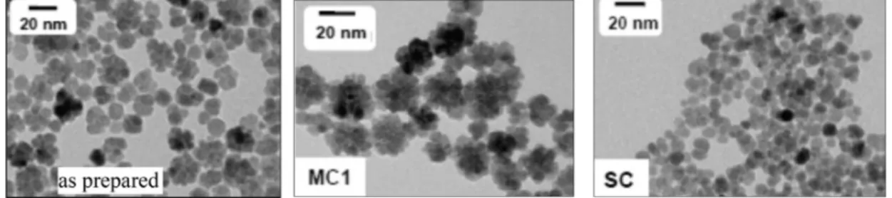
For example, the 1:2 mixture of Fe(II) and Fe(III) salts was hydrolyzed in organic solvent mixture (diethylene glycol and N-methyldiethanolamine) at high temperature; the clustering and coalescence of seeds took place during the longer period [84]. In this single step process, a big mixture of coalesced flower-like maghemite nanoparticles formed, which was fractionated by increasing salt content of aqueous system at low pH, taking advantage of electrostatic colloidal stability. Figure2 shows the nanoflowers formed in the one pot synthesis and two fractions selected as examples to distinguish the single and multicore IONPs. Different routes based on the partial oxidation of Fe(OH)2, polyol-mediated synthesis, or the reduction of iron acetylacetonate were used to obtain multicore iron oxide nanoflowers in the size range 25–100 nm [82]. The nanoparticles were either stabilized with well-known agents, such as dextran and citric acid, or, as an alternative, IONPs were embedded in polystyrene to ensure long-term colloidal stability. The first steps toward the standardization of the synthesis and characterization of nanoflowers have been attempted. By now, better quality of magnetite nanoflowers can be also synthesized by thermal decomposition in organic media [25].
magnetoactive fibrous nanocomposites and multi-responsive co-networks [80,81], which exhibit promising characteristics for magnetothermally or pH triggered drug delivery.
More recently nanoflower type composites came into the play [82], whose formation is due to exchange interactions between the cores favoring cooperative behavior and a crystal continuity at the core interfaces. The magnetic nanoflowers manifest enhanced susceptibility while maintaining superparamagnetic behavior; their structure (e.g., the contact between cores within a particle, having a strong impact on the collective magnetic properties [83]) critically depends on the synthesis process.
Two routes of the latter can be differentiated: the polyol method and thermal decomposition. For example, the 1:2 mixture of Fe(II) and Fe(III) salts was hydrolyzed in organic solvent mixture (diethylene glycol and N-methyldiethanolamine) at high temperature; the clustering and coalescence of seeds took place during the longer period [84]. In this single step process, a big mixture of coalesced flower-like maghemite nanoparticles formed, which was fractionated by increasing salt content of aqueous system at low pH, taking advantage of electrostatic colloidal stability. Figure 2 shows the nanoflowers formed in the one pot synthesis and two fractions selected as examples to distinguish the single and multicore IONPs. Different routes based on the partial oxidation of Fe(OH)2, polyol- mediated synthesis, or the reduction of iron acetylacetonate were used to obtain multicore iron oxide nanoflowers in the size range 25–100 nm [82]. The nanoparticles were either stabilized with well- known agents, such as dextran and citric acid, or, as an alternative, IONPs were embedded in polystyrene to ensure long-term colloidal stability. The first steps toward the standardization of the synthesis and characterization of nanoflowers have been attempted. By now, better quality of magnetite nanoflowers can be also synthesized by thermal decomposition in organic media [25].
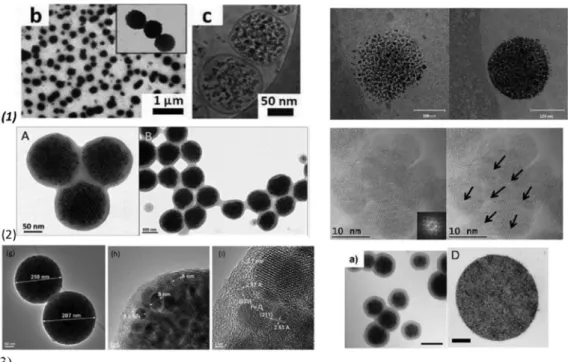
Figure 1. Magnetic multi-core particles obtained by different synthesis procedures: (1) encapsulation of magnetic nanoparticles (MNPs) into liposomes, polymersome: left—TEM (b) and cryo-TEM (c) micrographs of Ultra Magnetic Liposomes (UMLs) prepared by reverse phase evaporation process (REV) process. MNPs are trapped inside unilamellar vesicles (c) and dipole−dipole interaction can occur as exemplified by magnification (b) ([73]); right—Cryo-TEM image showing iron oxide nanoparticles incorporated in the polymersome membrane with 4.1% iron oxide (left), and 17.4% iron oxide (right) ([77]). (2) thermal decomposition: left—TEM images of polymer encapsulated colloidal ordered assemblies (polymer-COA) at higher (A) and lower (B) resolution. The dark pattern (A) results from the ordering of the closed packed assemblies within the nanobeads, while the brighter gray ring is caused by the polymer shell (lower electron density) of around 20 nm thickness ([59]);
right—High Resolution TEM of multi-core MNP showing the continuity of the crystal lattice at the Figure 1.Magnetic multi-core particles obtained by different synthesis procedures: (1)encapsulation of magnetic nanoparticles (MNPs) into liposomes, polymersome:left—TEM (b) and cryo-TEM (c) micrographs of Ultra Magnetic Liposomes (UMLs) prepared by reverse phase evaporation process (REV) process.
MNPs are trapped inside unilamellar vesicles (c) and dipole−dipole interaction can occur as exemplified by magnification (b) ([73]);right—Cryo-TEM image showing iron oxide nanoparticles incorporated in the polymersome membrane with 4.1% iron oxide (left), and 17.4% iron oxide (right) ([77]). (2)thermal decomposition:left—TEM images of polymer encapsulated colloidal ordered assemblies (polymer-COA) at higher (A) and lower (B) resolution. The dark pattern (A) results from the ordering of the closed packed assemblies within the nanobeads, while the brighter gray ring is caused by the polymer shell (lower electron density) of around 20 nm thickness ([59]);right—High Resolution TEM of multi-core MNP showing the continuity of the crystal lattice at the grain interfaces. The Fourier transform of this
Magnetochemistry2020,6, 2 6 of 36
high resolution image (see inset) shows the monocrystalline fcc structure of the multi-core nanoparticles, oriented along the [001] zone axis ([84]). (3) miniemulsion: left—TEM images of magnetic microgel with magnetite nanoparticles cluster as a core coated with two layers of cross linked polymer shells poly-N-isopropylacrylamide-polyacrylic acid ([63]);center—TEM image of magnetic clusters encapsulated in a copolymer hydrogel poly(N-isopropylacrylamide-acrylic acid). Scale bar:
100 nm ([62]);right—TEM image of cross section of superparamagnetic microparticles produced with ferrofluid nanoparticle concentrations of 1 g/L using oil-in water emulsion-templated assembly ([39]).
Reprinted with permission from Reference [41].
grain interfaces. The Fourier transform of this high resolution image (see inset) shows the monocrystalline fcc structure of the multi-core nanoparticles, oriented along the [001] zone axis ([84]).
(3) miniemulsion: left—TEM images of magnetic microgel with magnetite nanoparticles cluster as a core coated with two layers of cross linked polymer shells poly-N-isopropylacrylamide-polyacrylic acid ([63]); center—TEM image of magnetic clusters encapsulated in a copolymer hydrogel poly(N- isopropylacrylamide-acrylic acid). Scale bar: 100 nm ([62]); right—TEM image of cross section of superparamagnetic microparticles produced with ferrofluid nanoparticle concentrations of 1 g/L using oil-in water emulsion-templated assembly ([39]). Reprinted with permission from Reference [41].
Figure 2. TEM images of the polydisperse mixture of iron oxide nanoparticles (IONPs) (as prepared left side) and its fractions containing multicore (MC1 middle) and single core (SC right side) nanoparticles. Reprinted with permission from Reference [84].
3. Magnetic Nanoparticles in Aqueous Carrier
3.1. Ferrofluids vs. Bioferrofluids
The main distinctive feature of ferrofluids among the larger class of magnetic colloids is their long-term colloidal stability, even in strong and non-uniform magnetic fields specific to most of applications. In the carrier liquid, the overall particle interaction potential should be repulsive, i.e., the attractive van der Waals and magnetic forces have to be balanced by Coulombic, steric or other interactions, in order to keep particles apart from each other [17]. The stabilization of magnetic fluids impeding aggregate formation is more challenging for aqueous than for organic carriers. The necessary increase of magnetic particle concentration also involves an increase of the hydrodynamic volume fraction of surface coated particles determined by the stabilization procedure, electrostatic or electro-steric, which differentiate water-based ferrofluids, to attain high values of saturation magnetization. The steric stabilizing layer (usually a chemisorbed primary and a physisorbed secondary layer of surfactant molecules) has a much greater thickness than the electrostatic one, therefore the hydrodynamic volume fraction at the same magnetic volume fraction is much higher (approx. 7–8 times) for electro-steric (e.g., oleic acid double layer) than for electrostatic stabilized aqueous ferrofluids [85]. The significantly reduced interparticle distance produces colloidal stability issues that involve nanoparticle size and magnetic moment, dipolar interactions, excess surfactant, and agglomerate formation. Ferrofluids designed for biomedical applications—bio-ferrofluids [16]—
involve beside single-core particles, a large fraction of multi-core magnetic nanoparticles coated with single or multiple biocompatible surface layers [41], to be discussed in what follows.
3.2. Surface Coating of Magnetic Cores
The surface coating of magnetic iron oxide nanoparticles (IONPs) is inevitable to protect iron leaching, to optimize long term and in-use colloidal stability, to ensure biocompatibility, and to provide specific sites to graft biological functions as well. Therefore, the coating of magnetic nanoparticles should be carefully designed.
The different synthesis methods commonly produce IONP particles that are coated with a protective shell. The preparation of naked IONPs is relatively rare in the literature, probably for the reason that the surface properties of naked IONPs definitely depend on pH and nanoparticles
Figure 2.TEM images of the polydisperse mixture of iron oxide nanoparticles (IONPs) (as prepared left side) and its fractions containing multicore (MC1 middle) and single core (SC right side) nanoparticles.
Reprinted with permission from Reference [84].
3. Magnetic Nanoparticles in Aqueous Carrier
3.1. Ferrofluids vs. Bioferrofluids
The main distinctive feature of ferrofluids among the larger class of magnetic colloids is their long-term colloidal stability, even in strong and non-uniform magnetic fields specific to most of applications. In the carrier liquid, the overall particle interaction potential should be repulsive, i.e., the attractive van der Waals and magnetic forces have to be balanced by Coulombic, steric or other interactions, in order to keep particles apart from each other [17]. The stabilization of magnetic fluids impeding aggregate formation is more challenging for aqueous than for organic carriers.
The necessary increase of magnetic particle concentration also involves an increase of the hydrodynamic volume fraction of surface coated particles determined by the stabilization procedure, electrostatic or electro-steric, which differentiate water-based ferrofluids, to attain high values of saturation magnetization. The steric stabilizing layer (usually a chemisorbed primary and a physisorbed secondary layer of surfactant molecules) has a much greater thickness than the electrostatic one, therefore the hydrodynamic volume fraction at the same magnetic volume fraction is much higher (approx. 7–8 times) for electro-steric (e.g., oleic acid double layer) than for electrostatic stabilized aqueous ferrofluids [85]. The significantly reduced interparticle distance produces colloidal stability issues that involve nanoparticle size and magnetic moment, dipolar interactions, excess surfactant, and agglomerate formation. Ferrofluids designed for biomedical applications—bio-ferrofluids [16]—involve beside single-core particles, a large fraction of multi-core magnetic nanoparticles coated with single or multiple biocompatible surface layers [41], to be discussed in what follows.
3.2. Surface Coating of Magnetic Cores
The surface coating of magnetic iron oxide nanoparticles (IONPs) is inevitable to protect iron leaching, to optimize long term and in-use colloidal stability, to ensure biocompatibility, and to provide specific sites to graft biological functions as well. Therefore, the coating of magnetic nanoparticles should be carefully designed.
The different synthesis methods commonly produce IONP particles that are coated with a protective shell. The preparation of naked IONPs is relatively rare in the literature, probably for the reason that the surface properties of naked IONPs definitely depend on pH and nanoparticles strongly aggregate at neutral pHs, as discussed above [86,87]. The colloidal stability of IONPs under biorelevant conditions,
Magnetochemistry2020,6, 2 7 of 36
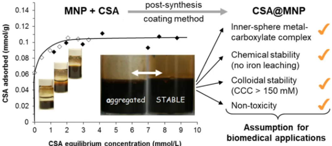
e.g., in blood, at pH~7.4 in physiological salts and protein concentration is the minimum requirement for biomedical applications [71,88]. Therefore, the aggregation of IONPs has to be prevented by protective coating, which can be created either during or after their synthesis. In the literature, in situ coating, post-synthesis adsorption, or post-synthesis grafting are distinguished [68]. In the latter, the functional groups of brush-like polymer chains are anchored to the IONP’s surface. Covalently bound molecules can more improve colloidal stability than adsorbed ones, as demonstrated, for example, in the work of Rinaldi and co-authors [89,90]. However, a great disadvantage of the former is the expensive purification process to remove impurities from organic synthesis to reduce the chemical hazard of the formulation. The multipoint adsorption of polyelectrolytes, especially natural polysaccharides, such as chondroitin-sulfate-A (CSA) bound chemically to≡Fe-OH surface sites of IONPs, is suitable for fabricating biocompatible magnetic fluid (MF) and magnetoresponsive nanocomposites [91–94].
Biopolymer coated magnetite nanoparticles fulfill all assumption of biomedical application, as shown in Figure3.
strongly aggregate at neutral pHs, as discussed above [86,87]. The colloidal stability of IONPs under biorelevant conditions, e.g., in blood, at pH~7.4 in physiological salts and protein concentration is the minimum requirement for biomedical applications [71,88]. Therefore, the aggregation of IONPs has to be prevented by protective coating, which can be created either during or after their synthesis. In the literature, in situ coating, post-synthesis adsorption, or post-synthesis grafting are distinguished [68]. In the latter, the functional groups of brush-like polymer chains are anchored to the IONP’s surface. Covalently bound molecules can more improve colloidal stability than adsorbed ones, as demonstrated, for example, in the work of Rinaldi and co-authors [89,90]. However, a great disadvantage of the former is the expensive purification process to remove impurities from organic synthesis to reduce the chemical hazard of the formulation. The multipoint adsorption of polyelectrolytes, especially natural polysaccharides, such as chondroitin-sulfate-A (CSA) bound chemically to ≡Fe-OH surface sites of IONPs, is suitable for fabricating biocompatible magnetic fluid (MF) and magnetoresponsive nanocomposites [91–94]. Biopolymer coated magnetite nanoparticles fulfill all assumption of biomedical application, as shown in Figure 3.
Figure 3. Adsorption isotherm of chondroitin-sulfate-A (CSA) on magnetite nanoparticles (MNP) at pH ~6.3 in aqueous NaCl solution (left side). With increasing CSA concentration, the colloidal state of samples changes characteristically from aggregated to stable, as seen in the vials. The inserted larger photos clearly show the difference between the well stabilized and aggregated magnetic fluids (the amount of CSA is expressed through the number of repeating units in mmol.) Some assumptions of biomedical application are listed in the right side of figure. Reprinted from Reference [94] under the terms of CC by 4.0.
In the literature, the biomedical use of citric acid stabilized IONPs is favored (e.g., the famous VSOP-C184 product) in [45,95,96] or of the multi-core samples in [97]). However, the citrated IONPs coagulate, even at low salt concentration and, moreover, their iron leaching is very high because citric acid has a reducing effect, and it forms complexes with the surface Fe ions [98]; the dissolved iron ions may cause oxidative stress besides the danger of particle aggregation in vivo. Amstad and coworkers [71] reported a similar iron dissolution effect of catechol derivatives (e.g., mimosine) grafted to Fe3O4 surfaces, causing the gradual dissolution of Fe3O4 nanoparticles through complexation. The other key point is the formation of protein corona of MNPs in biological fluids [99]. Up to now, the coating IONPs with hydrophilic agents is the most widely accepted method for overcoming this problem. Polyethylene oxides or glycols (PEG) and carbohydrates like dextran [3,71]
or carbohydrate derivatives (such as mannose, ribose, and rhamnose) [84,100] are the most common of many coating agents, which are chemically bound to the ≡Fe-OH surface sites or by multiple H- bonds, and make IONPs super hydrophilic, inhibiting the adsorption of proteins. However, the formation of protein corona on MNPs covered with dextran and its derivatives has been perceived [101]. Other types of PEG coating on IONPs (grafting with poly(ethylene glycol)-silane [90] or in situ forming in the poly(ethylene glycol) and poly(ethylene imine) mixture [102]), i.e., the PEGylation can generally improve the drug delivery, enhance the drug accumulation, and might improve the blood-
Figure 3.Adsorption isotherm of chondroitin-sulfate-A (CSA) on magnetite nanoparticles (MNP) at pH ~6.3 in aqueous NaCl solution (left side). With increasing CSA concentration, the colloidal state of samples changes characteristically from aggregated to stable, as seen in the vials. The inserted larger photos clearly show the difference between the well stabilized and aggregated magnetic fluids (the amount of CSA is expressed through the number of repeating units in mmol.) Some assumptions of biomedical application are listed in the right side of figure. Reprinted from Reference [94] under the terms of CC by 4.0.
In the literature, the biomedical use of citric acid stabilized IONPs is favored (e.g., the famous VSOP-C184 product) in [45,95,96] or of the multi-core samples in [97]). However, the citrated IONPs coagulate, even at low salt concentration and, moreover, their iron leaching is very high because citric acid has a reducing effect, and it forms complexes with the surface Fe ions [98]; the dissolved iron ions may cause oxidative stress besides the danger of particle aggregation in vivo. Amstad and coworkers [71] reported a similar iron dissolution effect of catechol derivatives (e.g., mimosine) grafted to Fe3O4 surfaces, causing the gradual dissolution of Fe3O4 nanoparticles through complexation.
The other key point is the formation of protein corona of MNPs in biological fluids [99]. Up to now, the coating IONPs with hydrophilic agents is the most widely accepted method for overcoming this problem. Polyethylene oxides or glycols (PEG) and carbohydrates like dextran [3,71] or carbohydrate derivatives (such as mannose, ribose, and rhamnose) [84,100] are the most common of many coating agents, which are chemically bound to the≡Fe-OH surface sites or by multiple H-bonds, and make IONPs super hydrophilic, inhibiting the adsorption of proteins. However, the formation of protein corona on MNPs covered with dextran and its derivatives has been perceived [101]. Other types of PEG coating on IONPs (grafting with poly(ethylene glycol)-silane [90] or in situ forming in the poly(ethylene glycol) and poly(ethylene imine) mixture [102]), i.e., the PEGylation can generally improve the drug delivery, enhance the drug accumulation, and might improve the blood-brain barrier transport of
IONPs [68]. A new design of PEGylated coating (P(PEGMA-co-AA)@MNPs) provides a non-fouling outer surface that helps the nanoparticles to remain “invisible” for the phagocytic mechanisms, while its free carboxylate moieties can be exploited for grafting specific biologically active molecules or proteins for theranostic applications [103].
Using the synthesis procedure of carboxylic (lauric, myristic or oleic) acid stabilized aqueous ferrofluids [49,104], bovine serum albumin (BSA) coating was applied to rise the colloidal stability of lauric acid-coated IONPs in biological media [105]. The coating greatly reduced the toxicity of nanoparticles and enhanced therapeutic potential of mitoxantrone drug-loaded system.
Further cross-linking of BSA coating was performed in order to improve colloidal stability [106], and monoclonal antibodies were covalently bound to BSA coated IONPs promising MRI contrast agents for glioma visualization in brain.
Nanoparticles interact with biological entities in a biological environment, and nano-bio interfaces form [107]. Only the particle surface can be modified to improve in vivo biocompatibility of nanoparticles. Seeing this issue, the size, the sign, and magnitude of surface charge (as manifested in the measurable zeta potential) and dispersibility in aqueous media (hydrophilic/hydrophobic feature) are the main options for change. Experiments have already shown that positively charged particles are probably more toxic than the larger hydrophobic ones clearing rapidly in the reticuloendothelial (RES) system. In biological systems, medium-sized particles with a neutral or weakly negatively charged surface generally tend to promote enhanced permeation and retention (EPR) [107].
A new generation of coating agents P(PEGMA-co-AA), which combine charged functional groups (i.e., carboxyl groups that are capable of anchoring both nanoparticles and bioactive molecules) and superhydrophilic uncharged segments (i.e., PEG chains in comb-like arrangement) has been reported last year [103]. In a post-coating process, these multifunctional molecules are able to spontaneously bind to MNPs’ surface sites≡Fe-OH; stabilize the particles electrostatically via the carboxylate moieties and sterically via the PEG moieties; provide high protein repellency via the structured PEG layer;
and, anchor bioactive molecules via chemical bond formation with the free carboxylate groups.
The electrosteric (i.e., combined electrostatic and steric) stabilization is efficient down to pH 4 and it tolerates saline media.
In biomedical applications, an optimized coating on SPION surface is required, via which IONPs can interact with different biological entities (proteins, cell membranes, etc.). Only the coating on the engineered NPs can be freely varied at the nanobio interface. The core of nanoparticles has almost all of the desired properties, such as chemical composition, shape and curvature, porosity and surface crystallinity, heterogeneity, and roughness, as listed by Nel and coworkers [107]. The coating layer of core-shell nanosystems provides optimal hydrophobicity/hydrophilicity in a given medium and active sites for anchoring biofunctions. In the same article, the other quantifiable properties of NPs’
interactions (dissolution, hydration, zeta potential, aggregation/dispersion, etc.), which are crucially influenced by the ionic strength, pH, temperature, and the presence of large organic molecules (e.g., proteins), or specifically adsorbing molecules or ions (e.g., detergents generally or phosphate ions) of the suspending media, are separately discussed. The composition and structure of interfacial layer on coated NPs, as well as its changes on the nanoscale, definitely affect the microscale and more the macroscale behavior of engineered nanoparticles. The quality of coating interrelates with colloidal stability under biorelevant conditions, as described in [108]. Sedimentation, freezing, and hemocompatibility tests (smears) are recommended for the qualification of good and bad SPION manufacturing for intravenous administration. Besides the colloidal stability of nanosystems, coatings also largely affect the functionality and biological fate of IONPs. Several different functions of NPs’
coating can be identified, namely: (i) colloidal stabilization under physiological conditions (protecting against aggregation at biological pHs and salty medium), (ii) inhibiting the corrosion and oxidation of magnetic core (passivation reducing the iron leakage), (iii) hindering non-specific protein adsorption in biological milieu, (iv) providing reactive groups for anchoring drugs and targeting molecules, and (v) controlling nano-bio interfacial interactions (bio/hemocompatibility, reticuloendothelial system
(RES) uptake, blood circulation time, IONP’s internalization efficiency, toxicity, targeting efficiency, in vivo fate, etc., as discussed in detail [3,41,68,71,107]). These functions of coating largely overlap with the general concerns of EMA (European Medicines Agency) [109] that should be considered in the development of nanomedicine products.
3.3. Stabilization Mechanisms
The dispersed nanoparticles move freely (thermal motion) in the carrier medium. The colloidal stability of dispersion is the question, whether nanoparticles can retain their separateness during collisions; i.e., whether the particle-particle interactions that are controlled by the frequency and efficiency of collision result in aggregate or not. The latter depends on the extent of attractive and repulsive contributions to the total interaction. The classical DLVO theory of colloidal stability describes the attractive (van der Waals) and repulsive (electrostatic) forces. In addition to these, the hydration, the hydrophobic interactions, and the steric hindrance should be also assessed [110,111]. In the case of magnetic particles, besides the short range exchange interaction especially relevant to formation of multi-core particles, such as nanoflowers [83], the magnetic dipole attraction having a fundamental effect on the collective magnetic properties must also be taken into account [112] to obtain reasonable theoretical stability predictions [90]. In these papers particle aggregation is used as a generic term for coagulation and flocculation independently of the inner structure of aggregates and the reversibility of their formation. Another term, agglomeration, has appeared in the relevant literature, with the same or different meanings as aggregation. Gutiérrez and coworkers [83] reviewed the aggregation of magnetic iron oxide colloids and definitely stated that “nanoparticles tend to form assemblies, either aggregates, if the union is permanent, or agglomerates, if it is reversible”, recalling a bit industrial terminology or that used in nanotechnology nowadays. However, there are certain inconsistencies with the classical colloid nomenclature (e.g., in refs. [110,111]), where aggregation involves coagulation and flocculation giving rise to compact and loose structures, respectively. Their reversibility depends on the magnitude of mechanical force against they should exist. For example, coagulum, the aggregate that forms in the coagulation process, is irreversible against thermal motion; however, it disintegrates when subjected to stronger shaking, stirring, or even mild ultrasonication, and, after this, coagulation restarts at rest, so its formation is reversible [111].
In [113], it was emphasized that nanomaterials should be characterized in the relevant medium, and not simply in water, especially in what concerns aggregation (agglomeration) processes. Referring to coagulation kinetics, IONPs’ colloidal stability should be acceptable under biorelevant conditions, i.e., at biological pH values, in the presence of salt and proteins, and also in cell culture media. In classical colloid science, coagulation kinetics can correctly characterize colloidal stability, also allowing for predicting the stability of SPIONs’ products both on storage and in use. However, the measurements require advanced instrumentation and a lot of time, thus a simpler method would be needed to test SPION preparations [108]. Particle aggregation tests (size evolution, filtration, sedimentation, etc.) under arbitrary conditions are often used [71]. Coagulation kinetics is useful for testing the salt tolerance of IONPs and predicting their resistance against aggregation under physiological condition [87].
A straightforward route of physicochemical (iron dissolution) and colloidal (pH-dependent charging and particle size, salt tolerance from coagulation kinetics) measurements was suggested for assessing the eligibility of IONPs for in vitro and in vivo tests [98].
4. Physical-Chemical Characterization
4.1. Chemical Composition of Magnetic Nanoparticles
X-ray Photoelectron Spectroscopy (XPS) is a very sensitive surface analysis method for the materials chemical composition. The method allows for the determination of the atomic concentrations, the chemical state of the emitting atoms (oxidation degree, valence states, chemical ligands, etc.).
This information results from the areas delimited by the photoelectron peaks and from the chemical
shifts of the peaks with respect to the elemental state, as induced by the chemical surrounding of the atoms. The electrostatic interaction between the nucleus and the electrons determine the core binding energies of the electrons. The electrostatic shielding of the nuclear charge from all other electrons in the atom reduces this interaction. The removal or addition of electronic charge will alter the shielding: withdrawal of valence electron charge (oxidation) increase in binding energy; addition of valence electron charge decrease in binding energy. Chemical changes can be identified in the photoelectron spectra.
In the case of magnetic nanoparticles, surface properties strongly influence their magnetic performance and their behavior in biological media. The core-shell type magnetic nanoparticle systems consist of the magnetic core and a shell around the core, usually a biocompatible polymer and additionally molecules fulfilling the roles of anchors, spacers, and various functionalities. XPS provides information regarding the chemical composition of the coating layers and, on the other hand, allow for determining the oxidation state of the metal in the magnetic core [63,114–117]. XPS allows for determining the oxidation states of iron and to quantify Fe2+and Fe3+ions in iron oxide nanoparticles.
These oxidation states of iron can be determined by Fe2p spectrum employing chemical shift and multiplet splitting and the characteristic satellites [85,118–122].
The organic coating layers of magnetic nanoparticles have major importance for biomedical applications of these nanomaterials. Coating layers ensures the chemical and colloidal stability of magnetic nanoparticles and allow for further functionalization [122–124]. Surface functionalization of magnetic nanoparticles for biomedical applications remains a major challenge. XPS is one of the most appropriate methods for the analysis of the functionalized organic coating of magnetic nanoparticles.
The optimization of the required properties for applications requires understanding the nature of the interface between the magnetic core and the shell and the influence of the surface complex formation on the nanoparticle’s magnetic properties.
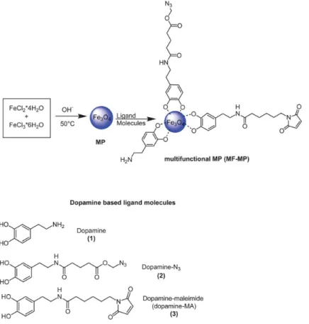
Mazur and coworkers reported a good strategy for surface functionalization of magnetic nanoparticles allowing for the simultaneous attachment of dopamine anchors bearing azide, maleimide, and alkyne terminal groups [125] (Figure4). This functionalization strategy of nanoparticles by using dopamine derivatives shells has the advantage that, besides the protection of the iron oxide core offering the possibility to integrate in a one-step reaction several reactive sites onto the nanoparticles, making these functionalized nanoparticles very promising for biomedical applications.
XPS allows for a detailed analysis of surface chemical composition of iron oxide nanoparticles before and after functionalization with dopamine derivatives (Figures5and6).
The ratio Fe/O=0.73 was calculated from XPS spectra, which is in-between that of Fe3O4(0.75) and Fe2O3 (0.66). This fact and the peak position and satellite peaks in Fe2p spectrum indicate that the magnetic core contains Fe3O4and Fe2O3. The presence of dopamine derivatives shells on the magnetic core was evidenced in the high resolution spectra of N1s and C1s core level spectra.
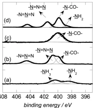
The deconvoluted N1s spectra of Fe3O4that were coated with dopamine derivatives (Figure6) evidence the characteristic groups of the organic shells. The calculated atomic ratio C/N is a good estimation for the success of the organic coating on magnetic nanoparticles. The XPS spectra and the calculated atomic concentrations for the elements C, O, N, and Fe evidence the coating of the magnetic nanoparticles with dopamine derivatives.
The coating layers largely influence the colloidal stability of magnetic nanoparticles under physiological conditions [3,68,71,107]. The protein corona’s effect differs significantly, depending on the surface chemistry of the nanoparticles. The surface chemistry strongly influences the formation of protein corona and the cellular uptake of the nanoparticles. Differences in protein corona formation have been observed for magnetic nanoparticles coated by different organic layers [126,127]. Szekeres et al. undertook a comparative study of the effect of protein corona formation on the colloidal stability of magnetic nanoparticles coated by polyelectrolyte shells, citrate (CA@MNP), and poly(acrylic-co-maleic acid) (PAM@MNP) [128]. PAM coating of MNP ensures a better stability at higher human plasma concentrations as compared with CA coated MNP. XPS determined the chemical composition (atomic
concentrations), as well as the chemical state of the atoms at the surface magnetic nanoparticles CA@MNP and PAM@MNP. The relevant differences between the nanoparticles CA@MNP and PAM@MNP can be observed in the XPS spectra for C1s core levels (Figure7). Both spectra of C1s contain the carboxylic groups and in the case of CA@MNP the C-OH group appears according to the characteristic coating shells.
Magnetochemistry 2020, 6, x FOR PEER REVIEW 11 of 36
Figure 4. Schematic illustration of the formation of magnetic fluid-multifunctional magnetic nanoparticles (MF-MPs) based on the use of differently functionalized dopamine derivatives.
Reprinted with permission from Reference [125].
Figure 5. (A) X-ray Photoelectron Spectroscopy (XPS) survey spectra of as prepared magnetic particles before (a, black) and after modification with dopamine (b, grey), dopamine-N3 (c, blue), dopamine- MA (d, green), and of MF-MP (e, red). (B) High resolution Fe2p spectrum of as-synthesized magnetic particles. Reprinted with permission from Reference [125].
Figure 4. Schematic illustration of the formation of magnetic fluid-multifunctional magnetic nanoparticles (MF-MPs) based on the use of differently functionalized dopamine derivatives. Reprinted with permission from Reference [125].
Magnetochemistry 2020, 6, x FOR PEER REVIEW 11 of 36
Figure 4. Schematic illustration of the formation of magnetic fluid-multifunctional magnetic nanoparticles (MF-MPs) based on the use of differently functionalized dopamine derivatives.
Reprinted with permission from Reference [125].
Figure 5. (A) X-ray Photoelectron Spectroscopy (XPS) survey spectra of as prepared magnetic particles before (a, black) and after modification with dopamine (b, grey), dopamine-N3 (c, blue), dopamine- MA (d, green), and of MF-MP (e, red). (B) High resolution Fe2p spectrum of as-synthesized magnetic particles. Reprinted with permission from Reference [125].
Figure 5.(A) X-ray Photoelectron Spectroscopy (XPS) survey spectra of as prepared magnetic particles before (a, black) and after modification with dopamine (b, grey), dopamine-N3 (c, blue), dopamine-MA (d, green), and of MF-MP (e, red). (B) High resolution Fe2p spectrum of as-synthesized magnetic particles. Reprinted with permission from Reference [125].
Magnetochemistry 2020, 6, x FOR PEER REVIEW 12 of 36
Figure 6. XPS N1s high resolution spectrum of magnetic particles modified with dopamine (a), dopamine-N3 (b), dopamine-MA (c), and MF-MPs (d). Reprinted with permission from Reference [125].
The coating layers largely influence the colloidal stability of magnetic nanoparticles under physiological conditions [3,68,71,107]. The protein corona’s effect differs significantly, depending on the surface chemistry of the nanoparticles. The surface chemistry strongly influences the formation of protein corona and the cellular uptake of the nanoparticles. Differences in protein corona formation have been observed for magnetic nanoparticles coated by different organic layers [126,127]. Szekeres et al. undertook a comparative study of the effect of protein corona formation on the colloidal stability of magnetic nanoparticles coated by polyelectrolyte shells, citrate (CA@MNP), and poly(acrylic-co- maleic acid) (PAM@MNP) [128]. PAM coating of MNP ensures a better stability at higher human plasma concentrations as compared with CA coated MNP. XPS determined the chemical composition (atomic concentrations), as well as the chemical state of the atoms at the surface magnetic nanoparticles CA@MNP and PAM@MNP. The relevant differences between the nanoparticles CA@MNP and PAM@MNP can be observed in the XPS spectra for C1s core levels (Figure 7). Both spectra of C1s contain the carboxylic groups and in the case of CA@MNP the C-OH group appears according to the characteristic coating shells.
Figure 6. XPS N1s high resolution spectrum of magnetic particles modified with dopamine (a), dopamine-N3 (b), dopamine-MA (c), and MF-MPs (d). Reprinted with permission from Reference [125].
Magnetochemistry 2020, 6, x FOR PEER REVIEW 13 of 36
Figure 7. C1s spectra of the core-shell MNPs and the schemes of Fe–O–C(O)–R binding between MNP iron sites and organic carboxylates. Peak positions for CA and PAM coated MNPs: C-C, CH 284.91 and 285.27 eV; O–C=O 289.05 and 288.9 eV, respectively, and C-OH 285.75 eV for CA@MNP.
Reprinted with permission from Reference [128].
4.2. Colloidal Stability. Zeta Potential and Hydrodynamic Size
The electrosteric (electrostatic+steric) stabilization has been shown to be quite effective; for example, polyelectrolyte (polyacrylic or polylactic acid, polyethylenimine, etc.) coating on IONPs provides excellent stability [108]. However, outstanding salt tolerance can be achieved through hydrophilic polymer coating, such as dextran, a polysaccharide, which is used commonly in aqueous magnetic fluids [68,95,129]. Silica materials [130], organic molecules (e.g., carboxylates, phosphates, phosphonate, sulfates, amines, alcohols, thiols, etc. [68,88,95]) are often applied as coating agents to ensure colloidal stability. The functional groups of organic agents are mostly chemically bound to the reactive (both charged and uncharged) sites on IONPs’ surface. For example, citrate through its OH and COOH groups are chemically linked to ≡Fe-OH sites in the citrated-electrostatic stabilized- magnetic fluids [98] or dopamine by two phenolic OH groups in the favored core-shell products [71,95]). The effect of surface coverage is hardly studied. IONP dispersions coagulate at a pH below PZC (point of zero charge), if polyacids, such as polyacrylic acid, are present in trace amounts, while their higher loading covers completely IONPs’ surface and improves the stability and salt tolerance of colloidal iron oxide dispersions [68,131]. Macromolecules adsorb at multiple sites of surface, the so-called multi-site bonding makes the coating layer resistant against dilution and the purification of equilibrium medium easy [110,132].
To illustrate the significance of the stabilization mechanism applied-electrostatic or electrosteric- the zeta potentials and hydrodynamic sizes were measured for citrate and oleate stabilized aqueous ferrofluid samples and are given in Figure 8 to show the characteristic pH-dependence due to the different dissociation behaviors of the acidic groups on the coating molecules [85].
Figure 7.C1s spectra of the core-shell MNPs and the schemes of Fe–O–C(O)–R binding between MNP iron sites and organic carboxylates. Peak positions for CA and PAM coated MNPs: C-C, CH 284.91 and 285.27 eV; O–C=O 289.05 and 288.9 eV, respectively, and C-OH 285.75 eV for CA@MNP. Reprinted with permission from Reference [128].
4.2. Colloidal Stability. Zeta Potential and Hydrodynamic Size
The electrosteric (electrostatic+steric) stabilization has been shown to be quite effective; for example, polyelectrolyte (polyacrylic or polylactic acid, polyethylenimine, etc.) coating on IONPs
provides excellent stability [108]. However, outstanding salt tolerance can be achieved through hydrophilic polymer coating, such as dextran, a polysaccharide, which is used commonly in aqueous magnetic fluids [68,95,129]. Silica materials [130], organic molecules (e.g., carboxylates, phosphates, phosphonate, sulfates, amines, alcohols, thiols, etc. [68,88,95]) are often applied as coating agents to ensure colloidal stability. The functional groups of organic agents are mostly chemically bound to the reactive (both charged and uncharged) sites on IONPs’ surface. For example, citrate through its OH and COOH groups are chemically linked to≡Fe-OH sites in the citrated-electrostatic stabilized- magnetic fluids [98] or dopamine by two phenolic OH groups in the favored core-shell products [71,95]).
The effect of surface coverage is hardly studied. IONP dispersions coagulate at a pH below PZC (point of zero charge), if polyacids, such as polyacrylic acid, are present in trace amounts, while their higher loading covers completely IONPs’ surface and improves the stability and salt tolerance of colloidal iron oxide dispersions [68,131]. Macromolecules adsorb at multiple sites of surface, the so-called multi-site bonding makes the coating layer resistant against dilution and the purification of equilibrium medium easy [110,132].
To illustrate the significance of the stabilization mechanism applied-electrostatic or electrosteric-the zeta potentials and hydrodynamic sizes were measured for citrate and oleate stabilized aqueous ferrofluid samples and are given in Figure8to show the characteristic pH-dependence due to the different dissociation behaviors of the acidic groups on the coating molecules [85].Magnetochemistry 2020, 6, x FOR PEER REVIEW 14 of 36
(a)
(b)
Figure 8. (a) pH dependence of the Z-average particle diameter (blue) and the zeta potential (red) for the citrated (MF/CA) sample. (b) pH dependence of the Z-average particle diameter (blue) and the zeta potential (red) for the oleic acid double layer stabilized (MF/OA) sample. Reprinted with permission from Reference [85].
The MF/CA sample loses colloidal stability below pH6 (Figure 8a) due to the difference in the charged state of citrated and oleic acid double layer coated MNPs, while the MF/OA ferrofluid below pH5 (Figure 8). This behavior is explained by the different dissociability values for citric acid (pKa1
= 3.13, pKa2 = 4.76, and pKa3 = 6.40) and oleic acid (pKa = 5.02). Additionally, from Figure 8a,b, it follows that over a broad range of pH values, where both type of samples are stable, the citrate covered particles have a smaller Z-average diameter than the oleic acid covered nanoparticles.
4.3. Magnetic Properties
The magnetic field dependence of the magnetization, i.e., the magnetization curve, provides information regarding the magnetic properties of single and multicore magnetic nanoparticle systems. The magnetization of single and multicore magnetic nanoparticle systems is conditioned by the magnetic relaxation processes that are specific to the composition, dimension, morphology, etc., as well as external conditions, like temperature.
The direct current (DC) magnetometry is the most frequently used magnetic characterization method. The DC magnetization can be measured by means of vibrating sample magnetometry (VSM), alternative gradient magnetometry (AGM), and superconducting quantum interference device (SQUID). Important characteristics of the sample can be directly obtained from the magnetization curve: initial susceptibility (χi), saturation magnetization (Ms), coercive field (Hc), and remanent magnetization (Mr). In the case of superparamagnetic systems, the DC first magnetization
Figure 8.(a) pH dependence of the Z-average particle diameter (blue) and the zeta potential (red) for the citrated (MF/CA) sample. (b) pH dependence of the Z-average particle diameter (blue) and the zeta potential (red) for the oleic acid double layer stabilized (MF/OA) sample. Reprinted with permission from Reference [85].
The MF/CA sample loses colloidal stability below pH6 (Figure8a) due to the difference in the charged state of citrated and oleic acid double layer coated MNPs, while the MF/OA ferrofluid below pH5 (Figure8). This behavior is explained by the different dissociability values for citric acid (pKa1=3.13, pKa2=4.76, and pKa3=6.40) and oleic acid (pKa=5.02). Additionally, from Figure8a,b, it follows that over a broad range of pH values, where both type of samples are stable, the citrate covered particles have a smaller Z-average diameter than the oleic acid covered nanoparticles.
4.3. Magnetic Properties
The magnetic field dependence of the magnetization, i.e., the magnetization curve, provides information regarding the magnetic properties of single and multicore magnetic nanoparticle systems.
The magnetization of single and multicore magnetic nanoparticle systems is conditioned by the magnetic relaxation processes that are specific to the composition, dimension, morphology, etc., as well as external conditions, like temperature.
The direct current (DC) magnetometry is the most frequently used magnetic characterization method. The DC magnetization can be measured by means of vibrating sample magnetometry (VSM), alternative gradient magnetometry (AGM), and superconducting quantum interference device (SQUID).
Important characteristics of the sample can be directly obtained from the magnetization curve: initial susceptibility (χi), saturation magnetization (Ms), coercive field (Hc), and remanent magnetization (Mr).
In the case of superparamagnetic systems, the DC first magnetization curve can be used to determine the statistic of the nanoparticles’ magnetic diameter by means of magnetogranulometry [133]. Figure9 presents the DC magnetization curve and the theoretical fit [24] for a sample of dried magnetic microgels with magnetite nanoparticles [35]. The inset of Figure9shows the nanoparticle size distributions that were obtained from magnetogranulometry and TEM.
Magnetochemistry 2020, 6, x FOR PEER REVIEW 15 of 36
curve can be used to determine the statistic of the nanoparticles’ magnetic diameter by means of magnetogranulometry [133]. Figure 9 presents the DC magnetization curve and the theoretical fit [24]
for a sample of dried magnetic microgels with magnetite nanoparticles [35]. The inset of Figure 9 shows the nanoparticle size distributions that were obtained from magnetogranulometry and TEM.
Several groups recently developed AC magnetometry [134,135] with the aim of determining the hysteresis loss in magnetic nanoparticles used in magnetic hyperthermia applications. The method is generally useful for characterizing the magnetic dynamic response of single and multicore magnetic nanoparticle systems at low and high frequencies in the range 1 kHz–1 MHz. Figure 10 presents the frequency dependent dynamic response for 9 nm (Sample I—Figure 10a) and 21 nm (Sample II—
Figure 10b) iron oxide nanoparticles, respectively [134]. The hysteretic loss is significantly larger in the bigger diameter nanoparticle sample.
Figure 9. Direct current (DC) magnetization curve and the theoretical fit for a sample of dried magnetic microgels with magnetite nanoparticles. Reprinted with permission from Reference [35].
Figure 9.Direct current (DC) magnetization curve and the theoretical fit for a sample of dried magnetic microgels with magnetite nanoparticles. Reprinted with permission from Reference [35].
Several groups recently developed AC magnetometry [134,135] with the aim of determining the hysteresis loss in magnetic nanoparticles used in magnetic hyperthermia applications. The method is generally useful for characterizing the magnetic dynamic response of single and multicore magnetic nanoparticle systems at low and high frequencies in the range 1 kHz–1 MHz. Figure10presents
![Figure 10b) iron oxide nanoparticles, respectively [134]. The hysteretic loss is significantly larger in the bigger diameter nanoparticle sample](https://thumb-eu.123doks.com/thumbv2/9dokorg/1098670.75883/14.892.217.672.624.1008/figure-nanoparticles-respectively-hysteretic-significantly-larger-diameter-nanoparticle.webp)
![Figure 10b) iron oxide nanoparticles, respectively [134]. The hysteretic loss is significantly larger in the bigger diameter nanoparticle sample](https://thumb-eu.123doks.com/thumbv2/9dokorg/1098670.75883/15.892.165.725.215.478/figure-nanoparticles-respectively-hysteretic-significantly-larger-diameter-nanoparticle.webp)