Regular Article
Elevated plasma neutrophil elastase concentration is associated with disease activity in patients with thrombotic thrombocytopenic purpura
Bálint Mikes
a, György Sinkovits
a, Péter Farkas
a, Dorottya Csuka
a, Ágota Schlammadinger
b, Katalin Rázsó
b, Judit Demeter
c, Gyula Domján
c, Marienn Réti
d, Zoltán Prohászka
a,⁎
a3rd Department of Internal Medicine, Semmelweis University, Budapest, Hungary
b2nd Department of Internal Medicine, University of Debrecen, Debrecen, Hungary
c1st Department of Internal Medicine, Semmelweis University, Budapest
dDepartment of Hematology and Stem Cell Transplantation, St István and St László Hospital, Budapest
a b s t r a c t a r t i c l e i n f o
Article history:
Received 21 October 2013
Received in revised form 7 January 2014 Accepted 27 January 2014
Available online 1 February 2014 Keywords:
Polymorphonuclear leukocyte neutrophil granulocyte elastase
thrombotic thrombocytopenic purpura disease activity
complement activation
Introduction:Genetic and autoimmune risk factors contribute to the development of thrombotic thrombocytopenic purpura (TTP) but triggers are needed to bring about acute disease.
The aim of the study was to investigate the association of neutrophil activation with acute TTP, to assess whether neutrophil activation changes during plasma exchange therapy and to show if complement- and neutrophil activation are parallel, characteristic processes in acute TTP.
Materials and Methods:Altogether 49 EDTA-plasma samples of 21 TTP patients with acute disease and 17 in remission were investigated along with 20 healthy controls.
A stable complex of PMNE-proteinase-inhibitor was measured by ELISA (Calbiochem, Merck-Millipore, Darmstadt, Germany).
Results:Acute disease was associated with significantly increased PMNE levels, the group medians were similarly low in TTP patients in remission and in healthy controls. Increased PMNE levels were characteristic for hematologically active and ADAMTS13 deficient form of TTP. PMNE concentration inversely correlated to disease activity markers platelet count (r =−0.349, p = 0.032) and hemoglobin levels (p =−0.382 p = 0.018).
Achievement of remission was associated with significant reduction of plasma PMNE levels (p = 0.031, Wilcoxon test). There was positive correlation between PMNE levels and complement activation markers C3a and Bb.
Conclusions:We report increased PMNE levels in acute TTP and showed its association to activity markers of acute TTP and complement activation. Effective treatment of an acute TTP episode resulted in marked decrease in PMNE levels. Our data support and extend previous observations that neutrophil extracellular traps may be released in acute TTP and potentially contribute to the pathophysiology of this disease.
© 2014 Elsevier Ltd. All rights reserved.
Introduction
Various etiological factors represent increased risk for the develop- ment of thrombotic thrombocytopenic purpura (TTP), a life-threatening disorder. Characteristic clinical features of TTP are microangiopathic hemolytic anemia, thrombocytopenia and various involvements of other organs, for example the kidneys and the nervous system[1,2]. In most cases development of functional inhibitors (autoantibodies) against the von Willebrand factor (VWF) cleaving protease (A Disintegrin and Metalloproteinase with ThromboSpondin-1 like motifs-13, ADAMTS13) predispose to disease. Inherited variations of ADAMTS13 may also confer increased risk of TTP development, whereas a minor part of TTP patients presents with non-ADAMTS13 deficient disease form[3]. Severe deficiency of ADAMTS13 leads to the increased presence of unusually
large multimers of VWF that promote the formation of platelet rich thrombi in the capillaries and small vessels leading to tissue ischemia and organ failure[4].
However, ADAMTS13 deficient patients may remain asymptomatic for several years[5,6]. Furthermore, several patients may go into sustained clinical remission despite deficient ADAMTS13 activity[7,8].
In addition, although patients with acute TTP often present without acute disease in history, in the majority of TTP patients infections or pregnancy precede the acute diseaseflare[9]. These well-known clinical observations collectively indicate that multiple hits may be necessary for the development of clinically active TTP, and ADAMTS13 deficiency, even though it is probably the most important predisposing risk factor, it is alone not sufficient to cause acute TTP.
Previously our group reported on the presence of complement acti- vation in patients with acute TTP[10]an observation recently con- firmed in an independent cohort [11]. Our data indicated that activation of the classical/lectin and alternative pathways that lead to the activation of the terminal pathway was present in TTP. It is known
⁎ Corresponding author at: H-1125 Budapest, Kútvölgyi st. 4. Tel.: +36 1 3251379, +36 20 8250962; fax: +36 1 225 3899.
E-mail address:prohoz@kut.sote.hu(Z. Prohászka).
0049-3848/$–see front matter © 2014 Elsevier Ltd. All rights reserved.
http://dx.doi.org/10.1016/j.thromres.2014.01.034
Contents lists available atScienceDirect
Thrombosis Research
j o u r n a l h o m e p a g e : w w w . e l s e v i e r . c o m / l o c a t e / t h r o m r e s
that activation of the complement system and of neutrophil granulocytes is characteristic for most infections and also in pregnancy, especially if complicated with preeclapmsia and/or fetal growth restriction[12,13].
Therefore, we hypothesized that neutrophil activation may associate with acute, ADAMTS13-deficient TTP and potentially contribute to the development of clinically active thrombotic microangiopathy.
Supporting this hypothesis were results of a recent study showing in- creased circulating DNA and myeloperoxidase levels in patients suffer- ing from thrombotic microangiopathies (TMAs)[14]. Hence, the aim of the current study was to formally investigate the association of neutro- phil activation with acute TTP, to assess whether neutrophil activation changes during plasma exchange therapy and to show if complement- and neutrophil activation are parallel, characteristic processes in acute TTP.
Materials and Methods Patients and Plasma Samples
Thirty-eight patients with TTP were enrolled in this single-research laboratory based investigation providing diagnostic services (ADAMTS13 and complement measurements) since August, 2007 for patients suspected to have HUS or TTP in Hungary. The patient enrolment for
this study was closed in January, 2011. The criteria used to guide patient stratification, diagnosis and sample selection have been described in de- tails[10]. Briefly, diagnosis of TTP was based on one or more episodes of Coombs-negative microangiopathic hemolytic anemia with thrombocy- topenia defined as serum lactate dehydrogenase (LDH) N450 U/L;
fragmented erythrocytes in the peripheral blood smear and platelet countb150 G/L; only patients with severely decreased ADAMTS13 activ- ity levels (b5%), or history of it, were included in this study. Patients with acute oligo-anuric renal failure were excluded from the study. Hemato- logical remission (HR) was determined when platelet counts were N150 G/L on two consecutive days without any sign of hemolysis even if there were any neurological, renal or other residual clinical symptoms, whereas complete remission (CR) was established when, platelet count remained above the lower limit continuously for at least 1 month.
Table 1shows clinical and laboratory data of the cohort who had available plasma samples for the current study. Using samples of this TTP cohort we measured plasma polymorphonuclear cell elastase (PMNE) levels during acute diseaseflare, in remission, and for compar- ison in controls. Samples of 38 TTP patients have been investigated.
Twenty-one patients with acute disease (mean age 40 years, SD 15, 17 women), 17 TTP patients in remission (mean age 42 years, SD 10, 14 women) and 20 healthy age- and sex matched controls (mean age 35 years, SD 17, 15 women) were enrolled. Eight patients with acute
Table 1
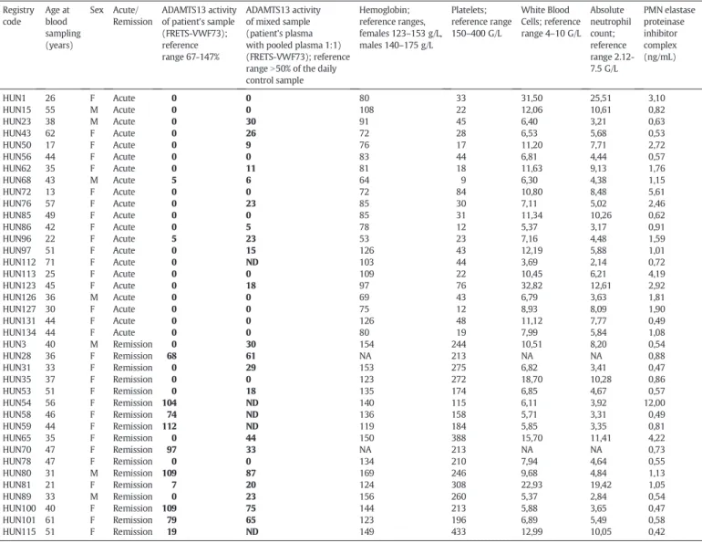
Clinical and laboratory data of the 38 patients with thrombotic thrombocytopenic purpura.
Registry code
Age at blood sampling (years)
Sex Acute/
Remission
ADAMTS13 activity of patient's sample (FRETS-VWF73);
reference range 67-147%
ADAMTS13 activity of mixed sample (patient's plasma with pooled plasma 1:1) (FRETS-VWF73); reference rangeN50% of the daily control sample
Hemoglobin;
reference ranges, females 123–153 g/L, males 140–175 g/L
Platelets;
reference range 150–400 G/L
White Blood Cells; reference range 4–10 G/L
Absolute neutrophil count;
reference range 2.12- 7.5 G/L
PMN elastase proteinase inhibitor complex (ng/mL)
HUN1 26 F Acute 0 0 80 33 31,50 25,51 3,10
HUN15 55 M Acute 0 0 108 22 12,06 10,61 0,82
HUN23 38 M Acute 0 30 91 45 6,40 3,21 0,63
HUN43 62 F Acute 0 26 72 28 6,53 5,68 0,53
HUN50 17 F Acute 0 9 76 17 11,20 7,71 2,72
HUN56 44 F Acute 0 0 83 44 6,81 4,44 0,57
HUN62 35 F Acute 0 11 81 18 11,63 9,13 1,76
HUN68 43 M Acute 5 6 64 9 6,30 4,38 1,15
HUN72 13 F Acute 0 0 72 84 10,80 8,48 5,61
HUN76 57 F Acute 0 23 85 30 7,11 5,02 2,46
HUN85 49 F Acute 0 0 85 31 11,34 10,26 0,62
HUN86 42 F Acute 0 5 78 12 5,37 3,17 0,91
HUN96 22 F Acute 5 23 53 23 7,16 4,48 1,59
HUN97 51 F Acute 0 15 126 43 12,19 5,88 1,01
HUN112 71 F Acute 0 ND 103 44 3,69 2,14 0,72
HUN113 25 F Acute 0 0 109 22 10,45 6,21 4,19
HUN123 45 F Acute 0 18 97 76 32,82 12,61 2,92
HUN126 36 M Acute 0 0 69 43 6,79 3,63 1,81
HUN127 30 F Acute 0 0 75 12 8,93 8,09 1,90
HUN131 44 F Acute 0 0 126 48 11,12 7,77 0,49
HUN134 44 F Acute 0 0 80 19 7,99 5,84 1,08
HUN3 40 M Remission 0 30 154 244 10,51 8,20 0,54
HUN28 36 F Remission 68 61 NA 213 NA NA 0,88
HUN31 33 F Remission 0 29 153 275 6,82 3,41 0,47
HUN35 37 F Remission 0 0 123 272 18,70 10,28 0,86
HUN53 51 F Remission 0 18 135 174 6,85 4,67 0,57
HUN54 56 F Remission 104 ND 140 115 6,11 3,92 12,00
HUN58 46 F Remission 74 ND 136 158 5,71 3,31 0,49
HUN59 44 F Remission 112 ND 119 184 5,85 3,35 0,81
HUN65 35 F Remission 0 44 150 388 15,70 11,41 4,22
HUN70 47 F Remission 97 33 NA 213 NA NA 0,73
HUN78 47 F Remission 0 0 134 210 7,94 4,64 0,55
HUN80 31 M Remission 109 87 169 246 9,68 4,84 1,13
HUN81 21 F Remission 7 20 124 308 22,93 19,42 1,05
HUN89 33 M Remission 0 23 156 260 5,37 2,84 0,54
HUN100 40 F Remission 109 75 144 213 5,88 3,65 0,47
HUN101 61 F Remission 79 65 123 196 6,89 5,49 0,58
HUN115 51 F Remission 19 ND 149 433 12,99 10,05 0,42
NA: not available; ND: not done.
TTP had available follow-up plasma samples. Six of them (patient’s reg- istry codes 1, 15, 62, 68, 86, 127) had plasma samples taken in hemato- logical remission, afterfinishing the PEX series. Five of them (registry codes 68, 86, 126, 127, 131) had plasma samples taken during during PEX series, before reaching hematological remission. Thus, altogether 49 EDTA-plasma samples were used for PMNE determination.
Blood samples (EDTA-anticoagulated plasma and sodium-citrate anticoagulated plasma) were taken by antecubital venipuncture or from central catheter, cells and supernatant separated by centrifugation and shipped in cooled packages (−20 degrees of Celsius) to the Research Laboratory, where aliquots were made and stored in ultrafreezers onb−70 degrees of Celsius until determinations.
From each subject, informed consent was obtained and the study was approved by the institutional Ethics Committee on human research.
Determination of Plasma Polymorphonuclear Cell Elastase (PMNE) Levels and other Laboratory Parameters
A stable complex of PMNE-alpha1-proteinase-inhibitor was mea- sured by sandwich type ELISA (QIA96, Calbiochem, Merck-Millipore, Darmstadt, Germany), according to the instructions given by the manufacturer.
Thefluorigenic substrate, FRETS-VWF73, was applied for the deter- mination of ADAMTS13 enzyme activity as described[15]. Briefly, citrated plasma was diluted 1:20 in assay buffer (5 mmol/l Bis-Tris, 25 mmol/L CaCl2, 0.005% Tween 20, pH 6.0) and mixed with 5μmol/L FRETS-VWF73 substrate solution (20μl each), in white 384-well plates.
Fluorescence was measured at 37 °C every 2 minutes for 1 hour in Cha- meleon microplate reader (Hidex, Turku, Finland) equipped with a 340 nm excitation and a 460 nm emissionfilter. The reaction rate was calculated by linear regression analysis offluorescence over time. A two-fold dilution series of normal human plasma (mixed from citrated plasma samples of 10 healthy blood donors) was applied as standard curve, 100% ADAMTS13 activity was set at the reaction rate observed in the 1:20 diluted sample. The intra-assay variation coefficient was b5%, the inter-assay CV% was 6-9% (measured at 60 and 100% activity levels). The presence of anti-ADAMTS13 inhibitors was determined by mixing 1 part of the patient’s sample with 1 part of normal pooled plas- ma, incubation at 37 degrees of Celsius for 2 hours, and measurement of ADAMTS13 activity of the sample. The presence of ADAMTS13 inhibi- tors was considered if the patient’s original sample hadb7% activity, and that of the mixed sample wasb50%.
Levels of complement activation products C3a (MicroVue C3a des- arg EIA A031, mean of healthy controls 129.6 ng/mL), Bb (MicroVue Bb EIA, A027, mean + 2SD range 0.49-1.42μg/mL) were determined with commercial Kits (Quidel, San Diego, California, USA) according to the manufacturer’s instructions in EDTA plasma samples.
Statistical Analysis
The continuous variables reported in this study showed skewed dis- tribution and according to results of Shapiro-Willk’s test deviated from the normal distribution. Therefore, for descriptive purposes the values of each measurement are given as median and 25th-75th percentile, or as numbers (percent) and non-parametric tests were used for group comparisons; continuous variables between two groups were compared with Mann–Whitney U test, for three or more groups with the Kruskal-Wallis ANOVA by ranks test, and for repeated measures with the Friedman test. Dunn’s post-test was used for group compari- sons after analysis of variance. Spearman’s correlation coefficients were calculated by non-parametric method. Statistical analyses were carried out using STATISTICA 7.0 (StatSoft Inc., Tulsa, OK, USA) and GraphPad Prism 4.03 (GraphPad Softwares Inc., CA, USA) softwares.
Two tailed p values were calculated and the significance level was put at a value of pb0.050.
Results
Acute TTP is Associated with Increased PMNE Levels
Table 1shows clinical and laboratory data of the cohort. All of the pa- tients in acute diseaseflare (n = 21) were ADAMTS13 deficient (activity b5%) and positive for anti-ADAMTS13 inhibitors. Samples for PMNE determinations were taken before the initiation of a series of plasma exchange sessions in 13 patients, whereas for the remaining 8 cases sampling was done during the PEX series. Seventeen patients were enrolled in complete remission, 8 of them were ADAMTS13 deficient (1 patient had 7% activity and was positive for inhibitor), 8 patients had ADAMTS13 activity within the reference range, and 1 patient had moderately decreased level. Increased absolute neutrophil counts were not associated with acute TTP (Table 1), the median (IQR) values for acute patients was 6.0 G/L (4.4-9.7) whereas for patients in remission 4.7 G/L (3.4-9.1; p = 0.223).
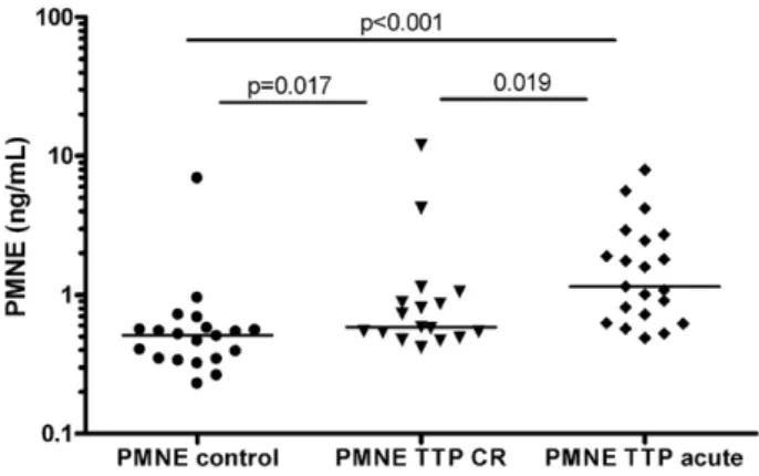
Acute disease was associated with significantly increased PMNE levels, whereas the group medians were similarly low in TTP patients in remission and in healthy controls (Fig. 1). Patients in remission with (0.54 ng/mL, 0.53-0.86) or without (0.73 ng/mL, 0.48-1.00) ADAMTS13 deficiency had similar PMNE levels (p = 0.680). Further- more, increased PMNE levels and deficient ADAMTS13 activity together characterized hematologically active disease (pltb150 G/L,Fig. 2, panel A). Note, that there were no patients in our cohort with non-deficient ADAMTS13 activity and decreased platelet counts. In line with this ob- servation there was a significant association between amounts of func- tional ADAMTS13 inhibitors (as assessed by measuring the ADAMTS13 activity of the mixed patient/normal plasma samples) and PMNE levels (Fig. 2, panel B). There was no significant correlation between ADAMTS13 activity (measured in patient’s own samples) and PMNE levels (data not shown). Similarly, no association between PMNE levels and neutrophil counts was observed (r =−0.381, p = 0.064, data not shown) and neutrophil counts were similar in acute and remission groups, excluding the possibility of the mere reflection of higher neutro- phil counts by increased PMNE levels. Finally, we investigated changes of PMNE levels during therapy. There were 5 patients with available samples before the initiation of PEX series, and during that. In 4 out of the 5 patients, PMNE levels decreased by 40-60%, whereas in one pa- tient it increased by 187%. Similarly, we investigated PMNE levels in samples of 6 patients (4 women) with available samples collected on the day of admission, before the initiation of PEX series, and also in hematological remission at least 1 week after the last session of the PEX series. As shown inFig. 3, achievement of remission was associated with significant reduction of circulating plasma PMNE levels in patients with TTP.
Fig. 1.Neutrophil elastase (PMNE) is marker of acute TTP.(A)Plasma neutrophil-elastase- alpha1-proteinase inhibitor complex (Calbiochem, Merck-Millipore, Darmstadt, Germany) was measured in EDTA-plasma samples of healthy controls, TTP patients in complete remission) and in TTP patients with acute diseaseflare. Horizontal lines indicate group median, P values were obtained by Mann–Whitney test.
Association between PMNE Levels, Activity Markers of TTP and Complement Activation Products
Next, correlations between TTP activity markers and PMNE levels were analyzed. The amount of PMNE showed inverse correlation to
platelet count and hemoglobin levels in the whole TTP cohort (Fig. 4, panel A and B). Fourteen patients with acute TTP received packed red blood cells before sampling for PMNE determinations. The significant inverse association is present between PMNE and hemoglobin levels if these subjects are excluded form the analysis presented onFig. 4B (r =−0.427, p = 0.037). Result of stratified correlation analysis (although underpowered due to low patient numbers) is presented in legend forFig. 4.
In a subset of these patients levels of complement factors and ac- tivation products have been measured previously. Increased levels of C3a and terminal pathway complex (TCC) were observed in acute TTP[10]. Therefore, we analyzed if any of the complement factors or activation products show association with activity markers of neutrophils. As shown inFig. 5, concentrations of the activity marker of the alternative pathway convertase’s enzymatic component Factor B (Bb, r = 0.392, p = 0.015) and anaphylatoxin C3a (r = 0.367, p = 0.024) showed significant, positive correlation to PMNE levels. No other significant correlation between complement components (C3, Factors B, H, I, r =−0.171, -0.287, 0.015, 0.255, respectively, all p N0.05) or activation products (C1r-C1s-C1-inhibitor, C4d, C3bBbP, TCC, 0.025, 0.058, 0.017, 0.199, respectively, all pN0.05) and PMNE concentration was observed.
Fig. 2. (A)Elevated PMNE level together with deficient ADAMTS13 activity is characteristic for hematologically active TTP. Analysis of 46 samples of patients with hematologically ac- tive TTP (platelet below 150 G/L) or in hematological remission (platelet above 150 G/L), as stratified by the presence of ADAMTS13 deficiency (activityb5%). P values were obtain- ed by Mann–Whitney test.(B)Correlation between functional ADAMTS13 inhibitor and PMNE concentrations. Inhibitors against ADAMTS13 were assessed in 33 samples of pa- tients with TTP by mixing studies as described in the Materials and Methods section. Sam- ples taken in acute TTP are marked with closed symbols, whereas those taken in remission are marked with open symbols. Horizontal lines indicate group median, correlation coefficient with p value was calculated by Spearman’s non-parametric method: pooled analysis r =−0.428, p = 0.013; acute TTP: r =−0,547, p = 0.012; TTP in remission:
r = 0.171, p = 0.577).
Fig. 3.Changes of plasma neutrophil elastase (PMNE) levels during treatment of acute diseaseflare in TTP. Comparison of PMNE concentrations in plasma from 6 patients (4 women) with TTP collected at the presentation with acute disease (acute TTP) or in hematological remission (HR). P value was obtained by Wilcoxon test.
Fig. 4.Association of plasma neutrophil elastase (PMNE) levels with disease activity markers in TTP. Correlation coefficients and p values between plasma PMNE levels and platelet counts(A)or hemoglobin levels(B)were calculated by the Spearman non-parametric method (Panel A, pooled analysis: r =−0.483, p = 0.006; acute TTP: -0.521, p = 0.015; TTP in remission: r =−0.086, p = 0.743; Panel B, pooled analysis: r =−0.382, p = 0.018, acute TTP: -0.353, p = 0.116; TTP in remission: r =
−0.257, p = 0.318). Samples taken in acute TTP are marked with closed symbols, where- as those taken in remission are marked with open symbols.
Discussion
Here we report on two novelfindings. First, in a well-characterized, homogenous ADAMTS13-deficient, inhibitor positive cohort of TTP pa- tients elevated neutrophil activation marker, PMNE, was shown to be increased in acute diseaseflare. PMNE levels were directly related to disease activity markers hemoglobin and platelet count, and correlated with the amounts of functional ADAMTS13 inhibitor. Second, according to the presented data, neutrophil- and complement activation are present parallel in acute TTP and correlate to each other.
The potential link between thrombotic microangiopathy and neu- trophils wasfirst studied and characterized inE. colicaused typical HUS. Increased initial neutrophil count was shown to be predictive for bad outcome in D + HUS[16]. Further, neutrophil markers (elastase, myeloperoxidase and IL-8) were elevated and linked to endothelial cell damage and thrombin activity in typical HUS[17]. Similar observa- tions have been reported for a few diarrhea negative HUS patients[18].
Recently, elevated myeloperoxidase and circulating DNA levels were shown in patients with various forms of TMA (ADAMTS13 deficient TTP, D + HUS, tumor-associated TMA and TMA of with unknown etiol- ogy). Increased plasma DNA and MPO levels together with ADAMTS13 deficiency characterized the acute disease state in patients with acquired TTP. Authors concluded that circulating DNA and histones in patients with TMA could have originated from neutrophil extracellular traps (NETs)[14].
Neutrophils release processed chromatin termed neutrophil extra- cellular traps (NETs) to trap and kill pathogens[19]. NETs are implicated in immune defense[20], sepsis[21]and autoimmunity[22]. NET is formed by a DNA-histones scaffold that contains granule proteins such as elastase and myeloperoxidase, as well as antimicrobial peptides [23]. Results obtained in PMNE knock-out mice showed that these ani- mals do not form NETs in a pulmonary model of bacterial infection [24]. These and other studies [24] collectively demonstrated that PMNE is essential for the initiation of NET formation and is externalized together with NETs after stimulation of neutrophils. Therefore, based on our results we conclude that the increased PMNE levels in acute TTP may originate from neutrophils in a process of cell activation and NET release. Our observations independently confirm the results of Fuchs et al., regarding the contribution of neutrophil activation to the patho- genesis of acute TMAs, and its correlation to disease activity.
Activation of neutrophils, as assessed by a specific biomarker, elastase, is characteristic for hematologically active TTP. Patients withN150 G/L platelet counts and deficient ADAMTS13 activity had similar mean PMNE level to those in remission without ADAMTS13 deficiency (Fig. 2), and also similar to healthy controls (Fig. 1). Fur- thermore, treatment of acute TTP by a series of PEX sessions resulted decrease of PMNE levels in the majority of patients during PEX, whereas the reduction was significant (Fig. 3) after reaching of he- matological remission. These results support the multiple hit con- cept in the pathogenesis of TMAs, specifically, TTP. Acute TMA flares often follow infections or precipitate during or after pregnancy and neutrophil activation is known to be increased in these clinical states[9]. In genetically (rare or common variants inADAMTS13) or immunologically (ADAMTS13 inhibitors) predisposed individuals, activation of neutrophils and release of NETs may precipitate acute TTP by multiple mechanisms. As shown in mouse model, DNA and histones stimulate thrombosis and promote cytotoxicity[25–27]. In addition, reactive oxygen species released by activated neutrophils have prothrombotic effect, mediated in part by inhibition of VWF cleavage by ADAMTS13[28]. Finally, leukocyte proteases including elastase cleave von Willebrand factor at or near the ADAMTS13 cleavage site and may therefore participate in the proteolytic regulation of VWF[29].
Furthermore, NETs may activate complement leading to C3b deposi- tion to NET components and generation of C5a[30]. Previously we[10]
and others[11]documented the activation of the classical/lectin, the al- ternative and the terminal pathways of complement in TTP. Our current results indicate the association between PMNE and C3 activation (C3a), and the presence of NETs may be a potential link between these factors.
The presence of correlation between PMNE and Bb levels point to the initiation of alternative amplification loop extending complement acti- vation. Complement activation attracts and activates neutrophils [31,32], and in turn may exacerbate NET release, initiating a positive feed-back loop. These observations are in line with the recent report on the association among circulating nucleosomes, activated neutro- phils (as indicated by increased neutrophil elastase-α1-antitrypsin complexes), and presence of deep vein thrombosis[33]. Taken together, evolutionarily conserved innate mechanisms acting in concert may stimulate thrombosis, endothelial damage[34,35]and contribute to the precipitation of acute TTP.
Currently PMNE measurement is possible only by manual immuno- and enzymatic assays, therefore this restricted availability and relatively high costs limit its potential clinical application. However, if these observations can be repeated in independent cohorts markers of neutrophil activation may potentially help to stratify patients between therapeutic modalities and to guide time and frequency of plasma exchange treatment.
We acknowledge potential limitations of our study. TTP is a rare disorder, and we were unable to enrol more patients in a reasonable short time-frame allowing the correct analysis of elastase and neutrophil activation. Therefore, due to the low number of patients some of the Fig. 5.Association of plasma neutrophil elastase (PMNE) levels with complement ac-
tivation products in TTP. Correlation coefficients and p values between plasma PMNE levels and activated factor B (Bb)(panel A)or anaphylatoxin C3a(panel B)levels were calculated by the Spearman non-parametric method (Panel A r = 0.392, p = 0.015;
Panel B r = 0.367, p = 0.024). Samples taken in acute TTP are marked with closed symbols, whereas those taken in remission are marked with open symbols.
analysis presented here are underpowered, there is a possibility for the presence of false negative or false positive conclusions in the results (especially for the correlation analysis), and therefore some of the con- clusions have to be taken as preliminary, until independent confirma- tion is published.
In conclusion, we report the association of increased plasma neu- trophil elastase levels with acute TTP. Increased PMNE levels were directly related to hematologically active TTP, to functional ADAMTS13 inhibitors, disease activity markers of acute TTP (hemoglo- bin concentrations and platelet counts) and complement activation markers of the alternative pathway. Effective treatment of an acute TTP episode resulted in marked decrease in PMNE levels. Our data lend credence to the hypothesis that neutrophil extracellular traps may be released in acute TTP and may potentially contribute to the pathophysiology of this disease. We identified complement activation products associated with high PMNE levels indicating for the presence of a potential positive feed-back loop between innate mechanisms behind the precipitation of acute TTP.
Conflicts of Interest Statement None to Declare.
Acknowledgements
The authors are grateful for the patients for their participation in the study and for the excellent technical support from Szigeti Antalné, Márta Kókai, Zsuzsanna Szendrei and Holeczky Rudolfné. The study wasfinan- cially supported by a grant from National Research Fund of Hungary (T100687 to ZP). The authors are grateful for the expert medical support and sharing of clinical data to Krisztina Madách, Csaba Bereczki, Attila J Szabó, György S Reusz, Gáspár Radványi, and Klára Gadó.
Addendum
Study concept and design: M Réti, P Farkas and Z Prohászka.
Experimental procedures: B
Mikes and G Sinkovits and D Csuka. Acquisition of data: M Réti, P Farkas, K Rázsó, Á
Schlammadinger, J Demeter, G Domján, Analysis and interpretation of data: Z Prohászka, D
Csuka, M Réti, P Farkas Critical writing of the manuscript: M Réti, P Farkas, Z Prohászka
Critical revision of the manuscript for important intellectual content:
B Mikes, M Réti, P
Farkas, D Csuka, K Rázsó, Á Schlammadinger, J Demeter, G Domján, Obtaining funding: Z
Prohászka Study supervision: M Réti, Z Prohászka
References
[1]Chapman K, Seldon M, Richards R. Thrombotic microangiopathies, thrombotic throm- bocytopenic purpura, and ADAMTS-13. Semin Thromb Hemost 2012;38(1):47–54.
[2]Clark WF. Thrombotic microangiopathy: current knowledge and outcomes with plasma exchange. Semin Dial 2012;25(2):214–9.
[3]Kremer Hovinga JA, Lammle B. Role of ADAMTS13 in the pathogenesis, diagnosis, and treatment of thrombotic thrombocytopenic purpura. Hematology Am Soc Hematol Educ Program 2012;2012:610–6.
[4]Tsai HM. von Willebrand factor, shear stress, and ADAMTS13 in hemostasis and thrombosis. ASAIO J 2012;58(2):163–9.
[5]Fujimura Y, Matsumoto M, Isonishi A, Yagi H, Kokame K, Soejima K, et al. Natural his- tory of Upshaw-Schulman syndrome based on ADAMTS13 gene analysis in Japan. J Thromb Haemost 2011;9(Suppl 1):283–301.
[6]Moatti-Cohen M, Garrec C, Wolf M, Boisseau P, Galicier L, Azoulay E, et al. Unexpect- ed frequency of Upshaw-Schulman syndrome in pregnancy-onset thrombotic thrombocytopenic purpura. Blood 2012;119:5888–97.
[7]Peyvandi F, Lavoretano S, Palla R, Feys HB, Vanhoorelbeke K, Battaglioli T, et al.
ADAMTS13 and anti-ADAMTS13 antibodies as markers for recurrence of acquired thrombotic thrombocytopenic purpura during remission. Haematologica 2008;93:232–9.
[8]Chaturvedi S, Carcioppolo D, Zhang L, McCrae KR. Management and outcomes for patients with TTP: analysis of 100 cases at a single institution. Am J Hematol 2013.
[9]Falter T, Alber KJ, Scharrer I. Long term outcome and sequelae in patients after acute thrombotic thrombocytopenic purpura episodes. Hamostaseologie 2013;33(2):113–20.
[10]Reti M, Farkas P, Csuka D, Razso K, Schlammadinger A, Udvardy ML, et al. Comple- ment activation in thrombotic thrombocytopenic purpura. J Thromb Haemost 2012;10:791–8.
[11]Westwood1J-P, Langley1K, Heelas1E, Machin1S, Scully2M. Complement activa- tion in thrombotic thrombocytopenic purpura. Mol Immunol December 15 2013;56(3):1.
[12]Derzsy Z, Prohaszka Z, Rigo Jr J, Fust G, Molvarec A. Activation of the complement system in normal pregnancy and preeclampsia. Mol Immunol 2010;47:1500–6.
[13]Hung TH, Chen SF, Lo LM, Li MJ, Yeh YL, Hsieh TT. Myeloperoxidase in the plasma and placenta of normal pregnant women and women with pregnancies complicated by preeclampsia and intrauterine growth restriction. Placenta 2012;33:294–303.
[14]Fuchs TA, Kremer Hovinga JA, Schatzberg D, Wagner DD, Lammle B. Circulating DNA and myeloperoxidase indicate disease activity in patients with thrombotic microan- giopathies. Blood 2012;120:1157–64.
[15]Gombos T, Mako V, Cervenak L, Papassotiriou J, Kunde J, Harsfalvi J, et al. Levels of von Willebrand factor antigen and von Willebrand factor cleaving protease (ADAMTS13) activity predict clinical events in chronic heart failure. Thromb Haemost 2009;102:573–80.
[16]Walters MD, Matthei IU, Kay R, Dillon MJ, Barratt TM. The polymorphonuclear leucocyte count in childhood haemolytic uraemic syndrome. Pediatr Nephrol 1989;3:130–4.
[17]Ishikawa N, Kamitsuji H, Murakami T, Nakayama A, Umeki Y. Plasma levels of gran- ulocyte elastase-alpha1-proteinase inhibitor complex in children with hemolytic uremic syndrome caused by verotoxin-producing Escherichia coli. Pediatr Int 2000;42:637–41.
[18]Fitzpatrick MM, Shah V, Filler G, Dillon MJ, Barratt TM. Neutrophil activation in the haemolytic uraemic syndrome: free and complexed elastase in plasma. Pediatr Nephrol 1992;6:50–3.
[19]Brinkmann V, Reichard U, Goosmann C, Fauler B, Uhlemann Y, Weiss DS, et al. Neu- trophil extracellular traps kill bacteria. Science 2004;303:1532–5.
[20]Brinkmann V, Zychlinsky A. Neutrophil extracellular traps: is immunity the second function of chromatin? J Cell Biol 2012;198(5):773–83.
[21]Clark SR, Ma AC, Tavener SA, McDonald B, Goodarzi Z, Kelly MM, et al. Platelet TLR4 activates neutrophil extracellular traps to ensnare bacteria in septic blood. Nat Med 2007;13:463–9.
[22]Kessenbrock K, Krumbholz M, Schonermarck U, Back W, Gross WL, Werb Z, et al. Net- ting neutrophils in autoimmune small-vessel vasculitis. Nat Med 2009;15:623–5.
[23]Urban CF, Ermert D, Schmid M, Abu-Abed U, Goosmann C, Nacken W, et al. Neutro- phil extracellular traps contain calprotectin, a cytosolic protein complex involved in host defense against Candida albicans. PLoS Pathog 2009;5:e1000639.
[24]Papayannopoulos V, Metzler KD, Hakkim A, Zychlinsky A. Neutrophil elastase and myeloperoxidase regulate the formation of neutrophil extracellular traps. J Cell Biol 2010;191:677–91.
[25]Massberg S, Grahl L, von Bruehl ML, Manukyan D, Pfeiler S, Goosmann C, et al. Recip- rocal coupling of coagulation and innate immunity via neutrophil serine proteases.
Nat Med 2010;16:887–96.
[26]Brill A, Fuchs TA, Savchenko AS, Thomas GM, Martinod K, De Meyer SF, et al. Neutro- phil extracellular traps promote deep vein thrombosis in mice. J Thromb Haemost 2012;10:136–44.
[27]Xu J, Zhang X, Pelayo R, Monestier M, Ammollo CT, Semeraro F, et al. Extracellular histones are major mediators of death in sepsis. Nat Med 2009;15:1318–21.
[28]Chen J, Fu X, Wang Y, Ling M, McMullen B, Kulman J, et al. Oxidative modification of von Willebrand factor by neutrophil oxidants inhibits its cleavage by ADAMTS13.
Blood 2010;115:706–12.
[29]Raife TJ, Cao W, Atkinson BS, Bedell B, Montgomery RR, Lentz SR, et al. Leukocyte proteases cleave von Willebrand factor at or near the ADAMTS13 cleavage site.
Blood 2009;114:1666–74.
[30]Leffler J, Martin M, Gullstrand B, Tyden H, Lood C, Truedsson L, et al. Neutrophil ex- tracellular traps that are not degraded in systemic lupus erythematosus activate complement exacerbating the disease. J Immunol 2012;188:3522–31.
[31]Jagels MA, Daffern PJ, Hugli TE. C3a and C5a enhance granulocyte adhesion to endothe- lial and epithelial cell monolayers: epithelial and endothelial priming is required for C3a-induced eosinophil adhesion. Immunopharmacology 2000;46(3):209–22.
[32]Peng Q, Li K, Sacks SH, Zhou W. The role of anaphylatoxins C3a and C5a in regu- lating innate and adaptive immune responses. Inflamm Allergy Drug Targets 2009;8:236–46.
[33]van Montfoort ML, Stephan F, Lauw MN, Hutten BA, Van Mierlo GJ, Solati S, et al. Cir- culating nucleosomes and neutrophil activation as risk factors for deep vein throm- bosis. Arterioscler Thromb Vasc Biol 2013;33(1):147–51.
[34]Karpman D, Tati R. Complement activation in thrombotic microangiopathy.
Hamostaseologie 2013;33(2):96–104.
[35]Noris M, Mescia F, Remuzzi G. STEC-HUS, atypical HUS and TTP are all diseases of complement activation. Nat Rev Nephrol 2012;8(11):622–33.