III./9.1. General structure of peripheral nerves; pathological forms of peripheral nerve damage and regeneration
Zsuzsanna Arányi
Peripheral nerves are made up of motor, sensory and autonomic fibers (axons).
Nerves fibers are organized into fascicles within a nerve. The nerve fibers, fascicles and the nerves themselves are surrounded by connective tissue, which is called endo-, peri-, and epineurium respectively (Fig. 1). Furthermore, most nerve fibers are also covered by a myelin sheath produced by Schwann cells (Fig. 2). The myelin sheath serves to increase nerve conduction velocity, as the action potential jumps from one node of Ranvier to the other (saltatory conduction). The nerve conduction velocity of unmyelinated nerve fiber is 1-5 m/s, whereas that of a myelinated nerve fiber may reach 120 m/s (in general 35-70 m/s).
Fig. 1: Cross section of a peripheral nerve. The axons are organized into fascicles. The outer sheath of the nerve is called epineurium (ep), the
connective tissue surrounding the fascicles is called perineurium (pe).
Fig. 2: The concentric layers of myelin sheath are made by Schwann cells.
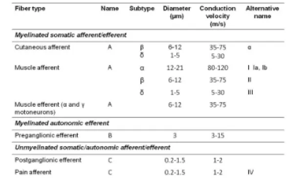
Several classifications of nerve fibers exist. They are summarized in Table 1. It is seen that nerve fibers may be grouped based on the following: 1) presence or absence of myelin sheath, 2) somatic or autonomic function, 3) afferent or efferent function, and 4) diameter. It is also seen that there is a direct relationship between fiber diameter and nerve conduction velocity: the greater the fiber diameter, the faster the conduction velocity.
Table 1:Classification of nerve fibers
Pathological forms of focal nerve lesions (Seddon):
1.) Focal (segmental) demyelination (neurapraxia): In the mildest form of nerve lesion, only the myelin sheath is damaged (e.g. due to a transient external compression), leading to a decrease of nerve conduction velocity.
Reduced nerve conduction velocity in itself doesn’t cause a functional deficit, only when the degree of myelin damage is such that conduction block occurs (Fig. 3): the action potential cannot pass across the site of lesion, causing a functional deficit (e.g. weakness, sensory loss). However, axonal continuity is preserved, muscles are not denervated, thus muscle atrophy doesn’t develop.
2.) Axonal damage (axonotmesis): In more severe damage (e.g. long standing compression), the axons are also damaged and axonal continuity is lost. The axon part distal to the lesion degenerates and disappears (Wallerian degeneration). In axonotmesis, functional deficit and muscle atrophy are both observed, because denervation also occurs. Muscle wasting due to denervation may become apparent quite quickly, even within days.
3.) Complete nerve damage (neurotmesis): Complete nerve damage, including the outer connective tissue (epineurium) is most often caused by lacerating traumas. Symptoms are the same as in axonotmesis, thus the occurrence of neurotmesis is suspected based only on the circumstances of the trauma.
Fig. 3: Conduction block due to focal demyelination
The type of nerve damage determines the type of regeneration, and the chances and time frame of recovery (Fig. 4). Therefore its knowledge is essential for determining prognosis after a nerve lesion.
Fig. 4: Types of nerve regeneration
In case of demyelination, remyelination is to be expected within weeks or 1-2 months, with complete clinical restitution. The internodal distances of the new myelin sheath are shorter than in the original, resulting in slower conduction
velocity without clinical consequences.
In case of axonal damage, two types of regeneration are possible. If most of the axons of a nerve are damaged, proximo-distal axonal regeneration is the
predominant type of regeneration. This means that axonal sprouting starts from the axon stumps at the site of lesion, which slowly grow towards the denervated areas.
The preserved basal lamina / endoneurium of the damaged axons show the way for the regenerating axons. This is a slow process, its average rate is 1 mm/day.
Knowing this value and the distance the regenerating nerve has to cover allows an estimation of the time needed for reinnervation and clinical recovery. For example, in case of a distal ulnar nerve lesion at the wrist, the reinnervation of small hand muscles located only a few centimeters away is expected already within a few months. On the other hand, in case of a sciatic nerve lesion at the gluteal region, the reinnervation of leg muscles may take – depending on height –up to 2-3 years.
Clinical restitution may be complete, however ʽaberrant reinnervation’ may also occur when regenerating axons miss the right way and reinnervate structures other than what they originally supplied. This is most often seen in facial nerve lesions (e.g. the mouth also moves when the eyes are closed), probably due to the fact that axons are not organized into fascicles in the facial nerve.
In case of an incomplete nerve damage, collateral reinnervation plays the greater role: axonal sprouting starts from intact motoneurons in the vicinity of the denervated muscle fibers, eventually forming axonal collaterals that reinnervate these muscle fibers. This is made possible by the anatomical situation that muscle fibers belonging to adjacent motoneurons are intermixed in a mosaic-like way.
Collateral reinnervation starts within 4-6 weeks and results in the increase in size of the motor unit performing the reinnervation (i.e. the number of muscle fibers of the unit increases). Therefore, the electromyographic evidence of large motor units is a sign of an old axonal lesion.
The differentiation of the demyelinative-axonal type of focal nerve damage is important, however they rarely occur in an isolated way. For example, a conduction block caused by focal demyelination is usually associated with some degree of axonal damage, and the time for recovery depends on its extent. If the axonal damage is significant, a two-phase recovery occurs: improvement is seen within weeks as remyelination takes place and the conduction block resolves, and further improvement is seen after months when reinnervation is completed.
In case of neurotmesis, regeneration is expected only when nerve suture is done and the proximal and distal stump of the nerve’s epineurium is joined to provide a pathway for regenerating axons. If the anatomical situation precludes regeneration (e.g. after an amputation), so called amputation neuromas may be formed, which may be rather painful. Amputation neuromas are benign tumors formed by a proliferation of Schwann cells, axons and connective tissue elements of the severed nerve.