III./9.4. Mononeuropathies – tunnel syndromes
Table 5 shows the possible causes of peripheral nerve damage. Mononeuropathy refers to the lesion of a single nerve. It is most often the consequence of a local lesion; the site of lesion along the course of the nerve can be well localized, however systemic disease (e.g. diabetes mellitus) may also lead to the lesion of a single nerve. In this case, exact localization of pathology on the given nerve is usually not possible due to diffuse damage.
Table 5: Causes of peripheral nerve damage
Compression of the nerve is one of the most common causes of focal nerve lesion.
Compression may be of external origin or nerves may be compressed while traversing through narrow areas bounded by bones-connective tissue-muscles (entrapment sites).
These latter are called tunnel syndromes or entrapment neuropathies.
The focal nerve lesions most commonly encountered in clinical practice are discussed below.
Median nerve
The median nerve supplies with motor fibers the wrist and finger flexors, and most of the thenar eminence. Its sensory fibers innervate the radial palmar aspect of the hand, including the first three fingers and the radial surface of the fourth finger. (See a textbook of anatomy for further details.)
Carpal tunnel syndrome: The tendons of flexor muscles and the median nerve pass from the forearm to the wrist through the carpal tunnel, which is an anatomical site bounded by the carpal bones and the transverse carpal ligament. The distal compression of the median nerve as a consequence of carpal tunnel syndrome (CTS) is clearly the most common focal neuropathy. It is also the most common tunnel syndrome and a very common neurologic condition. The incidence of CTS is approximately 200-500/100,000 /year. CTS is three times more common in females than in males.
Symptoms:
CTS usually affects both hands, but generally symptoms of the dominant hand appear first. Initial, highly characteristic first symptom is painful numbness of the hand during the night, which may disrupt sleep. Patients often report of referred pain and numbness of the whole arm or forearm, and symptoms are not necessarily restricted to the median innervated first three fingers. Other characteristic signs include stiffness and feeling of swelling of the fingers. Later, symptoms are also experienced during the day, especially during certain activities such as kitchen work, bicycling or car driving. In a more advanced stage, the numbness-paresthesia becomes constant and sensory loss may develop on the fingers resulting in reduced manual dexterity, clumsiness of the hand. In late stage, motor fibers are also damaged and denervation of the thenar occurs (Fig. 8), which leads to further clumsiness of the hand; patients may be unable to perform simple, everyday tasks such as buttoning or closure of a zipper.
Fig. 8: Symptoms of carpal tunnel syndrome
Causes:
CTS is most often idiopathic, which probably reflects a constitutionally narrow tunnel.
This affects mainly women of 40-60 years of age. Furthermore, any condition that contributes to the narrowing of the tunnel or leads to changes in wrist anatomy or is associated with an overuse of the hand are risk factors of CTS:
obesity
edema (pl. pregnancy, lymphedema, hypothyroidism) old or recent wrist fracture
arthrosis
rheumatoid arthritis or other forms of arthritis
diabetes mellitus (nerves already damaged by polyneuropathy are more sensitive to compression)
occupations (e.g. carpenter, mason, locksmith) sports (e.g. bicycling)
Diagnosis:
CTS can be usually diagnosed based on the highly characteristic symptoms. Symptoms may be provoked by the passive flexion of the wrist, which is called Phalen’ maneuver (Fig. 9). Tinel sign may be present (Fig. 10), which means that light pressure or tapping of the nerve at the site of lesion produces an electric sensation radiating along the course of the nerve.
Fig. 9: Phalen’s provocation maneuver in CTS Fig. 10: Tinel sign
CTS can be confirmed by EMG, which first shows focal demyelination in the carpal tunnel followed by axonal lesion. In accordance with clinical symptoms, sensory fibers are earlier and more severely affected than motor fibers. It is also important to know that EMG may be normal in early stages of CTS (i.e. there is no nerve damage, only
irritation of the nerve causing positive sensory symptoms).
Treatment:
Symptomatic treatment of CTS includes splinting of the wrist during the night or local steroid injection. However, the final solution is surgical decompression of the nerve (release of the nerve by cutting the transverse carpal ligament).
Proximal lesion of the median nerve is much less common (e.g. during sleep or
compression due to a tumor or compartment syndrome). In addition to distal symptoms (sensory loss of the fingers and thenar atrophy and weakness), finger flexors are also weak leading to the sign of oath hand: in an attempt to make a fist, the first three fingers remain extended (Fig. 11).
Fig. 11: Oath hand in proximal median nerve lesion
Ulnar nerve
The ulnar nerve supplies with motor fibers the adductor pollicis muscle of the thenar and all other small hand muscles, and the ulnar forearm flexors. Its sensory fibers supply the ulnar aspect of the hand, including the 4-5th finger. (See a textbook of anatomy for further details.)
Ulnar nerve lesion at the elbow (UNE): The ulnar nerve is most often damaged at the elbow, either in the ulnar groove behind the medial epicondyle (retroepicondylar lesion), or less commonly in the cubital tunnel (directly below the elbow, in the tunnel formed by the two heads of the flexor carpi ulnaris muscle and the aponeurosis connecting them (Fig. 12).
Fig.12: Anatomy of the ulnar nerve at the elbow
Causes of retroepicondylar lesion:
tardive ulnar nerve palsy: old elbow fractures or deformity arthrosis
rheumatoid arthritis diabetes mellitus
external compression, often of iatrogenic origin (e.g. during surgeries) Causes of cubital tunnel syndrome:
idiopathic
frequent habitual elbow flexion Symptoms:
As it is generally seen in compression neuropathies, sensory symptoms occur first:
numbness, sensory loss on the 4-5th finger and the ulnar aspect of the hand. It is characteristic that initially only the fingertips are affected. Motor symptoms occur later or in more severe lesions: paresis and atrophy of the hypothenar, interosseus muscles and the adductor pollicis muscle of thenar muscle group. This results in ʽclaw hand’
(Fig. 13). The hand also has the appearance of a ʽskeleton hand’ due to the atrophy of small hand muscles. Interestingly, flexion power of the 4-5th finger is often preserved.
Weakness of the adductor pollicis muscle can be demonstrated by Froment’s sign (Fig.
14): loss of adduction power of the thumb is compensated by the long, median nerve innervated flexors. Another typical sign in ulnar nerve lesion is Wartenberg’s sign (Fig.
15): due to the preferential involvement of the 3rd palmar interosseus muscle, the little finger stands apart from the other fingers.
Fig. 13: Claw hand in ulnar nerve lesion Fig. 14: Froment’s sign in ulnar nerve lesion
Fig. 15: Wartenberg’s sign in ulnar nerve lesion
Diagnosis:
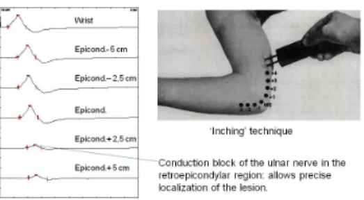
Ulnar nerve lesion can be confirmed by EMG (Fig. 16). Differential diagnosis includes C8 radicular lesion, where sensory symptoms are identical to those in ulnar nerve lesion, but other signs of cervical spondylosis may be seen and the distribution of motor
symptoms respects the C8 myotome (not only the small hand muscles, but for example the radial nerve innervated extensor indicis muscle is also affected).
Fig. 16: Electrophysiological diagnosis of UNE
Treatment:
avoiding elbow flexion, surgical release
Distal ulnar nerve lesion at the wrist is much less common than UNE. The area at the wrist where the ulnar nerve passes from the forearm to the hand is called Guyon tunnel.
Real Guyon tunnel syndrome is however very rare and a questionable entity, and it is not the synonym of distal ulnar nerve lesion. In clinical practice, the distal, deep, purely motor branch in the palm is most often affected by compression (e.g. during bicycling, weight lifting, in certain occupations). This branch originates distally to the branch supplying the hypothenar and to the sensory branch, therefore weakness and atrophy of all small hand muscles are seen with the exception of the hypothenar, and there is no sensory loss. These symptoms are often erroneously attributed to motoneuron disease or cervical myelopathy.
Radial nerve
The radial nerve supplies with motor fibers the triceps muscle and the wrist and finger extensors. Its sensory fibers innervate the radial dorsal aspect of the hand. (See a textbook of anatomy for further details.)
Compression of the radial nerve on the upper arm: The radial nerve is most often damaged at the level of the middle part of the humerus, where the nerve lies directly over the bone in the spiral groove. Compression palsy usually develops during sleep, as a result of the pressure exerted by the head of the patient or of the partner. This is more likely to occur when sleep is stuporous due to exhaustion or alcohol or drug
consumption. Therefore, it is sometimes referred to as Saturday night’s palsy.
Symptoms:
Wrist drop is seen due to the weakness of wrist and finger extensors (Fig. 17). Triceps muscle is spared, because the branch innervating the triceps muscle leaves the main trunk at the level of the axilla. Sensory loss affects the radial dorsal aspect of the hand.
Fig. 17: Wrist drop in radial nerve lesion
Diagnosis:
Clinical symptoms alone usually allow diagnosis of radial nerve palsy. Weakness due to upper motoneuron lesion also results is distally pronounced weakness of the arm, resembling that of a wrist drop, however in this case finger flexion and small hand muscles are also weak.
Treatment:
In case of an external compression, spontaneous recovery is expected in a time frame depending on the severity of lesion (neurapraxia-axonotmesis).
Radial nerve lesion on the forearm:
The distal, pure motor branch of the radial nerve, the posterior interosseus nerve may be compressed at the level of the supinator muscle, which is penetrated by the nerve (supinator tunnel syndrome). It is usually caused by overuse, activities that involve frequent supination-pronation of the forearm. There is no sensory loss and only the finger extensors are weak (finger drop), wrist extension is largely spared. It is typical that weakness first affects the 4-5th finger.
The distal, pure sensory branch of the radial nerve, the superficial radial nerve may be damaged in the distal third of the forearm where the nerve becomes superficial.
Compression of the nerve may result from tight handcuffs, or it may be damaged by surgery of de Quervain’s tenosynovitis.
Common peroneal nerve
The common peroneal nerve supplies with motor fibers the muscles performing dorsiflexion of the foot and toes, and the peroneal muscles. Its sensory fibers innervate the dorsum of the foot and the lateral aspect of the calf. (See a textbook of anatomy for further details.)
Compression of the common peroneal nerve at the fibular head: The common peroneal nerve runs forward from the popliteal fossa passing around the fibular head on the outside where it lies directly over the bone covered only by skin. Therefore, the nerve is very vulnerable to compression at this site. Diabetes mellitus is risk factor for
compression palsy.
Causes:
lying on the side for longer time (e.g. in a comatose state, bed-bound patients, during sleeping on a hard surface)
leg crossing for longer time
squatting for longer time (e.g. strawberry pickers, tilers) during operations (patients are fixed too tightly at the knees) cast
The peroneal nerve passes through the peroneal tunnel formed by the head of the long peroneal muscle as it originates on the fibular head. Compression in this tunnel (real peroneal tunnel syndrome) is however very rare and may be suggested by slowly progressing symptoms.
Symptoms:
The typical symptom of peroneal nerve lesion is foot drop (Fig. 18) with resulting steppage gait (during walking, the patient lifts his/her leg higher on the affected side in order to avoid tripping). The patient is unable to stand on heel on the affected side.
Sensory loss involves the lateral aspect of the calf and the dorsum of the foot.
Differential diagnosis includes the common L5 radicular lesion, which may also cause a foot drop. However, in this case, other (non-peroneal nerve innervated ) muscles
supplied by the L5 segment may also become weak, e.g. inversion of the foot (posterior tibial muscle) and abduction of the hip (gluteus medius muscle). Furthermore, other
signs of lumbar spondylosis may be present.
Symptoms identical to common peroneal nerve lesion may be produced by more proximal lesions, e.g. sciatic nerve lesion or lumbosacral trunk lesion in the pelvis (see plexopathies). In the sciatic nerve, fibers eventually forming the common peroneal nerve run separately from tibial nerve fibers, thus they may be selectively affected.
Fig. 18: Foot drop in common peroneal nerve lesion
Diagnosis:
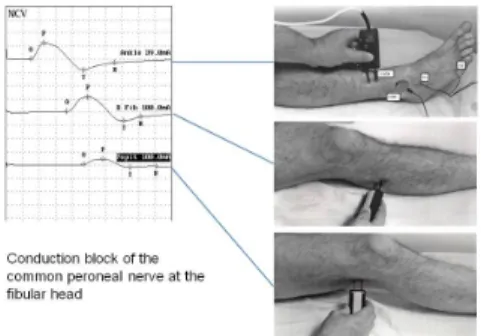
In addition to clinical symptoms, common peroneal nerve lesion at the fibular head may be well localized by EMG (Fig. 19). Furthermore, EMG has a major role in
differentiating common peroneal nerve lesion from the other conditions mimicking peroneal nerve lesion, as mentioned above.
Fig. 19: Conduction block of the common peroneal nerve at the fibular head
Treatment:
In case of an external compression, spontaneous recovery is expected in a time frame depending on the severity of lesion (neurapraxia-axonotmesis). In the rare peroneal tunnel syndrome, surgical release is recommended.
Lateral cutaneus femoral nerve
The lateral cutaneus femoral nerve is a pure sensory nerve, which innervates the skin on the lateral aspect of the thigh. The nerve exits the pelvis by penetrating the inguinal ligament close to the anterior superior iliac spine, where it is very often compressed or damaged. The condition is called meralgia paresthetica.
Causes:
idiopathic obesity pregnancy tight garments
diabetes mellitus crista biopsy Symptoms:
Burning, tingling pain and hypesthesia on the lateral aspect of the thigh (Fig. 20).
Symptoms are more pronounced in a standing position. The area of sensory loss has sharp borders, which helps in differentiating sensory loss of similar distribution due to the rare L2 radiculopathy.
Fig. 20: Sensory innervation area of the lateral cutaneus femoral nerve
Diagnosis:
Diagnosis is usually possible based on clinical symptoms.
Treatment:
Weight loss, loose garments. Rarely local steroid injection may be considered, but spontaneous remission is typical.