High-Resolution Ultrasonography of Peripheral Nerves:
Measurements on 14 Nerve Segments in 56 Healthy Subjects and Reliability Assessments
Hochauflösender Ultraschall peripherer Nerven: Messungen an
14 Nervensegmenten bei 56 gesunden Personen und Untersuchungen zur Reliabilität der Methode
Authors J. Boehm1, E. Scheidl2, D. Bereczki2, T. Schelle3, Z. Arányi2 Affiliations 1 Dept. of Neurology, Freiberg County Hospital, Freiberg
2 Dept. of Neurology, Semmelweis University, Budapest
3 Dept. of Neurology, Municipal Hospital Dessau
Key words
●
" high-resolutionultrasonography
●
" cross-sectional area●
" peripheral nerves●
" normal values●
" reliabilityreceived 26.1.2013 accepted 23.11.2013
Bibliography DOIhttp://dx.doi.org/
10.1055/s-0033-1356385 Published online: April 24, 2014 Ultraschall in Med 2014; 35:
459–467 © Georg Thieme Verlag KG Stuttgart · New York · ISSN 0172-4614
Correspondence Dr. Zsuzsanna Arányi Dept. of Neurology, Semmelweis University Balassa u. 6
1083 Budapest Hungary
Tel.: ++36/12/10 03 30 Fax: ++36/12/10 13 68 aranyi.zsuzsanna@med.
semmelweis-univ.hu
Abstract
!
Purpose:The aim of this study was to assess dif- ferent aspects of reliability in high-resolution ul- trasonography (HRUS) of the peripheral nerves and to establish reference values for the most fre- quently examined nerve segments.
Materials and Methods:A nerve size parameter, the cross-sectional area (CSA) of the C5, C6 and C7 cervical roots, the median, ulnar, radial, super- ficial radial, peroneal, tibial, and the sural nerves was measured using HRUS at a total of 14 prede- fined anatomical sites in two different cohorts of healthy subjects (n = 56), and the inter-rater, in- tra-rater and inter-equipment reliability of meas- urements was assessed.
Results:The mean CSA of the 14 nerve segments ranged from 2 to 10 mm2. The intra-rater, inter- rater and inter-equipment reliability was high with intraclass correlation coefficients of 0.93, 0.98, and 0.86, respectively. The CSA values showed no consistent correlation with age, height, and body weight, but males had signifi- cantly larger values than females for nerve seg- ments on the arm after correcting for age, weight and height in multivariate analysis. CSA values did not differ when two independent cohorts were compared.
Conclusion:Peripheral nerve ultrasonography is a reliable and reproducible diagnostic method in the hands of experienced examiners. Normal val- ues for several upper and lower extremity nerves are provided by our study.
Zusammenfassung
!
Ziel:Beurteilung verschiedener Aspekte der Relia- bilität des hochauflösenden Ultraschalls (HRUS) bei der Untersuchung peripherer Nerven und die Bestimmung von Referenzwerten der Nerven- querschnittsfläche (CSA- cross sectional area) an verschiedenen Nervenabschnitten.
Material und Methoden:Mittels HRUS wurde bei zwei Normalkollektiven an 14 vorher anato- misch definierten Nervenabschnitten die Ner- venquerschnittsfläche (CSA) der Rami ventrales C5, C6, C7, des Nervus medianus, N. ulnaris, N. radialis, Ramus superficialis n. radialis, N. pe- roneus, N. tibialis und N. suralis (n = 56) gemes- sen und die Inter-rater, Intra-rater und Inter- equipment Reliabilität bestimmt.
Ergebnisse: Die durchschnittliche Nervenquer- schnittsfläche (CSA) an den 14 Nervenabschnitten betrug 2 bis 10 mm2. Die Korrelationskoeffizienten der Intra-rater, Inter-rater und Inter-equipment Reliabilität waren mit 0,93 versus 0,98 und versus 0,86 hoch. Dabei zeigten die Nervenquerschnitts- flächen (CSA) keine konstante Korrelation mit dem Alter, der Körpergröße und dem Körperge- wicht. Andererseits ergab eine multivariante Ana- lyse mit Korrektur dieser Faktoren bei Männern signifikant höhere Werte als bei Frauen. Die Ner- venquerschnittsflächen (CSA) unterschieden sich nicht signifikant in den beiden Normalkollektiven.
Schlussfolgerung: Der hochauflösende Ultra- schall peripherer Nerven ist eine zuverlässige und reproduzierbare Untersuchungsmethode in den Händen erfahrener Untersucher. Normwerte für mehrere Nervenabschnitte an der oberen und unteren Extremität wurden in der vorliegenden Studie bestimmt.
Downloaded by: IP-Proxy Semmelweis_Uni, Semmelweis University-Central Library. Copyrighted material.
Introduction
!
High-resolution ultrasonography is an emerging non-invasive technique for the investigation of peripheral nerves and is in- creasingly used worldwide in the diagnosis of peripheral nerve disorders. Neurosonography provides a reliable diagnosis and lo- calization in entrapment neuropathies, traumatic peripheral nerve injuries and tumors of the peripheral nerves, and it has be- come a useful supplementary tool for electrodiagnostic studies in these conditions [1–4, 30, 31]. Characteristic nerve size changes in polyneuropathies have been reported as well [5–9]. Further- more, ultrasonography allows precise structural analysis and quantitative measurements of the nerves, which makes compar- ison of different studies possible. Nerve width (medial to lateral diameter), thickness (anterior to posterior diameter) and cross- sectional area (CSA) measured on transverse scans, and antero- posterior diameter (LAPD) measured on longitudinal scans are the most frequently used quantitative parameters for the ultra- sound investigation of peripheral nerves. Furthermore, ratios of CSA between different segments of the same nerve have also been used. Several reports have been published on reference val- ues for the cross-sectional areas of the median and ulnar nerves [10–17], with good agreement among the measurements. On the other hand, data are less abundant concerning normal values for cervical roots, radial nerve, lower limb nerves and pure sen- sory nerves [15–25], and they show more variation among stud- ies. Some studies have reported values for intra- and inter-rater reliability [24–28], but inter-equipment reliability has not been hitherto addressed.
The aim of our study was to establish a set of normal CSA values for C5, C6, and C7 cervical roots, and several upper and lower limb nerves, including some pure sensory nerves, at pre-defined anatomical sites, and to assess whether the CSAs correlated with age, gender, height, and body weight. Furthermore, to test if such measurements are reliable in routine clinical practice, the intra- rater, inter-rater and inter-equipment reliability of peripheral nerve ultrasound measurements was assessed. CSA values of two independent cohorts from the two study sites were also compared in order to determine the external validity of collected normal values.
Subjects and Methods
!
Subjects
Prior to the start of our study, approval of the institutional review board at both study sites was obtained, and the participants signed informed consent. Between May 2011 and December 2011, 56 healthy subjects were investigated with high-resolution nerve ultrasound at the Dept. of Neurology of Semmelweis Uni- versity in Budapest (Hungary) and at the Dept. of Neurology of the County Hospital in Freiberg (Germany). Subjects were re- cruited from the hospital staff and patients. None of the study subjects had symptoms or signs suggesting polyneuropathy or systemic diseases potentially associated with polyneuropathy, nor any history of neuromuscular disease. Demographic data (age, gender, height, and body weight) were recorded. All sub- jects were of Caucasian ethnicity.
Ultrasound examination
For ultrasound examinations, a Philips HD15XE ultrasound de- vice with a small part imaging software and a 15 MHz 3 cm
“hockey stick”linear array transducer was used for 25 subjects in Budapest. In Freiberg, the same device was used for 10 sub- jects, and an additional 21 subjects were examined with a Toshi- ba Aplio SSA-700A device with small part imaging software and a 12 MHz PLT-1204 4.5 cm linear array transducer. In both devices, compound imaging software (SonoCTfor the Philips HD15XE and ApliPurefor Toshiba Aplio SSA-700A) was used to improve image quality.
The following 14 CSA measurements on the upper and lower ex- tremities were carried out, all on the left side: C5, C6 and C7 cer- vical roots; median, ulnar and radial nerves at the mid-upper arm; ulnar nerve at the elbow at the level of the medial epicon- dyle, median, ulnar and superficial radial nerves at the distal third of the forearm; median nerve at the proximal entrance of the carpal tunnel; peroneal nerve at the fibular neck; tibial nerve at the ankle; and sural nerve at the proximal calf. These sites in- cluded common areas of nerve entrapment (ulnar nerve in the ulnar groove, median nerve in the carpal tunnel), sites largely in- accessible for electrophysiologic studies (cervical roots), as well as sites corresponding to those usually evaluated by electrodiag- nostic studies. The superficial radial and the sural nerves were chosen as pure sensory nerves. Subjects were examined mostly in a supine position, with the exception of the peroneal nerve ex- amined with the subject lying on one side, and the sural nerve examined in a prone position.
For brachial plexus sonography, the following technique de- scribed earlier for determining root level was used [29]: The C7 root was identified in the oblique transverse plane of the C7 ver- tebra, which appeared as a hyperechoic structure characterized by the presence of only a posterior tubercle on its transverse pro- cess, the anterior tubercle being absent. When the transducer was moved slightly upward, the C6 and C5 vertebrae were suc- cessively identified by the presence of both anterior and poster- ior tubercles, the C5, C6 roots appearing as hypoechoic structures between the tubercles. Color Doppler sonography was used to differentiate roots from blood vessels.
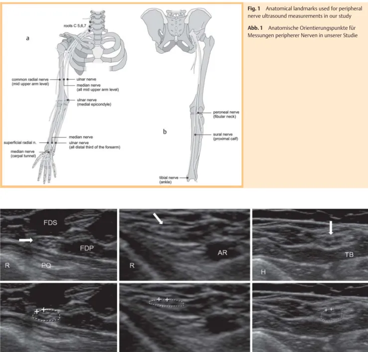
The nerves of the upper and lower extremities were identified on transverse scans using the same typical anatomic landmarks as described before (
●
" Fig. 1) [1]. On the upper arm, the median nerve was identified adjacent to the brachial artery between the biceps and triceps muscles at the midpoint of the line connecting the axilla and the medial epicondyle. The ulnar nerve was then identified at the same level by moving the probe more medially.The radial nerve was assessed at the same level directly on the surface of the humerus in the radial nerve groove, accompanied by the deep brachial artery. At the elbow, the ulnar nerve was measured in the ulnar groove, with the elbow in a slightly flexed position, between the medial epicondyle and the olecranon. On the distal forearm, the median nerve was measured first at the level of the proximal third of the pronator quadratus muscle:
after the pronator quadratus muscle was visualized, the median nerve was identified between the tendons of the flexor pollicis longus and flexor digitorum superficialis muscles. From this point, the transducer was moved medially to the ulnar nerve, which is accompanied at this level by the ulnar artery. Next, the transducer was moved radially to identify the superficial radial nerve, lying between the extensor carpi radialis longus and flexor carpi radialis muscles, just above the palpable bony prominence of the radius, and adjacent to the radial artery [21]. At the wrist, the median nerve was examined at the proximal entrance of the carpal tunnel using the pisiform bone as an anatomic landmark.
Downloaded by: IP-Proxy Semmelweis_Uni, Semmelweis University-Central Library. Copyrighted material.
On the lower limb, a transverse scan of the peroneal nerve was obtained at the level of the fibular neck with the subject lying on the side with the knee propped up and slightly flexed (20° to 30°) [5]. The tibial nerve was examined at the level of the medial mal- leolus, just posterior to the tibial artery. The sural nerve was ex- amined at the proximal dorsal calf, identified superficially be- tween the two heads of the gastrocnemius muscle. If necessary for correct identification, the nerve was followed more distally.
The cross-sectional area (CSA) of the nerves was measured using the trace function of the ultrasound device by manually tracing inside the hyperechoic rim of each nerve (
●
" Fig. 2). The angle of insonation was adjusted perpendicular to the nerve where the nerve appeared the brightest with the best discernible outer margins. The CSA of each nerve segment was measured three times. The three measurements were averaged and the mean val- ue was used for analysis.Fig. 2 Normal ultrasound images of three different nerves. On the lower images, the tracing used to measure the cross-sectional area is shown.
Left: Median nerve at the distal forearm (CSA: 7.7 mm2); R = radial bone, PQ = pronator quadratus muscle, FDS = flexor digitorum superficialis mus- cle, FDP = flexor digitorum profundus muscle. Arrow points to the median nerve. Middle: Superficial radial nerve at the distal forearm (CSA: 1.9 mm2);
R = radial bone, AR = radial artery. Arrow points to the superficial radial nerve. Right: Ulnar nerve at the upper arm (CSA: 6.8 mm2); H = humerus, TB = medial head of the triceps brachii muscle. Arrow points to the ulnar nerve.
Abb. 2 Normale Ultraschallbilder von drei unterschiedlichen Nerven. An den unteren Abbildungen wird die Messung der Nervenquerschnittsfläche (CSA) durch Umfahren der Faszikel mit dem Cursor gezeigt. Links: N. me- dianus am distalen Unterarm (CSA:7.7 mm²), R = Radius, PQ = M. pronator quadratus, FDS = M. flexor digitorum superficialis, FDP = M. flexor digitorum profundus. Das Pfeil zeigt den N. medianus. Mitte: Ramus superficialis N. radialis am distalen Unterarm (CSA:1.9 mm2); R = Radius, AR = A. radialis.
Das Pfeil zeigt den Ramus superficialis n. radialis. Rechts: N. ulnaris am Oberarm (CSA: 6.8 mm2); H = Humerus, TB = Medialer Kopf des M. triceps brachii. Das Pfeil zeigt den N. ulnaris.
Fig. 1 Anatomical landmarks used for peripheral nerve ultrasound measurements in our study Abb. 1 Anatomische Orientierungspunkte für Messungen peripherer Nerven in unserer Studie
Downloaded by: IP-Proxy Semmelweis_Uni, Semmelweis University-Central Library. Copyrighted material.
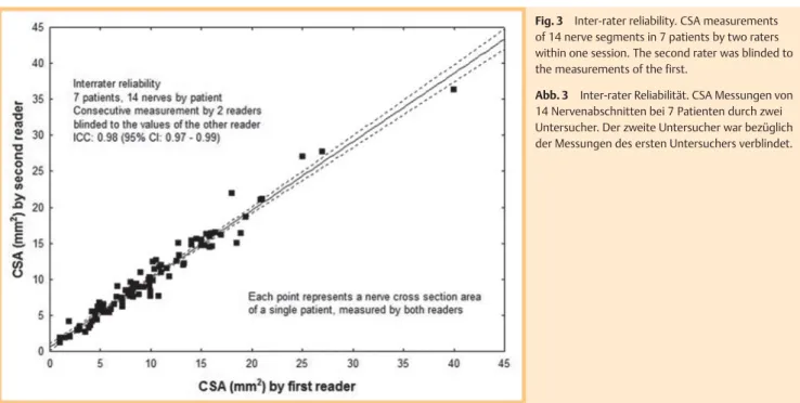
Theinter-rater reliabilitywas assessed at the start of the study.
Two ultrasonographers measured nerve cross-sectional areas in 7 subjects (on all 14 sites in each subject, as described above).
Both examiners are neurologists and clinical neurophysiologists who perform neuromuscular ultrasound in a clinical setting on a daily basis. Both ultrasonographers received training for this study prior to the initiation of data collection. The repeated measurements were done in one session: the examination of all 14 nerve segments by one rater was repeated in the same session by the other rater who was blinded to the results of the first.
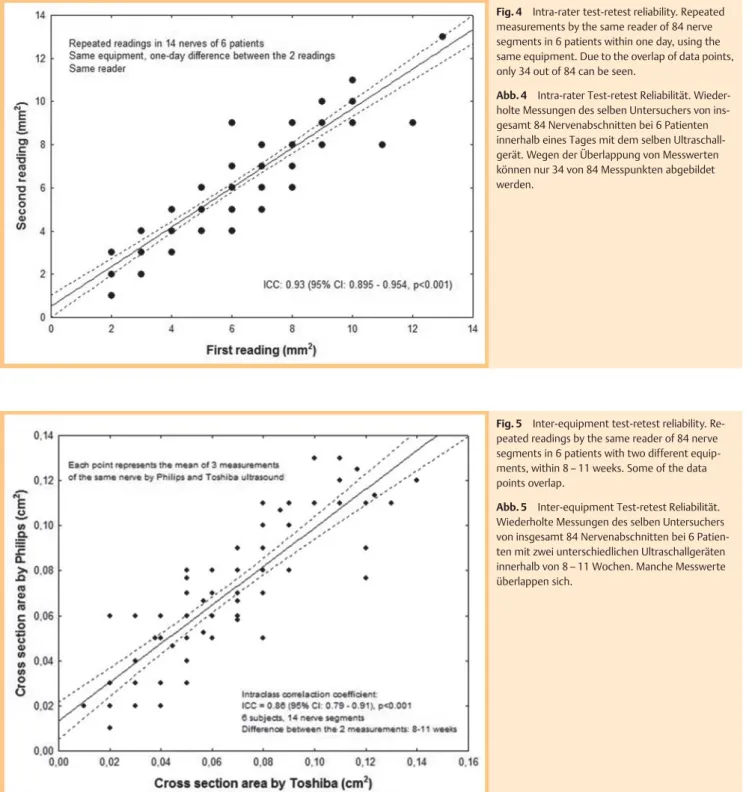
To assessintra-rater reliability, 6 subjects in Freiberg were re-ex- amined with the same Toshiba device by the same investigator 24 hours after the first sonographic examination.
To assessinter-equipment reliability, 6 subjects in Freiberg were examined by the same examiner first with the Philips device, and 8–11 weeks later with the Toshiba ultrasound device.
Thevalidity of normal valueswas tested by comparing the CSA values of the 14 nerve segments in the two independent cohorts of the two study sites.
Statistical analysis
Descriptive statistics were used to present basic demographic data of the study population. The following parameters were cal- culated and presented for normal CSA values of the 14 nerve seg- ments: mean, median, standard deviation (SD), 95 % confidence intervals of the mean, and the coefficient of variation. The nor- mality of variables was checked by the Shapiro-Wilk test. The correlation of CSA measurements with age, gender, height and body weight was tested using the Spearman correlation coeffi- cients. Values between genders were compared by the Kruskal- Wallis ANOVA. The general linear model (GLM) was used to test if gender remains a significant predictor of CSA when age, height and body weight are also considered. Intraclass correlation coef- ficients and corresponding 95 % confidence intervals were calcu- lated to define values for intra-rater, inter-rater, and inter-equip- ment reliability. The validity of our normal values was tested in two independent cohorts using repeated measure ANOVA for the comparison of the CSA values of the 14 nerve segments.
Results
!
Basic demographic features of the study population are given in
●
" Table 1. The univariate relationships between CSA and age,body weight, height and gender are presented in
●
" Table 2. The results of multivariate testing for the effect of gender are present- ed in the last column of●
" Table 2. CSA measurements showed mostly normal distribution in both genders and in pooled data.Descriptive statistics of CSA measurements of all 14 nerve seg- ments for all subjects are presented in
●
" Table 3. The mean CSA values of these 14 nerve segments ranged from 2 to 10 mm2(
●
" Table 3,●
" Fig. 2).Theinter-raterreliability,intra-ratertest-retest reliability, andin- ter-equipment test-retest reliability are presented in
●
" Fig. 3–5,respectively. The intraclass correlation coefficients in all three ana- lyses of reproducibility were remarkably high (0.86–0.98). When the CSA values of the 14 nerve segments were compared between two independent cohorts, no significant difference was found
(
●
" Fig. 6).Discussion
!
In the past decade, high-resolution ultrasonography has become an effective tool for the investigation of peripheral nerve disor- ders. It has been demonstrated that peripheral nerve pathology results in focal or diffuse thickening of the nerves together with
Table 1 Demographic data of the two study cohorts.
parameter germans hungarians p
n 31 25 –
age (years) 51.8 ± 16.4 48.5 ± 15.6 0.45
gender (M:F) 15:16 11:14 0.74
weight (kg) 75.4 ± 13.0 79.6 ± 18.2 0.31
height (cm) 171 ± 9 168 ± 6 0.12
No difference in demographic features between the Hungarian and the German study groups
Table 2 Univariate Spearman correlations of peripheral nerve CSA values with age, body weight, height, and Kruskal-Wallis ANOVA test for gender, and mul- tivariate testing (GLM) for gender.
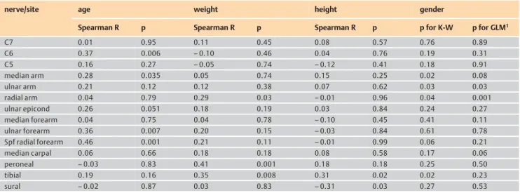
nerve/site age weight height gender
Spearman R p Spearman R p Spearman R p p for K-W p for GLM1
C7 0.01 0.95 0.11 0.45 0.08 0.57 0.76 0.89
C6 0.37 0.006 –0.10 0.46 0.04 0.76 0.19 0.31
C5 0.16 0.27 –0.05 0.74 –0.12 0.41 0.18 0.91
median arm 0.28 0.035 0.05 0.74 0.15 0.25 0.02 0.08
ulnar arm 0.21 0.12 0.12 0.38 0.07 0.62 0.03 0.03
radial arm 0.04 0.79 0.29 0.03 –0.01 0.96 0.04 0.001
ulnar epicond 0.26 0.051 0.18 0.19 0.03 0.84 0.24 0.27
median forearm 0.04 0.75 0.04 0.78 –0.10 0.45 0.41 0.11
ulnar forearm 0.36 0.007 0.20 0.15 –0.03 0.84 0.61 0.78
Spf radial forearm 0.46 0.001 0.21 0.11 –0.01 0.99 0.06 0.21
median carpal 0.06 0.66 0.18 0.18 0.08 0.58 0.17 0.06
peroneal –0.03 0.83 0.41 0.001 0.18 0.18 0.25 0.50
tibial 0.19 0.16 0.35 0.008 0.31 0.02 0.02 0.23
sural –0.02 0.87 0.03 0.83 –0.31 0.03 0.27 0.53
CSA = cross-sectional area; Spf = superficial; values are uncorrected for multiple comparisons. K-W: Kruskal–Wallis univariate ANOVA for comparing CSA values between genders.
1GLM: general linear model analysis, taking gender, age, weight and height as possible predictors of CSA. P-values for gender are presented with correction for age, height and
Downloaded by: IP-Proxy Semmelweis_Uni, Semmelweis University-Central Library. Copyrighted material.
a pathological change of echostructure. These changes can be quantified by measuring nerve size parameters, such as the cross-sectional area (CSA) of the nerve. The increase of the CSA of the involved nerve allows precise localization in entrapment neuropathies and peripheral nerve tumors [1–4]. Moreover, the enlargements of multiple nerves in acquired and hereditary polyneuropathies are also described [5–9]. Therefore, it is essen- tial to compare nerve size parameters measured in patients to re- ference values. However, reference values are still lacking for some nerves and those published tend to show variability prob- ably due to factors such as measurement accuracy, expertise of the examiner, equipment, location of the nerve, and patient- specific factors (ethnicity, age, gender, body mass, height). Our aim was to contribute a large set of reference values to the pool of normative data currently being amassed in the literature by measuring the cross-sectional areas of 10 upper and lower limb nerves at a total of 14 sites in 56 healthy individuals. Other stud- ies usually assessed fewer nerve segments (
●
" Table 4). We alsowished to examine the reliability of measurements. Our study subjects represented a broad range of age and a balanced gender distribution from two different European countries, but the ethnicity (Central-European Caucasian) was homogeneous.
Although the sample size could be larger, the narrow range of 95 % confidence intervals for the mean, the relatively low coeffi- cient of variation (generally between 20–30 %) (
●
" Table 3) and the normal distribution of values of a given nerve, gender groups examined separately or combined and with different resolutions (analysis not shown), all support that the sample size of our study is acceptable. Furthermore, no significant differences were found when comparing two independent cohorts (i. e. German and Hungarian populations), which also supports the validity of col- lected normal values. We found no consistent correlations be- tween CSA values and age, height, or body weight, but males had significantly larger values than females for nerve segments in the upper arm. This finding is similar to some earlier reports but data in the literature are not fully consistent in this respect.Fig. 3 Inter-rater reliability. CSA measurements of 14 nerve segments in 7 patients by two raters within one session. The second rater was blinded to the measurements of the first.
Abb. 3 Inter-rater Reliabilität. CSA Messungen von 14 Nervenabschnitten bei 7 Patienten durch zwei Untersucher. Der zweite Untersucher war bezüglich der Messungen des ersten Untersuchers verblindet.
Table 3 CSA values (mm2) of 14 nerve segments in 56 healthy subjects.
nerve/site valid n mean
(mm2)
median (mm2)
SD (mm2)
95 % CI for the mean (mm2)
coeff. of var.
(%)
C7 50 10.0 10.0 2.9 9.1–10.8 29.5
C6 50 9.5 8.7 2.7 8.7–10.2 28.1
C5 52 5.6 5.3 1.6 5.1–6.0 29.1
median arm 56 8.9 8.9 1.8 8.4–9.4 20.7
ulnar arm 56 6.3 6.3 1.7 5.8–6.8 27.1
radial arm 56 4.2 4.1 1.0 3.9–4.5 24.2
ulnar epicond 56 7.6 7.3 2.1 7.0–8.1 27.3
median forearm 56 5.7 5.9 1.3 5.4–6.0 22.2
ulnar forearm 56 5.2 5.0 1.3 4.9–5.6 25.7
spf radial forearm 56 2.3 2.0 0.7 2.1–2.5 31.2
median carpal 56 8.5 8.4 1.8 8.0–9.0 21.4
peroneal 56 8.9 8.8 2.0 8.3–9.4 23.1
tibial 56 9.6 9.1 2.2 9.0–10.2 23.4
sural 50 1.8 2.0 0.6 1.6–1.9 35.7
CSA = cross-sectional area; spf = superficial; SD = standard deviation; CI = confidence interval; Coeff. of var. = Coefficient of variation
Downloaded by: IP-Proxy Semmelweis_Uni, Semmelweis University-Central Library. Copyrighted material.
Similarly to our results, Heinemeyer et al. found no correlation between nerve size parameters and age, height and body weight, but reported thicker nerves on the upper limbs in males [15].
Cartwright et al. [16] reported that nerve size correlated with body weight and body mass index, and that these correlations were most pronounced in the nerves of the proximal leg. They also found that females had smaller nerves than males. No differ- ence in nerve size parameters was found when dominant and non-dominant sides were compared. According to Zaidman et al., CSAs of the ulnar and median nerves are larger with increas- ing height, at proximal sites and at sites of entrapment, but are independent of age and no side difference was observed [5].
●
" Table 4shows, together with our results, some of the most im-portant studies that published normative values for the CSA of peripheral nerves. Among these studies, our study is the first to evaluate most of the important nerves of the upper and lower limbs in the same person, including pure sensory nerves and cer- vical roots in a healthy Central-European population. The CSA of most nerves, as in our study, ranges from 2 to 10 mm2. However, we did not measure femoral and sciatic nerves, which are consid- erably larger, with a CSA as high as 41 mm2in the case of the scia- tic nerve [25].
●
" Table 4also shows that CSA values across these studies are consistent for the major upper limb nerves, with the exception of the radial nerve in the spiral groove reported byFig. 5 Inter-equipment test-retest reliability. Re- peated readings by the same reader of 84 nerve segments in 6 patients with two different equip- ments, within 8–11 weeks. Some of the data points overlap.
Abb. 5 Inter-equipment Test-retest Reliabilität.
Wiederholte Messungen des selben Untersuchers von insgesamt 84 Nervenabschnitten bei 6 Patien- ten mit zwei unterschiedlichen Ultraschallgeräten innerhalb von 8–11 Wochen. Manche Messwerte überlappen sich.
Fig. 4 Intra-rater test-retest reliability. Repeated measurements by the same reader of 84 nerve segments in 6 patients within one day, using the same equipment. Due to the overlap of data points, only 34 out of 84 can be seen.
Abb. 4 Intra-rater Test-retest Reliabilität. Wieder- holte Messungen des selben Untersuchers von ins- gesamt 84 Nervenabschnitten bei 6 Patienten innerhalb eines Tages mit dem selben Ultraschall- gerät. Wegen der Überlappung von Messwerten können nur 34 von 84 Messpunkten abgebildet werden.
Downloaded by: IP-Proxy Semmelweis_Uni, Semmelweis University-Central Library. Copyrighted material.
Cartwright et al. [16]. However, in the most recent study of Won et al. [17] and in previous studies reporting reference values for the radial nerve [8, 19], the CSA ranged from 3.1 mm2to 5 mm2, which is consistent with our results. Thus, the radial nerve values reported by Cartwright et al. can be considered as an‘outlier’, probably due to methodological reasons. Concerning the superfi- cial radial nerve at the distal forearm, the few studies available [21, 22] published CSA values of 2–3 mm2, similarly to ours.
However, there seems to be a discrepancy across studies with re- spect to the CSA values of cervical roots. The ultrasonographic vi- sualization and measurement of the cervical roots of the brachial plexus are limited by the deep position and oblique course of the roots and also by body habitus, accurate measurement becoming sometimes impossible. Tagliafico et al. [25] emphasizes that the deep position of the nerve affects measurement accuracy due to
poorer visualization. They have found that the minimum detect- able difference for between-limb comparisons for the sciatic nerve is much higher than that of superficial nerves, mainly ex- plained by the poorer visualization. Further studies with high- end equipment are needed to obtain more accurate and consis- tent normal values for the cervical roots. Nonetheless, available data are consistent in that the C5 root is the smallest among the cervical roots. Concerning lower limb nerves, the variability ap- pears to be higher. In the study of Tagliafico et al. [25] of side-to- side comparison of lower limb nerves, they found that the stand- ard error of measurement and minimum detectable side-to-side difference are relatively high for the peroneal nerve at the fibular head and the sural nerve (among the nerves also examined in our study). It is a general experience of those performing ultrasono- graphy of peripheral nerves that–due to the echogenic proper- Fig. 6 Measurements in two independent cohorts.
CSA values of the 14 nerve segments in Hungarian (n = 25) and German (n = 31) healthy subjects. Re- peated measure ANOVA revealed no significant country effect. When pairwise comparisons were done by the Mann-Whitney-test, no significant dif- ference was found between the CSAs in any of the nerve segments after correction for multiple com- parisons.
Abb. 6 Messungen bei zwei unterschiedlichen Kollektiven. CSA-Werte von 14 Nervenabschnitten bei ungarischen (n = 25) und deutschen (n = 31) ge- sunden Probanden. ANOVA zeigte keinen signifi- kanten Ländereffekt. Der paarweise Vergleich mit- tels Mann-Whitney-Test zeigt keinen signifikanten Unterschied der CSA-Werte in keinem der Nerven- abschnitte nach Korrektur für multiple Vergleiche.
Table 4 Comparison of mean (±SD) peripheral nerve CSA values (mm2) from our study to data in the literature.
nerve/site present study n = 56
Cartwright et al. 2008 n = 60
Zaidman et al. 2009 n = 100
Haun et al. 2010 n = 33
Tagliafico et al. 2012 n = 60
Won et al. 2013 n = 97
C7 10.0 ± 2.9 6.3 ± 2.4
(combined mean value of the three trunks)
12.1 ± 4.1
C6 9.5 ± 2.7 10.6 ± 4.3
C5 5.6 ± 1.6 7.1 ± 4.1
median arm 8.9 ± 1.8 8.9 ± 2.1 8.9 ± 2.0 9.4 ± 1.4 (R)
ulnar arm 6.3 ± 1.7 6.2 ± 1.4 5.9 ± 1.1 (R)
radial arm 4.2 ± 1.0 7.9 ± 2.7 4.6 ± 0.9 (R)
ulnar epicond 7.6 ± 2.1 7.3 ± 1.7 7.2 ± 1.4 (R)
median forearm 5.7 ± 1.3 7.5 ± 1.6 7.9 ± 2.4 6.5 ± 1.1 (R)
ulnar forearm 5.2 ± 1.3 5.5 ± 1.4 6.3 ± 1.0 (R)
spf radial forearm 2.3 ± 0.7 2.0 ± 0.5 (R)1
median carpal 8.5 ± 1.8 9.8 ± 2.4 9.7 ± 1.9 8.3 ± 1.5 (R)
peroneal 8.9 ± 2.0 11.2 ± 3.3 13.2 ± 142
tibial 9.6 ± 2.2 13.7 ± 4.3 9.6 ± 42
sural 1.8 ± 0.6 5.3 ± 1.8 (at the distal calf) 3.6 ± 112(at the distal calf)
CSA = cross-sectional area; spf = superficial; SD = standard deviation;
1measured at the elbow.
2mean values are provided with standard error of measurement; R = right side.
Downloaded by: IP-Proxy Semmelweis_Uni, Semmelweis University-Central Library. Copyrighted material.
ties of surrounding tissues–lower limb nerves are less clearly de- marcated and thus their borders, especially on cross-sectional images, may be difficult to discern. This of course accounts for measurement inaccuracy. Furthermore, the peroneal nerve has an oblique course around the fibular head and even slight tilting of the probe may affect nerve size measurements on cross-sec- tional scans. The sural nerve measurements in our study are not comparable with other studies, because we made measurements at the proximal calf rather than at the distal calf. Further studies are needed for the sural nerve.
Theinter-rater reliabilitywas analyzed in several reports, how- ever, the intraclass correlation coefficient (ICC) in our study was high (0.98) when compared to previous studies. Impink et al.
found only a moderate reliability in measuring the parameters of the median nerve [26], whereas other authors reported mostly good or excellent results but had lower ICC values than calculated in our study [16, 18, 28]. This may be explained by the fact that our two investigators worked closely together for several weeks in the training session, and the measurements were taken using precisely predefined anatomical landmarks.
Intra-rater reliability has been rarely reported in peripheral nerve ultrasound measurements [27, 28]. We analyzed 14 meas- urements of 6 patients repeated by the same investigator within one day. The high ICC value (0.93) reflects the excellent reliability and reproducibility of neurosonography in the hands of an ex- perienced and well-trained examiner. Other authors reported high concordance as well, but mostly the median nerve was ex- amined, whereas several nerves of the upper and lower limbs and cervical roots were measured in our study, including nerves that are more difficult to examine.
A novel and noteworthy element of our study is the evaluation of inter-equipment reliability. Similarly to the intra-rater reliability, it was also aʽtest-retest’assessment carried out by the same in- vestigator but on two different devices, with linear array transdu- cers of different frequencies (Philips vs. Toshiba, 15 MHz vs.
12 MHz transducers). Statistical analysis showed a high overall concordance (ICC = 0.86), suggesting a good reproducibility of measurements carried out on different ultrasound equipments.
The difference between the resolution of a 12 and a 15 MHz transducer did not prove to be significant with respect to nerve size measurements. Only a single study reporting good concor- dance of median nerve measurements performed in different la- boratories [28] was found in the literature, but inter-equipment reliability involving the same investigator and the same patients on multiple nerves has not been studied previously.
In conclusion, the good reliability and reproducibility of neuroso- nography in the examination of peripheral nerve disorders in the hands of experienced investigators is highlighted by our study.
The use of predefined anatomical landmarks is essential to obtain comparable data. The excellent reliability of our measurements serves as a basis for the acceptance of the normal values provided by our study. Nonetheless, it is important to note that ultrasono- graphic measurements of peripheral nerves should be put in the context of clinical and electrophysiological data, and caution is advised when interpreting a minor deviation from normative data.
Acknowledgements
!
This study was partly supported by a grant from TAMOP-4.2.1.B- 09/1/KMR (Semmelweis University). Z. Arányi was partly suppor- ted by WAP-B.
References
01Peer S,Bodner G. High-Resolution Sonography of the Peripheral Ner- vous System. 2nd Revised Edition. Springer Sonographic Anatomy of the Peripheral Nervous System
02Kele H. The potential value of ultrasonography in the evaluation of car- pal tunnel syndrome. Neurology 2003; 61: 389–391
03Chiou HJ,Chou YH,Cheg SP et al.Cubital tunnel syndrome: diagnosis by high-resolution ultrasonography. J Ultrasound Med 1998; 17:
643–648
04Beekman R,Schoemaker MC,Van Der Plas JP et al.Diagnostic value of high resolution sonography in ulnar neuropathy at the elbow. Neurol- ogy 2004; 62: 767–773
05Zaidman CM,Al-Lozi M,Pestronk A. Peripheral nerve size in normals and patients with polyneuropathy an ultrasound study. Muscle Nerve 2009; 40: 960–966
06Imamura K,Tajiri Y,Kowa H et al.Peripheral nerve hypertrophy in chronic inflammatory demyelinating polyradiculoneuropathy detect- ed by ultrasonography. Intern Med 2009; 48: 581–582
07Cartwright MS,Brown ME,Eulitt P et al.Diagnostic nerve ultrasound in Charcot-Marie-Tooth disease type 1B. Muscle Nerve 2009; 40: 98–102 08Beekman R. Ultrasonography shows extensive nerve enlargement in
multifocal motor neuropathy. Neurology 2005; 65: 305–307 09Scheidl E,Böhm J,Simó M et al.Ultrasonography of MADSAM neuro-
pathy: focal nerve enlargements at sites of existing and resolved con- duction blocks. Neuromuscul Disord 2012; 22: 627–631
10 Cartwright MS,Shin HW,Passmore LV et al.Ultrasonographic reference values for assessing the normal median nerve in adults. J Neuroima- ging 2009; 19: 47–51
11 Yao L,Gai N. Median nerve cross-sectional area and MRI diffusion characteristics: normative values at the carpal tunnel. Skeletal Radiol 2009; 38: 355–361
12 Klauser S,Halpern E,De Zordo T et al.Carpal tunnel syndrome assess- ment with US: value of additional cross-sectional area measurements of the median nerve in patients versus healthy volunteers. Radiology 2009; 250: 171–177
13 Nakamichi K,Tachibana S. Ultrasonographic measurement of median nerve cross-sectional area in idiopathic carpal tunnel syndrome: diag- nostic accuracy. Muscle Nerve 2002; 26: 798–803
14 Cartwright MS,Shin HW,Passmore LV et al.Ultrasonographic findings of the normal ulnar nerve in adults. Arch Phys Med Rehabil 2007; 88:
394–396
15 Heinemeyer O,Reimers CD. Ultrasound of radial, ulnar, median and sciatic nerves in healthy subjects and patients with hereditary motor and sensory neuropathies. Ultrasound Med Biol 1999; 25: 481–485 16 Cartwright MS,Passmore LV,Yoon JS et al.Cross sectional area refer-
ence values for nerve ultrasonography. Muscle Nerve 2008; 37: 566–
571
17 Won SJ,Kim BJ,Park KS et al.Reference values for nerve ultrasonogra- phy in the upper extremity. Muscle Nerve 2013; 47: 864–871 18 Haun DW,Cho JC,Kettner NW. Normative cross-sectional area of the
C5-C8 nerve roots using ultrasonography. Ultrasound Med Biol 2010;
36: 1422–1430
19 Foxall GL,Skinner D,Hardman JG et al.Ultrasound anatomy of the ra- dial nerve in the distal upper arm. Reg Anest Pain Med 2007; 32:
217–220
20 Gruber H,Peer S,Meirer R et al.Peroneal nerve palsy associated with knee luxation: evaluation by sonography—initial experiences. Am J Roentgenol 2005; 185: 1119–1125
21 Visser LH. High resolution sonography of the superficial radial nerve with two case reports. Muscle Nerve 2009; 39: 392–395
22 Marx SC,Kumar PSD,Marx CA et al.Histological and ultrasonographi- cal study of the human superficial branch of the radial nerve at distal forearm and its clinical implications. Rom J Morphol Embryol 2010;
51: 751–758
Downloaded by: IP-Proxy Semmelweis_Uni, Semmelweis University-Central Library. Copyrighted material.
23 Wilmes M,von Piekartz H. The sural nerve as a contributing factor in chronic achillodynia–an explorative ultrasound study. Sportverletz Sportschaden 2010; 24: 212–217
24 Alshami AM,Cairns CW,Wylie BK et al.Reliability and size of the meas- urement error when determining the cross-sectional area of the tibial nerve at the tarsal tunnel with ultrasonography. Ultrasound Med Biol 2009; 35: 1098–1102
25 Tagliafico A,Cadoni A,Fisci E et al.Reliability of side-to-side ultrasound cross-sectional area measurements of lower extremity nerves in heal- thy subjects. Muscle Nerve 2012; 46: 717–722
26 Impink BG, Gagnon D,Collinger JL et al.Repeatability of ultrasono- graphic median nerve measures. Muscle Nerve 2010; 41: 767–773 27 Kluge S,Kreutziger J,Hennecke B et al.Inter- and intraobserver reliabil-
ity of predefined diagnostic levels in high-resolution sonography of
the carpal tunnel syndrome–a validation study on healthy volunteers.
Ultraschall in Med 2010; 31: 43–47
28 Hobbson-Webb LD,Padua L. Median nerve ultrasonography in carpal tunnel syndrome: findings from two laboratories. Muscle Nerve 2009; 40: 94–97
29 Martinoli C,Bianchi S,Santacroce E et al.Brachial plexus sonography: a technique for assessing the root level. Am J Roentgenol 2002; 179:
699–702
30 Plaikner M,Loizides A,Loescher W et al.Thickened Hyperechoic Outer Epineurium, a Sonographic Sign Suggesting Snapping Ulnar Nerve Syndrome? Ultraschall in Med 2013; 34: 58–63
31 Loizides A,Peer S,Plaikner M et al.Punched Nerve Syndrome: Ultraso- nographic Appearance of Functional Vascular Nerve Impairment. Ul- traschall in Med 2012; 33: 352–356
Downloaded by: IP-Proxy Semmelweis_Uni, Semmelweis University-Central Library. Copyrighted material.