High resolution sonography of peripheral nerves:
normal values in healthy individuals and the role of sonography in rare disorders of peripheral nerves
PhD Thesis Josef Böhm, MD
Semmelweis University
János Szentágothai Doctoral School of Neurosciences
Consultant: Dániel Bereczki, MD, DSc, professor Referees:
Committee for PhD examination:
Chairman: István Bitter, MD, DSc, professor Members: Anita Kamondi, MD, DSc, professor
Attila Valikovics, MD, PhD, chief neurologist
Budapest
2014
TABLE OF CONTENTS
ABBREVIATIONS ... 3
1. Introduction and background ... 4
1.1. Peripheral nerve imaging ... 4
1.2. High resolution ultrasound ... 5
1.3. Technical requirements of HRUS ... 7
1.4. Examination technique with HRUS ... 8
1.5. Ultrasound anatomy of the normal peripheral nerve ... 10
2. OBJECTIVES ... 12
3. METHODS AND PATIENTS ... 13
3.1. Normal values and reliability assessments ... 13
3.2. Ultrasonography of patients with rare neuropathies ... 18
4. RESULTS ... 19
4.1. Normal values and reliability assessments ... 19
4.2. Ultrasonography of patients with rare neuropathies ... 24
4.2.1. Rare tumours and focal tumour-like lesions ... 24
4.2.2. Nerve torsion ... 28
4.2.3. Rare diseases mimicking carpal tunnel syndrome ... 30
4.2.4. Thoracic outlet syndrome (TOS)... 35
4.2.5. Neuralgic amyotrophy (Parsonage-Turner syndrome, PTS) ... 37
4.2.6. Rare polyneuropathies ... 39
5. DISCUSSION ... 43
5.1. Normal values and reliability assessments ... 43
5.2. Ultrasonography of patients with rare neuropathies ... 51
5.2.1. Rare tumours and focal tumour-like lesions ... 51
5.2.2. Nerve torsion ... 54
5.2.3. Rare diseases mimicking carpal tunnel syndrome ... 55
5.2.4. Thoracic outlet syndrome (TOS)... 58
5.2.5. Neuralgic amyotrophy (Parsonage-Turner syndrome, PTS) ... 59
5.2.6. Rare polyneuropathies ... 60
6. CONCLUSIONS AND FURTHER DIRECTIONS ... 63
7. SUMMARY ... 64
8. ÖSSZEFOGLALÁS ... 65
9. REFERENCES ... 66
10. PUBLICATIONS ... 74
10.1. Publications relating to the thesis ... 74
10.2. Other publications ... 75
11. ACKNOWLEDGMENTS ... 76
ABBREVIATIONS
ADM abductor digiti minimi muscle APB abductor pollicis brevis muscle CSA cross sectional area
CT computer tomography CTS carpal tunnel syndrome EDX electrodiagnostic testing EMG electromyography
HRUS high resolution ultrasound MAP muscle action potential MRI magnetic resonance imaging
NCAM medial antebrachial cutaneous nerve NCV nerve conduction velocity
ONSD optic nerve sheath diameter PIN posterior interosseous nerve PTS Parsonage-Turner syndrome SD standard deviation
SNAP sensory nerve action potential
T Tesla
TOS thoracic outlet syndrome UNE ulnar neuropathy at the elbow
US ultrasound
1. Introduction and background
1.1. Peripheral nerve imaging
Before the introduction of imaging methods the diagnostic work-up of peripheral nerve disorders was based only on evaluation of the clinical history, neurological examination and standard electrodiagnostic examination, consisting of nerve conduction studies, recording of late responses (F-waves) and needle electromyography (1). Nerve imaging became an important method in patient management by providing information on lesion morphology, anatomic location, relationship of lesions to surrounding soft tissue, and evaluation of areas difficult to access with electrodiagnostic methods. Imaging can also identify peripheral nerve lesions that are not apparent on electrodiagnostic testing. High resolution ultrasound (HRUS) and magnetic resonance imaging (MRI, MR-Neurography) are the most commonly used methods for visualizing peripheral nerves. Ultrasound (US) modified diagnostic and therapeutic management beyond the electrodiagnostic findings in as many as 43% of patients and had a confirmatory role in 40% of the patients. US complements neurophysiological assessment even in routine practice, and this confirms the increasing interest in US in a multidimensional evaluation of peripheral nervous system diseases (2). Types of peripheral nerve abnormalities suited for visualization by HRUS include changes in nerve caliber, continuity, echogenicity, echotexture, and vascularisation. Imaging can identify peripheral nerve tumours, traumatic lesions, entrapments with nerve damage, inflammation, demyelinating features, infections, and it can be used for imaging-guided interventions, such as nerve blocks, biopsies or therapeutic application of drugs. Intraoperative HRUS can show the extent of traumatic peripheral nerve lesions, it appears to be capable of assessing the type (intraneural/perineural) and grade of nerve fibrosis, and in combination with intraoperative neurophysiological studies it is an important tool for the non-invasive assessment of the regenerative potential of a nerve lesion (3).
HRUS and MRI are complementary methods, each having its advantages and disadvantages (4). For example, deep situated structures of the peripheral nervous system such as the lumbosacral plexus and the intrapelvic part of the sciatic nerve can be examined with much higher quality by MRI than with high resolution ultrasound.
Furthermore, MRI has a much higher contrast resolution, and contrast agents can be administered as well. However MRI is less available, time-consuming, and expensive, and normal values are hardly available. On the other hand, ultrasound has a higher spatial resolution, can be modified and tailored immediately to the pathology seen by the examiner, and it allows dynamic assessment.
1.2. High resolution ultrasound
High resolution sonography (HRUS) of peripheral nerves is a relatively new imaging field. First studies date back to the mid-1980s. In 1985, Solbiati et al. studied the recurrent laryngeal nerve by sonography (5), and Fornage published in 1988 the sonographic features of the median nerve in the carpal tunnel (6). However, with the previously available ultrasound equipment (Fornage used an Aloka device SSD-210 with 5-7.5 MHz linear array transducer, Fig. 1) only larger pathological nerve alterations could be detected.
Since then, all major manufacturers have improved the resolution and quality of their ultrasound images dramatically by means of high-frequency and high-resolution broad band linear array transducers up to 18 MHz, as well as by introduction of new image processing technologies. With these modern equipments, the number of publications in this field increase continuously, reaching more than 250 publications only the last year PubMed search for 2013 with the terms sonography and peripheral nerves. US can readily be used for the detection of nerve abnormalities caused by trauma, tumors, inflammation, and a variety of non-neoplastic conditions, including compressive neuropathies and polyneuropathies.
A
B
Figure 1. A.Aloka ultrasound device Echo Camera SSD- 210, 1983. (Reproduced from www.ob-ultrasound.net anu acturers.ht l ) B. US images of the median nerve on
transverseand longitudinal scan (Fornage, 1988).
Advantages of the method, such as the possibility of dynamic examination, assessment of long nerves segments in a short time, bed-side-availability, non-invasivity and low cost, make US the ideal imaging tool in peripheral nerve disease (7).
In a retrospective study, the accuracy, sensitivity and specificity of MRI and US in the evaluation of mononeuropathies and brachial plexopathies was assessed in patients with focal peripheral nerve disease who had undergone both tests. Nerve
pathology was diagnosed in 47/53 patients, with US detecting abnormalities in 93% and MRI detecting only 67%. It was therefore concluded that US may be the better choice for examining peripheral nerves (4). The superiority of US was critically analysed and it was added that US is an operator-dependent technology, and the results for US would be probably worse in the hands of a less skilled examiner. Similarly, most MRI studies were performed using a 1.5-Tesla scanner, but many MR-neurographers recommend using a 3-Tesla magnet (8).
1.3. Technical requirements of HRUS
Today, many different scanners are available, including high-quality portable systems, enabling high-end sonographic imaging. Contrast and resolution are the basic physical characteristics of B-mode imaging, and therefore high-resolution linear array transducers are an essential prerequisite for nerve imaging. The scanning frequency used depends on the examined nerve and the clinical question. For superficial nerves, 12-18 MHz is recommended. For imaging of common compression neuropathies, a 12 MHz transducer is usually sufficient, but fine details (such as in post-operative conditions or nerve injuries, e.g. partial dehiscence of a coaptation, small intraneural neuroma) require higher frequencies up to 18 MHz, which gives an axial resolution of 250-500 µ (9). The lateral resolution ay reach 0.6 mm. Optimal are multi-frequency transducers with a wide frequency range up to 18 MHz. Image quality depends on spatial and contrast resolution.
Contrast or tissue resolution is defined as the ability to distinguish different normal tissues and normal from abnormal tissues, while spatial resolution refers to the ability to depict and distinguish small objects that are in close proximity. Spatial resolution can be divided into axial and lateral resolution. Axial resolution is defined as the ability to separate two objects lying in tandem along the axis of the ultrasound beam, and lateral resolution as the ability to separate two adjacent objects (10). The use of a 22-MHz high-frequency transducer, which is not usual in the clinical practice, allows the visualization of the inner part of small cutaneous nerves (e.g. the sural nerve) and the measurements of the fascicles, and it could be a valuable tool for evaluating cutaneous nerve neuropathy (11). In dermatology, even higher frequency scanners
operating at requencies between 20 MHz and 1-2 GHz are available. The optimal requency range or der atological questions is between 20 and 100 MHz. Using a 20 MHz transducer, it is possible to visualize structures up to 6-7 in depth (12).
Due to the limitation of the penetration depth of high frequencies, for deeper lying nerves (e.g. sciatic nerve) lower frequencies (down to 5 -7.5 MHz) are required.
With low ultrasound frequencies, resolution diminishes and the differentiation of nerves from surrounding tissues, as well of their internal structure becomes difficult. Modern ultrasonic scanners allow an assessment of subtle changes up to a depth of about 2.5 cm.
Depending on the type of probe and focusing, highest resolution is achieved at a depth of approximately 0.5–1.5 cm from the skin (13). To improve image quality, ultrasound devices are equipped with various software tools. Compound imaging is a tool, which allows the simultaneous recording of several images under different sub-apertures in real time. These single frames are mathematically compounded, resulting in the final image with significant noise reduction and better representation of the tissue boundaries. Furthermore, tissue harmonic imaging (THI) can improve contrast and lateral resolution, and it is therefore especially helpful to visualize deep situated tissue structures. Extended field of view imaging creates a panorama image from numerous individual images, and it can demonstrate complex lesions and document the true extent of the lesion (13). Moreover, high-quality colour-Doppler and duplex function are required to assess nerve vascularisation. This can be useful in nerve tumours, inflammatory nerve diseases, and probably in compressive neuropathies as well. For colour Doppler, a small-flow-setting of the ultrasound device is recommended (pulse repetition frequency 500 Hz, band-pass filter 50 Hz) (7).
The examiner should have a thorough knowledge of the musculoskeletal topographic anatomy, including cross-sectional anatomy. The examiner's expertise in diseases of the peripheral nervous system and electrophysiological knowledge facilitates the correct interpretation of ultrasound nerve pathologies.
1.4. Examination technique with HRUS
Peripheral nerves can be easily identified by means of anatomical landmarks.
First, the investigator has to locate anatomical structures such as bones, muscles or
blood vessels lying close to the affected nerve. The examination of peripheral nerves is usually started with transverse sections. Once found, the nerve can be simply traced upwards and downwards in cross-sections. The site of underlying pathology should be examined on longitudinal scans as well.
Nerves and tendons are sonographically characterized by anisotropy. For that reason, the optimal echogenicity of a nerve tissue can only be obtained by a strict perpendicular insonation. In order to further improve the image quality of diagnostic ultrasound, particularly in regions with an uneven body surface (e.g. the elbow), the use of aqueous coupling devices is recommended. As peripheral nerves change their depth during their course in the extremity, the continuous adjustment of the electronic focus as well as of the transmission frequency of the probe is very important. With increasing transmission frequency (and resolution) the penetration depth decreases, and vice versa.
Therefore, for example the intrapelvic section of the sciatic nerve cannot be examined with high-resolution sonography. Magnetic resonance tomography here provides superiority. Pathological alterations are always documented in two planes, in longitudinal as well as in transverse scans. Recording short video sequences may provide better understanding of the pathologies. Special questions even require passive movement of the limb to be examined (e.g. snapping triceps syndrome).
In normal-weight people, all major nerves of the extremities, e.g. the median, ulnar, radial, tibial, peroneal, and sciatic nerves can be visualized in their entire course in the extremities. Even smaller nerves of the upper extremity, e.g. the posterior interosseus, superficial radial, musculocutaneous and cutaneous antebrachii nerves, the palmar branch of the median nerve, the superficial and deep branch of the ulnar nerve at the wrist level can be readily displayed. The imaging of the axillary nerve is troublesome because of its rather complex course through several soft tissue compartments of the shoulder and axilla. However, using reliable anatomical landmarks it could be detected in all 15 examined persons (14). Even nerves of the trunk and abdominal wall, e.g. thoracodorsal nerve, long thoracic nerve, intercostal nerves, ilioinguinal nerve, iliohypogastric and genitofemoral nerves, obturator nerve and pudendal nerve can be examined (9). The spinal nerves C4-C8 and the supraclavicular brachial plexus can also be visualized, but especially the inferior trunk and the fascicles are not constantly imaged in good quality. The visualization of the infraclavicular and
infrapectoral brachial plexus is restricted by the clavicle and the depth of the structures.
Cranial nerves, such as the vagal and accessory nerves, can be easily visualized at the level of the neck. Particularly in obese patients, the examination of the sciatic nerve in the thigh and the tibial nerve on the proximal lower leg is difficult or even impossible.
In thin individuals, however, even small sensory nerves on the lower extremity, such as the saphenous nerve, sural nerve, superficial peroneal nerve, lateral femoral cutaneous nerve and lateral cutaneus suralis nerve can be assessed (9).
1.5. Ultrasound anatomy of the normal peripheral nerve
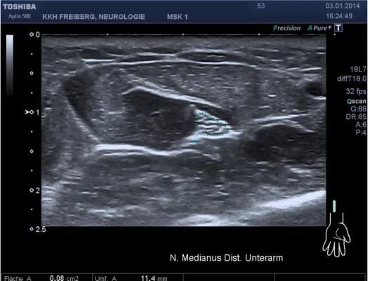
The cross sectional appearance of the normal peripheral nerve is round to oval and reminiscent of a honey comb (Fig .2).
Figure 2. Honey comb appearance of the median nerve at the distal forearm on transverse scan 1 cm under the surface. Cross-sectional area is (CSA) 0.08 c ² (Toshiba
Aplio T-500, 18 MHz linear array).
The hypoechogenic fascicles are surrounded by an echogenic rim representing the interfascicular epineurium and the perineurial sheath. Fascicles are the smallest
structure to be visualized by HRUS, individual axons and the endoneurium are not visible. The outer hyperechogenic layer is the outer epineurium. Particularly in large nerves, a clear cable-like fascicular echotexture can be seen.
The number of fascicles depends on the type of the nerve (amount of motor and sensory fibres), its location and its size. The amount of the fascicles varies in the course of the nerve, because they repeatedly unite and divide. Depending on the transmission frequency and resolution of the probe, the number of fascicles in the ultrasound image can differ from that determined histologically (15). This may be explained by the coalescence of some adjacent fascicles in a single image (13). In longitudinal sections, peripheral nerves have a cable-like appearance, in contrast to the more fibrillar echotexture of tendons. In addition, tendons can be easily distinguished from nerves sonographically, because they end in a muscle proximally.
Peripheral nerves at the root level have a mono-fascicular architecture, subdividing into a fascicular group arrangement more distally, eventually reaching a multi-fascicular arrangement in the periphery. Parallel with this changing group arrangement, the amount of connective tissue between the fascicles (inter fascicular epi- and perineurium) increases. This explains the fact why cervical roots demonstrate only one individual fascicle in sonography, whereas the number of fascicles for example in the median nerve increases from proximal to distal.
With the colour Doppler and duplex function of current ultrasound equipments, epineural vessels can also be identified. As quantitative diagnostic criteria for
“hypervascularization” are not yet available, the a ected side should be co pared with the unaffected one.
Many disorders of peripheral nerves result in an increase of nerve size. These conditions include entrapment, other mononeuropathies of various aetiologies, polyneuropathies, trauma, and nerve tumours. Ultrasonography allows precise structural analysis and quantitative measurements of the nerves, which makes comparison of different studies possible. Nerve width (medial to lateral diameter), thickness (anterior to posterior diameter) and cross-sectional area (CSA) measured on transverse scans, and antero-posterior diameter (LAPD) measured on longitudinal scans are the most frequently used quantitative parameters for the ultrasound investigation of peripheral nerves. Furthermore, ratios of CSA between different segments of the same nerve have
also been used (16, 17). CSA reference values for some peripheral nerves and the brachial plexus have been reported in previous studies (18-22, 33).
2. OBJECTIVES
Nerve size change is one of the most important ultrasonographic features of nerve pathology. Therefore it is crucial to have normal (reference) values of all nerves routinely assessed. Furthermore, in order to be able to use these reference values with confidence in everyday practice, it is also important to examine the reliability of these normal values, i.e. the congruence of values obtained by different examiners, ultrasound devices and by the same examiner at different time points.
Several reports have been published on reference values for the cross-sectional area of the median nerve. This resulted in an evidence-based guideline stating that ultrasound may be used as a diagnostic test for carpal tunnel syndrome (Level A). There are also several reports on reference values for the CSA of the ulnar nerve with good agreement among the measurements, among them some recent studies (24, 25). On the other hand, data are less abundant concerning normal values for cervical roots, radial nerve, lower limb nerves and pure sensory nerves, as mentioned above, and they show more variation. The first objective of our study was to establish a set of normal CSA values for C5, C6, and C7 cervical roots, and several upper and lower limb nerves, including some pure sensory nerves, at pre-defined anatomical sites, and to assess whether CSAs correlated with age, gender, height, and body weight. Our second objective was to systematically assess the reliability of these measurements on several nerves in the upper and lower limbs, with respect to intra-rater, inter-rater and inter- equipment variation. CSA values of two independent cohorts from the two study sites were also compared in order to determine the external validity of collected normal values.
A large body of ultrasonographic literature is available on common neuropathies such as carpal tunnel syndrome and ulnar neuropathy at the elbow, but literature data on uncommon conditions are lacking. As the third objective of our study, we analysed
cases of rare neuropathies assessed by ultrasonography in order to establish the role of HRUS in rare disorders of the peripheral nerves.
3. METHODS AND PATIENTS
3.1. Normal values and reliability assessments
Subjects
Prior to the start of our study, approval of the institutional review board at both study sites was obtained, and participants signed informed consent. Between May 2011 and December 2011, 56 healthy subjects were investigated with high-resolution nerve ultrasound at the Dept. of Neurology of Semmelweis University in Budapest (Hungary) and at the Dept. of Neurology of the District Hospital in Freiberg (Germany). Subjects were recruited from the hospital staff and patients. None of the study subjects had symptoms or signs suggesting polyneuropathy or systemic diseases potentially associated with polyneuropathy, nor any history of neuromuscular disease.
Demographic data (age, gender, height, and body weight) were recorded. All subjects were of Caucasian ethnicity. For the inter-rater reliability assessments in addition to the healthy subjects patients from a polyneuropathy study in Budapest have been included.
Ultrasound examination
For ultrasound examinations, a Philips HD15XE ultrasound device with a small part imaging software and a 15 MHz 3 c “hockey stick” linear array transducer was used for 25 subjects in Budapest. In Freiberg, the same device was used for 10 subjects, and an additional 21 subjects were examined with a Toshiba Aplio SSA-700A device with small part imaging software and a 12 MHz PLT-1204 4.5 cm linear array transducer. In both devices, compound imaging software (SonoCT for the Philips HD15XE and ApliPure for Toshiba Aplio SSA-700A) was used to improve image quality.
Normal value measurements
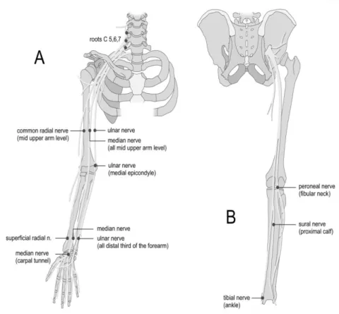
The following 14 CSA measurements (Fig. 3) on the upper and lower extremities were carried out, all on the left side: C5, C6 and C7 cervical roots; median, ulnar and radial nerves at the mid-upper arm; ulnar nerve at the elbow at the level of the medial epicondyle, median, ulnar and superficial radial nerves at the distal third of the forearm; median nerve at the proximal entrance of the carpal tunnel; peroneal nerve at the fibular neck; tibial nerve at the ankle; and sural nerve at the proximal calf. The measurements were time-consuming (approximately 45-55 minutes) and therefore they have been performed only on the left side. These sites included common areas of nerve entrapment (ulnar nerve in the ulnar groove, median nerve in the carpal tunnel), sites largely inaccessible for electrophysiologic studies (cervical roots), as well as sites corresponding to those usually evaluated by electrodiagnostic studies. The superficial radial and the sural nerves were chosen as pure sensory nerves. Subjects were examined mostly in supine position, with the exception of the peroneal nerve examined with the subject lying on one side, and the sural nerve examined in prone position.
Figure 3. Anatomical landmarks used for peripheral nerve ultrasound measurements in the study.
For brachial plexus sonography, the following technique described earlier for determining root level was used (26): The C7 root was identified in the oblique transverse plane of the C7 vertebra, which appeared as a hyperechoic structure characterized by the presence of only a posterior tubercle on its transverse process, the anterior tubercle being absent. When the transducer was moved slightly upward, the C6 and C5 vertebrae were successively identified by the presence of both anterior and posterior tubercles, the C5, C6 roots appearing as hypoechoic structures between the tubercles. Colour Doppler sonography was used to differentiate roots from blood vessels.
The nerves of the upper and lower extremities were identified on transverse scans using the same typical anatomic landmarks as described before (27). On the upper arm, the median nerve was identified adjacent to the brachial artery between the biceps and triceps muscles at the midpoint of the line connecting the axilla and the medial epicondyle. The ulnar nerve was then identified at the same level by moving the probe more medially. The radial nerve was assessed at the same level directly on the surface of the humerus in the radial nerve groove, accompanied by the deep brachial artery. At the elbow, the ulnar nerve was measured in the ulnar groove, with the elbow in a slightly flexed position, between the medial epicondyle and the olecranon. On the distal forearm, the median nerve was measured first at the level of the proximal third of the pronator quadratus muscle: after the pronator quadratus muscle was visualized, the median nerve was identified between the tendons of the flexor pollicis longus and flexor digitorum superficialis muscles. From this point, the transducer was moved medially to the ulnar nerve, which is accompanied at this level by the ulnar artery. Next, the transducer was moved radially to identify the superficial radial nerve, lying between the extensor carpi radialis longus and flexor carpi radialis muscles, just above the palpable bony prominence of the radius, and adjacent to the radial artery (28). At the wrist, the median nerve was examined at the proximal entrance of the carpal tunnel using the pisiform bone as an anatomic landmark.
On the lower limb, transverse scan of the peroneal nerve was obtained at the level of the fibular neck with the subject lying on the side, and the knee propped up and slightly lexed (20° to 30°) (27). The tibial nerve was exa ined at the level o the
medial malleolus, just posterior to the tibial artery. The sural nerve was examined at the proximal dorsal calf, identified superficially between the two heads of the gastrocnemius muscle. If necessary for correct identification, the nerve was followed more distally.
The CSA of the nerves was measured using the trace function of the ultrasound device by manually tracing inside the hyperechoic rim of each nerve (Fig. 4). The angle of insonation was adjusted perpendicular to the nerve where the nerve appeared the brightest with the best discernible outer margins. The CSA of each nerve segment was measured three times. The three measurements were averaged and the mean value was used for analysis.
Reliability assessments
The inter-rater reliability was assessed at the start of the study. Two ultrasonographers measured nerve cross-sectional areas in 7 subjects (on all 14 sites in each subject, as described above). Both examiners are neurologists and clinical neurophysiologists who perform neuromuscular ultrasound in a clinical setting on a daily basis. Both ultrasonographers received training for this study prior to the initiation of data collection. The repeated measurements were done in one session: the examination of all 14 nerve segments by one rater was repeated in the same session by the other rater who was blinded to the results of the first.
To assess intra-rater reliability, 6 subjects in Freiberg were re-examined with the same Toshiba device by the same investigator 24 hours after the first sonographic examination.
To assess inter-equipment reliability, 6 subjects in Freiberg were examined by the same examiner first with the Philips, and 8-11 weeks later with the Toshiba ultrasound device.
The validity of normal values was tested by comparing CSA values of the 14 nerve segments in the two independent cohorts of the two study sites.
Statistical analysis
Descriptive statistics were used to present basic demographic data of the study population. The following parameters were calculated and presented for normal CSA values of the 14 nerve segments: mean, median, standard deviation (SD), 95%
confidence intervals of the mean, and the coefficient of variation. Normality of variables was checked by the Shapiro-Wilk test. Correlation of CSA measurements with age, gender, height and body weight was tested using the Spearman correlation coefficients.
Values between genders were compared by the Kruskal-Wallis ANOVA. The general linear model (GLM) was used to test if gender remains a significant predictor of CSA when age, height and body weight are also considered. Intraclass correlation coefficients and corresponding 95% confidence intervals were calculated to define values for intra-rater, inter-rater, and inter-equipment reliability. The validity of our normal values was tested in two independent cohorts using repeated measure ANOVA for the comparison of CSA values of the 14 nerve segments. Statistica for Windows v.
11 (StatSoft, Tulsa, OK) was used for data analysis.
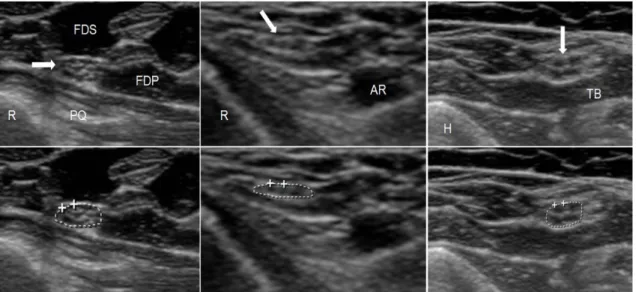
Figure 4. Normal ultrasound images of three different nerves. On the lower images, the tracing used to measure the cross sectional area is shown.
Le t: Median nerve at the distal orear (CSA: 7.7 ²); R= radial bone, PQ= pronator quadratus muscle, FDS= flexor digitorum superficialis muscle, FDP= flexor digitorum
profundus muscle. Arrow points to the median nerve.
Middle: Super icial radial nerve at the distal orear (CSA: 1.9 ²); R= radial bone, AR= radial artery. Arrow points to the superficial radial nerve.
Right: Ulnar nerve at the upper ar (CSA: 6.8 ²); H=hu erus, TB= edial head o the triceps brachii muscle. Arrow points to the ulnar nerve.
3.2. Ultrasonography of patients with rare neuropathies
Subjects
Between January 2009 and December 2013, about 3500 predominantly outpatients were investigated with HRUS at the Dept. of Neurology of the District Hospital in Freiberg (Germany). The vast majority of the patients were adults. The patients were referred to the outpatient consultation with a suspected peripheral nerve disease from different faculties, mostly surgeons (trauma surgeons, neurosurgeons, hand surgeons), neurologists and general practitioners. All patients were examined neurologically and by standard electrodiagnostic examination, consisting of nerve conduction studies, recording of late responses (F-waves) and needle electromyography.
Demographic data (age, gender, height, and body weight) were recorded.
Ultrasound examination
For ultrasound examinations between January 2009 and November 2012, a Toshiba Aplio SSA-700A device with small part imaging software and a 12 MHz PLT- 1204 4.5 cm linear array transducer and from November 2012 a Toshiba Aplio T-500 SSA-700A device with small part imaging software and a 18 MHz array transducer was used. In both devices, compound imaging software (ApliPure) was used to improve image quality.
Ultrasound measurements
Depending on the clinical question, either a single nerve was examined, mostly bilaterally in order to ascertain side differences, or in suspected generalized diseases several nerves were examined on different segments along the course of the nerve.
Pathological alterations were documented in two planes, in longitudinal as well as in transverse scans. Occasionally, short video sequences were recorded for better understanding of the pathology.
The CSA of the nerves was measured using the trace function of the ultrasound device by manually tracing inside the hyperechoic rim of each nerve.
4. RESULTS
4.1. Normal values and reliability assessments
Basic demographic features of the study population are given in Table 1.
Table 1. Demographic data of the two study cohorts
Parameter Germans Hungarians P
N 31 25 -
Age (years) 51.8±16.4 48.5±15.6 0.45
Gender (M:F) 15:16 11:14 0.74
Weight (kg) 75.4±13.0 79.6±18.2 0.31
Height (cm) 171±9 168±6 0.12
No difference in demographic features between the Hungarian and the German study groups
The univariate relationships between CSA and age, body weight, height and gender are presented in Table 2. The results of multivariate testing for the effect of gender are presented in the last column of Table 2. CSA measurements showed mostly normal distribution in both genders and in pooled data.
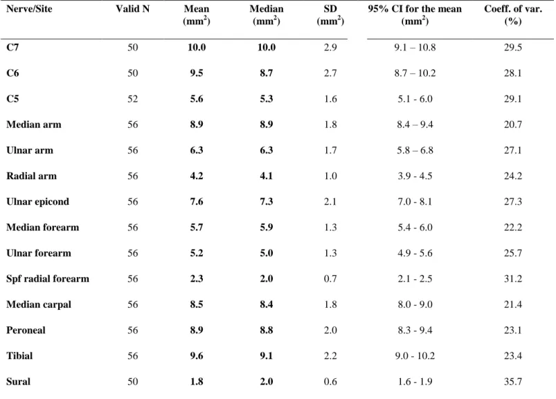
Descriptive statistics of CSA measurements of all 14 nerve segments for all subjects are presented in Table 3. Mean CSA values of these 14 nerve segments ranged from 2 to 10 mm2 (Table 3 and Fig. 8).
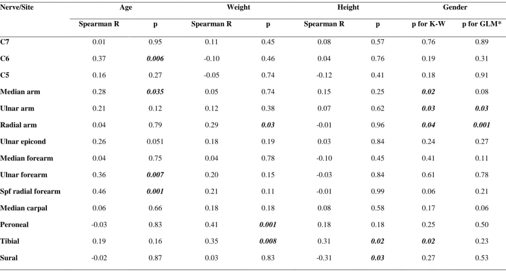
Table 2. Univariate Spearman correlations of peripheral nerve CSA values with age, body weight, height, and Kruskal-Wallis ANOVA test for gender, and multivariate testing (GLM) for gender
Nerve/Site Age Weight Height Gender
Spearman R p Spearman R p Spearman R p p for K-W p for GLM*
C7 0.01 0.95 0.11 0.45 0.08 0.57 0.76 0.89
C6 0.37 0.006 -0.10 0.46 0.04 0.76 0.19 0.31
C5 0.16 0.27 -0.05 0.74 -0.12 0.41 0.18 0.91
Median arm 0.28 0.035 0.05 0.74 0.15 0.25 0.02 0.08
Ulnar arm 0.21 0.12 0.12 0.38 0.07 0.62 0.03 0.03
Radial arm 0.04 0.79 0.29 0.03 -0.01 0.96 0.04 0.001
Ulnar epicond 0.26 0.051 0.18 0.19 0.03 0.84 0.24 0.27
Median forearm 0.04 0.75 0.04 0.78 -0.10 0.45 0.41 0.11
Ulnar forearm 0.36 0.007 0.20 0.15 -0.03 0.84 0.61 0.78
Spf radial forearm 0.46 0.001 0.21 0.11 -0.01 0.99 0.06 0.21
Median carpal 0.06 0.66 0.18 0.18 0.08 0.58 0.17 0.06
Peroneal -0.03 0.83 0.41 0.001 0.18 0.18 0.25 0.50
Tibial 0.19 0.16 0.35 0.008 0.31 0.02 0.02 0.23
Sural -0.02 0.87 0.03 0.83 -0.31 0.03 0.27 0.53
CSA=cross-sectional area; Spf=superficial Values are uncorrected for multiple comparisons. K-W: Kruskal –Wallis univariate ANOVA for comparing CSA values between genders.
*GLM: general linear model analysis, taking gender, age, weight and height as possible predictors of CSA. P values for gender are presented with correction for age, height and weight.
20
Table 3. CSA values (mm2) of 14 nerve segments in 56 healthy subjects
Nerve/Site Valid N Mean
(mm2)
Median (mm2)
SD (mm2)
95% CI for the mean (mm2)
Coeff. of var.
(%)
C7 50 10.0 10.0 2.9 9.1 – 10.8 29.5
C6 50 9.5 8.7 2.7 8.7 – 10.2 28.1
C5 52 5.6 5.3 1.6 5.1 - 6.0 29.1
Median arm 56 8.9 8.9 1.8 8.4 – 9.4 20.7
Ulnar arm 56 6.3 6.3 1.7 5.8 – 6.8 27.1
Radial arm 56 4.2 4.1 1.0 3.9 - 4.5 24.2
Ulnar epicond 56 7.6 7.3 2.1 7.0 - 8.1 27.3
Median forearm 56 5.7 5.9 1.3 5.4 - 6.0 22.2
Ulnar forearm 56 5.2 5.0 1.3 4.9 - 5.6 25.7
Spf radial forearm 56 2.3 2.0 0.7 2.1 - 2.5 31.2
Median carpal 56 8.5 8.4 1.8 8.0 - 9.0 21.4
Peroneal 56 8.9 8.8 2.0 8.3 - 9.4 23.1
Tibial 56 9.6 9.1 2.2 9.0 - 10.2 23.4
Sural 50 1.8 2.0 0.6 1.6 - 1.9 35.7
CSA=cross-sectional area; Spf=superficial; SD=standard deviation; CI=confidence interval; Coeff. of var.=Coefficient of variation
21
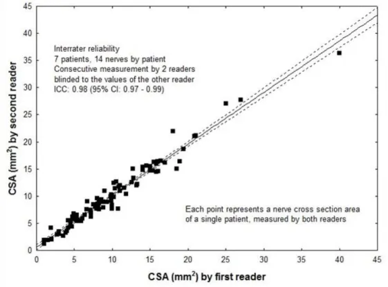
Inter-rater reliability, intra-rater test-retest reliability, and inter-equipment test- retest reliability are presented in Figs. 5-7. Intraclass correlation coefficients in all three analyses of reproducibility were remarkably high (0.86 – 0.98).
Figure 5. Inter-rater reliability. CSA measurements of 14 segments by two raters within one session. The second rater was blinded to the measurements of the first.
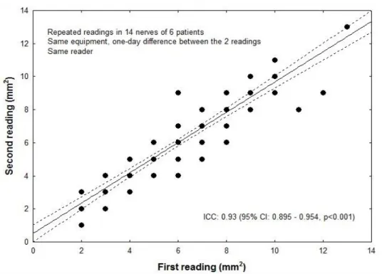
Figure 6. Intra-rater test-retest reliability. Repeated measurements by the same reader of 84 nerves in 6 patients one day apart. Due to the overlap, only 34 out of 84 datapoints
appear in the plot.
Figure 7. Inter-equipment test-retest reliability. Repeated readings by the same reader of 84 nerves in 6 patients with two different equipments, within 8-11 weeks. Some of
the datapoints overlap.
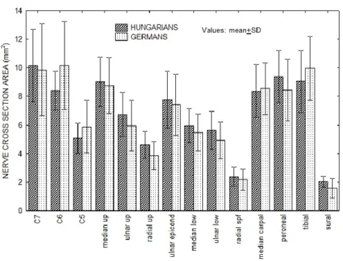
When CSA values of the 14 nerve segments were compared between two independent cohorts, no significant difference was found (Fig. 8).
Figure 8. Measurements in two independent cohorts. CSA values of 14 nerve segments in Hungarian (N= 25) and German (N=31 healthy subjects). Repeated measure ANOVA
revealed no significant country effect. When pairwise comparisons were done by the Mann-Whitney-test, no significant difference was found between the CSAs in any of the
nerve segments after correction for multiple comparisons.
4.2. Ultrasonography of patients with rare neuropathies
4.2.1.RARE TUMOURS AND FOCAL TUMOUR-LIKE LESIONS
Case 1. Amyloidosis
In a 68-year-old female patient with progressive radial nerve lesion with paresis distal to the triceps muscle, electrodiagnostic testing revealed axonal motor and sensory lesion, but the exact level of the lesion could not be ascertained. 1.5 T-MRI findings were negative. By means of high resolution ultrasound, a hypoechoic lesion of the
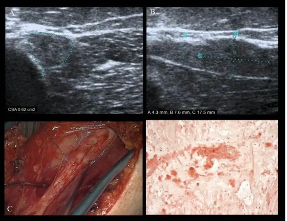
common radial nerve was identified, involving its complete course from the axilla to the spiral groove, with arked seg ental enlarge ent (CSA=0.62 c ², dia eter 7.6 ) at the distal part of the spiral groove. In this segment, sonography indicated the presence of calcification which was later histologically confirmed to be a small metaplastic ossification. Furthermore, fascicular biopsy revealed amyloid deposition using congo red stain and polarized light microscopy (Fig. 9). Because neither laboratory findings nor histology (nerve and rectum) could reveal a primary light chain amyloidosis (AL), we could not confirm a primary amyloidosis in this case. Curative surgical treatment by means of microsurgery was impossible.
Figure 9. Affection of the common radial nerve by amyloidoma. HRUS on transverse scan (A) and longitudinal scan (B) demonstrates a focal, hypo-echoic nerve enlargement
with effacement of the fascicles. Intraoperative image (C) shows a fusiform nerve enlargement. Fascicular biopsy was carried out. Courtesy of TN Lehmann, MD (Department of Neurosurgery, Bad Saarow/Germany) Histological examination (D) revealed amyloid deposition. Courtesy of S Koch, MD PhD (Department of Pathology,
Bad Saarow/Germany).
Case 2. Intraneural lymphoma
In a 70-year-old female patient after 2 years of progressive ulnar neuropathy, unsuccessful neurolysis of the nerve at the elbow was performed. MRI (1.5 T) at the elbow showed an enlarged ulnar nerve. HRUS demonstrated a large inhomogeneous, mostly hypoechogenic nerve lesion. Its contour on longitudinal scan was slightly irregular with some hypervascularisation of the nerve from the axilla to the elbow (Fig.
10). Another vascularized tumour was seen within the flexor carpi ulnaris muscle.
Histology revealed primary non-Hodgkin’s B-cell lymphoma with additional axillary and supraclavicular lymph node metastases.
Figure 10. Affection of the ulnar nerve by intraneural lymphoma. HRUS (A) demonstrates a large hypoechoic nerve enlargement without fascicular structure on longitudinal view at the elbow (A) and at the middle-arm (b). At the mid-arm level on
transverse scan (C) and on longitudinal scan (D) intraneural hypervascularisation.
Case 3. Radial nerve lesion in rheumatoid arthritis
In a 32-year-old female patient during the first trimester of pregnancy, after discontinuation of the treatment with sulfalazin and dose reduction of prednisolone from 7.5 mg to 5 mg/day given for rheumatoid arthritis, painless radial nerve palsy without
involvement of the triceps muscle developed. No signs of inflammation were present.
Electrodiagnostic testing showed axonal radial nerve lesion of distal upper arm type.
EMG demonstrated denervation signs not only in the muscles innervated by the posterior interosseus nerve but also in the brachioradial and the extensor carpi radialis muscles, which indicated that the lesion started somewhat distal to the triceps muscle.
HRUS demonstrated (Fig. 11) short focal enlargement of the radial nerve on the upper ar distal to the spiral groove with pro inent ascicles (CSA: 0.11 c ²;
diameter: 2.7 mm; diameter on the contralateral side: 1.5 mm). Spontaneous recovery ensued over 6 months without increasing the dose of the drugs again.
Figure 11. Radial nerve distal to the spiral groove between the brachioradial and the brachial uscles (H= Hu erus) with a CSA o 0.11 c ² on transverse scan (A) with
prominent fascicular structure and longitudinally enlarged nerve (2.7 mm) (B).
Case 4. Plexiform neurofibromatosis
In a 27-year-old male patient, progressive painless radial nerve palsy developed over the course of one year. Steroids and immunoglobulins were ineffective. Clinical examination and electrodiagnostic testing indicated a lesion distal to the triceps muscle.
1.5 T-MRI was negative. HRUS demonstrated (Fig. 12) a lesion of the radial nerve without vascularization - more proximally than suggested clinically and electrophysiology - between the axilla and the spiral groove (CSA: 0.42 c ²; extensive
hypoechogenic elongation: 4 mm). Histology after operation demonstrated a plexiform neurofibroma.
Figure 12. Extensive enlargement (4 mm) of the radial nerve proximal to the spiral groove without evidence o vascularization on duplex (A).CSA (0.42 c ²) was increased (B). Operation site (C), markedly enlarged nerve nerve after epineurotomy (D). Image (C, D) courtesy of TN Lehmann, MD (Department of Neurosurgery, Bad
Saarow/Germany).
4.2.2.NERVE TORSION
A 42-year-old male patient had a 15-month-long history of numbness on the ventral upper arm with severe pain for 2 weeks in the mid-upper arm, radiating one week later to the thumb. Possibly mechanical stress (intensive training with dumbbells) was reported. Neurological examination showed sensorimotor deficit of the radial nerve of distal upper arm type (triceps muscle intact, brachioradial muscle without voluntary activation). Electrodiagnostic testing after 3.6 and 12 months showed no signs of
reinnervation on EMG. 1.5 T-MRI was negative. HRUS demonstrated an enlarged nerve (CSA= 0.20 c ²), a caliber change with an hourglass-like constriction and a change in the continuity of the epineurium indicating a possible torsion of the nerve with a long segment increase of the diameter, proximally (3.6 mm) more than distally (Fig. 13A). On the contralateral side, the diameter of the radial nerve was normal (1.7 mm). The deep brachial artery just above the constriction showed normal flow characteristics.
Intraoperatively, the radial nerve was found to be twisted and constricted after the removal of the adventitia (Fig.13B). A pseudoneuromatous bulging mostly proximal to it was discernable. The surgical approach consisted of microsurgical exploration by means of epineurotomy as a first step. A careful derotation followed, a procedure, which was already reported rather early as helpful (29), but this case needed subsequent segmental resection and repair by grafts in accordance with literature cases (30).
Histology showed pronounced neural fibrosis at the level of the torsion and some inflammatory cell infiltration of the neighbouring nerve fascicles.
The operation was carried out 15 months after onset of the symptoms. Slight electrophysiological and clinical recovery of the radial nerve was noticed 4 months after surgery. HRUS showed a new torsion distal (Fig. 13C) to the site of the first torsion, which was again confirmed by neurosurgical exploration.
Figure 13. HRUS with multiple torsions of the radial nerve. A: Substantial calibre change and hourglass-like constriction (arrows) with deep brachial artery just above the
constriction as well as long-segmental enlargement of nerve diameter proximal to the constriction (3.6 mm). B: Corresponding intraoperative image. The twisted constriction
and proximal bulging of the nerve was clearly seen only after removal of adventitia.
Image courtesy of TN Lehmann, MD (Department of Neurosurgery, Bad
Saarow/Germany). C: A second distally located spontaneous torsion of the radial nerve in the same patient 12 months after surgical repair of the torsion depicted in A and B.
4.2.3.RARE DISEASES MIMICKING CARPAL TUNNEL SYNDROME
Case 1. Thrombosis of the persistent median artery mimicking carpal tunnel syndrome A 40-year-old right-handed locksmith complained of pain in the whole hand during the day mainly after manual work with pain relief during the night for the last three months. He reported mild dysesthesia on the median nerve innervated fingertips.
Phalen-test was negative. The pulse of radial and ulnar artery was normal.
Electrodiagnostic testing showed normal values. HRUS showed a normal median nerve in the carpal tunnel with normal echotexture and CSA on transverse scan. No compression signs (no caliber change on the longitudinal scan) (Fig. 14A), but an enlarged, partly coloured persistent median artery was seen on duplex-sonography (Fig.
14B). The vessel was not compressible and showed no pulsation .
Figure 14. HRUS of the median nerve at the proximal level of the carpal tunnel.
Longitudinally (A) no compression, on transverse scan enlarged median artery, only partly coloured (B). PW–mode on duplex scan showed decreased flow velocities (C) compared to the contralateral side (D) and absent flow in the middle and distal part of
the persistent median artery, indicating thrombosis of the artery.
To clarify whether the thrombosed artery participated on the supply of the palmar arch, a 3 T–MRI with small hand coils was carried out, which showed the partly thrombosed vessel and sufficient collateral circulation (Fig. 15).
Figure 15. MR-angiography on the left side shows a thrombosis of the median artery and normal median artery on the contralateral right side. Image courtesy of Thomas
Schelle (Department of Neurology, Dessau-Roßlau Ger any).
Case 2. Acute carpal tunnel syndrome after hyperventilation tetany (31)
A 37-year-old female patient collapsed after gastroenteritis and exposition to heat followed by hyperventilation. During the hyperventilation attack, her hand was in a flexed position for approximately 60 minutes. Immediately afterwards, she complained of hypaesthesia in the sensory area of the left median nerve. Electrophysiological assessment 8 days later showed a prolonged distal motor latency (4.8 ms) and reduction of the sensory nerve conduction velocity of the median nerve on the left side (39 m/s), consistent with carpal tunnel syndrome. On HRUS, the median nerve was slightly enlarged (Fig. 16) only on the left side, without segmental compression in the carpal tunnel (CSA = 0.12 c ², dia eter on longitudinal scan 2.7 ; on the right side CSA=0.08 c ², dia eter 2 ). The electrophysiologic alterations were nor alized in weeks and the CSA value in 6 weeks.
Figure 16. Acute CTS, longitudinal scan. The left median nerve is enlarged in the carpal tunnel (diameter =2.7 mm) without segmental caliber change. On the right side,
normal value (diameter=2 mm).
Case 3. Schwannoma of the median nerve distal to the carpal tunnel mimicking carpal tunnel syndrome
A 53-year-old female patient complained of numbness and tingling in the three middle fingers of the left hand without pain. Electrophysiology was performed elsewhere and the results were unknown. Operation at the carpal tunnel brought no relief of her complaints. HRUS showed a normal median nerve in the carpal tunnel, but more distally a homogeneous hypoechoic ovoid mass was seen (Fig. 17) with hypervascularisation (not documented). The nerve entered and left the centre of the tumour. Histology confirmed the suspected diagnosis of schwannoma.
Figure 17. Homogeneous hypoechoic ovoid mass in the continuity of the median nerve distal to the carpal tunnel. Colour-duplex (not documented) has shown marked
hypervascularisation of the tumour.
Case 4. Schwannoma of the median nerve proximal to the carpal tunnel mimicking carpal tunnel syndrome
A 72- year-old male patient was operated without success after complaints for more than 10 years with clinically suspected progressive carpal tunnel syndrome. He had a trauma at the distal upper arm as a young man with persistent slight sensory disturbance of the median nerve. The median nerve was normal in the carpal tunnel but more proximally, at the level of the former nerve injury more than 50 years previously, a mostly homogeneous hypoechoic fusiform mass with slight vascularisation was demonstrated (Fig. 18). Histology confirmed the suspected diagnosis of schwannoma with regressive changes.
Figure 18. Homogeneous mostly hypoechoic fusiform mass in the continuity of the median nerve proximal to the carpal tunnel. Colour-duplex showed slight
vascularisation of the tumour.
4.2.4.THORACIC OUTLET SYNDROME (TOS)
A 49-year-old female patient was referred to us from another hospital. She suffered for 15 years diffuse pain in the right arm, progressive paresis of the hand muscles and atrophy of the abductor pollicis brevis (APB) muscle. Electrophysiology showed slightly prolonged F-wave latency of the ulnar nerve (with normal motor nerve conduction velocity and motor action potential of the ulnar nerve), decreased sensory nerve action potential of the ulnar nerve, and absent sensory potential of the medial cutaneous antebrachii nerve. Myography showed chronic neurogenic changes only in the APB. Conventional X-ray images of the cervical spine were negative and MRI was not performed. CT scan of the cervical spine showed an enlarged transverse process of the right C7 vertebra with a rib stub and fibrous band attached to it. HRUS showed the so-called “wedge sickle-sign”: hyperechoic tip o the edial border o the scalenus medius muscle on the right side and thickened inferior trunk of the brachial plexus (Fig.
19a and 19b).
Figure 19a. HRUS showing compression of the inferior trunk (C8 root) between the scalenus medius and anterior muscles. AS= subclavian artery. Dotted lines =pleura and
first rib.
Figure 19b. HRUS showing the hyperechoic tip of the medial border of the scalenus medius muscle (arrow) causing compression of the inferior trunk (C8 root).
During surgery, compression of the inferior trunk of the brachial plexus by the medial fibrous edge of scalenus medius muscle was confirmed and resected (Fig. 20).
Follow-up 6 months after surgery showed clinical improvement in terms of muscle power, pain and atrophy of ADM.
Figure 20. Intraoperative findings in TOS caused by compression of the inferior trunk (A) of the brachial plexus by the white medial fibrous edge of scalenus medius muscle (B). Partial resection of the fibrous edge (C) and total resection (D). Image courtesy of
TN Lehmann, MD (Department of Neurosurgery, Bad Saarow/Germany).
4.2.5.NEURALGIC AMYOTROPHY (PARSONAGE-TURNER SYNDROME,PTS)
Case 1. Predominant radial nerve palsy in PTS
A 47-year-old female patient developed few weeks after severe shoulder pain a radial nerve palsy on the right side as a leading clinical symptom with additional minor signs of brachial plexus involvement on EMG. Electrophysiology showed isolated axonal motor (motor action potential 1.7 mV) and sensory (sensory nerve action potential 1 µV) lesion o the right radial nerve. EMG indicated involve ent o the triceps brachii muscle and the more distal muscles innervated by the radial nerve. MRI
of the cervical spine was normal. HRUS demonstrated 3 months after the onset of symptoms segmental enlargement of the radial nerve from the level of the spiral groove down to the supinator tunnel, which persisted even 1.5 years after symptom onset (Fig.
21). On follow–up (1.5 years later), almost complete clinical recovery was seen. In this case, the typical clinical course with sudden onset and recovery without specific treatment helped to differentiate neuralgic amyotrophy from a true focal compressive neuropathy.
Figure 21. HRUS of the radial nerve in PTS on the right side 1.5 years after symptom onset. The radial nerve on the right side (A, C) is slightly enlarged at the spiral groove
(diameter 1.9 mm vs. 1.3 mm) and the supinator tunnel (1.5 mm vs. 1.1 mm).
Case 2. Bilateral interosseus anterior nerve paresis in PTS
A 52-year-old male patient developed 2 weeks after severe bilateral shoulder pain paresis of the long flexors of the thumb and the index finger on both sides. 1.5 T MRI of the cervical spine was negative. Electrophysiology showed evidence of bilateral anterior interosseus nerve lesion. Two months after onset, HRUS demonstrated bilateral segmental enlargement of the median nerve and its fascicles at the upper arm from the mid-arm level to the elbow (Fig. 22). The ulnar and radial nerve and the cervical roots
C5-C7 were normal. On follow–up, 1.5 years later, almost complete clinical recovery was seen. Similar to the above mentioned first case, the typical clinical course with sudden onset, recovery without specific treatment and bilateral involvement helped in the diagnosis of neuralgic amyotrophy.
Figure 22. PTS with anterior interosseous nerve palsy on both sides. HRUS: long segmental swelling of the median nerve and its fascicles at the upper arm from the mid-
arm level to the elbow. CSA of single fascicles 0.05-0.07 c ², CSA on the right side from proximal (mid-ar ) 0.16 c ² (A) to distal (elbow) 0.23 c ² (C) , resp. on the le t
side 0.21 c ² ( id-ar ) (B )and 0.31 c ² (elbow) (D).
4.2.6.RARE POLYNEUROPATHIES
Case 1. Paraneoplastic multiple mononeuropathy (32)
A 58-year-old male patient, who worked as a master bricklayer and with a history of cigarette smoking (50 pack-years), presented with a 1.5-year history of slowly progressive, asymmetric, especially right-sided numbness of the 1-3rd fingers on both sides and a 1-year history of numbness of the 4-5th fingers on the left side. Strength and
muscle volume of his left hand slowly diminished. One year before presentation, he was operated for suspected right carpal tunnel syndrome, but without subjective benefit.
Later numbness of the left 4th and 5th fingers, as well as diminished strength in abduction of left 5th finger developed. Entrapment neuropathy of the left ulnar nerve at the elbow was suspected and operated by anterior transposition of the nerve. In addition, the right carpal tunnel was surgically revisited. Symptoms persisted and eventually increased. On examination, severe neuropathy of the left ulnar and moderate neuropathy of the right median nerves were found. Specifically, severe paresis of left ulnar- innervated hand muscles (MRC grade 2) and slight paresis of the right thumb abduction (MRC grade 4) were observed. The strength of other muscles and lower extremities was normal. Left ulnar-innervated hand muscles were atrophic, and the hand had a claw appearance. Hypaesthesia to light touch was present in left 4-5th fingers, the ulnar aspect of the hand and forearm, the lateral upper arm, the volar side of the right 1-3rd fingers, as well as the lateral side of the lower right leg. Electrodiagnostic studies from both median nerves showed axonal sensory–motor neuropathy without conduction block, and ulnar nerve conduction studies showed severe, left, non-localizing axonal motor–
sensory neuropathy and neuropathy of left ulnar nerve at the elbow. Electromyography showed fibrillation potentials and neurogenic changes in ulnar-innervated muscles in the left hand. Electrophysiological studies of the lower extremities were normal. HRUS showed focal enlargement and hypoechogenicity of the left ulnar nerve in the entire upper arm, focal enlargement, and hypoechogenicity of left median nerve at the elbow over a 10 cm-long segment (Fig. 23). Multiple entrapment neuropathies could therefore be ruled out, and multiple mononeuropathy with focal nerve hypertrophy was diagnosed.
Figure 23. HRUS of the left ulnar nerve in paraneoplastic multiple mononeuropathy.
The nerve at the elbow (CSA 0.24 c ², dia eter 3.5 ) and at the upper ar (CSA=0.12 c ², dia eter 2.2 mm) is hypoechoic and enlarged.
Extensive work-up demonstrated only slightly elevated cerebrospinal fluid protein (607 mg/L, normal <500 mg/L). All other parameters (including tests for syphilis, borreliosis, thyroid function, porphyria, Refsum disease, sarcoidosis, vasculitis, collagenosis, and vitamins B1, B6, B12, E, and folate deficiencies) were normal.
Testing for paraneoplastic serum antibodies revealed high titers of anti-Hu antibodies on immunofluorescence testing on primate cerebellum and intestine. Immunoblot with recombinant antigen confirmed specificity. No other paraneoplastic antibodies were positive. CT confirmed the suspected lung cancer. Biopsy histologically revealed small- cell lung cancer.
Case 2. MADSAM polyneuropathy
A 65-year-old male patient had a 10-year-long history of slowly progressive asymmetric paresis with atrophy, predominantly on the upper arm without sensory
disturbance. GM1-antibodies were positive, cerebrospinal fluid findings were normal.
Electrophysiology showed severe, asymmetric motor axonal damage without localizing findings. In another hospital, amyotrophic lateral sclerosis was supposed and the patient was referred for HRUS. HRUS showed markedly enlarged nerves of the upper arms on both sides, predominantly on the most involved side with marked changes of the fascicle calibers and of the cervical roots bilaterally (Fig. 24).
Figure 24. HRUS in MADSAM. Enlarged (diameter: 5.3 mm) median nerve at the upper arm (right side) with caliber change of the fascicles.
Case 3. Polyneuropathy in vasculitis
A 72-year-old male patient had for 3 months generalised arthralgia, for 3 months burning feet sensation more on the right side and violet-red skin changes on the lower leg and on the back of the foot. Laboratory values showed thrombocythaemia (128 G/l), decreased red blood cell count (3.9 T/l; N > 4.6 T/l), in differential blood cell count 54% lymphocytes, slightly elevated C-reactive protein (8.8 mg/L), and markedly elevated rheumatoid factor 436 IU/l (N<15). ANA, pANCA and cANCA were negative.
Electrophysiology showed an axonal distal-symmetric motor and sensory neuropathy.
HRUS demonstrated the isolated enlargement of the tibial nerve (Fig. 25). Histologic
examination of the sural nerve revealed leukocytoclastic vasculitis. Further rheu atologic assess ent showed Sjögren syndro e and parotid biopsy confirmed chronic B-cell lymphoid leukaemia.
Figure 25. HRUS of the left ankle. The CSA o the tibial nerve is enlarged (0.17 c ²).
5. DISCUSSION
5.1. Normal values and reliability assessments
High resolution ultrasonography has become an effective tool for the investigation of peripheral nerve disorders. It has been demonstrated that peripheral nerve pathology results in focal or diffuse thickening of the nerves together with a pathological change of echostructure and echogenicity (2). Examination of pathologic peripheral nerve in HRUS is based mainly on changes in nerve size, and in clinical practice the contralateral side is often used as an internal control. The most common method used to quantify nerve size is the measurement of the cross-sectional area (CSA) of the nerve.
The increase of CSA of the involved nerve allows precise localization in entrapment neuropathies and peripheral nerve tumours (27). Moreover, enlargements of multiple nerves in acquired and hereditary polyneuropathies are also described (19) Therefore, it is essential to compare nerve size parameters measured in patients to reference values. However, reference values are still lacking for some nerves and those published tend to show variability probably due to factors such as measurement accuracy, expertise of the examiner, equipment, location of the nerve, and patient specific factors (ethnicity, age, gender, body mass, height). Our aim was to contribute a large set of reference values to the pool of normative data currently being amassed in the literature by measuring the cross sectional areas of 10 upper and lower limb nerves at altogether 14 sites in 56 healthy individuals. Other studies usually assessed fewer nerve segments (Table 4). In more recently published studies in 2013, nerves were measured bilaterally and not only unilaterally at more sites (26) but only at the upper extremity (22) and at 15 sites bilaterally (33), but partly at different segments (no measurement at the mid upper arm level, instead of that at the axilla and no measurement of the cervical roots C5-C7).
Our study subjects represented a broad range of age and a balanced gender distribution from two different European countries, but ethnicity (Central-European Caucasian) was homogeneous. Although sample size could be larger, the narrow range of 95% confidence intervals for the mean, the relatively low coefficient of variation (generally between 20-30%) (Table 3) and the normal distribution of values of a given nerve, gender groups examined separately or combined and with different resolutions (analysis not shown), all support that the sample size of our study is acceptable.
Furthermore, no significant differences were found when comparing two independent cohorts (i.e. German and Hungarian populations), which also supports the validity of collected normal values. We found no consistent correlations between CSA values and age, height, or body weight, but males had significantly larger values than females for nerve segments in the upper arm. Regarding the age dependent correlations, our study had some limitations. Due to only 56 examined mostly middle-aged subjects, the correlations could not been investigated in sufficient numbers in young and old subjects.
Likewise, these different age subgroups could not be compared.