CHAPTER 12
Inhibition of Isozymes
Elliot S. Vesell
I. Introduction 383 II. Metabolic Significance of Isozymes and the Role of Inhibitors in Their
Study 384 III. The Role of Inhibitors in Elucidating the Molecular Mechanisms Respon-
sible for the Generation of Isozymes 388 IV. Inhibition of LDH Isozymes 399
V. Conclusions 419 References 420
I. INTRODUCTION
In 1959 Markert and M0ller (1) introduced the word "isozyme" to call attention to the growing number of reports of enzymes that existed within an organism in multiple molecular forms. To qualify as isozymes, these multiple molecular forms also had to exhibit similar substrate speci- ficity (1). Excluded from consideration as isozymes were enzymes with numerous substrates of which only a few were shared (1); although subse- quent work established true isozymes within the esterases and phospha- tases, neither of these two classes of enzymes with very broad, overlap- ping substrate specificity was initially considered to fit the definition
(1). After much work in many laboratories on the genetic control and chemical structure of various isozymes, Markert (2) suggested the possi- bility of adding descriptive modifications to the word to indicate genetic and chemical information on the nature of the isozyme; prefixing such terms as allelic, nonallelic, homopolymeric, conformational, hybrid, or conjugated to the word isozyme was suggested. Whether or not this notation will ever become commonly employed by workers in the field, it is recommended that when the word isozyme is used the tissue and
383
the species from which the multiple forms were derived be indicated to avoid misunderstanding and to insure clarity and reproducibility.
This recommendation is made because isozyme patterns vary markedly not only among species but even among the tissues of an organism.
For some biochemists the definition of isozyme remains disturbingly vague, in spite of the extensive work on chemical structure that has established and supported many isozymic systems. These biochemists draw attention to the lack of precise criteria of chemical structure or substrate specificity that must be fulfilled for two proteins to be con- sidered isozymes. Moreover, some biochemists have felt no necessity to elaborate new nomenclature for enzymes that obey the same biochemical laws that other enzymes follow. The virtue of the term isozyme is in its illustration of the general biological principle that many metabolic reactions can be more finely regulated if several catalysts, rather than a single catalyst, participate. Before the word isozyme (also spelled isoenzyme) was coined, only a few examples of this biological principle were available, but the popularity of the term isozyme helped to draw attention to the principle and to stimulate investigation in the field.
Subsequently, more than 200 different isozymic systems have been de- scribed, and the existence of multiple forms of an enzyme is now recog- nized as a relatively common means by which cells can achieve various metabolic requirements.
This review offers several examples of differential isozyme inhibition in vitro under certain narrow and unphysiological experimental condi- tions which, when slightly altered, produce dramatic changes in the ex- tent of inhibition. Therefore, two main points of this review are that, in attempting to elucidate the function of isozymes and the role of inhibi- tors, the investigator should systematically alter certain physiologically important physicochemical variables and he should attempt to keep these variables and reagent concentrations as close as possible to the conditions prevailing in vivo.
II. METABOLIC SIGNIFICANCE OF ISOZYMES AND THE ROLE OF INHIBITORS IN THEIR STUDY
Several distinct functions of isozymes in the biochemical modulation of intracellular reactions have been identified. The first example is that of the same biochemical reaction required within morphologically distinct subcellular organelles, each possessing a different electrical charge; under
such circumstances, it would be advantageous for several forms of the same enzyme to exist, each having a different charge to permit easier attachment on differently charged subcellular particles. Such situations have been described for lactate dehydrogenase (LDH) (3, 4) and malate dehydrogenase ( M D H ) (5) isozymes. Even within an organelle, the same catalytic activity might have to function in association with macro- molecules of different charge, in which case the existence of differently charged forms of an enzyme might be useful in their attachment. A second advantage conferred by multiple forms of an enzyme involves reversible reactions in which one isozyme is preferentially adapted to one substrate, whereas the other isozyme serves as a better catalyst for the second than for the first substrate. Aldolase isozymes provide an example of this situation. Class A aldolases favor cleavage of fructose 1,6-diphosphate to dihydroxyacetone phosphate and glyceraldehyde 3-phosphate and, therefore, glycolysis. On the other hand, class Β aldolases are better geared for fructose 1,6-diphosphate synthesis and, thus, gluconeogenesis (6). As yet, no specialized physiological function has been associated with class C aldolases (6).
A third regulatory advantage of isozymes, differential feedback inhibi
tion, is particularly germane to the subject of this book. The role of isozymes in regulating rates of metabolite flow at branched metabolic pathways in bacteria is one of the most elegant and biologically signifi
cant functions yet ascribed to them (7). The situation in which several end products are derived from a common precursor controlled by a single enzyme is potentially dangerous for the cell. If the concentration of precursor is regulated by a feedback mechanism, an accumulation of one of the end products could lead to a deficiency of the others. In E. coli, aspartyl phosphate is an intermediate in the biosynthesis of methionine, threonine, and lysine. Aspartokinase converts aspartate to aspartyl phosphate in this organism, and the aspartokinase activity in crude extracts is inhibited by threonine, lysine, and methionine. Thus, by feedback inhibition, excessive accumulation of threonine, for example, would be expected to lead to a deficiency of lysine and methionine.
However, such a deficiency fails to occur because the organism possesses three aspartokinase isozymes, one sensitive only to threonine, another only to lysine, and a third only to methionine. These observations with aspartokinase were extended to other bacterial enzymes, and it was con
cluded that many isozymic systems in bacteria function in a similar fashion at branched pathways to regulate the relative flow of metabo
lites over each pathway (7). Thus far in mammals such elegant control of branched metabolic pathways has not been demonstrated.
A fourth function of multiple forms of an enzyme available to perform a given task may involve differential rates of isozyme degradation in different tissues (8-10). If a particular enzymic activity is critical within a tissue where one molecular form is rapidly degraded, an alternative form of the enzyme not subject to such rapid degradation would be required. Isozymes might fulfill this requirement if, as shown for the L D H isozymes (8-10), various tissues of an organism exhibited wide differences in the rate constant for degradation of a single protein. Ac
cordingly, some isozymic systems might have arisen from the biochemical necessity of maintaining the activity of critical enzymes in the face of rapid rates of degradation in particular tissues. The mechanisms re
sponsible for the intracellular catabolism of enzymes are not understood, but several types of inhibitory processes could be involved. In the case of L D H isozymes, it may be pertinent to the degradative process that two intracellular LDH-inhibiting peptides have been identified; one in
hibits L D H - 1 , and the other inhibits L D H - 5 (11, 12). Both inhibitors appear in urine. In studies performed in vitro, differences between L D H - 1 and L D H - 5 in rates of proteolytic degradation and thermal denaturation have been reported (13); furthermore, certain common in
tracellular metabolites differentially protected L D H isozymes from degradation (18). Large differences among tissues in rates of L D H iso
zyme degradation suggest that perhaps differences among tissues in cer
tain physicochemical conditions and in concentrations of various metabo
lites that affect isozyme decay in vitro might play a role in regulating their catabolism in vivo. From an evolutionary standpoint, development of isozymes to protect critical catalytic activities from destruction would represent an interesting, new type of selective pressure in which the goal would be maintenance of a sufficient variety of isozymic forms to permit survival of the enzymic activity required in each tissue.
In the case of the L D H isozymes, it has been suggested that gene duplication with subsequent mutations at both the parent and daughter loci is the biological mechanism for producing multiple molecular forms of an enzyme from a single primordial catalyst (Π). Such a mechanism has previously been proposed for the evolution of the a and β chains of the hemoglobin molecule (15). Recent work suggests that the genes coding for the two different A and Β subunits of the L D H isozymes are not linked; that is, they are not present on the same chromosome (16, 17).
Additions to biological knowledge through use of isozymes as a labora
tory tool, although too diversified to enumerate in detail here, should be identified in broad outline. Isozymes provide the clinical pathologist
with a sensitive diagnostic tool; the geneticist with a fruitful method for discovering and investigating polymorphic systems, marking chromo
somes, and following changing gene action; the biochemist with a new approach to understanding the control and regulation of intermediary metabolism; and the student of growth and development with precise markers along the stages of ontogeny.
Regardless of the functional significance of multiple molecular forms of enzymes (and the functional role apparently differs somewhat for each isozymic system thus far investigated from this standpoint), the existence of physicochemically distinguishable forms of an enzyme pre
sumably confers specific biochemical advantages on an organism. Other
wise, it is difficult to explain how isozymes became so common a feature of cellular organization, occurring from bacteria through primates, and also how their patterns came to be so specific for different tissues (Fig.
1) and at various stages of the embryonic development of each tissue.
Rather than being the exception, as initially thought when the first examples of isozymes were reported, multiple molecular forms of enzymes now are accepted as the rule. When sought for with sufficient diligence in enough tissues of a large enough number of species, most enzymes can be shown to exist in physicochemically separable forms. In many of these isozymic systems, inhibitors have been utilized to distinguish the chemical properties of these multiple forms of an enzyme or to identify the relative contributions of each form to the total activity of a mixture containing several isozymes. Inhibitors also have been em-
4 3 2 1 Isozymes
Lung
Heart
υ ο c
Ο
Kidney Ό a>
Ο c
(J D Liver <
Muscle Skin
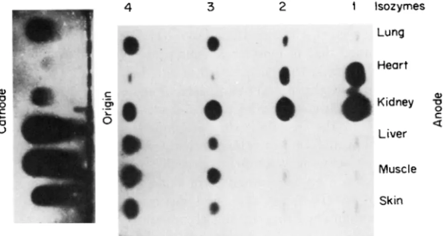
FIG. 1. Photograph of a starch gel treated to reveal the LDH isozymes in homogenates of six human tissues obtained at autopsy from a healthy subject killed in an accident. Note the presence of five LDH isozymes in each homogenate.
ployed to help establish the physiological significance of these different forms, particularly in situations in which some forms may be sensitive, whereas others may be resistant, to endogenously occurring inhibitors.
Since so many enzymes exist within an organism in multiple molecular forms, to review briefly but comprehensively the subject of the inhibi- tion of isozymes would be an almost impossible task. Instead, I have been highly selective in choosing only a few of the numerous examples of iso- zyme inhibition. Therefore, this discussion has been confined to those examples of inhibition that illustrate, in my view, particularly interesting properties of isozymes. This chapter deals largely with L D H isozymes because L D H isozymes have been most intensively investigated and be- cause they have served as an effective and stimulating model on which many other studies of different isozymic systems were later based.
III. THE ROLE OF INHIBITORS IN ELUCIDATING THE MOLECULAR MECHANISMS RESPONSIBLE
FOR THE GENERATION OF ISOZYMES
Before 1957 enzyme heterogeneity was regarded with suspicion and displeasure by enzymologists, who generally interpreted it as an artifact introduced by the laboratory procedures commonly employed for protein purification. Enzymologists attempted to make their proteins as homogeneous as possible by all physiochemical criteria. Colvin et al.
(18) were exceptions in their recognition that enzyme heterogeneity re- flects biological realities rather than laboratory artifacts. Colvin et al.
in 1954 maintained that in most organisms proteins actually exist within cells in multiple forms; they believed that this heterogeneity arises from occasional lapses in the protein-synthesizing machinery. Although this concept was far in advance of its time and although it has been shown that ambiguity in the genetic code does indeed lead to heterogeneity of the hemoglobin molecule in rabbit reticulocytes (19, 20), recent work on many diverse isozymic systems has revealed that errors at the level of protein synthesis are an exceedingly rare cause of protein heterogeneity.
Meister (21) and Neilands (22) first described two forms of bovine L D H . When human erythrocytes and serum were demonstrated to con- tain several electrophoretically distinguishable LDH's changing indepen- dently in various disease states, multiple molecular forms of an enzyme were recognized as reflections of biological events occurring in vivo (23).
Early studies on the L D H isozymes in tissues of various vertebrates
include those of Wieland and Pfleiderer (24), Sayre and Hill (25), and Hess (26). The introduction of rapid and convenient techniques for analysis of isozymes by histochemical staining after starch gel electro- phoresis greatly facilitated studies on tissue, ontogenetic, and species specificity of various isozymes (1, 27) and consequently led to a vast increase in the number of investigations on isozymes. This zymogram technique (1, 27), in which various histochemical stains are used to visualize isozymes after their electrophoretic separation on starch gels (28), has proved unequaled for rapid, qualitative identification of the multiple forms of many different enzymes (29, 30) and for studying the differential effects of various inhibitors on these forms. As an example of the zymogram technique, Fig. 1 shows the different proportions of the five L D H isozymes in homogenates of six human tissues.
Several problems have arisen concerning the legitimacy of extrapolat- ing to in vivo situations from bands observed on the starch gel after separation of enzymes present in crude homogenates of tissues. The finer the physicochemical techniques devised for the resolution of different forms of an enzyme, the greater the care that must be exercised to exclude artifacts. It has been demonstrated that, simply by changing certain electrophoretic conditions, additional bands can be generated from a single macromolecule through interactions with an uncharged constituent of the solvent medium (31). Cann and Goad (31) described how, with certain precautions, fractionation of macromolecules by starch gel electrophoresis can provide an unambiguous method for distinguishing between interactions and heterogeneity. However, certain other instances of anomalous bands (32) and subbands (14, 82-37) observed on the starch gel after histochemical staining for isozymes have never been adequately resolved. Therefore, vigilance in search of possible artifacts is highly recommended in experiments on new isozymic systems. In this context, artifacts signify the generation in vitro of several bands from a single molecular species that exists in vivo. For example, differences in the zymogram pattern of mouse kidney and liver glucose 6-phosphate dehydrogenase (G-6-PD) disappeared after dialysis, which apparently removed a small molecular weight substance from mouse liver ho- mogenate (38). An initial interpretation of the zymogram pattern of undialyzed mouse liver and kidney homogenates would have led to the erroneous conclusion that biochemically and genetically distinct isozymes of G-6-PD existed in these two tissues of the mouse.
Another interesting example of an environmentally induced alteration in isozyme pattern is that of alcohol dehydrogenase (ADH) from Drosophila melanogaster (39, 40); A D H isozymes change in mobility
on addition of N A D to the acrylamide gel and increase in number after passage through a D E A E Sephadex column.
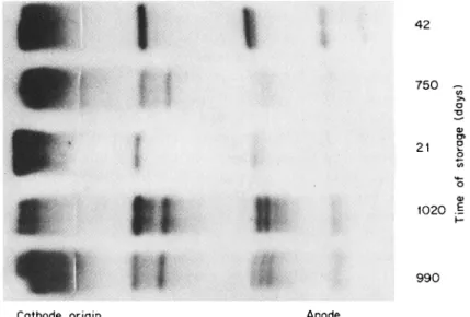
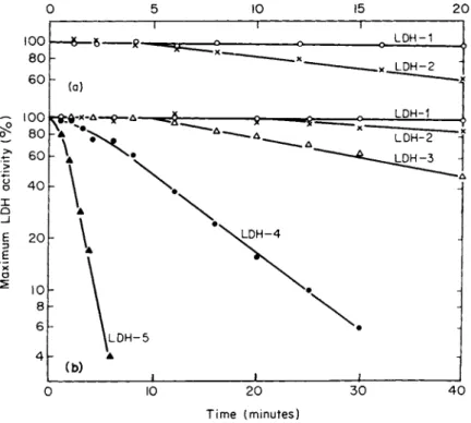
Splitting of the major L D H bands on the starch gel with increasing time of storage of human skeletal muscle at —25°C (82) is shown in Fig. 2. The explanation for the acquisition of these additional bands remains uncertain (32, 85-37), but it may reflect a type of degradation or inhibition of L D H isozymes since this subbanding occurs mainly in the anodal L D H isozymes as their activity decreases with age (32).
Thus, in skeletal muscle the normal loss of L D H activity proceeds from anodal to cathodal bands. Later studies confirmed this type of anodal band splitting with age (37) but attributed it to conformational re
arrangements among the L D H subbands, influenced by a macromolecular factor in tissues, probably neither substrate nor coenzyme, but possibly an activator or an inhibitor such as the peptides mentioned earlier (11, 12).
Another type of subbanding is observed in fresh tissues of most species; this subbanding characteristically occurs in the cathodal L D H isozymes and decreases progressively with increasing anodal mobility ( L D H - 1 exhibits no subband). Various explanations for the appearance of these subbands have been offered (38-37). Neither differential binding of each L D H isozyme to N A D nor the existence of two different forms
4 2
7 5 0 ~ ω >»
Τ3 α
α>
ο α>
102 0 . §
9 9 0
Cathod e origi n Anod e
FIG. 2 . Photograph of a starch gel showing L D H isozymes in human skeletal muscles from five individuals. These muscles were treated identically except for length of storage at — 25° C. Note that splitting of bands increases with length of storage of the whole muscle. [From Vesell and Brody (32) λ
of the A subunit (34) adequately fits the data. Altering the concentration of N A D in the gel or homogenate may (33) or may not (35) change the pattern of subbands, and the theory of two forms of A subunit would require five subbands for L D H - 5 , whereas several more than five subbands of this isozyme have been observed in some systems (35).
When one of the subbands of L D H is removed by electrodialysis after starch gel electrophoresis and resubmitted to electrophoresis under identi- cal conditions, other new bands, in addition to the original band that was isolated and rerun, are generated (37). The L D H subbands may be metastable polymers, produced in vitro by interactions between L D H isozymes and ions in the supporting medium used during electrophoresis.
If this proves to be true in vitro, then conformational rearrangements of the main L D H isozymes might arise in vivo from interactions of the major isozymes with certain ions or small molecules. Although these ions or small molecules might be considered activators or effectors in terms of the subbands that are generated, they might also be considered to function as inhibitors of the main isozyme whose activity decreases with the generation of the satellite subbands. In this connection, the existence of conformational or configurational isozymes has previously been suggested to explain multiple forms of M D H (41) and of electro- phoretically undetectable but isomeric forms of L D H that might exist within a single L D H band on the starch gel (42).
The first isozyme system to have its chemical nature elucidated was L D H ; the discovery was made with the use of two irreversible inhibitors of L D H activity, urea and guanidine hydrochloride. However, these agents were not employed according to the conventional techniques of enzyme inhibition, but rather with the specific chemical purpose of rup- turing the hydrogen bonds that help hold the isozyme in its three-dimen- sional configuration (43). Enzyme inhibition occurred as a result of this process and also as a consequence of the fact that the products of this treatment, the constituent subunits of the tetrameric L D H iso- zymes, are enzymically inactive (43). After treatment with urea or guanidine hydrochloride, L D H was quickly denatured with complete loss of its activity. The molecular weight of the undissociated active isozyme was approximately 140,000, but on treatment with urea (12 M) or guanidine hydrochloride (5 M), the molecular weight was shown by sedimentation rates observed in the analytical ultracentrifugation to decrease to 35,000 (43). Thus, after highly purified preparations of the L D H isozymes were treated with urea or guanidine hydrochloride, two electrophoretically distinguishable, enzymically inactive bands of protein having a molecular weight of 35,000 were observed. This infor-
mation provided the following hypothesis for the structure of the L D H isozymes. Each of the five L D H isozymes is a tetramer composed of varying proportions of two differently charged subunits or monomers, A and Β (43). According to this hypothesis, L D H - 5 = A4; L D H - 4 = A3B ; L D H - 3 = A2B2; L D H - 2 = A B3; and L D H - 1 = B4 (43). Abundant evidence from genetic, biochemical, and immunological investigations reviewed extensively elsewhere (2, 4, 30, 43-45) has been gathered to support this hypothesis, which is now generally accepted.
However, some investigators believe that the monomers of the L D H molecule may have a molecular weight of 17,000 rather than 35,000.
In this view, the L D H isozymes are octamers rather than tetramers (46, 47).
Regardless of whether the L D H isozyme is a tetramer or an octamer, the subunit structure of the molecule is firmly established. Markert pro
duced one of the most convincing pieces of evidence for this hypothesis when he froze purified preparations of LDH-1 and L D H - 5 in sodium chloride (1 M) and demonstrated after the mixture thawed that all five L D H isozymes were in a proportion of 1:4:6:4:1 (48). Generation of the hybrid isozymes (LDH-2, L D H - 3 , and LDH-4) suggested that freezing in sodium chloride dissociated LDH-1 and L D H - 5 into their constituent monomers (48). The monomers then reassociated randomly in all possible combinations of 4 to form the five L D H isozymes in a mathematically predictable ratio of 1:4:6:4:1 (48). Furthermore, this experiment suggested to Markert (48) that the tissue patterns of L D H isozymes (Fig. 1) arose in vivo by random recombination of the A and Β monomers and that the different tissue isozyme patterns reflected the relative intracellular abundance of A and Β subunits. In turn, the relative abundance of A and Β subunits indicated the relative activity in each tissue of the a and b structural genes responsible for the production of the A and Β subunits. In apparent support of this view of the intracel
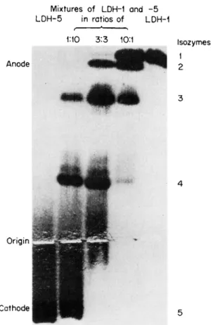
lular genesis of tissue-specific L D H isozyme patterns, Vesell was able to produce in vitro different patterns resembling the L D H isozyme profiles of various tissue by freezing in sodium chloride (1 M) mixtures composed of different proportions of purified LDH-1 and LDH-5 (Fig.
3) (49). However, it was emphasized that diverse tissue isozyme patterns could theoretically still arise in vivo even if no differences existed be
tween the activities of the a and b structural genes (49). Given equal intracellular concentrations of A and Β subunits, tissue-specific patterns could arise from the operation of numerous epigenetic factors to be dis
cussed in detail later. In brief, L D H isozymes differ markedly in response to many factors including temperature, pH, dilution, and concentration
Mixtures of LDH-1 and - 5 LDH-5 in ratios of LDH-1
1Ί0 3:3 10M Isozymes 1
Anode 2
Origin
Cathode
FIG. 3. Photograph of a starch gel in which LDH-1 (far right) and LDH-5 (far left) were mixed in the proportions indicated and frozen in 1 Μ NaCl to produce isozyme patterns characteristic of various human tissues. [From Vesell (49).]
of pyruvate, lactate, and other intermediary metabolites. Therefore, the different environments existing within various tissues might operate at a post translational level to influence the proportions in which A and Β subunits combine to form the five L D H isozymes. That this recombi
nation of subunits is not invariably random in vivo but can be influenced by tissue-specific epigenetic factors is illustrated by the fact that several vertebrate tissues do not exhibit L D H isozyme patterns entirely con
sistent with the theory of random association of subunits. Particularly striking examples of this failure to fit the concept of random recombina
tion occur in certain fish, notably the alewife (30, 50), where the pattern in vivo shows only L D H - 1 , -3 and -5, although the missing L D H iso-
3
4
5
zymes can be produced in vitro by freezing tissue homogenates in sodium chloride. Why certain fish completely lack specific L D H isozymes that can be generated so easily in vitro by recombination of the subunits present in their tissues is uncertain. However, explanation of this unusual isozyme pattern seems to require postulation of environmental conditions operative in vivo in certain species that prevent the formation or accel
erate the degradation of particular isozymes.
A theory to explain the formation and decay of the three hybrid L D H isozymes (LDH-2, L D H - 3 , and LDH-4) by exchange of subunits among active tetramers at a primarily posttranslational level has been developed (9). According to this theory, once the homopolymers A4 and B4 are synthesized from A and Β subunits, respectively, three simple reversible reactions might occur in which the synthesis of one isozyme is accomplished simultaneously with the decay of other isozymes (9).
A4 + B4 ;=± 2 A2B2
A4 + A2B2^ 2 A3B B4 + A2B2^ ± 2 A B3
According to this view, synthesis and catabolism, a specialized form of inhibition if inhibition can be equated simply with processes causing loss of enzymic activity, are reciprocal events. Types of tetrameric inter
change in addition to those indicated above are possible, but these formulas represent the simplest model for such a system. The idea of a finely modulated epigenetic control of isozyme patterns is at the heart of this theory designed to introduce regulatory possibilities at a post
translational level (9). Although evidence for the existence of such tetrameric interactions was presented (9), the intracellular mechanisms responsible for such interactions have not been established.
In the recombination experiment in which L D H - 1 and L D H - 5 were frozen in sodium chloride with the generation of LDH-2, -3 and -4, free A and Β subunits were not isolated and identified (48); therefore, these results could have arisen from subunit interchange as well as from complete dissociation of active tetramers into inactive monomers. How
ever, subsequently, reassociation experiments demonstrated that, after dissociation in either urea, guanidine hydrochloride, or acid, L D H iso
zymes were reformed (51, 52). The results suggested that, rather than subunit interchange, the active tetramers had indeed, under the influence of these inhibiting agents, unfolded and come apart into monomers which then recombined to form new active L D H isozymes.
In addition to LDH, many other isozymic systems have been shown to arise from subunit interaction. These include aldolase (6), catalase
(53), creatine phosphokinase (54), and alkaline phosphatase (55), among others. Furthermore, under certain circumstances an enzyme that is usually homogeneous because it is composed of several identical sub- units (such as LDH-1) may exhibit multiple forms. These multiple forms are generated when a single mutant allele is present at the genetic locus controlling the production of the subunit. For example, L D H - 1 is gen
erally observed to be a single band on zymograms of tissues from most individuals because L D H - 1 represents a homopolymer of four identical Β subunits; however, in certain rare subjects, five bands are observed at the L D H - 1 position in the starch gel. This unusual pattern, explained on the basis of family studies of these subjects, indicates that instead of two normal b alleles at the genetic locus coding for the Β subunit these individuals have one normal b allele and an abnormal, mutant allele, which can be designated b'. If the abnormal Β ' subunit arose from a point mutation producing substitution of a charged for an un
charged amino acid, then when the Β and B' subunits associate in all possible combinations of 4, five electrophoretically distinguishable bands in the L D H - 1 position of the starch gel will occur: B4, B3B / , B2B2' , ΒιΒ3', B4'. These five forms will occur in a proportion of 1:4:6:4:1 if the recombination is entirely random and if there are an equal number of Β and B ' subunits (5). Such individuals do exist. Their unusual L D H isozyme patterns have been described for mutations affecting not only L D H - 1 , but also L D H - 5 (4)- For this reason, mutations affecting the genes that produce the subunits from which enzymes are assembled must be recognized as a cause of multiple molecular forms of an enzyme in a few rare individuals. Although such aberrant forms of an enzyme may be rare (4), their incidence can show marked differences in geo
graphical distribution. For example, mutations at the a locus of L D H , relatively rare in most areas, have been described in a frequency of 4% in certain regions in southern India (56). Furthermore, if one con
siders the large number of different enzymes and isozymes in man, the likelihood is high that each person is heterozygous at several of the multiple genetic loci controlling the synthesis of these enzymes. Thus, theoretically, one would expect to encounter multiple molecular forms of certain enzymes on this basis of genetic heterozygosity. A discontinu
ous or variable distribution of an enzymic pattern in a single population when it reaches a frequency of more than 1% has been called a poly
morphism by Ford (57). In fact, a surprisingly high frequency of poly
morphisms was reported in independent studies of several isozymes in man (58) and Drosophila melanogaster (59). Between 30 and 50% of the isozymes examined were polymorphic (58, 59).
In addition to the association of normal subunits to form active iso- zymes and the association, in heterozygous subjects, of normal subunits with mutant, differently charged subunits to form isozyme patterns with extra bands, several other molecular mechanisms have been proposed as explanations for the existence of certain isozymes. For example, in the discussion of L D H subbands, the possibility of multiple forms of an enzyme arising from differences in conformational state was raised as an explanation of these subbands. Hotchkiss (42) initially proposed the existence of configurational isozymes, likened them to chemical iso- mers, and suggested that they might not be electrophoretically distin- guishable, but separable by other chemical methods. In essence, Hotch- kiss (42) stated that a mutant L D H isozyme of the structure A B B B ' might also exist in the alternative forms ABB'B and AB'BB. At the time, this suggestion offered a new chemical basis for isozymes. Subse- quently, the word "conformers" was coined to refer to different conforma- tional states of an enzyme all with the same primary amino acid se- quence (60). Since the primary amino acid sequence of any isozyme has not yet been established, proof that two molecular forms of an en- zyme differ only in conformation must be inferential. The different forms of chicken heart mitochondrial M D H observed on zymograms were claimed to be conformers, since all the bands exhibited similar catalytic properties and total amino acid composition (60). However, on refolding after denaturation in guanidine hydrochloride, the M D H isozymes did not assume a random pattern, as would be expected if they were simply conformational isozymes. Since on refolding they resumed their initial nonrandom pattern, it was concluded that, rather than being "con- formers," the M D H isozymes of chicken heart mitochondria probably had convalent differences in their structure (61, 62).
Other isozymic systems arise from different polymeric states of a single subunit, as is apparently the case for certain forms of phosphorylase (63) and glutamic dehydrogenase (GDH) (64)· The polymeric states of the G D H molecule range from molecular weights of 250,000 to 1,000,000 and are regulated by an amazing variety of endogenously oc- curring compounds including cations, coenzymes, nucleotides, and steroid hormones (65-67). The polymeric state even changes according to the enzyme concentration. At different polymeric states, the relative activity of the G D H molecule toward the substrates glutamate and alanine shifts; the lower molecular weight form exhibits most activity toward alanine, whereas the higher molecular weight polymer has more avidity for glutamate than for alanine (64)- Thus, in the regulation of specific metabolic pathways the state of aggregation of a protein can play an
important role, and, as shown in the case of G D H , this regulation can be maintained intracellularly by a multiplicity of endogenously occurring substances, which have been designated allosteric effectors (68).
The terms "allosteric protein" and "allosteric effector" were introduced by Monod and his associates to call attention to the fact that enzymes contain sites different from the catalytic active site for the binding of various small molecules (69, 70). These small ligands by virtue of being bound at the allosteric site were considered to cause a change in the conformation of the protein, thereby altering the kinetics of the substrate interaction at the active site and, hence, effectively regulating the activ- ity of the enzyme (68-70). Clearly, the possibilities for regulation of enzyme activity by allosteric ligands are enormous, but from the point of view of enzyme inhibition it should be mentioned that allosteric effec- tors have been shown to change the rate at which enzymes are inacti- vated by proteolytic enzymes (71, 72) and that they themselves can inhibit enzyme activity. For example, it has been shown that aspartate transcarbamylase can become unresponsive to feedback inhibitors with- out impairment of its catalytic activity (73), that the catalytic and regulatory sites of this enzyme are on different chains (74) (to qualify as allosteric, a protein must be composed of more than one chain), and that on a single enzyme such as G D H several different allosteric sites for the accommodation of chemically distinct ligands can exist, acting either antagonistically or in concert (68). Also from the point of view of inhibition, the state of aggregation of the G D H molecule, which is a polymer constructed of identical subunits (75), can be altered by nucleotide inhibitors and stimulators (76-78).
Another mechanism for the generation of isozymes of particular inter- est with respect to inhibition is the proteolytic removal of a few amino acids of a protein. Although this is a rather common occurrence in vitro during the purification and chemical treatment of enzymes, it also prob- ably occurs in vivo. The conversion of a few inactive enzymes, such as chymotrypsinogen, trypsinogen, pepsinogen, and procarboxypeptidase, to their active forms (chymotrypsin, trypsin, pepsin, and carboxypep- tidase, respectively) does not fulfill the definition of an isozyme, because the active and inactive forms differ in substrate specificity, the inactive form being incapable of catalyzing the reaction. Another type of cleav- age, not of a peptide chain but of a sugar residue, sialic acid, by the enzyme neurominidase has been reported to produce isozymes of alkaline phosphatase (79) and acid phosphatase (80, 81). Since the several iso- zymes of alkaline phosphatase in rat kidney can be reduced to a single more slowly migrating form on electrophoresis after treatment with
neuraminidase, the initial differences in electrophoretic mobility of al- kaline phosphatase isozymes are attributable to the binding of a different number of sialic acid residues to each isozyme (81). An analogous exam- ple is provided by the two isozymes of glutamine synthetase in E. coli.
Possessing the same molecular weight and total amino acid composition, these enzymically interconvertible isozymes apparently differ only in whether A M P is entirely unbound or bound (to the extent of 12 molecules of A M P per mole of glutamine synthetase) (81). Acetylation, which has been demonstrated to occur in vivo in the albumin of individuals taking aspirin (82), has been shown in vitro to alter the physicochemical and immunological properties of L D H isozymes (83, 84). Acetyl groups have also been demonstrated in certain L D H isozymes not subjected to acetylation in vitro (85). In addition to acetylation, phosphorylation has produced alternate forms of glycogen phosphorylase (63), glycogen synthetase (86-88), and phosphoglucomutase (89). Here, again, the two forms consist of a phosphorylated and unphosphorylated isozyme; the shift between these forms might play a significant role in the regulation of the specific metabolic reactions over which each enzyme presides.
The extent of intracellular inhibition of phosphorylation could exert a significant metabolic function.
As a final illustration of isozymes produced by alteration of specific chemical groups on a protein and of the possible metabolic role that inhibitors of this alteration could theoretically play in vivo, two examples involving the disulfide bond and sulfhydryl groups will be cited. It has been reported that citrate synthetase exhibits two isozymes interconverti- ble in vitro by reagents that either break or form disulfide linkages
(90). Several catalase isozymes are also claimed to be interconvertible by such sulfhydryl reagents (91).
In the discussion of possible metabolic roles of isozyme inhibitors only very general conclusions were drawn since several of the specific examples cited for the production of isozymes probably applied to situa- tions existing only in vitro. For example, cleavage of proteins, including removal of whole residues or of amino, carboxyl, hydroxyl, or sulfhydryl groups, would constitute a hazard of various purification steps or other treatments in vitro and would be hard to establish as a legitimate cause for the production of isozymes in vivo. Nevertheless, the possibilities that such mechanisms for isozyme production do exist in vivo and that their inhibition could play a significant metabolic role merit attention.
The next section of this review describes studies of specific agents and physicochemical factors that cause differential inhibition of L D H iso-
zymes in vitro, but an important conclusion of that section should be
stated here as a reason for having kept the discussion of isozyme inhibi- tion in this section as general as possible: Extreme caution ought to be exercised before conclusions about inhibition in vivo are drawn from results obtained in vitro, particularly when these results are derived at temperatures, pH values, or ionic strengths different from those exist- ing in vivo, and also when the concentrations in vitro and in vivo of enzyme, substrate, cofactors, activators, and inhibitors differ markedly.
IV. INHIBITION OF LDH ISOZYMES
The primary structure of the L D H isozymes remains to be elucidated.
However, determination of their total amino acid composition in various tissues and species (92-94), analysis of the X-ray diffraction pattern of dogfish L D H - 5 at 5 A resolution (95, 96), and elucidation of the structure of the enzyme at 2.8 A resolution (97) and the sequence of a dodecapeptide containing an essential thiol group (98) in dogfish L D H - 5 have all been accomplished. As might have been anticipated from similarities in substrate specificity between L D H - 1 and LDH-5, the sequence of the active site in these molecules is almost identical not only in a given organism but also in different organisms, indicating little change in the catalytic properties of L D H isozymes during evolu- tion (99).
Despite the similarity of the primary structure of the active sites of L D H - 1 and LDH-5, the kinetic properties of these isozymes differ markedly, as shown in Table I
(100).
Because the Km values of theseT A B L E I
VALUES OF Km WITH « - K E T O ACIDS AND ^-HYDROXY ACIDS AS SUBSTRATES0
Km (moles X 104/liter)
LDH-1 LDH-5
Substrate Heart Brain Liver Brain
Pyruvate 1.2 1.4 4 . 6 4 . 0
a-Ketobutyrate 17 16 63 63
a-Ketovalerate 54 51 101 89
L-Lactate 41 37 143 111
L-a-Hydroxybutyrate 44 64 48 52
° From Nisselbaum et al. (100).
isozymes are so different for both pyruvate and lactate, it is not surpris
ing that a variety of inhibitors, including oxalate (101, 102), oxamate (101, 102) sulfite (103), phenazine methosulfate (104), 2-mercapto
ethanol (105), the carbonate ion (106), and urea (107, 108), act differ
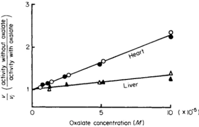
entially on L D H - 1 and L D H - 5 (Fig. 4). Whereas oxalate, oxamate, sulfite, and phenazine methosulfate inhibit L D H - 1 more than LDH-5, urea and the carbonate ion preferentially inhibit LDH-5. Several chemi
cal analogs of urea also differentially inhibit L D H - 5 ; these include hy- dantoic acid and methylurea, which is a particularly potent inhibitor of LDH-5 (109). Although oxalate behaves as a noncompetitive inhibitor with pyruvate as substrate, oxamate is a competitive inhibitor (102).
The extent of oxalate inhibition correlates with the content of Β subunits and thus can be directly related to the decreasing electrophoretic mobili
ties of the isozymes from L D H - 1 to LDH-5. Urea apparently directly inhibits a site on the A subunit, so that there is progressively less inhibi
tion of the L D H isozymes from 5 to 1, as their content of A subunits decreases (107).
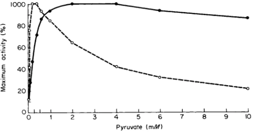
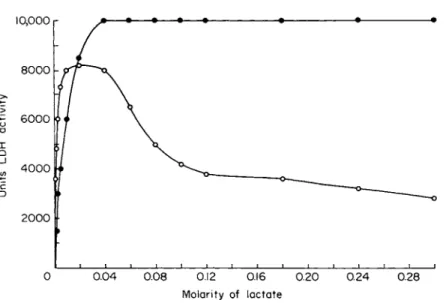
The best-known inhibitors of L D H are the substrates for the enzyme:
pyruvate and lactate (109-115). These substrates at high concentrations, which are apparently above the physiological range as judged by deter
minations made in vivo, differentially inhibit L D H - 1 , as shown in Figs.
5 and 6.
On the basis of the inhibition of L D H - 1 by pyruvate concentrations to which L D H - 5 is resistant, Kaplan and his associates proposed a theory designed to explain tissue-specific isozyme patterns (41, 99, 114, 115).
3 r
I 1 .
Ο 5 Ι Ο ( Χ Ι Ο " 5)
Oxalat e concentratio n {M)
FIG. 4. Differential inhibition by oxalate of human LDH from heart and liver.
Substrates: Ο, A , 0.7 mM pyruvate; # , A, 3.3 mM 2-oxobutyrate. [From Plummer and Wilkinson (102) Λ
This theory, termed the aerobic-anaerobic theory, maintains that L D H - 1 is the main isozyme in so-called "aerobic" tissues, whereas L D H - 5 pre
dominates in so-called "anaerobic" tissues. Quantitative oxygen tensions that would be required to place a tissue in either the "aerobic" or
"anaerobic" category have not been established. Thus, tissues are con
sidered either "aerobic" or "anaerobic" to suit their L D H isozyme pat
terns, which are then interpreted as conforming to the predictions of the theory. For example, a recent textbook of genetics states that liver is an anaerobic tissue and contains almost exclusively L D H - 5 (116).
Liver does contain almost exclusively LDH-5, but it is one of the most aerobic tissues. The fact that its isozyme pattern is contrary to the predictions of the theory is evidence against the theory. Other tissues whose isozyme patterns do not conform to the predictions of the theory are mature erythrocytes, platelets, and lens fibers (117).
The theory has been supported by the kinetic difference between LDH-1 and LDH-5. At 25°C LDH-1 is inhibited by pyruvate concentra
tions to which L D H - 5 is resistant (Figs. 5 and 6), but this great differ
ence between L D H - 1 and L D H - 5 in capacity to withstand substrate inhibition at 25°C is substantially reduced at temperatures more physio
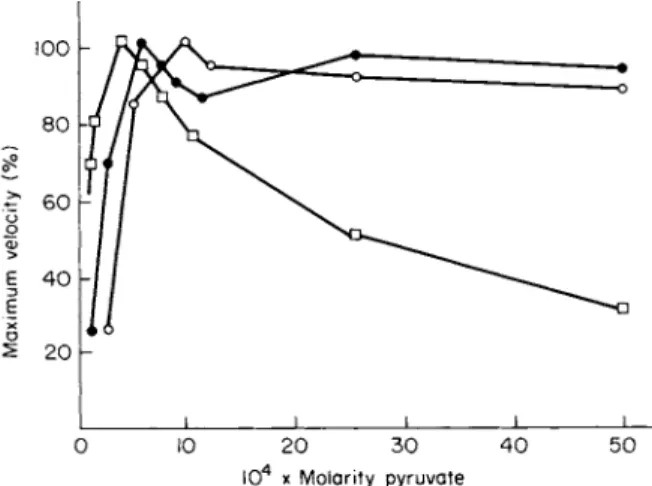
logical for mammals. This fact has been known since 1961, when the curve reproduced in Fig. 7 was published (118). Figure 7 shows that, although L D H - 1 is very sensitive to substrate inhibition at 6°C, at the more physiological temperature of 40°C, its sensitivity to the concen
trations of pyruvate used in the experiment is negligible.
Pyruvat e (n\M)
FIG. 5. Differential inhibition of human heart LDH-1 (O) and skeletal muscle LDH-5 ( · ) by pyruvate at 2 5 ° C . The final N A D H concentration was 2 X 10~4 M, and the LDH concentration was approximately 1 χ 10~9 M.
0 0.0 4 0.0 8 0.1 2 0.1 6 0.2 0 0.2 4 0.2 8 Molarit y o f lactat e
FIG. 6 . Differential inhibition of human heart LDH-1 (O) and skeletal muscle LDH-5 ( · ) by lactate at 25°C. The final N A D concentration was 5 χ ΙΟ"3 M, and the LDH concentration was approximately 1 χ 10"9 M.
I 1 1 ι ι ι ι
0 0.3 0.6 0 . 9 1.2 1.5 1.8 Pyruvate (m/Jf)
FIG. 7. Effect of temperature on pyruvate inhibition of rabbit LDH-1. Note that at physiological temperature for the rabbit substrate inhibition is negligible com
pared to that observed at 6 °C. [From Plagemann et al. (118).]
Another question relating to the "aerobic-anaerobic" theory is whether, under even the most anaerobic circumstances, concentrations of pyruvate sufficiently high to inhibit L D H - 1 are ever attained in vivo.
Ooncentrations of pyruvate and lactate in various tissues have been available for many years (119-122). Concentrations of pyruvate and
lactate in canine skeletal muscle under the most anaerobic conditions never exceed 2.0 and 25 mM, respectively (122). Even under unphysio- logical conditions of temperature (25°C), L D H - 1 is not significantly inhibited by these substrate concentrations (122).
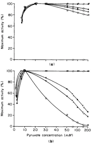
Furthermore, L D H - 1 inhibition is dependent on the degree of dilution of the enzyme (Fig. 8 ) . Intracellular concentrations of L D H isozymes have been estimated (123). At these intracellular isozyme concentrations
no pyruvate inhibition of LDH-1 can be detected even at pyruvate concentrations as high as 20 m M (123, 124). It should be emphasized that intracellular concentrations of L D H isozymes, pyruvate, and lactate are not uniform. The enzyme and its substrates, as mentioned in the section on subcellular localization, exhibit specific subcellular distribu- tions; hence, estimations of concentrations based on tissue homogenates are probably at best only approximations. Because the concentrations of L D H isozymes at certain regions within the cell are much higher than at other regions, the concentrations of L D H isozymes in certain subcellular sites are higher than those calculated from tissue homogenates (123). Similarly, substrate concentrations are also probably higher at certain sites within the cell than calculations made from tissue ho- mogenates would indicate. However, such differences that may occur between estimations based on tissue homogenates and the local concen- trations actually existing within cells do not alter the conclusion that substrate inhibition of L D H - 1 as measured by the usual spectro- photometric methods is probably an artifact of dilution. With stopped- flow spectrophotofluorometric techniques (125), no LDH-1 inhibition could be detected when the isozyme was present at physiological concen- trations (123, 124).
Inhibition is not observed with excess pyruvate and lactate at physio- logical enzyme concentrations because, under these conditions, the abor- tive ternary complex composed of L D H , N A D , and pyruvate is not formed. It is believed that inhibition of L D H by pyruvate in vitro results from formation of this ternary complex, which is a time- dependent reaction (123-127). The time for formation of this complex is considerably longer at the high concentrations of the enzyme that exist in vivo (126). Therefore, whether this ternary complex ever forms in vivo or under physiological conditions in vitro is at the core of the current dispute concerning the relationship between intracellular pyr- uvate concentrations and tissue-specific isozyme patterns. When the reac- tion is run immediately after mixing physiological concentrations of L D H , pyruvate, and N A D H without prior incubation, the results suggest that ternary-complex formation and inhibition do not occur (123, 124).
0 1.0 2.0 3.0 4.0 5.0 10.0 20.0 Pyruvate concentration {mM)
( b )
FIG. 8. Effect of increasing pyruvate concentration on the activity at 25°C of several concentrations of (a) LDH-5 and (b) LDH-1 purified from rat kidney. All reagents were prepared in 0.1 Μ sodium phosphate buffer, pH 7.0, and the final NADH concentration was 0.56 mM. Molar concentrations of partially purified LDH-1 and LDH-5 were calculated from turnover numbers (94). The LDH-1 was purified 130-fold, and LDH-5 was purified 98-fold. The maximum specific activity remained constant over the range of enzyme concentrations examined. With 1.0 mM pyruvate, approximately 3.2 /umoles of N A D H were oxidized per second per milli
gram of LDH-1, and approximately 4.5 /imoles of N A D H were oxidized per second per milligram of LDH-5 with 2.0 mM pyruvate. Concentrations of LDH-5 (a): φ, 7.0 χ ΙΟ"6 M; O, 1-8 Χ ΙΟ"8 M; A, 9.0 χ ΙΌ"9 Μ ; Δ , 4.5 X 10~9 M ; • 1.8 χ ΙΟ"9 M.
Concentrations of LDH-1 (b): · , 7.0 χ 1 0β M; O, 3.5 χ ΙΟ"7 Μ; A, 1-8 Χ ΙΟ"7 M ; Δ , 9.0 Χ ΙΟ"8 M; • , 3.5 χ ΙΟ"9 M. [From Wuntch et al. (124)-1
However, Everse et al. (126) demonstrated that, when L D H and pyr- uvate are preincubated for 30 minutes prior to addition of N A D H , sub- strate inhibition does occur. It must be said that their experiment is unphysiological for a variety of reasons. It ignores the fact that intracel- lularly several dehydrogenases and other proteins compete with L D H for N A D H (127). Accordingly, when incubated in a mixture containing L D H , N A D H , and pyruvate, these dehydrogenases, as well as other proteins and the coenzymes themselves, decrease the extent of substrate inhibition (Tables II and III) (127). Furthermore, concentrations of pyruvate and N A D H in the cell are in a state of dynamic flux (128, 129). They do not remain at the constant concentrations assumed in the model proposed by Everse et al. (126) where L D H , pyruvate, and N A D H are incubated in a cuvette for 30 minutes unexposed to any of the numerous metabolites normally present intracellularly. Since N A D and N A D H concentrations change continuously within cells (128, 129), their concentrations at actual sites of the L D H isozymes and other N A D - linked dehydrogenases are difficult to determine, as is the extent of abor- tive ternary-complex formation.
Because the concentration of many enzymes is so high in vivo that dilution of tissue homogenates several hundredfold is required prior to spectrophotometric assay of these enzymes, Srere (130) has emphasized that kinetic and inhibitory studies at unphysiologically low enzyme con- centrations should not be extrapolated to intracellular situations until the effects of enzyme dilution are ascertained. For this reason, we per- formed stopped-flow experiments that permitted kinetic studies on L D H at the high concentrations present in vivo. These experiments permitted a new approach to the question whether substrate inhibition exists intracellularly (123, 124, 127). The results revealed that, under more physiological conditions of enzyme concentration than have previously been employed, substrate inhibition probably does not constitute a major threat to L D H - 1 activity and, hence, that tissue distributions of L D H isozymes are not adequately explained solely on the basis of the relative intracellular abundance of pyruvate. For different reasons, Griffin and Criddle (131) believe that the L D H tetramer is probably not subject to inhibition in vivo; they reported that the monomeric subunit of L D H and not the tetramer is required for ternary-complex formation in vitro
(131). Finally, the experiments of Coulson and Rabin (132) are pertinent to the subject of substrate inhibition. They suggested that L D H - 1 inhibi- tion by pyruvate is attributable to the enol form of pyruvate present in commercial preparations as an impurity (132) ; the enol and keto forms exist in vivo in equilibrium, but the actual extent of intracellular
L D H activity6 (%) remaining after incubating L D H with
Final N A D and pyruvate N A D , pyruvate, N A D , pyruvate, N A D , pyruvate,
pyruvate in only and G - 3 - P D and M D H and B S AC
cuvette Incubation
(mM) (minutes) L D H - 1 L D H - 5 L D H - 1 L D H - 5 L D H - 1 L D H - 5 L D H - 1 L D H - 5
1.0 10 18 32 47 50 25 33 27 34
30 6 10 15 17 15 20 10 13
5.0 10 7 13 52 53 20 16 15 17
30 3 5 18 17 8 11 5 13
« The incubating mixtures contained 3.5 Χ 10"6 Μ LDH-1 or LDH-5, 14.0 μΜ N A D , 2.0 or 10.0 m M pyruvate, 7 Χ ΙΟ"6 Μ G-3-PD or 3.5 Χ 10~6 Μ M D H or 2.5 mg BSA/ml, all made up in 0.05 Μ tris-HCl buffer, pH 7.4. Final concentrations in the cuvette were 1.75 Χ 10"6 Μ LDH-1 or LDH-5, 7.0 μΜ N A D , 1.0 or 5.0 m M pyruvate, 3.5 Χ 10"6 Μ G-3-PD or 1.75 Χ 10"6 Μ M D H or 1.25 mg BSA/ml, and 0.7 mM N A D H . Reactions were initiated by the addition of reduced coenzyme.
6 Activity compared to that of control where N A D is deleted from the incubation medium.
c Bovine serum albumin.
406
T A B L E III
EFFECT OF INCUBATING SEVERAL PROTEINS WITH L D H - 1 , N A D , AND N A D HA
Initial L D H - 1 activity (%) remaining after incubating L D H - 1 with
rmiu pyruvate
in cuvette
(mM)
Incubation (minutes)
N A D , N A D H ,
and G-3-PD
N A D , N A D H ,
and M D H
N A D , N A D H ,
and BSA6
N A D and
N A D H N A D
0.5 10 100 100 100 75 51
30 100 100 60 25 51
1.0 10 100 100 100 63 45
30 100 100 57 20 47
5 . 0 10 100 100 85 25 44
30 100 100 57 6 44
10.0 10 100 100 57 13 46
30 100 85 43 3 45
a The incubating mixtures contained 3.5 Χ 10"6 Μ LDH-1, 14.0 mM N A D , 1.4 mM NADH, and 7.0 Χ 10"6 Μ G-3-PD or 3.5 Χ 10"6 Μ M D H or 2.5 mg BSA/ml, all made up in 0.05 Μ tris-HCl buffer, pH 7.4. Final concentrations in the cuvette were 1.75 Χ ΙΟ"6 Μ LDH-1, 7.0 m M NAD, 0.7 m M N A D H , 3.5 Χ 10"6 Μ G-3-PD or 1.75 Χ ΙΟ"6 Μ M D H or 1.25 mg BSA/ml, and 0.5, 1.0, 5.0, and 10.0 m M pyruvate.
Reactions were initiated by the addition of pyruvate and, in the case of N A D alone (last column), by pyruvate and N A D H .
6 Bovine serum albumin.
L D H inhibition would be restricted by the enol-keto tautomerization rate of pyruvate. According to this view, only the intracellular pyruvate present in the enol form could inhibit L D H , and this amount must be less than the total intracellular pyruvate measured in the experiments described earlier [119-122).
Several additional L D H inhibitors have been identified within the reagents employed to assay L D H activity. For example, impurities capable of inhibiting various dehydrogenases have been identified in N A D H (133, 134) and in N A D (135, 136). By chromatography on DEAE-cellulose, Dalziel (135) separated a competitive inhibitor of A D H from several commercial preparations of N A D . These impurities were present in sufficient quantity, approximately 3-4% of the total nucleo
tide, to produce large errors in the estimation of initial rate parameters for liver A D H and were not removed by recrystallization as the quinine salt (135).
Recognizing that at physiological concentrations of pyruvate and lac-
tate substrate inhibiton of neither L D H - 1 nor L D H - 5 occurs, Stambaugh and Post (137) demonstrated a difference between L D H - 1 and L D H - 5 in degree of inhibition by the end product lactate. They suggested that L D H isozymes might be distributed in tissues not according to inhibition by substrate, but rather by the end product lactate. With pyruvate as substrate, L D H - 1 , when assayed in dilute concentrations at pH 7.4 and 25°C, was inhibited significantly more by physiological concentra- tions of lactate placed in the reaction mixture than was L D H - 5 (137).
Although these observations were confirmed (138), the difference between L D H - 1 and L D H - 5 in end-product inhibition diminished substantially
(Fig. 9). Furthermore, as the pH was lowered from 7.4 to 6.8, no signifi- cant difference between LDH-1 and L D H - 5 in end-product inhibition
LDH- 5
~i 1 1 1 1 1 1 1 1 1 10 2 0 3 0 4 0 5 0 6 0 7 0 8 0 9 0 10 0
L a c t a t e (m/W )
FIG. 9. Effect of lowering pH on product inhibition of human LDH-1 and LDH-5 at 25°C. Two concentrations of pyruvate (circles for 0.1 mM and triangles for 0.2 mM) were employed to assay LDH activity in the absence and presence of 20, 40, and 90 mM lactate. [From Vesell (138).]