Growth Is Affected
ARMIN C . BRAUN
The Rockefeller Institute, New York, New York
I. Introduction 189 II. Harmonious Changes Involving Exaggerated Growth Responses . . 192
A. Generalized Stimulation 192 1. Bakanae Effect 192 B. Growth Changes in Leaves and Petioles 196
1. Growth Movements 196 2. Curling and Distortion 198 3. Frenching and Shoestringing 199 C. Growth Changes in Stems and Branches 201
1. Organs Arising in Unusual Places 201
a. Adventitious Roots 201 b. Adventitious Shoots 203 2. Witches'-Brooms 203 D. Growth Changes in Floral Organs 205
1. Alterations of Floral Parts 205 2. Overcoming of the Normally Arrested Development of Floral Parts 207
E. Modification in Fruiting Structures 208
F. Fasciation 209 III. Amorphous Changes in Growth Pattern 211
A. Self-Limiting Overgrowths 211
1. Intumescences 211
2. Galls 212 a. Insect Galls 212
b. Root Nodules 216 c. Root Knot 219 d. Clubroot 220 B. Non-Self-Limiting Tumors 221
1. Crown Gall 225 2. Wound Tumor 234 3. Genetic Tumors 236 Acknowledgments 240 References 241
I. INTRODUCTION
One of the most striking characteristics of higher plants and animals is the extraordinary way in which all of their functional parts fall into a coherent and flexible, but definitely limited pattern. In the normal organ-
189
ism, morphogenetic laws are strictly obeyed and processes concerned in metabolism, growth, cellular differentiation, and organogenesis are pre- cisely regulated. These processes start and stop in harmony to give the organism certain proportions that remain constant from one generation to the next.
The normal plant may be looked upon as a precisely balanced com- plex of interrelated reactions that are in a state of dynamic equilibrium.
Any deviation in this complex of reactions will tend to disrupt the bal- anced system and may lead to atypical, abnormal, or pathological growth.
It is not surprising, therefore, that growth patterns of plants may be readily modified by external environmental conditions such as heat, cold, light, and humidity, by nutritional deficiencies and excesses, by radiation, by changes in genetic constitution, as well as by pathogenic organisms of the most diverse types. The borderline between the normal and the abnormal in plants is, therefore, often quite indistinct and may represent merely quantitative changes which are evidenced either by a harmonious but somewhat exaggerated manifestation of the normal developmental processes or, as is more commonly found to be true, by an arrest or in- hibition of the processes concerned with growth and development. At other times, orderly qualitative changes result which in their most inter- esting form reveal growth and developmental potentialities in a plant far beyond any realized in the past history of the species. In the most extreme instances, plant cells undergo far-reaching qualitative changes and may, as a result, become permanently modified into new cell types in which continued unregulated and unorganized growth, rather than differentiation, characterizes the behavior of the affected cell.
Of particular concern to the present discussion is the influence that parasites of many different kinds exert on the growth and development of a parasitized plant.
Gaumann (1954) has indicated that organisms are pathogenic only if they are toxigenic. Although this sweeping generalization probably contains a large element of truth, little is actually known yet about the physiological and chemical mechanisms underlying disease. It is never- theless true that the growth of systemically diseased plants is generally inhibited. Even in those instances in which the disease is very mild and symptoms are largely masked (latent virus infections of potato, masked strains of tobacco mosaic virus), a statistically significant inhibition of growth of the host plant is found to occur. More often, pronounced stunt- ing is a characteristic manifestation of disease in plants. This may result either from systemic infection, or from the diffusion of specific chemical compounds elaborated by a pathogen, which itself remains localized in
the host. These chemical substances fall essentially into two categories:
(1) enzymes; (2) soluble metabolites, many of which are capable of reproducing perfectly the toxic manifestations of disease and some of which have been characterized chemically. (See further discussion in Chapter 13 (Volume II of this treatise).
Of far greater biological interest than the substances which act in such an unsophisticated way to damage, kill, or otherwise inhibit growth of the cells of the host, are those substances that stimulate cells to excessive growth.
The regulation of growth, differentiation, and organ formation in higher plants appears to result, among other things, from a very pre- cisely balanced series of growth-regulating substances or hormones, on the one hand, and from inhibitory systems or other compensatory mech- anisms that control the synthesis and response of plant cells to such biologically active substances, on the other. Certain metabolites elab- orated by pathogens appear to be quite similar or identical in their physiological action to growth-regulating hormones found to occur naturally in a plant. When these are produced in excess by the pathogen during the course of infection, growth responses occur—in a host—that simulate those found following the artificial application of excessive amounts of such growth-promoting substances. At other times, the chemical stimuli produced by the pathogen are quite different from any thus far recognized in the normal plant and, being different, tend to divert the normal growth pattern of the plant into new and unusual directions. Such morphogenetic stimuli appear to be of many different types. They are commonly highly specific in their action and regularly induce unusual growth responses in an affected plant. It must be recog- nized, however, that whatever form the growth abnormality takes, the potentialities for this form must have been present in the cells of the host. These growth potentialities are simply called into activity by the inciting stimulus. Thus, the morphogenetic stimulus produced by the pathogen as well as the protoplasmic substrate of the host upon which the stimulus acts are critical in determining the type of growth that results.
Finally, the metabolite elaborated by the pathogen may not itself be a hormone or a morphogenetic stimulus of the type referred to above which directly influences the growth pattern of the host. It may instead affect specifically the regulatory mechanism of the host cells by elim- inating either temporarily or permanently those cellular systems that are concerned with the regulation of growth or, alternatively, it may activate within the affected cell growth-substance-synthesizing systems whose
products are concerned specifically with growth and developmental processes. In these instances, it is the affected cell itself that elaborates growth-promoting substances—in greater than regulatory amounts—in response to a specific stimulus transmitted by a pathogen.
II. HARMONIOUS CHANGES INVOLVING EXAGGERATED GROWTH RESPONSES
A. Generalized Stimulation 1. Bakanae Effect
Intensified manifestations of normal developmental potentialites have been recorded in a number of plant species following infection with a variety of fungi, many of which are obligate parasites. The common houseleek, Sempervivum hirtum, grows as a rosette. The leaves of this plant are broadly ovate in form and are about twice as long as they are broad. Following infection with the rust Endophyllum sempervivi the appearance of the leaves is altered. They grow strongly in length and may be seven times as long as they are broad. As a result, the leaves assume a linear shape. Infected leaves stand erect and are much paler in color than are their normal counterparts.
Elongation of the internodes with a resulting significant increase in size appears to be a characteristic response of certain plants to infection by specific pathogenic fungi. Following infection of one of the spurges, Euphorbia cyparissias, by the aecial stage of the rust Uromyces pisi, the stem of the host elongates greatly. The distances between successive leaves in healthy Euphorbia plants are about 0.5 mm. while those of infected plants are 2-3 mm. The foliage leaves of the normal plant are thin, flexible, and about twelve times as long as they are broad. In diseased specimens, the leaves are thick, brittle, and only about two to three times as long as they are broad. Infected plants have an etiolated appearance. Similar responses have been observed in Vaccinium vitis- idaea infected with the teleutospore stage of Melampsora goeppertiana, in sugar cane with the downy mildew Sclerospora sacchari, and in Bromus erectus with the smut fungus Usttlago hypodytes. Alterations produced on the shoots of the periwinkles Vinca herbacea, Vinca major, and Vinca minor by the uredospore stage of the rust Puccinia vincae and on the shoots of Cirsium arvense by the teleutospore stage of Puc- cinia suaveolens are also very similar to those described above for Euphorbia. The stems of the infected plants become much elongated, while the leaves are shorter, broader, yellow in color, and brittle when compared with the normal. Frequently, the shoots blossom prematurely and the flowers are more or less abortive. When, for example, Primula clusiana and P. minima are infected with Uromyces primulae integri-
foleae, not only do the rosette leaves elongate but the flowers open in the autumn of the same year rather than in the following spring as hap- pens in the normal uninfected specimens.
Pilet (1952, 1953) studied the auxin content of healthy leaves of Sempervivum sp. infected with Endophyllum sempervivi as well as of healthy and diseased leaves of Euphorbia cyparissias infected with Uromyces pisi in an attempt to account—on a physiological level—for elongation of the stem as a result of infection. Parasitized leaves of both plant species revealed a much higher auxin content than normal. On the basis of these observations, Pilet (1953) suggested (1) that the causal fungus elaborates auxins, (2) that the increased auxin level found in the host is produced by the host in response to the parasite, or (3) that the parasite produces something which activates precursors of auxins in the host cell and which in turn are converted into auxin. It has not yet been shown, however, that the application of auxin to normal plants results in an elongation of leaves and internodes comparable to that obtained after infection by specific rust fungi. Brian (1957) has, therefore, suggested that the increased auxin found in the leaves and shoots of such plants may be coincidental rather than causal since quite different metabolic products of fungi, the gibberellins, have been shown to be responsible for stem elongation in the bakanae disease of rice as well as in many other plant species when these substances are applied artificially.
The bakanae or foolish seedling, disease of rice is the best under- stood of this type of disease. It has been studied by Japanese pathologists for many years. A comprehensive review of this disease has recently been presented by Stowe and Yamaki (1957). The bakanae disease, which is caused by Fusarium moniliforme (imperfect stage) or Gibberella fuji- kuroi (perfect stage), is widely distributed and found in most rice- growing regions of the world. Fusarium moniliforme has a large host range, is soil-borne, and attacks the roots and basal portions of the stem not only of rice plants but of maize, cotton, sugar cane, and other plant species as well. In the case of rice, a browning of the tissue usually occurs at the site of infection, the leaves tend to yellow and curl inward and, in many instances, growth of affected seedlings is arrested. How- ever, some of the affected plants grow more rapidly than do healthy ones and are conspicuous in the field because of their height and etiolated appearance. The dry weight of the elongated seedlings was found to be significantly greater than that of the healthy plants. This overgrowth effect—occurring naturally—has been reported only in rice, although artificial inoculation has resulted in increased growth in maize, barley, sugar cane, sorghum, millet, wheat, and oats.
Kurosawa (1926) showed that the overgrowth or bakanae effect could be reproduced in rice seedlings by treating them with cell-free culture filtrates on which the causal fungus had grown. These filtrates reproduced all of the characteristic manifestations of the bakanae dis
ease such as lengthening of the internodes and leaves, chlorosis (except under conditions of nutrient excess), and reduced tillering. This pioneer work of Kurosawa was followed by the isolation and chemical character
ization of several biologically active substances. Two powerful growth- promoting substances, which were given the trivial names of gibberellin A and B, were isolated in crystalline form by Yabuta and Sumiki (1938), and Yabuta and Hayasi (1939a, b, 1940). It was found later that gib
berellin A is converted to Β by warming in dilute acid at 50°-70° C. Boil
ing under acid conditions converted both substances into a biologically inactive compound named gibberic acid. A third biologically active substance, gibberellin C, was isolated by Yabuta et al. (1941a) from acid-treated gibberellin A. The nature of the gibberellin skeleton was established by Yabuta et al. (1941b). It was demonstrated in these studies that gibberene obtained by selenium dehydrogenation of gib
berellin A, B, or gibberic acid is a fluorene derivative.
Another quite distinct but chemically related, biologically active com
pound was isolated by American workers headed by F. H. Stodola and by British investigators under P. W. Brian. This substance was named gibberellin X by the Americans and gibberellic acid by the British group.
The latter name is now commonly used for this compound. Although gibberellic acid, whose empirical formula is C1 9H2 206, resembles gib
berellin A in biological activity and degradation products, it differs from this compound in infrared spectra, optical rotation, in certain deriva
tives, as well as in the empirical formula. The basic skeleton of gib
berellic acid, like gibberellin A, was found to be gibberene, which Mulholland and Ward (1954) identified as 1,7-dimethylfluorene. This led Cross et al. (1956) to suggest the tentative structure for gibberellic acid shown in Fig. 1. It is reasonably certain that the formula shown in Fig. 1 is essentially correct, although the exact points of attachment of the lactone grouping to the cyclohexenol ring of gibberellic acid are not known as yet.
Recently, Takahashi et al. (1955) reexamined gibberellin A and found this compound to be essentially homogeneous by countercurrent distri
bution, paper and partition chromatography. Esterification followed by chromatography on alumina led to the isolation of three methyl esters which were named gibberellin A1 ? A2, A3. Stodola et al. (1957) believe that their gibberellin A is identical with A^ Gibberellic acid was shown to be identical with A3. Gibberellin A2 has been isolated only in Japan
but Stodola (1956) reports its corrected empirical formula as L>»i9 H2 606. Thus, the existence of three distinct gibberellins appears to be established.
Evidence is now available suggesting that the gibberellins elaborated by the fungus Gibberella fujikuroi correspond closely (in their biological activity) to growth-regulating compounds found to occur naturally in higher plants. West and Phinney (1957) have isolated—but have not yet identified chemically—an ether-extractable substance from wild cucum- ber seeds which, like the gibberellins of fungus origin, promotes the active growth of dwarf mutants of maize. Since these genetic dwarfs appear to be due to single-gene defects, they may be explained as result- ing from blocks in the biosynthetic pathway leading to the formation of a natural gibberellin-like substance. The gibberellins appear to substitute in the plant for the missing product of that reaction. Very recently Mac- Millan and Suter (1958) have obtained high yields of gibberellin AT from the seeds of runner bean plants. The isolation and characterization of gibberellin AT from a higher plant indicate further that this com- pound, in all probability, participates directly in the growth-regulating system of higher plants and, therefore, represents a new type of en- dogenous growth regulator.
COOH FIG. 1. Gibberellic acid.
The gibberellins of fungal origin have been applied to a large number of different plant species and have produced a variety of responses. The most consistent and striking response is a marked elongation of plant stems. In one survey of 42 different plant species belonging to many different families, only 3, white pine, gladiolus, and onion, failed to respond with stem elongation. In many instances, not only do the stems grow longer but also they are proportionately thicker. Gibberellin-treated oak and maple trees are not only taller but have a diameter more than twice as great as that of untreated control specimens. Brian and Hem- ming (1955) have reported remarkable stimulation of dwarf varieties of
peas, broad beans, and French bean by gibberellic acid. The treated dwarfs and untreated normal plants grew equally well.
In the bakanae disease of rice as well as in certain related diseases described earlier in this discussion, infected plants that reached maturity flowered earlier than did normal plants of the same variety. With the availability of pure gibberellic acid for experimental purposes, Lang (1956) found that a biennial variety of Hyoscyamus treated with that substance bolted and flowered in the first year. In a later study, Lang (1957) found that in annual varieties of Hyoscyamus, Silene, and Sam- olus the long-day requirement for flowering could also be replaced by gibberellic acid. Wittwer et al. (1957) found that gibberellic acid-treated lettuce bolted and flowered under photoperiodically adverse conditions.
These same workers reported the gibberellins to be 500 times more effec- tive than indoleacetic acid in inducing parthenocarpy in tomatoes.
Reduced fruit yields are characteristic of the bakanae disease. Hayashi et al. (1953) found that the gibberellins reduced rice grain production by 32% although the yield of straw was increased by 14%.
Stowe and Yamaki (1957) suggest that the one property common to the gibberellins is their capacity to remove certain limitations on cell enlargement, while incidentally releasing other responses. Despite this similarity to the auxins, the gibberellins are not auxins but appear to depend for their activity on the presence of auxins. The biological activ- ity of these substances has been attributed (Brian, 1958) to the neutral- ization of a growth-inhibitory system which normally limits growth.
Certain other plant diseases give indication that infection interferes with the synthesis or utilization of gibberellin-like substances. Carrot plants infected with the tomato big-bud virus of California, as well as with a related virus found to occur naturally in carrots in the State of Washington, were shown by Kunkel (1951) to bolt and flower prema- turely. These findings suggest that virus infection may increase the synthesis of gibberellin-like substances by the plant. On the other hand, in the rosette diseases, as exemplified by peach and peanut rosette, infec- tion may interfere with the normal synthesis or utilization of these biologically active substances by the plant. The stunting effects, but not other symptoms associated with aster yellows, corn stunt, and wound tumor diseases, have in fact been reversed with gibberellic acid (Mara- morosch, 1957).
B. GROWTH CHANGES IN LEAVES AND PETIOLES
1. Growth Movements
Nastic responses refer to changes in position of a bilaterally sym- metrical organ as a result of differential growth. Epinasty of leaf petioles
has been found by Wellman (1941) to be a characteristic early symptom of the Fusarium wilt disease of tomato plants. Hunger (1901) and Smith (1920) found it to be associated with Granville wilt, while Grieve
(1941) recorded this condition as a primary symptom in roses affected with the rose wilt virus. Pronounced epinasty has also been produced experimentally by Locke et al. (1938) following inoculation of tomato plants with a highly virulent strain of the crown gall bacterium. Hypo- nasty, on the other hand, has been observed in plants infected with Erwinia phytophthora.
Grieve investigated the question of epinasty of the leaf petiole as it occurs in the Granville wilt disease (1936, 1939, 1940). In this instance, the growth response was found to be an irreversible one and the invasion of one lateral trace by the bacteria was sufficient to induce it. Grieve reported that a growth substance which appeared to be indoleacetic acid was elaborated by the bacteria in culture and that this substance induced a characteristic epinastic response when applied to tomato plants. How- ever, virulent and avirulent cultures of the bacterium produced approxi- mately equal amounts of the growth-promoting substance. Furthermore, no significant difference between the growth-substance content of com- parable control and infected stem segments could be detected. Studies of the growth-substance distribution in upper and lower halves of reflex- ing petioles, on the other hand, showed a significantly greater concen- tration in the upper halves. In normal petioles, the concentration of growth substance was found to be greatest in the lower halves of the petioles. Grieve (1939) reached no definite conclusion as to how the redistribution of growth substance in the basal part of the petiole is accomplished. He pointed out (Grieve, 1943), however, that the balance of the normal growth-controlling mechanism at the base of the petiole is very delicate, as evidenced by the fact that ethylene, in one part in ten million parts of ambient air, as well as very minute amounts of growth substance from bacterial cultures will disturb it. Grieve considers that even a small stimulus from the invading organisms can initiate a chain of reactions that leads to a redistribution of growth substance with a resulting nastic response.
More recently, Dimond and Waggoner (1953) made a detailed inves- tigation of the cause of epinastic symptoms in Fusarium wilt of tomato plants. From these studies it was concluded that ethylene is responsible for the characteristic growth response. This conclusion was based on the finding that ethylene is produced in culture by the causal agent of this disease, Fusarium oxysporum f. lycopersici. Ethylene production by in- fected tomato plants was demonstrated, moreover, by confining such plants with healthy indicator plants. Under these conditions, epinastic responses developed to a greater degree than when the indicator plants
were confined with healthy tomato plants. In these studies, ethyl alcohol, which is capable of causing epinasty in tomato and which is produced by the causal fungus in culture as well as in the infected host, was also considered a possible cause of the growth response. This compound was ruled out, however, because the amounts produced by the fungus in the hosts were insufficient to account for the observed epinastic responses.
Another characteristic response—involving growth movements—which is associated with certain disease conditions is concerned with the up- right growth habit of leaves. This is one of the most characteristic symptoms of the aster yellows disease in many different plant species.
Leaves on a normal aster plant take approximately a horizontal position in relation to the main axis of the stem. In virus-infected plants, on the other hand, the petioles elongate and the leaves assume an extremely upright habit of growth which approximately parallels the main axis of the plant. It is this upright habit of growth that suggested the name
"rabbit ears" for lettuce plants infected with the aster yellows virus.
Kunkel (1954) has shown that twigs of peach trees infected with peach yellows virus characteristically assume an upright growth habit.
In this instance, the plants can be cured of the disease by thermal treat- ment. After cure, the new growth of such twigs again takes a normal position with respect to the main stem. This change necessitates a
change in the direction of growth of the terminal bud.
2. Curling and Distortion
The curling and distortion of leaves are characteristic of many dis- eases. Needless to say, such well known maladies as curly top of sugar beet, peach leaf curl, and leaf roll of potatoes have been assigned their trivial names because curling of leaves is the most conspicuous symptom of the disease. The most extreme form of leaf rolling is found in the so-called scroll galls produced by certain insects. Under the influence of specific insects, the leaves curl lengthwise to form tightly rolled scrolls in which the insects live.
Peach leaf curl caused by Taphrina deformans is perhaps the most studied of this type of disease. The causal fungus commonly infects very young leaves which soon become either quite red or paler in color than are the normal leaves. Such infected leaves soon become curled and puckered, increase greatly in thickness, and have a firm consistency.
The host cells which are in contact with the invading fungus are stimu- lated to abnormal activity. This may involve isolated regions of a leaf or in extreme instances most of the leaf. Such cells increase in size and number and produce marked changes in the form and structure of the leaf. Loss of chlorophyll is almost complete in the stimulated cells. The
increase in size of the cells on either side of the midrib results in a puckering of the leaf. At the same time, the leaves tend to curl and become concave on the lower side. The cells of the palisade parenchyma respond much more actively to the stimulus of the fungus than do the subjacent cells of the spongy parenchyma, thus producing the curling.
Link et al. (1937) have reported that a substance having the properties of an auxin and which was presumably indoleacetic acid was extractable from Taphrina deformans culture filtrates. The role, if any, that this sub
stance plays in the development of the disease picture as described above is as yet not clear. It is nevertheless true that auxins are capable of stimulating cell enlargement of the type described above in paren
chymatous cells of many different plant species.
3. Frenching and Shoestringing
Frenching, a well-known deformity of tobacco, occurs in most tobacco-growing regions of the world. This condition is, in an advanced stage, characterized by a cessation of terminal bud and stem growth, and by a reticular type of chlorosis in the slowly expanding new leaves.
This chlorosis may disappear as the young leaves develop and become strap-shaped (sword and string) as a result of the failure of the leaf lamina to expand. As apical dominance in such diseased plants is lost, the axillary buds develop and an unusually large number of leaves—
which may be as high as 300—appears on a plant. These leaves assume an upright growth habit and are commonly sword- or string-shaped.
Such plants have the appearance of a rosette or, in extreme instances, of a witches' broom. Root growth is also somewhat inhibited, although the effect on the leaves appears to be the most characteristic feature of the disease.
The extent to which frenching modifies the morphology of a leaf can be seen from some figures that are given by Wolf (1935). Normal tobacco leaves average 55 cm. in length and 30 cm. in width. The cor
responding measurements for sword leaves and string leaves are 40 cm.
X 10 cm., and 27 cm. χ 1.8 cm., respectively. Histological studies show, moreover, that the diseased leaves are two to three times as thick as normal leaves. This increase in thickness is due to an enlargement of cells of all the leaf tissue except cells of the vascular system. Schweizer (1933) has indicated that the xylem is markedly reduced in develop
ment in frenched leaves. In mature string leaves, the parenchyma remains quite juvenile with little or no evidence of dorsiventrality.
Although the cause of frenching is not clearly established, it is now believed to be a toxicity disease rather than the result of a nutritional deficiency. No parasitic organism of any kind has yet been implicated
in this condition. McMurtrey (1932) described briefly the similarity of frenching symptoms to those of thallium toxicity in tobacco. Spencer
(1935) found that one part of soil obtained from a field in which frenching occurred mixed with 2,000 parts of sand produced typical frenching in the test plants. Thus, a toxic factor, effective in very low concentrations, was present in frenching soils. Spencer (1935) tested 33 different elements on tobacco and found, as had McMurtrey, that only thallium at a concentration of 5 p.p.m. or less produced chlorosis, strap- shaped leaves and other symptoms characteristic of frenching. Later, Spencer and Lavin (1939) indicated that frenching and thallium toxicity are probably two distinct physiological conditions.
Steinberg (1947, 1950) has suggested that frenching of tobacco is caused by the action of diffusates from the presumably nonpathogenic soil bacterium Bacillus cereus. The effectiveness of B. cereus diffusates in eliciting typical frenching symptoms in tobacco varied with the kind and quantity of peptone used and the concentration of inorganic nitrogen in the test medium.
Bacillus cereus is a widely distributed soil microorganism. The pro- gressive development and type of symptom produced by it in tobacco (in aseptic culture) largely parallel those that appear in plants subject to this abnormality under field conditions. Moreover, rhizosphere and rhizoplane counts of this organism increased by 65% and 200%, respec- tively, when frenching occurred in the field. The nature of the toxic substance is not yet known.
Steinberg (1952) has shown that slight excesses of certain amino acids caused production of symptoms resembling frenching in tobacco seedlings. Frenching symptoms were, however, limited to the natural isomers of alloisoleucine and isoleucine in tobacco and to these com- pounds and leucine in Nicotiana rustica. Alloisoleucine was most effec- tive in both species and as little as 2 to 8 p.p.m. resulted in chlorosis and strapping of leaves in tobacco. Leucine, which was ineffective when applied to tobacco, was more effective than isoleucine in N. rustica.
Analytical studies indicated, moreover, that frenching was found to be accompanied by a marked increase in isoleucine and other free amino acids in the leaf lamina of field-grown plants (Steinberg et al, 1950).
The absence of free amino acids in frenching soil and the marked increase of free isoleucine in frenched leaves indicate that Bacillus cereus toxin and isoleucine are not identical. The conclusion was therefore drawn that the accumulation of excessive quantities of free amino acids in the strapped leaves was a probable chemical factor involved in the production of the morphological symptoms in the plant. The stages sug- gested by Steinberg (1952) leading to abnormal growth in frenching of
field tobacco were, therefore, as follows: bacterial soil toxin —» receptor -» excessive accumulation of isoleucine and other free amino acids in the leaves —> frenching.
A condition resembling frenching may also be caused by certain strains of the tobacco mosaic and cucumber mosaic viruses or a mix- ture of these viruses, as well as by a gene mutation. Kunkel (1954) has reported an extreme instance of shoestringing caused in tomato plants by a mixture of tobacco mosaic virus and the virus of carrot yellows of Texas. Leaf-blade development in this instance was completely sup- pressed. Just how these viruses suppress or prevent leaf-blade growth is not understood.
The so-called "wiry" tomato plants described by Lesley and Lesley (1928) are not unlike those which result from the virus diseases of the shoestring type reported above. Wiry plants appear, however, to result from genie mutation. This condition is recessive to the normal and the plants are completely sterile. The leaves of these plants are variable in shape but have a strong tendency toward reduction of the leaf lamina and in extreme instances the leaves consist merely of a tapering midrib.
C. Growth Changes in Stems and Branches 1. Organs Arising in Unusual Places
a. Adventitious Roots. The development of adventitious roots on stems of plants has been reported to be a characteristic host response associated with certain bacterial, fungal, and viral diseases.
A number of investigators (Bryan, 1915; Grieve, 1936, 1940; Hunger, 1901; Smith, 1914, 1920) have described this phenomenon following infection of tomato and certain other hosts with Pseudomonas solan- acearum. Smith (1914, 1920) reported it to be associated with the Grand Rapids disease of tomato which is caused by Corynebacterium mich- iganense. Adventitious root formation was found to occur on tomato
(Locke et al., 1938) and Kalanchoe (Price and Gainor, 1954) following inoculation of those host species with the crown gall bacterium. It has also been observed by Wellman (1941) and Dimond and Waggoner (1953) in tomato plants diseased with Fusarium wilt. Certain viruses, such as the cranberry false blossom virus in tomato as well as that implicated etiologically in the sereh disease of sugar cane, character- istically stimulate root formation in the stems of their respective hosts.
Grieve (1936, 1940) studied the development of adventitious root formation in tomato plants infected with Pseudomonas solanacearum.
This investigator found that the new roots arise commonly over a span of several internodes along the path of the primary bundle except in
those instances in which the disease progresses with great rapidity, in which case these adventitious structures do not develop. Histological examination of transverse and longitudinal sections of infected plant stems demonstrated that adventitious roots usually develop in regions where large primary bundles are affected. The initiation of root pri- mordia often precedes the advancing bacteria, indicating action at a distance. Grieve applied indoleacetic acid to the stems of tomato plants and found that the roots initiated were similar to those resulting from infection. This finding suggested that the bacteria are inducing root formation either directly as the result of the production of a growth- promoting substance, or indirectly through their interference with the metabolism of the plant. In attempting to distinguish between these two possibilities, Grieve found that Pseudomonas solanacearum produces in- doleacetic acid from tryptophan in culture. However, virulent and aviru- lent strains produce approximately equal amounts of this substance. The possibility that the bacteria produce an auxin in the xylem by acting on naturally occurring or artificially introduced tryptophan was examined but no evidence for such production was obtained. No difference in auxin concentration could be detected, moreover, between healthy and diseased plants by bioassay methods. Furthermore, adventitious roots could be produced by cutting or blocking the bundles. These findings led Grieve to question whether the bacteria induce the formation of adventitious roots directly through the elaboration of indoleacetic acid.
He suggested (Grieve, 1943) that the production of these structures more likely results from disturbances of normal auxin transport in the plant as a result of mechanical blocking of the vessels by the bacteria.
Price and Gainor (1954) reported a striking correlation between adventitious root development and the number of leaves present on a Kalanchoe plant inoculated with crown gall. These workers suggested that a substance responsible for stimulation of adventitious roots is organ- ized in leaves and is transported downward to the site of the tumor.
That the biologically active substance is not indoleacetic acid was indi- cated by the finding that 2% of this substance in lanolin—applied to stumps of petioles above the points of inoculation—did not stimulate the development of adventitious roots in Kalanchoe daigremontiana.
De Ropp (1947a) has shown, moreover, that a powerful root-stimulat- ing substance is elaborated by sterile crown gall tumor cells. In crown gall it appears likely, therefore, that the stimulus for adventitious root formation originates in the cells of the host rather than directly from the causal bacteria.
The course of Fusarium wilt of tomato is not unlike that of the bacterial wilt of this host as described above. Wellman (1941) reported
adventitious roots associated with this fungus disease. Since epinasty of the leaf petioles often appears on plants showing adventitious root formation, Dimond and Waggoner (1953) have suggested that in the case of Fusarium wilt, adventitious root formation, like epinasty, may result from the production of ethylene either by the causal fungus or by the cells of the host as a result of the interaction of the host and the pathogen.
b. Adventitious Shoots. The witches'-broom virus in potato causes the infected plant to produce numerous buds at the nodes in the above- ground stems of potato plants. Long slender stolons that resemble aerial roots but which are covered with trichomes develop from these adven- titious buds. Aster yellows virus and carrot yellows virus from Texas, on the other hand, stimulate the production of small aerial tubers in the axils of the leaves of potato plants.
An extreme example of adventitious shoot formation was reported about 100 years ago by von Martius. This worker described an unusual plant that possessed a mania for forming innumerable leaves and shoots.
This species has been appropriately named Begonia phyllomaniaca.
Plantlets develop spontaneously in incredible numbers from the super- ficial cell layers of the leaf lamina, petioles, and stems. Erwin Smith
(1920) studied phyllomania and believed it to be conditional on shock such as is encountered during the repotting of plants. Smith indicated, moreover, that the cells of leaves and internodes are susceptible to such shock only during a relatively brief period of meristematic growth. The adventitious shoots do not arise from preformed buds but develop from totipotent cells at the base of the trichomes and especially from botryose glands which are found in great abundance in young stems and leaves of this species. These embryo plants develop a vascular system of their own but the vast majority never succeed in connecting this with the vascular system of the plant. They must, therefore, be considered not as branches but rather as independent organisms.
2. Witches'-Brooms
Witches'-brooms, or "hexenbesens," are closely grouped, much branched structures commonly found to occur on a number of different species of trees and shrubs. The stimulus necessary for their formation is supplied in different plant species by pathogens of the most diverse types. Fungi of the genus Taphrina and various rust fungi are effective in inducing witches'-broom, while distinct virus species have been shown to cause the formation of such an abnormality in alfalfa, potato, peach, and the black locust. Bos (1957) has recently presented a detailed account of the virus-induced witches'-broom. An eriophyid mite belong-
ing to the genus Aceria, followed by a powdery mildew fungus, is said to be involved in the development of branch knot so commonly found on the hackberry.
Typical witches'-brooms caused by the fungi appear to live a more or less independent life and act as parasites on the plants from which they are derived. In accordance with their independent existence, witches'-brooms tend to break away from the correlations of the parent plant. Instead of branching out horizontally, the brooms stand as more or less erect clusters of branches. A normal dorsiventral symmetry is thus changed to a radial symmetry. Witches'-brooms as a rule do not produce flowers, indicating further a breakaway from the correlations of the parent plant. Heinricher (1915) has shown, more- over, that a twig of a witches'-broom caused by Taphrina cerasi grafted to a healthy sweet cherry tree develops again into a typical witehes'- broom. The independence of these structures is further shown in an impaired periodicity. The vegetative buds found on the brooms of the sweet cherry, for example, open several weeks earlier than do those present on healthy branches (Schellenberg, 1915). Gaumann (1946) has pointed out that this premature unfolding of the buds is probably asso- ciated with the fact that the shoots and buds comprising the broom never achieve a true winter dormancy since the pathogen never becomes entirely quiescent. This incomplete winter dormancy may result in a winter killing of the first year twigs present on a broom.
The witches'-broom of the silver fir caused by Melampsorella caryo- phyllacearum is typical of this peculiar type of growth. The primary infection is said to occur in the young bark of branches surrounding buds. In the spring when the buds develop, the fungus mycelium grows into the epidermis of the developing shoot and penetrates the cortex so that by fall a slight swelling of the shoot axis is found. During the fol- lowing year, an overgrowth of considerable size may be formed and buds embedded in this growth develop to produce the characteristic deformation. The twigs of this particular type of broom are found to develop in whorls. They are short, thick, soft, and pliable. This results from the fact that the cortical parenchyma is spongy and the wood is not well developed. The buds on the broom open earlier in the spring than do those present on a healthy twig, while the leaves found on the diseased specimens remain short, are yellowish in color, and fall off when a year old. The leaves present on a normal twig are, on the other hand, long, straight, dark green on the upper side, and commonly remain in position 5 or more years. The longevity of the diseased twigs them- selves is limited and they die within a few years, giving rise to the dry, bristling brooms characteristically found on the silver fir.
The witches'-broom-like effects that occur in certain plant species following virus infection appear to result from the excessive stimulation and development of secondary shoots. This condition is not accompanied by swellings or overgrowths characteristic of many of the fungus-induced brooms.
Brian (1957) has suggested that the diseases characterized by exces- sive branching may well be due to auxin deficiency rather than to auxin excess. Lacey (1948) has, in fact, shown that cultures of Coryne- bacterium fascians, the causal agent in leafy gall development on certain plants, are capable of rapidly destroying indoleacetic acid and other auxins present in plant tissues.
D. Growth Changes in Floral Organs 1. Alterations of Floral Farts
Under the stimulus of pathogens, the sepals, petals, stamens, or pistils of a flower may be transformed into structures that are very different in appearance from those found normally. Kerner von Marilaun
(1891) has reported that double flowers are produced in Valerianella carinata, the common corn salad, as a result of infestation by a mite.
This doubling results from a retrograde alteration of the stamens into a whorl of petals. These petals, under the stimulus of the mite, enlarge to more than fifty times their original size and finally appear as fleshy lobes which fuse with one another into a disc. The greatly enlarged lobes bend backward and are concave on the lower side. It is in the cavities thus formed that the gall mites live.
In the capitula of certain of the milfoils, Achillea millefolium and A. nana, the peripheral ray florets and the central tubular ones become leaf-like in appearance and assume remarkable forms as a result of mite infestation. A capitulum is often subdivided into several stalked sub- capitula, while the flowers are altered into green funnel-shaped structures with jagged mouths and into small flat-lobed green foliage leaves, while short green scale-like leaflets, which represent modified stamens, develop from the midribs of these leaves. Sometimes, however, the changes in growth pattern following mite injury are not so extreme as the two instances cited above might suggest. In Veronica saxatilis, for example, the development of numerous hairs on the rachis of the raceme, the pedicels, and the bracts appears to be the only characteristic host re- sponse. Hairs are not present in the corresponding normal structures of this plant species.
Doubling of the flowers of the alpine rose, Rhododendron fer- rugineum, has been reported by Kerner von Marilaun (1891) to result
from gnat infestation. In this instance the stamens and carpels are trans- formed into red petals. Since flowers of this species normally have 10 stamens and 5 carpels, there should be only 15 red petals in the center of each but there are often two or three times that many present. It thus appears that not only metamorphosis but multiplication takes place.
According to Tschirch (1890), the aphid Astegopteryx stryacophila induces remarkable changes in young flowers of Styrax benzoin. The calyx, corolla, and androecium are transformed into large abnormal leaves that form bag-like pockets. The pistils, however, appear to be unaffected in this instance.
Metamorphoses of floral parts of higher plants are also brought about by certain species of fungi. The transformation of stamens into petals commonly follows infection of Viola sylvestris by Puccinia violae. Sim- ilarly, Peronospora violacea has been reported to encourage stamen primordia in the flowers of Knautia arvensis to develop into petal-like structures. An extreme instance of phyllody is found when flowers of the Japanese plum are infected with a rust of the species Caeoma makinoi.
In this instance all of the floral parts are transformed into foliage leaves.
In the alder the bracts of the pistillate flowers are changed by Taphrina alni'incanae (Amentorum) into greatly elongated purple-red spatulate lobes which are twisted and bent.
Von Tubeuf (1895) has reported that when Albugo Candida infects the inflorescences of the radish, the ovary, calyx, and corolla enlarge and the androecium assumes a leaf-like appearance. Stamens may become green and leaf-like in the downy mildew disease of Pennisetum glaucum and other grasses. Thus, this disease, which is caused by Sclerospora graminicola, is popularly known as green ear. In the case of head smut of maize, caused by Sorosporium reilianum, the whole staminate head or the ear may become a leafy structure.
In the United States, there are at least three different viruses, the aster yellows virus, the cranberry false blossom virus, and tomato big bud virus, that produce gigantism in the floral organs and more particularly in the sepals and calyxes of tomato and certain other solanaceous plants.
The tomato big bud virus appears to affect the sepals rather specifically.
These structures enlarge greatly under the influence of this virus and fuse to form huge bladder-like growths that conceal the inner parts of the flower. The stamens and pistils do not appear to be greatly affected.
The aster yellows virus and the cranberry false blossom virus cause symptoms in the flower trusses of tomato that are almost identical with those caused by the big bud virus. Although these three viruses cause the sepals of certain of the solanaceous plants to enlarge greatly, they
do not have this effect on sepals of species of certain other families. They do not, for example, cause gigantism in the sepals of the flower trusses of the periwinkle, Vinca rosea, but they bring about a virescence in the petals, stamens, and styles. There is, in this instance, a retrograde devel- opment of floral parts into foliage leaves. It is, therefore, clear that the type of symptom produced depends as much on the species of plant in which the virus multiplies as on the nature of the virus.
The aster yellows, cranberry false blossom, and carrot yellows virus of Texas stimulate the formation of adventitious buds in the stigmas of flowers of a number of different hosts. Such buds may give rise to either flowers or to leafy stems which in turn may bear flowers.
Bos (1957) has recently attempted to interpret the development of the flower and its component parts on the basis of the phenomenon of antholysis accompanying virus infections in Crotalaria and certain other hosts. This investigator has suggested, on the basis of the homology of the reproductive and vegetative parts of a plant and their morphogenetic development, that the sexual and vegetative activities in the plant are mutually antagonistic. A growing point can develop only into an inflor- escence or into a vegetative shoot. During flower initiation the vege- tative characters are suppressed and sexual characters prevail. The sexual characters remain suppressed, on the other hand, during vegetative growth. From the manner of appearance of antholysis in virus-infected Crotalaria plants, Bos has concluded that flower induction is stopped suddenly and that subsequent development of the floral parts proceeds exclusively in a vegetative manner. The ultimate result obtained depends upon the stage of development of the primordia in the bud at the time of suppression of flower induction by the virus. Thus, flowers initiated in succession, produce a series of flowers showing increasing antholysis.
These—in reverse order—clearly show a macroscopically recognizable picture of the morphogenesis of the flower and of its component parts.
Therefore, according to Bos, antholysis supports the theory that the flower must be regarded as a modified leafy branch.
2. Overcoming of the Normally Arrested Development of Floral Parts The anther smut, Ustilago violacea, produces its spores only on the anthers of certain hosts. When this fungus infects the female flowers of Melandrium album or M. dioicum, the stamens, which are normally arrested in their development and are present only in a rudimentary form in the pistillate flowers, grow to full size but when mature are filled with smut spores instead of pollen.
A similar process is said to occur when pistillate flowers of Knautia
arvensis and K. sylvatica are infected with Ustilago scabiosae. The nature of the morphogenetic stimulus which overcomes the arrested develop- ment of male sex organs in female flowers is still unknown.
E. Modification in Fruiting Structures
Modification of the fruiting bodies of higher plants occurs quite commonly as a result of infection. Such descriptive names as little peach, bladder plum, phony peach, etc., have been applied to conditions of this type. Hypertrophied fruits are commonly produced in certain species of the genus Prunus as a result of infection by Taphrina pruni. In this instance, the tissue of the diseased ovary is stimulated to growth, but not in the same way as in the normal fruit. The resulting body is flat- tened on two sides, is brittle, yellow in color, and much longer than the normal fruit. The seed within is abortive and a hollow space is left in its place. These hypertrophied growths, which are commonly called
"bladder plums," fall from the trees at the end of May and are said to be eaten in certain areas. A somewhat similar condition is found when Taphrina aurea infects the pistillate flowers of the poplar. This fungus, like the one described above, stimulates the growth of the ovaries with the resulting development of golden yellow capsules that are more than twice the normal size. The smut fungus UstiMgo zeae also stimulates growth of the tissue of the pistillate flowers of maize. As a result, the grains are replaced by irregular cushion-like structures with a diameter of up to 7 cm. The resulting growths contain more auxin than do normal tissues. Ustfago zeae, moreover, produces an auxin in culture (Moulton, 1942) which was identified as indoleacetic acid by Wolf (1952). The fruit size may be greatly dwarfed as in the case of two virus diseases, little peach and phony peach, or the fruits may assume an asymmetrical shape as is found characteristically in the xyloporosis disease of oranges—which is also of viral origin.
The seed capsules of Datura stramonium normally bear numerous conspicuous spines. When plants of this species are infected with the severe etch virus, spine formation may be completely inhibited (Kunkel, 1944). The aster yellows virus, on the other hand, causes a bursting of the abnormal fruits produced by Cajophora lateritia plants infected with this agent. The seed-like structures present in these fruits are green in color and have been transformed, under the influence of the virus, into short stems which bear leaves.
The entire inflorescence may, in certain disease conditions, be altered to quite a different type. The rust fungus, Aecidium esculentum, is said to cause the normal inflorescence in Acacia to change from a head to a spike. Similarly, the stinking smut fungus, Tilletia tritici, may change the
growth pattern of the head of the club types of wheat to the elongated or "vulgare" type.
Individual floral parts or even entire organs may be replaced by new structures in certain diseases. Clamceps purpurea invades and destroys certain of the ovaries of rye and other grasses. In place of the seeds, elongated dark purple sclerotia are produced. These are composed of dense aggregates of fungus mycelium.
In sheep sorrel infected with Ustilago oxalidis the seeds are replaced by the spores of the pathogen. In this instance, the spores are forcibly expelled from the seed capsule as if true seeds were present.
F. Fasciation
A condition known as fasciation can be classified between that group of growth abnormalities described above (involving harmonious changes in growth pattern) and the amorphous changes to be considered later in this discussion. Fasciation is a morphological term that has been used to describe a series of abnormal growth phenomena resulting from many different causes, any of which result at the morphological level in a flat- tening of the main axis of the plant. Although this ribbon-like expansion of the stem is often the most striking feature of this condition, all parts of the plant may be affected. Fasciation often results in alterations in the arrangement of foliar and floral structures. White (1948) points out that, when fasciation occurs, the early seedling growing stages are normal. As the plant develops, however, the growing point becomes broader, and the unregulated, distorted tissue growth results in significant increases in weight and volume of plant tissue. The apical growing region becomes linear and comb-like in some instances or develops numerous growing points, producing a witches'-broom effect. In still other instances, the growing points may be coiled and resemble a ram's horn or they may be highly distorted into a grotesque tangle of coils. Fasciations are wide- spread, both geographically and taxonomically. Examples have been recorded in 102 families of vascular plants. Fasciations found in certain plant species such as the common cockscomb, Celosia cristata, as well as the cristates in the cacti are highly prized by gardeners.
Fasciations have been classified on morphological grounds—based on such physical features as form, color, and anatomical structure. Linear, bifurcated, multi-radiate, and ring or annular types have been listed (de Vries, 1909-1910; White, 1948). This condition has also been classi- fied from a causal standpoint. In this type of classification the conditions and agencies necessary to produce the character as well as the modify- ing factors that affect its development are considered. White (1948) has placed these essentially into five categories (1) fasciations which breed
true and which in crosses with the normal obey genetic laws so that their genie basis is known; (2) noninherited forms in which the fasciation is due to environmental causes and the character is not reproduced in the selfed seed; (3) fasciations that occur spontaneously and which have been propagated vegetatively but in which neither the initial cause nor the question as to whether the condition is transmitted through the seed is known; (4) fasciations which are induced artificially by known pro- cedures; (5) fasciations which have been imperfectly investigated.
Fasciations resulting from modifications of the normal gene comple- ment, and which breed true, have been known for centuries in such plants as Celosia as well as in the mummy or crown type of garden pea.
Gregor Mendel (1866) found fasciation in the mummy pea to be reces- sive in the Fx when crossed with normal varieties. Such F/s showed a ratio of 3 normals to 1 fasciate in the F2. A gene-fasciated race in Nico- tiana tabacum when crossed with nonfasciated strains, on the other hand, gives 1 : 2 : 1 F2 ratios. In other instances, very complicated results were obtained, showing the effects of modifying factors and giving rise to nonfasciated segregates which carry the fasciated gene. Fasciation is not correlated with changes in chromosome numbers but it may produce meiotic irregularities.
Aside from gene mutation, fasciation has been reported to result from environmental effects such as frost, pressure, alteration of food and water relationships, pruning, mutilation, as well as by more specific etiological agencies such as a bacterium, Corynebacterium fascians, and X-irradiation.
There seems little doubt but that nutritional changes due to correla- tive disturbances in growth-substance relationships play a role in fascia- tion. Bloch (1938) has reported that this condition can in certain instances be produced experimentally by treating plants with auxin- containing pastes. On the other hand, high doses of X-rays have been shown to inactivate auxins and yet disturbances caused by them also result in fasciation. The effects of X-irradiation on auxin metabolism in the growing plant are obviously complex. These nongenetic types of fasciation are not transmitted through the seed but may, in some in- stances, be propagated asexually. Both inherited and noninherited types of fasciations which are phenotypically indistinguishable have been described for at least 3 different plant species.
Orland E. White believes that the basic cause of fasciation is a dis- turbed metabolism involving excessive nutriment which mobilizes energy that must be used. This energy, once accumulated, goes, according to White (1948), into growth and becomes "wildly" expended in abnormal and unpredictable tissue production. Heslop-Harrison (1952) points out,
however, that these conclusions are descriptive rather than truly ex- planatory since excessive nutrition in itself cannot account for the abnor- mal element in fasciation. He believes, rather, that a proximate cause of these abnormalities is probably to be found in maldistribution of the auxins in the plant. Jones (1935) and Orland White (1948) have sug- gested that fasciation bears an analogy to animal cancer. In the opinion of the writer, however, this condition does not represent a true tumor at all but might better be placed in that category of teratological abnor- malities known as monstrosities.
III. AMORPHOUS CHANGES IN GROWTH PATTERN
Amorphous changes involving either the temporary or permanent loss of typical organization are found to occur very commonly in plants.
These overgrowths range from somewhat exaggerated but self-limiting wound-healing responses at the one extreme, to rapidly growing non- self-limiting tumors which have no characteristic size or structure, at the other.
A. Self-Limiting Overgrowths 1. Intumescences
Intumescences, which are among the simplest and most innocuous of this type of growth, are found to occur most commonly on leaves but are also found on stems and fruits of plants. These blister-like pustules usually result from the abnormal elongation of groups of cells with or without increased cell division. In a typical leaf intumescence found in many plant species, the palisade parenchyma cells elongate considerably in localized areas to give rise to the pustules. Intumescences have been found to be produced experimentally in a number of ways. No pathogens, however, appear to be concerned in their development. Sorauer (1886) believed them to result from an excess of moisture in the air and soil.
Harvey (1918) produced intumescences artificially on the undersurface of cabbage leaves by subjecting such leaves to slight freezing. Smith (1920), on the other hand, induced blister-like pustules in cauliflower leaves with the use of such irritating chemicals as formic and acetic acids. Wolf (1918) has shown that intumescences may be produced in cabbage leaves by means of a sandblast and this worker has indicated that such overgrowths found naturally in the field result from sand driven against the leaves by wind. More recently, La Rue (1933a, b, 1935) was able to reproduce perfectly the intumescences which arise under conditions of high humidity on the leaves of poplar with the use of low concentrations of indoleacetic acid. Much larger self-limiting over-
growths were produced artificially by Brown and Gardner (1936) on bean plants with the use of higher concentrations of that substance. It is not unlikely that certain of the burls as well as the overgrowths that sometimes arise at graft unions result from a hormonal imbalance of this type.
2. Galls
Localized overgrowths in which the host cells are stimulated to exces- sive growth by pathogens are known as galls. In these instances, the continued growth of the host cells is dependent upon continued stimula- tion by the pathogen. There are unusually large numbers of distinct overgrowths of this type to be found in plants of which only a few representative examples have been selected for discussion.
a. Insect Galls. Among the most interesting types of self-limiting growth abnormalities found in plants are those that result from the activity of certain of the gall-forming insects. The whole subject of insect galls as reviewed in detail by Kiister (1911), Ross and Hedicke (1927), and more recently by Felt (1940) suggests that many of these highly specialized overgrowths represent beautiful examples of dependent differentiation.
Insect galls may result either from a mechanical or chemical stimulus.
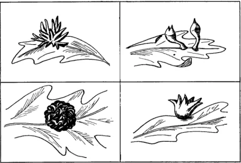
Stem swellings on roses caused by the closely placed spiral galleries of the rose stem girdler, Agrilus viridis, are believed to be an example of the former type. Chemical stimulation, however, is probably far more important than mechanical irritation in the production of many insect galls, although the chemical stimulus may in some instances be supple- mented by directive feeding of the insect. An examination of the litera- ture leaves the unmistakable impression that highly specific morpho- genetic stimuli, of chemical nature, and elaborated by insects, are cap- able of initiating, stimulating, and directing most precisely the develop- ment and differentiation of plant cells. There is no question about the fact that the morphological form that a gall assumes depends upon the nature of the pathogenic insect. This is evidenced by the fact that the same host species or even the same organ of the same plant attacked by different but closely related insect species produces morphologically very different galls. Figure 2 illustrates this point and shows drawings of 4 galls of quite different morphology induced on leaves of the California white oak by 4 closely related species of cynipids. The morphology of the gall is so specific that it is considered by some to be a more reliable criterion for distinguishing between closely related species of insects than are the morphological characters of the adult insects. The morphol- ogy of the gall does not, moreover, appear to be a function of the part
of the plant from which it arises. Currant galls of the oak, which are found on both flower stalks and leaves, have similar morphological structure at both points of origin. The same insect species may, more- over, produce the same gall type on different species of plants. The saw- fly, Micronematus gallicola, produces bright red galls of similar mor- phology on four different species of willow. Findings such as those reported above suggest in the strongest possible manner that a large number of highly specific chemical substances are elaborated by insects and that these substances have specific morphogenetic effects on the cells
FIG. 2. Four morphologically distinct galls produced on leaves of the California white oak by four closely related species of insects. (Drawings by R. J. Mandlebaum.) and tissues of a plant. However, only moderate success has thus far been achieved in establishing experimentally the truth of this view. That a morphogenetic stimulus is elaborated by certain insects is suggested by the fact that the Norway spruce aphid, Adelges abietis, attaches itself firmly by its beak to the scale of a fir bud and can directly influence only a few cells of the young shoot. Nevertheless, thousands of cells on this shoot are affected and soon begin to assume an altered form and are stimulated to growth, indicating that a diffusible stimulus is active.
Plumb (1953) has, in fact, induced the formation of this gall by inject- ing a glycerol extract of the salivary glands of the insect into the bases of spruce buds. Beijerinck, as early as 1888, killed the eggs of a sawfly with
a hot needle immediately after they had been deposited in the host and found that the galls developed normally. Beijerinck called the biologically active materials "growth enzymes/' and in 1897 applied this concept to the development of form in general. Parr (1940) showed that the gall- forming coccid, Asterolecanium variolosum, stimulates the host tissue to both hyperplastic and hypertrophic activity. He demonstrated further that the effect of the salivary secretion of this homopteron continues to stimulate plant cells even after the insect itself is gone. It was possible to reproduce galls similar to those produced by the insect by injecting glycerol extracts of the salivary glands. When the salivary extracts were heated to 60° C. they did not produce galls, indicating that a heat-labile, enzyme-like substance was involved in gall formation.
Rossig (1904) investigated various organs of gall wasps and believed that he could trace the site of formation of the gall-forming substances to the Malpighian vessels. Similar findings were reported by Trigger son
(1914) for the cynipid which produces the oak-hedgehog gall. Lewis and Walton (1947) reported some very interesting histological and cyto- logical findings dealing with a biologically active substance believed to be involved in the growth and differentiation of cell development of the cone gall of witchhazel. This gall is produced by the aphid species Hormaphis hamamelis Fitch. In this instance the stem mother inserts her stylets into and between the cells of an immature leaf and injects very minute droplets of a substance secreted by glands opening into the stylar canal. This stinging process is not to be confused with feeding and the sting substance is quite different in its nature from substances found in the salivary secretions. When about 150 stings have been made in a small circular area by the stem mother, the cells in this region at first become etiolated. Almost immediately thereafter, cells receiving injected sting material dedifferentiate, undergo rapid mitotic divisions, and then redifferentiate into gall cells rather than typical leaf cells.
The injected sting material consists of a ground substance in which minute crystalloids are embedded. These crystalloids show a reddish purple cast in such stains as gentian violet, Congo red, and acid fuchsin.
They are capable of passing readily from cell to cell as well as of enter- ing cells when they are deposited by the insect in the intracellular spaces.
Whether the injection is inter- or intracellular, the end result is the entry of the crystalloids into the nucleolus. There they may fuse to form a large crystalloid which again breaks up into smaller ones as mitosis begins. During nuclear division, the crystalloids are distributed to the daughter nuclei where they are again found in the nucleolus. Since these bodies are incapable of self-propagation and since they are apparently used up during growth of the cells, fresh sting material must be injected
repeatedly by the stem mother during the entire growth of the gall. It would indeed be interesting to know the chemical nature of the "crystal- loids" since they possess some very unusual regulatory properties.*
J. P. Martin (1942) induced galls on three-month-old sugar cane plants by injecting extracts obtained from macerated leaf hoppers of the species Draculacephala mollipes. These findings again appear to impli- cate hormone-like substances in the initiation and development of insect galls. Anders (1958) reported that he was able to reproduce the swell- ings associated with the Phylloxera disease by applying to grape plants secretions obtained from the aphid responsible for this condition. A similar type of swelling was obtained when certain amino acids, used in the same proportions found in the insect secretions, were applied to grape roots.
The larva of the moth, Gnorimoschema gallaesolidaginis, induces an eliptical monothalmous gall on the stem of the Solidago host. The larva in this instance burrows into the terminal bud of the plant and then down the stem to a point 2 cm. below the growing point, where it eats away the central tissues and induces the formation of a simple spindle- shaped gall. The gall stimulus, according to Beck (1954), appears to be associated with a silky substance secreted by the feeding larva. This silky material induces anatomical changes in normal stems similar to those found in the galls. The stimulus is, however, short-lived and uniform and continued deposition of the silky substance over the surface of the larval chamber appears necessary for the formation of typical galls.
Boysen-Jensen (1952) studied, with the use of rather ingenious methods, the development of a midge (Mikiofo fagi) gall on beech leaves. Evidently the formation of the gall in this instance is caused by growth substances given off by the larva. These as yet uncharacterized substances produce cell enlargement and cell division but not organized growth. It was, therefore, suggested that cell enlargement and cell divi- sion are regulated by the larva which moves rapidly about the gall chamber and secretes the growth-promoting substances in definite places, thereby making the gall assume its special form. According to this interpretation, the growth-promoting substance does not have special organizing properties but the shape of the gall is dependent upon the distribution of a rather nonspecific type of growth substance by the larva. The growth substances are, in other words, tools which are used
* After this paper had been submitted to the editors, a comprehensive account of the studies of Lewis and Walton (1958) appeared. In that investigation the diagnostic crystalloids were found to be Feulgen-positive, and it was concluded that the gall results from the activities of a virus. On the basis of the evidence pre- sented, this conclusion is, in the opinion of the author, unwarranted.