Peptidases
Heinz Wiist
Peptidases hydrolyse terminal peptide bonds. Unlike the proteinases they do not attack native pro
teins. Their substrates are di- and polypeptides.
Peptidases are to a large extent substrate specific (see the Appendix, p. 836), are usually activated by heavy metals and hydrolyse the peptides o f the naturally occurring L-amino acids. They are found in varying concentrations in practically all tissues of the body. Liver i), kidney
2
\ pancreas and erythro
c y t e s
1 - 4
.
5
) have a relatively high peptidase activity.
Peptidases can be subdivided according to their substrate into dipeptidases, aminopeptidases and carboxypeptidases. Dipeptidases hydrolyse dipeptides which have a free carboxyl and amino group.
Aminopeptidases hydrolyse peptides containing free terminal amino groups. Carboxypeptidases require free terminal carboxyl groups.
T o obtain the optimum experimental conditions for the assay of peptidases, it is usually necessary to choose the right activators and the most suitable substrate (as well as buffer, p H and substrate con
centration). Table 1 gives a survey of the optimum assay conditions.
Peptidases can be determined by a number different m e t h o d s :
1. Acidimetric determination of the amino acids liberated by titration in the presence of alcohol
5
*
6
) 2. Colorimetric determination o f the amino acids liberated with ninhydrin
7
.
8
>
3. A m i n o acid determination by manometric O2 m e a s u r e m e n t s
9
*
1 0
)
4. Spectrophotometric determination of individual amino acids (applicable to purified e n z y m e s )
1 1
) 5. Colorimetric determination of the reaction products by means o f the formation o f azo dyes from the corresponding substrates, for example carboxypeptidase with naphthoxycarbonyl-phenyl- a l a n i n e
1 3
.
2 7
) , leucine aminopeptidase with L-leucyl-naphthylamine hydrochloride 12,14-17,25,26).
A. Acidimetric Determination of Di- and Polypeptidases
56)
The m e t h o d of Grassmann and Heyde
6
) has been modified by Smith
5
)', it allows the estimation of the enzymatic hydrolysis of peptides and is largely independent of the type of substrate.
Principle
The amino acids liberated o n the hydrolysis o f the peptide are determined acidimetrically with thymolphthalein as indicator. The basicity of the amino group liberated at the same time is suppressed by use of ethanol as solvent in the titration. The increase of acidity per unit time is a measure of the enzyme activity.
D H. Wiist, F. Bruns and H. Schmitz, unpublished.
2) R. Ammon and W. Dirscherl: Fermente, Hormone, Vitamine. Thieme, Stuttgart 1959.
3) E. L. Smith and D. H. Spackman, J. biol. Chemistry 212, 255, 271 [1955].
4
) R. Merten and M. Winschuh, Z. Vitamin-, H o r m o n - u. Fermentforsch. 7, 35 [1948].
5
> E. L. Smith, G. E. Cartwright, F. H. Tyler and M. M. Wintrobe, J. biol. Chemistry 185, 59 [1950].
6) W. Grassmann and W. Heyde, Hoppe-Seylers Z. physiol. Chem. 188, 69 [1930].
7) T. B. Schwartz and F. L. Engel, J. biol. Chemistry 184, 197 [1950].
8) G. A. Fleisher, H. R. Butt and K. A. Huizenga, Proc. Staff Meetings M a y o Clinic 32, 410 [1957].
9) H. Herken, A. Schmitz and R. Merten, Hoppe-Seylers Z. physiol. Chem. 275, 29 [1942]; Natur
wissenschaften 29, 670 [1941].
10) H. Herken and H. Erxleben, Hoppe-Seylers Z. physiol. Chem. 264, 251 [1940].
u) F. Binkley and C. Torres, Arch. Biochem. Biophysics 86, 201 [I960].
12) H. E. Arst, R. T Manning and M. Delp, Amer. J. med. Sci. 238, 598 [1959].
13) H. A. Ravin and A. M. Seligman, J. biol. Chemistry 190, 391 [1951].
1
4
) / . A. Goldbarg, E. P. Pineda and A. M. Rutenburg, Amer. J. clin. Pathol. 32, 571 [1959].
1
5
) O. Braun-Falco and K. Salfeld, Arch. klin. exp. Dermat. 204, 407 [1957].
16) B. Rassler and H. Schon, Clin. chim. Acta 6, 583 [1961].
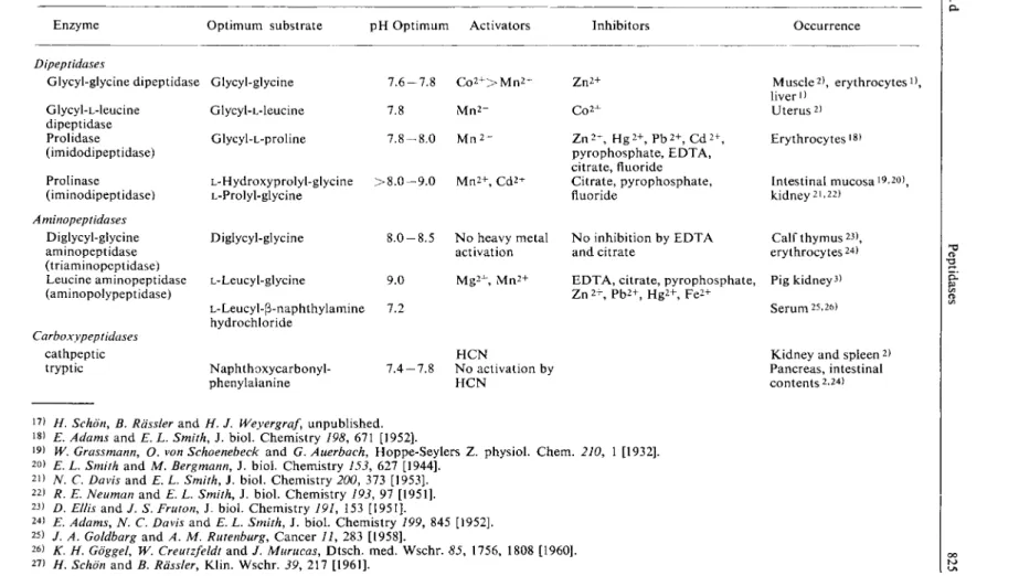
Table 1. Occurrence of peptidases and optimum conditions for their assay
Enzyme Optimum substrate p H Optimum Activators Inhibitors Occurrence
Dipeptidases
Glycyl-glycine dipeptidase Glycyl-L-leucine
dipeptidase Prolidase (imidodipeptidase) Prolinase (iminodipeptidase) Aminopeptidases
Diglycyl-glycine aminopeptidase (triaminopeptidase) Leucine aminopeptidase (aminopolypeptidase)
Carboxypeptidases cathpeptic tryptic
Glycyl-glycine Glycyl-L-leucine Glycyl-L-proline
L-Hydroxyprolyl-glycine > £ L-Prolyl-glycine
Diglycyl-glycine
7 . 6 - 7 . 8 C o 2 + > M n 2 + 7.8 M n 2 + 7 . 8 - 8 . 0 M n 2 +
. 0 - 9 . 0 M n 2 + , C d 2 +
L-Leucyl-glycine
L-Leucyl-P-naphthylamine hydrochloride
1 . 0 - 8 . 5
>.0 K2
Naphthoxycarbonyl- phenylalanine
7 . 4 - 7 . 8
N o heavy metal activation M g 2 + , M n 2 +
H C N
N o activation by H C N
Z n
2
+ C 0 2 + Z n
2 +
, H g 2 + , P b 2 + , C d 2 + , pyrophosphate, E D T A , citrate, fluoride Citrate, pyrophosphate, fluoride
N o inhibition by E D T A and citrate
M u s c l e
2
) , erythrocytes
1
), liver
1
) Uterus
2
) Erythrocytes
l g
)
Intestinal mucosa i9,20)
j
kidney
2
1 -
2 2
) Calf thymus
2 3
>, erythrocytes
2 4
>
E D T A , citrate, pyrophosphate, Pig kidney
3
) Zn
2
+ , P b
2 +
, H g
2 +
, F e
2+
Serum 25,26)
Kidney and spleen
2
) Pancreas, intestinal c o n t e n t s
2
-
2 4
)
17
) H. Schon, B. Rdssler and H. J. Weyergraf, unpublished.
18) E. Adams and E. L. Smith, J. biol. Chemistry 198, 671 [1952].
19
> W. Grassmann, O. von Schoenebeck and G. Auerbach, Hoppe-Seylers Z. physiol. Chem. 210, 1 [1932].
2
0) E. L. Smith and M. Bergmann, J. biol. Chemistry 153, 627 [1944].
21
) N. C. Davis and E. L. Smith, J. biol. Chemistry 200, 373 [1953].
22
) R. E. Neuman and E. L. Smith, J. biol. Chemistry 193, 97 [1951].
23
) D. Ellis and / . S. Fruton, J. biol. Chemistry 191, 153 [1951].
24
) E. Adams, N. C. Davis and E. L. Smith, J. biol. Chemistry 199, 845 [1952].
25) / . A. Goldbarg and A. M. Rutenburg, Cancer 11, 283 [1958].
2
6) K. H. Goggel, W. Creutzfeldt and / . Murucas, Dtsch. med. Wschr. 85, 1756, 1808 [I960].
27) H. Schon and B. Rdssler, Klin. Wschr. 39, 217 [1961].
826
Optimum Conditions for Measurements
D u e to the different p H optima of peptidases and the various activators required (see Table 1), the buffer, activators and other experimental conditions (e.g. pre-incubation o f activator and substrate) must be adjusted to suit the enzyme to be determined. The assay o f glycyl-glycine dipeptidase (acti
vated by C o
2 +
) in serum is described here. The m e t h o d can also be applied to tissue homogenates.
The p H optimum for the enzyme from serum and erythrocytes is 7.6. Optimum activity requires the addition of C o
2+
ions. Other divalent ions have little or no effect. Whilst prolidase must be pre- incubated for 2 to 3 hours with M n
2 +
, glycyl-glycine dipeptidase is active immediately after the addi
tion of C o
2 +
. The enzyme is saturated with substrate at 0.05 M glycyl-glycine
5
*. A s the hydrolysis of the substrate requires a relatively long incubation time, the initial rate of the reaction cannot be deter
mined. The hydrolysis is a first order reaction.
Reagents
1. Glycyl-glycine
2. Diethylbarbituric acid (veronal), sodium salt 3. Cobalt sulphate, C o S 0 4 - 7 H 2 0
4. Sodium hydroxide, A. R., pellets 5. Ethanol, 96% (w/v)
6. Thymolphthalein indicator
Preparation of Solutions
I. Substrate solution (0.125 M glycyl-glycine):
Dissolve 822 mg. glycyl-glycine in 50 ml. distilled water and adjust to pH 7.8 with 0.1 N NaOH.
II. Veronal buffer (0.1 M; pH 7.8):
Dissolve 2.06 g. Na veronal in distilled water and make up to 100 ml. Mix 66.2 ml. of this solution with 33.8 ml. 0.1 N HC1. Check the pH.
III. Cobalt sulphate (0.01 M):
Dissolve 281 mg. C 0 S O 4 7 H 2 0 in distilled water and make up to 100 ml.
IV. Alcoholic sodium hydroxide (0.01 N):
Shake 2 g. NaOH (pellets) in a 500 ml. volumetric flask with pure 96 % ethanol until dissolved and allow to stand for 24 hours. A fine precipitate settles out. Decant the clear supernatant (0.1 N). Dilute 10 ml. of this solution to 100 ml. with ethanol freshly each day and adjust the titre.
V. Thymolphthalein indicator (0.1 %):
Dissolve 100 mg. indicator in 100 ml. 96% ethanol.
Stability of the solutions
Store the glycyl-glycine solution in a refrigerator or better still in a deep-freeze. Store the substrate solution (I) in a refrigerator at 4° C and prepare freshly every 6 to 8 days. The 0.1 N alcoholic N a O H is relatively stable if protected from atmospheric CO2. Store the buffer solution (II) in a refrigerator.
II.4.d Peptidases 827
Procedure
A s s a y
Preliminary remarks:
Use fresh serum. In contrast to the activity in several animal sera (rat, mouse) the activity in human serum is low. With very low activity, 0.4 ml. instead of 0.2 ml of the incubation solution should be taken for the titration. In this case add 3.6 ml. ethanol for the deproteinization. To obtain a greater hydrolysis of the substrate, the incubation time can be increased to 4 hours. All alterations should be allowed for in the calculations.
Method:
Reaction volume: 2.5 ml.; temperature: 37°C (water bath). Pipette successively into test tubes:
0.75 ml. buffer (solution II) 0.25 ml. cobalt solution (III)
1.00 ml. substrate solution (I).
Equilibrate for ca. 10 min., then start the reaction by the addition of 0.50 ml. serum.
Mix and note the time. Immediately (0 min.) remove an 0.2 ml. sample (zero value); remove other 0.2 ml. samples at 30, 60, 120 or more min. Pipette into 10 ml. centrifuge tubes:
1.8 ml. 96% ethanol 0.2 ml. sample.
Centrifuge*) for about 10 min. at 3000 g. Pour the supernatant into a 50 ml. Erlenmeyer flask, wash the precipitate once with a little 96 % ethanol and also pour the washings into the flask. Add
3 drops thymolphthalein solution (V)
and titrate with 0.01 N alcoholic NaOH until the colour changes to blue. The colour change is not very sharp.
Calculations
The enzyme activity is proportional to the amount o f amino acids liberated, and this in turn is proportional to the N a O H required (difference between the zero value and the sample). T h e enzyme activity is expressed as either the % hydrolysis of the substrate per unit time, or better defined as the reaction rate constant ki (first order reaction):
1 , 100 X l o g -
"
A
t[min.] ° 100 - (% hydrolysis)
Under the given conditions complete hydrolysis corresponds to the consumption of 12.5 ml. 0.01 N N a O H for 2.5 ml. of the enzymatic reaction mixture, i.e. 1.0 ml. 0.01 N N a O H for the 0.2 ml. sample.
Therefore % hydrolysis = (consumption of 0.01 N N a O H [ml.]) X 100 Example
H u m a n hepatitis serum. 0.2 ml. samples were taken from the enzymatic reaction mixture.
Titration values: 0 min. 1.24 ml. 0.01 N N a O H 60 min. 1.56 ml. 0.01 N N a O H Increase in acidity: 0.32 ml. 0.01 N N a O H
% Hydrolysis 3 2 %
kl = io
x l o g
T o r ^ = I x l o g L 4 7
= To 'x 2 9
=0 6 X 1 0 - 3 3
"m i n
*) Filtration has not proved successful; the losses due to evaporation of the ethanol are too great.
Section C: Measurement of Enzyme Activity
B. Colorimetric Determination of Carboxypeptidase
27)
Carboxypeptidase is an SH-enzyme. It occurs in high concentrations in pancreas as the inactive proenzyme which can be activated autocatalytically or by t r y p s i n
1 3
*
2 8
) . A carboxypeptidase differing from the pancreatic enzyme has been found in kidney and spleen
2
\ A substrate with a terminal carboxyl group must be used for the measurement of the activity of carboxypeptidase. N a p h t h o x y - carbonyl-phenylalanine is a specific substrate
1 3
).
Principle
Naphthoxycarbonyl-phenylalanine *) is hydrolysed by carboxypeptidase to phenylalanine and naphth- oxycarboxylic acid. The latter decomposes spontaneously to p-naphthol and CO2. The p-naphthol can be determined colorimetrically as an azo dye by coupling with tetrazotized odianisidine.
Optimum Conditions for Measurements
The optimum p H lies between 7.4 and 7.8. With a naphthoxycarbonyl-L-phenylalanineconcentration of 6 x 1 0
-4
M the reaction is linear for the first 30 min. (ca. 10% conversion of the substrate). In older m e t h o d s
1 3
^ which use a substrate concentration of 2 x 10~
4
M there is no linear relationship between the amount of substrate hydrolysed and the amount of enzyme present.
T o obtain optimum activity in duodenal juice calcium ions (0.005 M) are r e q u i r e d
2 7
) . Recovery of crystalline carboxypeptidase is only quantitative after the addition of calcium ions.
Reagents
1. Naphthoxycarbonyl-DL-phenylalanine * *) 2. Tris-hydroxymethyl-aminomethane, tris 3. Calcium chloride, CaCl2-6 H2O
4. Tetrazotized o-dianisidine
5. Perchloric acid, A. R.; sp. gr. 1.67; ca. 70% (w/v) 6. Ethyl acetate
7. Sodium sulphate, anhydrous 8. p-Naphthol
9. Methanol
Preparation of Solutions I. Substrate solution (ca. 3 x 10~
3
M naphthoxycarbonyl-L-phenylalanine):
Dissolve 20 mg. of the racemate in 10 ml. pure methanol. Prepare freshly each day.
II. Tris buffer (0.05 M; pH 7.8):
Dissolve 6.08 g. tris-hydroxymethyl-aminomethane in distilled water and make up to 250 ml., a d d 342 ml. 0.1 N HC1 and dilute to 1000 ml. with distilled water. Check the p H and adjust if necessary.
III. Calcium chloride (0.25 M):
Dissolve 5.48 g. CaCl2-6 H2O in distilled water and make up to 100 ml.
*) Also called carbonaphthoxy-phenylalanine ( C N P A )
**) e.g. from Mann Research Laboratories, N e w York, U.S.A.
2
«) W. Rick, Klin. Wschr. 38, 408 [I960].
II.4.d Peptidases
829 IV. Colour reagent (0.4 % tetrazotized o-dianisidine):
Dissolve 200 mg. tetrazotized o-dianisidine in 50 ml. distilled water. Prepare freshly each day.
V. P-Naphthol(50[xg./ml.):
Dissolve 50 mg. P-naphthol in ca. 100 ml. methanol and dilute to 1000 ml. with distilled water.
Stability of the s o l u t i o n s
Apart from solutions (I) and (IV), which should be prepared freshly each day, the other substances and solutions are stable practically indefinitely.
Procedure
Experimental material
The method described below is suitable for duodenal juice. Dilute the juice 1:10 with buffer (solution II) before the measurements. Carboxypeptidase in pancreas can be determined with the same method
1 3 )
. However, in this case the heat treatment is not sufficient to inactivate the sample (blank, see below). Instead use 0.1 mg. crystalline trypsin/ml. sample; this in
activation takes ca. 3 m i n . 2 8 )
.
A s s a y
Reaction volume: 5.5 ml.; duration of reaction: 20 min.; temperature: 37°C (water bath).
Colorimetric measurements at 546 mu.; light path: 1 cm.
For each series of measurements prepare a blank containing inactivated sample: heat the diluted (see above) sample for 5 min. in a boiling water bath. The optical density of the blank should lie between 0.100 and 0.500, otherwise repeat the measurements with more (0.5 ml.) or less (0.1 ml.) sample.
Pipette successively into 25 ml. centrifuge tubes:
0.2 ml. dilute duodenal juice 0.1 ml. CaCl 2 solution (III) 4.7 ml. buffer (solution II).
Equilibrate for about 10 min. at 37° C, add 0.5 ml. substrate solution (I),
mix and note the time. After exactly 20 min. add 1.0 ml. colour reagent (IV),
mix and start a stopwatch. After 1 min. pipette in 1.0 ml. 70 % perchloric acid.
Small amounts of protein precipitate out of solution. Without centrifuging, stopper and shake with
10.0 ml. ethyl acetate.
Centrifuge for a few minutes to separate the phases. Carefully decant the upper layer and dry by the addition of
1 spatula tip of
Na2SC>4(anhydrous).
Read the optical density of the dry dye solution against a cuvette containing ethyl acetate.
830
Standard curve
Dilute 0.1 to 1.0 ml. (3-naphthol solution (V) (corresponding to 5 to 50 u.g.) with buffer (solution II) to 5.5 ml., add 1 ml. colour reagent and then proceed as described above.
Plot the optical densities (ordinate) against the u.g. (3-naphthol (abscissa). The Lambert-Beer Law is obeyed up to 50 [xg. p-naphthol
27
>. An optical density (546 mu.) of 1.000 corresponds to 36.5 u.g. p-naphthol.
Calculations
The enzyme activity is defined as the u.g. p-naphthol liberated/min./ml. sample.
Calculate the difference in optical density between the sample and the blank, and obtain the u.g.
P-naphthol corresponding to this value from the standard curve. To convert to 1 ml. serum multiply
by ^ X 10 = 50 and to obtain the rate per min. divide by 20.
Therefore
[jimoles P-naphthol per tube X 2.5 = [jimoles (3-naphthol liberated/min./ml.
The enzyme activity in duodenal juice lies between 2.1 and 51 [jimoles (3-naphthol liberated/min./ml.
Example
0.2 ml. of a 1:10 dilution of duodenal juice was taken for assay. After 20 min. incubation the following optical densities were measured:
Sample 0.257 Blank 0.178 Difference 0.079
This corresponded to 2.85 [ig. (3-naphthol on the standard curve. Hence the enzyme activity is:
2 . 8 5 x 2 . 5 = 7.1 [jig. P-naphthol liberated/min./ml.
C. Colorimetric Determination of Leucine Aminopeptidase in Serum
The determination of leucine aminopeptidase activity (LAP) in human serum is superior to older methods (e.g. the assay of alkaline phosphatase) for the differential diagnosis of jaundice i2,i7,26)
j
particularly in intra and extrahepatic obstructive jaundice
1 7
»
2 6
l Raised values are found in serum in acute h e p a t i t i s
1 7
»
2 5 )
, in p a n c r e a t i t i s
1 2
'
3 0
) , c a r c i n o m a
1 2
.
3 0
) and in p r e g n a n c y
1 2
.
3 1
) .
The enzyme was first described by Linderstrdm-Lang in 1930. The optimum substrates are L-leucin- amide, L-leucyl-glycine, L-leucyl-glycyl-glycine and L-leucyl-(3-naphthylamine
1 4
»
3 2
).
(a) Method according t o
1 4 1 6 1 7 2 5 2 9>
Principle
L A P catalyses the reaction:
(1) L-Leucyl-p-naphthylamine + H 2 O —>• leucine -f- (3-naphthylamine
P-Naphthylamine is diazotized and coupled with naphthylethylenediamine (Bratton and Marshall r e a c t i o n
1 6
.
2 9
) ) . The dye has an absorption maximum at 578 mu,
1 6
). The amount of P-naphthylamine liberated per unit time is a measure of the L A P activity.
29
) M. N. Green, K. Tsou, R. Bressler and ,4. M. Seligman, Arch. Biochem. Biophysics 57, 458 [1955]-
3
°) A. L. Miller and L. Worsley, Brit. med. J. 1419 [I960].
31
) R. Bressler and B. R. Forsyth, N e w England J. Med. 261, 746 [1959].
32) E. L. Smith and W. J. Polglase, J. biol. Chemistry 180, 1209 [1949].
IIAd
Peptidases831 Optimum Conditions for Measurements
The p H optimum for the enzyme in h u m a n serum
1 4
>
1 ?
) is around 7.2 — 7.3; the same activity is found in veronal, tris or phosphate buffer
1 7
). A substrate concentration of 200 u.g. L-leucyl-p-naphthyl- amine/ml. is sufficient. Under the conditions given here the hydrolysis of the substrate is linear with time on addition of up to 1 ml. of a 1:5 dilution of serum.
Reagents*)
1. Potassium dihydrogen phosphate, KH2PO4 2. Disodium hydrogen phosphate, Na2HPC>4 3. L-Leucyl-(3-naphthylamine hydrochloride * *>
4. Sodium nitrite, N a N 0 2 , A. R.
5. Perchloric acid, A. R.; sp. gr. 1.67; ca. 70% (w/v) 6. Ammonium sulphamate, A. R.
7. JV-(l-Naphthyl)-emylenediamine dihydrochloride, A. R.
8. Methanol, A. R.
9. P-Naphthylamine hydrochloride, A. R.
10. Sodium chloride, A. R.
Preparation of Solutions
I. Phosphate buffer (0.1 M; pH 7.2):
a) Dissolve 9.078 g. K H 2 P 0 4 in doubly distilled water and make up to 1000 ml.
b) Dissolve 11.876 g. N a 2 H P 0 4 in doubly distilled water and make up to 1000 ml.
Mix 27.4 ml. solution a) with 72.6 ml. solution b).
II. L-Leucyl-(3-naphthylamine hydrochloride, substrate (0.2% w/v):
Dissolve 200 mg. L-leucyl-P-naphthylamine hydrochloride in methanol and make up to 100 ml.
III. (3-Naphthylamine standard solution (50 and 5 u.g. p-naphthylamine/ml.):
a) Stock solution: dissolve 62.75 mg. p-naphthylamine hydrochloride in doubly distilled water and make up to 1000 ml.
b) Immediately before use dilute 10 ml. solution a) to 100 ml. with 90 ml. phosphate buffer (solution I).
IV. Perchloric acid (ca. 20% w/v):
Dilute 17.5 ml. 70% HCIO4 to 100 ml. with doubly distilled water.
V. Sodium nitrite (0.2% w/v N a N 0 2 ) :
Dissolve 2 g. NaNC>2 in doubly distilled water and make up to 1000 ml.
VI. Ammonium sulphamate (0.5% w/v):
Dissolve 500 mg. ammonium sulphamate in doubly distilled water and make up to 100 ml.
*) Complete reagent kits are available commercially, see p. 1036.
**) e.g. obtainable from Mann Research Laboratories, N e w York, U.S.A., or Dr. Th. Schuchardt, Munich, Germany.
1 2 3
4
5 6(3-naphthylamine standard solution (Ilia) - - - 0.2 0.5 ml. -
P-naphthylamine solution (Illb) 0.2 0.5 1.0 ml.
— — —dilute serum 0.5 0.5 0.5 0.5 0.5 0.5 ml.
phosphate buffer (solution I) 1.3 1.0 0.5 1.3 1.0 1.5 ml.
Immediately deproteinize with perchloric acid (solution V). Proceed as described under
"Assay".
VII. A r
-(l-Naphthyl)-ethylenediamine dihydrochloride (0.05% w/v):
Dissolve 50 mg. of the dihydrochloride in methanol and make up to 100 ml.
VIII. Physiological saline (0.9% w/v NaCl):
Dissolve 9 g. NaCl in doubly distilled water and make up to 1000 ml.
Stability of the solutions
The substrate solution (II) is stable at 4 ° C for two m o n t h s
1 4 )
, the A^l-naphthyl)-ethylenediamine hydrochloride solution (VII) is stable in a brown bottle for at least 14 d a y s
1 7
) , and the P-naphthyl- amine standard solution ( I l i a ) is stable for 7 d a y s
1 7
) . The solutions of sodium nitrite and a m m o n i u m sulphamate must be prepared freshly each day.
Procedure
A s s a y
Wavelength: 578 mu.; light path: 1 cm.; incubation at 37°C; measure at room temperature against a reagent blank containing 1.8 ml. phosphate buffer (solution I) + 0.2 ml. substrate solution (II). Treat the blank like the sample.
Dilute 1 ml. serum with 4 ml. physiological saline (solution VIII).
Pipette successively into a test tube:
1.3 ml. phosphate buffer (solution I) 0.5 ml. dilute serum
0.2 ml. substrate solution (II).
Incubate in a water bath at 37° C. After exactly 30 min. add 1.0 ml. perchloric acid (solution V),
shake and centrifuge. Mix in a test tube:
1.0 ml. supernatant 1.0 ml. nitrite solution (V).
Incubate for 10 min. at 37° C and then destroy the excess nitrite by the addition of 1.0 ml. sulphamate solution (VI).
Then 2 min. later pipette in
2 ml. naphthylethylenediamine solution (VII),
mix and incubate for 30 min. at 37° C in the absence of direct sunlight. Cool to room tem
perature and measure the optical density.
Standard curve
The p-naphthylamine standard solution (Ilia) contains 50 u,g. (3-naphthylamine/ml., solution (Illb) contains 5 [xg. p-naphthylamine/ml. Take 1 to 25 pig. (3-naphthylamine.
Pipette into test tubes:
II.4.d
Peptidases 833 CalculationsOne unit is the amount of enzyme contained in 1 000 ml. serum which hydrolysis 1 (xmole of substrate in 1 min. With an incubation period of 30 min. and a molecular weight for ^-naphthylamine of 143.18, it follows that:
ug. naphthylamine x 10 x 1000 , , . .. . _. , , / — „ — — = umoles/min./l 000 ml. serum 1 4 3 . 1 8 x 30 ^
1 1
u.g. naphthylamine X 2.33 = units/1000 ml. serum where
10 = dilution factor for the serum 1000 = conversion of 1 ml. serum to 1000 ml.
30 = incubation time in min.
Example
Normal serum was analysed. The optical density measured against the reagent blank was 0.142. This value corresponded to 12 u.g. ^-naphthylamine on the standard curve. Hence the enzyme activity is:
12 u,g. naphthylamine X 2.33 = 28 units/1000 ml. serum.
N o r m a l v a l u e s
The following normal values have been found in m a n : cf 18.3 — 36.7 ( ± 9 . 0 ) units/1000 ml. serum;
$ 1 6 . 3 - 2 9 . 2 ( ± 6 . 4 ) units/1 000 ml. serum.
b) Method according t o
1 5 2 9)
Erich Bernt and Hans-Ulrich Bergmeyer Principle
For the equation for the reaction, see p. 830. The P-naphthylamine liberated is coupled with diazotized 3-chloro-4-nitroaniline to give a red dye. This is extracted with ethyl acetate and measured colori
metrically. The amount of p-naphthylamine liberated per unit time is a measure of the enzyme acti
vity.
Optimum Conditions for Measurements
See p. 831.
Reagents *)
1. Potassium dihydrogen phosphate, KH2PO4 2. Dipotassium hydrogen phosphate, K2HPO4 3. L-Leucyl-p-naphthylamine hydrochloride**) 4. Diazotized 3-chloro-4-nitroaniline (Echtrot 3 GL)**) 5. Perchloric acid, A. R., sp. gr. 1.67; ca. 70% (w/w) 6. Ethyl acetate
7. p-Naphthylamine 8. Ethanol, 96%
*) Complete reagent kits are available commercially, see p. 1036.
**) Dr. Th. Schuchardt, Munich, Germany.
Preparation of Solutions
(for ca.25
determinations)I. Phosphate buffer (0.05 M; pH 7.2):
Dissolve 0.720 g. K 2 H P 0 4 and 0.155 g. K H 2 P 0 4 with distilled water and make up to 50 ml.
II. L-Leucyl-P-naphthylamine hydrochloride (6 mg./ml.):
Dissolve 15 mg. L-leucyl-[3-naphthylamine hydrochloride in 2.5 ml. distilled water.
III. LAP reagent:
Pour solution II into solution I with vigorous stirring.
IV. Chromogen (1 mg. diazotized 3-chloro-4-nitroaniline/ml.):
Just before a series of estimations dissolve 20 mg. diazotized 3-ehloro-4-nitroaniline in 20 ml. distilled water.
V. (3-Naphthylamine standard solution (200 u.g./ml.):
Dissolve 100 mg. p-naphthylamine in 100 ml. 96% ethanol in a 500 ml. volumetric flask and dilute to the mark with distilled water.
VI. Perchloric acid (ca. 0.6 M):
Dilute 5.2 ml. 70% HC10 4 to 100 ml. with distilled water.
Stability of the solutions
The L A P reagent (solution III) is stable for at least 4 weeks at 4 ° C in a dark bottle. The chromogen (solution IV) is unstable and should be prepared shortly before use. All the other solutions are stable at room temperature.
Procedure
A s s a y
Assay volume: 2.55 ml.; incubation temperature: 37°C (constant); wavelength: 546 mu., 492 mu. or an adjacent wavelength; light path: 1 cm.
Each series of estimations requires a reagent blank without serum, which is treated like the sample. Measure against this blank.
Pipette successively into 10 ml. centrifuge tubes:
2.50 ml. LAP reagent (solution III) 0.50 ml. serum.
Mix and allow to stand for 1 hour at 37° C. Then add 2.50 ml. perchloric acid (solution VI),
mix and centrifuge for 5 min. at ca. 3000 g. It is unnecessary to centrifuge the reagent blank since it contains no protein. Pipette into 10 ml. centrifuge tubes:
1.00 ml. supernatant or reagent blank solution 1.00 ml. chromogen solution (IV).
Mix and allow to stand for 10 min. at room temperature.
Add
5.00 ml. ethyl acetate,
stopper the centrifuge tubes, shake vigorously for 1 min. and centrifuge the stoppered tubes
for 3 min. at ca. 3000 g. Transfer the red solution with a pipette to a dry cuvette or one which
has been rinsed out with ethanol and measure the optical density against the blank. Obtain
the [Jig. (3-naphthylamine corresponding to the measured optical density from the standard
curve.
IIAd
Peptidases 835 Standard curveThe (3-naphthylamine standard solution (V) contains 200 fig./ml. Take 0.05 to 0.15 ml.
corresponding to 10 to 30 fig. p-naphthylamine. Also prepare a reagent blank like the stan
dards but containing no [3-naphthylarnine.
Pipette successively into 10 ml. centrifuge tubes:
0.50 ml. distilled water
0.05—0.15 ml. ^-naphthylamine standard solution (V) 0.50 ml. perchloric acid (solution VI)
1.00 ml. chromogen solution (IV).
Mix and allow to stand for 10 min. at room temperature. Add 5.00 ml. ethyl acetate
and proceed as described under "Assay".
Plot the optical densities (ordinate) against the fig. ^-naphthylamine (abscissa).
Calculations
One L A P unit is defined as the amount o f enzyme contained in 1 ml., which liberates 1 fig. {3-naphthyi- amine in 1 hour at 37° C. With 0.05 ml. serum in an assay volume of 2.55 ml. and a dilution on deproteinisation of 1: 2, the measured fig. (3-naphthylamine must be multiplied by 102.
fig. p-naphthylamine X 102 = L A P units/ml. serum.
To convert to fimoles/min./ml. (International Units): the molecular weight of (3-naphthylamine is 1 4 3 , s o l fig. = 7 X 1 0
-3
fimoles; 1 fig./hour/ml. corresponds to 0.117 (imoles/min./l 000 ml. Therefore fig. P-naphthylamineX 1 0 2 x 0 . 1 1 7 = fig. ^-naphthylamine X 11.9 = International Units/1 000 ml.
Example
N o r m a l serum was analysed. The following optical densities were measured at 546 mu.: serum: 0.026;
standard curve: 0.126 for 10 fig. ^-naphthylamine. Therefore the L A P in the serum liberated 2.1 fig.
naphthylamine. Hence the L A P activity is:
2.1 X 102 = 214 L A P units/ml. serum or
2.1 X 11.9 = 25 International Units/1000 ml. serum.
836
Section C : Measurement o f Enzyme ActivityAppendix
Substrate specificity of p e p t i d a s e s
1
"
3 1 9
^ )
Substrate
Glycyl- Glycyl
glycine L-leucine Proli- dipepti- dipepti- nase
dase dase
Enzyme
Proli- A - i - ^ Carboxy- dase ^ P * * *
1
- pepti- PfP'
1
"
d a s e
dase
d a s e
0 0
+ + 0 0 0 0
(+)
+ + +
0+ + + + + + (+)
Glycyl-glycine | + + + | Glycyl-L-leucine
Glycyl-L-proline -f -f- Glycyl-L-hydroxyproline
Glycyl-glycyl-glycine 0 0 Glycyl-glycyl-L-proline
Glycyl-glycyl-L-hydroxyproline
Glycyl-L-prolyl-glycine 0 L-Prolyl-glycyl-glycine 0 L-Hydroxyprolyl-glycyl-glycine 0 L-Hydroxyprolyl-L-alanine - f - f
L-Alanyl-glycyl-glycine L-Leucyl-glycyl-glycine
L-Prolyl- glycine I+ + +I L-Hydroxyprolyl-glycine + + + L-Prolyl-L-tyrosine + -f- +
L-Prolyl-L-aspartic acid 0 L-Prolyl-L-proline - f - f
L-Hydroxyprolyl-L-tyrosine + +
L-Hydroxyprolyl-glutamic acid 0 L-Prolyl-L-hydroxyproline + L-Phenylalanine-L-hydroxyproline
L-Leucyl-glycine L-Leucyl-L-leucine L-Histidyl-glycine Tyrosyl-tyrosine
L-Prolinamide 0 L-Hydroxyprolinamide 0 L-Leucinamide
Glycyl-glycinamide
Carbobenzoxy-glycyl-L-proline Carbobenzoxy-L-leucyl-glycyl-glycine Carbobenzoxy-glycyl-glycine Carbobenzoxy-glycyl-tryptophan Carbonaphthoxyphenylalanine
L-Leucyl-P-naphthylamine hydrochloride
The following are not attacked by the above-mentioned peptidases: glycyl alanine; DL-alanyl-glycine; glycine-DL-phenylalanine
0 = no hydrolysis ( + ) = uncertain hydrolysis
+ = slight hydrolysis - f + = significant hydrolysis
+ + + = optimum hydrolysis, recommended substrate
0 ( + ) 0 (+)
F++I
o
Em Em
++
0 0
Em
0
F++1
- D L - v a l i n e glycyl-DL-