A STATUS REPORT
MICHAEL F, BARILE
DHEW, Food and Drug Administration Bureau of Biologies
Division of Bacterial Products Mycoplasma Branch Bethesda, Maryland
Robinson et al. (1956) reported the first isolation of a mycoplasma from a contaminated cell culture. Subsequently, myco- plasmas have been shown to be common contaminants capable of al- tering the activity of cells. Because many of the viral vaccines prepared for human use are produced in cells, we have maintained a continuing study for the past 18 years to examine the effect of mycoplasmas as contaminants in cell cultures. In this report, I will review our findings and present an updated status report on
(1) the epidemiological aspects of cell culture contamination, (2) discuss the incidence, prevalence and sources, as well as the procedures recommended for prevention and elimination of myco- plasma contamination? (31 review the general aspects of mycoplas^
ma-virus-cell interactions; (4) discuss some specific effects of mycoplasmas on cell function and virus propagation; and (5) sum-
291
292 M I C H A E L F. B A R I L E
marize the advantages and disadvantages of procedures used for primary isolation and detection of mycoplasmas (Barile, 1973a, b; 1974; Barile et al., 1973).
I. EPIDEMIOLOGICAL ASPECTS OF MYCOPLASMA CONTAMINATION
A. Incidence of Contamination
Primary cell cultures are rarely contaminated (zero to four percent), whereas, continuous, stable cell lines are frequently contaminated (57 to 92 percent. Table I ) . Therefore, the data shows that the original tissues used to prepare the primary cell culture are not major sources of contamination, and that most con- tamination occurs during cell propagation and originates from out- side sources.
I. Effect of Cell Volume on Contamination
Because contamination comes from outside sources, the risk and/or frequency of contamination should be directly proportional to the volume of cells and the number of containers used. Be- cause the laboratories that use cells for virus propagation and the commercial suppliers use or produce large volumes of cells, they should have the highest risk and, indeed, the highest inci- dence of contamination; and, in fact, this is precisely what our studies showed many years ago (Table I I ) . The virologist had 76%
and the suppliers had 91% contamination. In recent years, how- ever, the suppliers have shown a marked improvement in their ef- forts to provide mycoplasma-free cultures to the scientific com- munity. Nonetheless, these data clearly show that large scale production of cell cultures presents a greater risk of mycoplasma contamination, and that the large volume producer or user must be cognizant of these risks and establish vigorous quality control procedures to prevent contamination (see Prevention I.G.).
TABLE I Incidence of Mycoplasma, Contamination; A Survey*
Cell cultures Number positive/
Number tested Percent Investigators 0/37 0. Rothblatt and Morton, 1959
PRIMARY CELLS 12/>1500 0/26
0.8 0
Barile et al., 1 9 6 2 , 1 9 6 3 , 1 9 6 8 , 1 9 7 2 Herderschee et al., 1963
? 3-4
(0-4%)
Levashov and Tsilinskîi, 1964
22/37 59 Rothblatt and Morton, 1959
94/166 57 Pollocketal., 1960
CONTINUOUS CELLS 140/234 32/55
57 58
Barile, et al., 1 9 6 2 , 1 9 6 3 , 1 9 6 8 , 1 9 7 2 Herderschee et al., 1963
45/49 92 Rakavskaya, 1965
52/60 87
(57-92%)
Ogata and Koshemizu, 1967
•From: Barile, (1973)
TABLE II The Effect of Cell Usage on the Rate of Mycoplasma Contamination*
Nature of Cell Culture
Studies
Number of Laboratories
No. Positive No. Tested
Percent Positive
All
Laboratories 15 48/92 52
Nutritional
Studies 4 0/31 0
Virus
Propagation 9 38/50 76
Commercial Cell Culture Suppliers
2 10/11 91
1. The larger the number of containers & volume of cells, the greater the risk 2. Investigators w h o examine cell daily for morphological 8( nutritional changes
have lower rates of contamination because problem detected earlier
* From: Barile et a7, (1962)
294 M I C H A E L F. BARILE
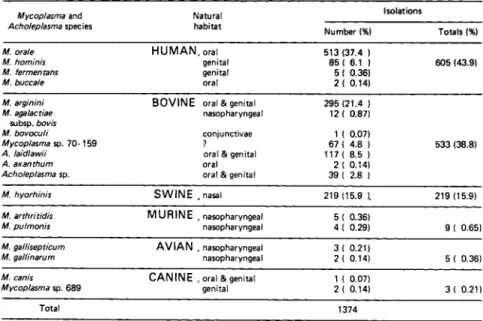
B, Identification and Sources of Mycoplasma Contamination Table III summarizes our findings on the identification and prevalence of mycoplasma species isolated from contaminated cell cultures. To date, of 11,000 primary and continuous cell culture specimens examined, 1374 mycoplasmas have been isolated and iden- tified. About 8 percent of the cells had mixed contamination with two or more species. Ninety nine percent of the mycoplasma contaminants were either human, bovine, or swine species (Barile et al., 1973).
1. Human Sources
The human oral and genital mycoplasma species represent the largest group of contaminants (Table III), and therefore, the laboratory personnel are a major source of contamination. Mouth pipetting is the major vehicle of human oral contamination. Sa- liva is introduced into the cell culture: the antibiotics present in the medium destroy the bacteria, but do not inhibit mycoplasma.
In cells grown without antibiotics, the bacteria destroy the cells and the culture is discarded. Thus, antibiotics tend to mask mycoplasma contamination and should be avoided. Contamina- tion by human genital strains is probably due to inadequate sterile procedure.
2. Bovine Sources
The bovine species of mycoplasmas are the second major group of contaminants. At least eight distinct bovine species have been isolated from contaminated cell cultures (Table III).
3. Contaminated Bovine Sera
Barile and Kern 0-9711 have shown that commercial bovine se^
rum is frequently contaminated with, mycoplasmas and that it is the major source of bovine mycoplasma contamination of cell cul^
tures. In these studies (Barile et al,, 1973), 285 mycoplasmas
TABLE III Identification and Speciation of Mycoplasmas Isolated from Contaminated Cell Cultures*
Mycoplasma and
Acholeplasma species Natural habitat
Isolations
Number (%) Totals (%)
M. orale M. hominis M. fermen tans M. buccale
H U M A N , oral genital genital oral
513 (37.4 ) 85 ( 6.1 ) 5 ( 0.36) 2 ( 0.14)
605 (43.9)
M. arginini M. agalactiae
subsp. bovis M. bovoculi Mycoplasma sp. 7 0 - 1 5 9 A. laidlawii
A. axanthum Acholeplasma sp.
B O V I N E oral & genital nasopharyngeal conjunctivae
? oral & genital oral oral & genital
295 (21.4 ) 12 ( 0.87)
1 ( 0.07) 67 ( 4.8 ) 117 ( 8.5 ) 2 ( 0.14) 39 ( 2.8 )
533 (38.8)
M. hyorhinis S W I N E .nasal 219 (15.9 ). 219 (15.9)
M. arthritidis
M. pulmonis M U R I N E .nasopharyngeal nasopharyngeal
5 ( 0.36)
4 ( 0.29) 9 ( 0.65)
M. gallisepticum
M. ga/linarum A V I A N , nasopharyngeal nasopharyngeal
3 ( 0.21)
2< 0.14) 5 ( 0.36)
M. canis
Mycoplasma sp. 689 C A N I N E , oral & genital genital
1 ( 0.07)
2 ( 0.14) 3 ( 0.21)
Total 1374
* F r o m : Barile et.al., 1973
TABLE IV Isolation of Mycoplasmas from Contaminated Commercial Bovine S e r aa
S e r ab
Source Origin Isolations0
Supplier Supplier Manufacturer Totals
Calf
Fetal bovine Fetal bovine
22/55 159/438 104/395 285/888
aFrom: Barile et al, C1973K
^Supplier denotes unprocessed sublots of raw sera and manufac'- turer denotes final lots of sera produced for market,
cNumber positive/number tested. Lots were not selected at ran^
dorn and data do not reflect incidence of contamination.
296 M I C H A L E F. B A R I L E
TABLE V Mycoplasma, Contamination of
Commercial Bovine Sera; Identification & Speciationt
Species Natural
Habitat
Numbers of
Isolations Percent
M.arginini bovine: oral & genital 6 5 33.7
M.alkalescans bovine: oral & genital 6 3.1
M. agalactiae bovine, genital 5 2.6
subsp. bovis
M.bovoculi bovine, conjunctivae 3 1.6
Mycoplasma sp 7 0 - 1 5 9 * (cell cultures) 3 1.6
M.hyorhinis swine, nasal 1 0.5
A. laid/a wii bovine, genital 8 7 45.1
A.axanthum (cell cultures) 1 0.5
Acholeplasma sp* ? 2 2 11.4
* Distinct unspeciated serotypes unrelated to established species, t From: Barile e t al, (1973)
were isolated from 888 lots of raw, unprocessed sera obtained from suppliers or from final lots of sera produced for market
(Table I V ) . Successful isolations were made with our most sensi- tive large volume broth culture procedure (Barile and Kern, 1971).
4. Mycoplasma Species Isolated from Contaminated Bovine Sera
The bovine mycoplasmas isolated from contaminated bovine sera were similar or identical to the bovine species isolated from contaminated cell cultures (Table V) and support the findings that contaminated bovine sera are the major source of bovine mycoplasma contamination. In addition, one mycoplasma was a swine strain of Mycoplasma hyorhinis, suggesting that bovine serum may also be a source of swine mycoplasma contamination (Barile et al., 1973).
5. $&£ne Sources
The third major group of contaminants are the swine myco- plasmas (Table ΙΙΓ1, Although trypsin has been incriminated, all attempts to grow out mycoplasmas from commercial trypsin have failed. Because swine and cattle are frequently processed through the same abattoir, bovine sera may also become contaminated with swine mycoplasmas during collection and manufacture.
6. Original Tissue Sources
Occasionally primary cell cultures are contaminated with my- coplasmas derived from murine, avian, and canine origin (Table III). Because these mycoplasmas were only isolated from primary cells derived from murine, avian and canine tissues, and were host specific, the probable source of these contaminants were the original tissues used to produce the primary cell culture (Barile, 1973a; Barile et al., 1973).
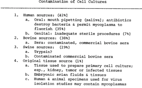
7. Sources of Mycoplasma Contamination
To summarize, the major sources of contamination are of human origin - the investigator, his colleagues and his environment; of bovine origin - primarily contaminated bovine sera; and of swine origin (Table V I ) . About one percent of the tissues used to pro- duce primary cell cultures are also contaminated (Barile et al., 1973). In addition, specimens collected for virus isolation studies may also contain mycoplasmas, and thereby contaminate the cell culture. Moreover, many of these mycoplasma contaminants have been mistaken for viruses because they produce cytopathic effects, are filterable, can hemadsorb, and are neutralized by antisera (Barile, 19731.
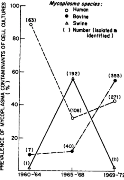
C. Prevalence of Contamination
The amount of contamination from human, bovine and swine sources was determined for the period 1960 through 1972 (Fig. 1 ) .
298 M I C H A E L F. BARILE
TABLE V I Sources of Mycoplasma Contamination of Cell Cultures
1. Human sources: C42%î
a. Oral: mouth, pipetting C s a l i v a î; antibiotics destroy bacteria & permit mycoplasma to fluorish (35%)
b. Genital: inadequate sterile procedures (7%) 2. Bovine sources: (38%)
a. Sera: contaminated, commercial bovine sera 3. Swine sources: (19%)
a. Trypsin?
b. Contaminated commercial bovine sera 4. Original tissue source (1%)
a. Tissue used to prepare primary cell culture;
esp.f kidney, tumor or infected tissues b. Embryonic avian fluids & tissues
c. Human & animal specimens used for virus isolation studies may contain mycoplasmas
The major source of contamination in the early I960's was the hu- man oral strains; in the mid-19601s it was the swine strains, and in the early 1970's it was the bovine strains (Barile, 1973a).
Today, the most prevalent contaminants (55%) are the human spe- cies, followed by the bovine and swine species which cause an equal amount of contamination.
D. Spread of Contamination
In addition to the original sin, mycoplasma contamination can spread from cell to cell by aerosols of contaminated cell culture material or by contaminated saliva or nasal secretions, genital and intestinal droppings of man and animals. Contamination can spread also by contaminated virus pools, antisera, and other re- agents, or by contaminated equipment commonly used in cell cul^
ture studies ( B a r i l e , 1973a,b).
Mycoplasma species:
O Human
• Bovine Δ Swine
( ) Number (isolated a identified )
1 9 6 0 - ' 6 4 1 9 6 5 - ' 6 8 1 9 6 9J7 2
FIGURE 1 The prevalence of mycoplasma contamination from human, bovine and swine sources during the period 1960-1972. The numbers in parentheses are the numbers of mycoplasmas isolated from contaminated cell cultures for each time period.
E. Cell Susceptibility to Mycoplasma Contamination
Cell cultures derived from all tissues, organs and animals examined were found to be contaminated, including cells from mam- malian, avian, reptilian, fish, insect and plant origin. All types of cells were found subject to contamination, including cells from normal, infected or neoplastic tissues, primary or continuous; diploid, heteroploid, fibroblastic and epithelial cells, and cells grown in monolayer or suspension. Most myco- plasma contaminants produce titers in most cell cultures which range from 1 05 to 1 09 logs per ml of medium fluid (Barile, 1973a).
300 M I C H A E L F. B A R I L E
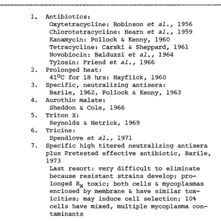
TABLE yil Procedures Used to Eliminate Mycoplasmas from Contaminated Cell Cultures
1. Antibiotics :
Oxytetracycline: Robinson et al,, 1956 Chlorotetracycline: Hearn et al., 1959 Kanamycin: Pollock & Kenny, 1960 Tetracycline: Carski & Sheppard, 1961 Novobiocin: Balduzzi et al., 1964 Tylosin: Friend et al., 1966 2. Prolonged heat:
41°C for 18 hrs: Hayflick, 1960 3. Specific, neutralizing antisera:
Barile, 1962, Pollock & Kenny, 1963 4. Aurothio malate:
Sheddon & Cole, 1966 5. Triton X:
Reynolds & Hetrick, 1969 6. Tricine:
Spendlove et al., 1971
7. Specific high titered neutralizing antisera plus Pretested effective antibiotic, Barile, 1973
Last resort: very difficult to eliminate because resistant strains develop; pro- longed Rjç toxic; both cells & mycoplasmas enclosed by membrane & have similar tox- icities; may induce cell selection; 10%
cells have mixed, multiple mycoplasma con- taminants
F. Eliminating Mycoplasma Contamination
Because mycoplasma contamination is a common event, we have maintained a continuing study to examine and to develop procedures for eliminating mycoplasmas from a contaminated cell. It has been our experience that there is no single, fail-safe procedure for curing cells (Table VII; Barile, 1973aî, Not only is it very dif- ficult to eliminate mycoplasmas, but it is also very time^consum^
ing. Prevention is much more effective than is elimination of contamination. However, if the cell culture is irreplaceable, we recommend the combined use of specific, high^titered neutralizing antisera and a pretested effective antibiotic.
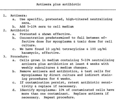
Antisera plus antibiotic
1. Antisera:
a. Use specific, pretested, high^titered neutralizing sera.
b. Add 5~10% sera to cell medium 2. Antibiotic
a. Pretested & shown effective.
b. Concentration predetermined to fall between ef- fective dose for mycoplasma & toxic dose for cell culture.
c. We have found 10 pg/ml tetracylcine & 100 yg/ml kanamycin, effective.
3. Procedure :
a. Cells grown in medium containing 5-10% neutralizing antisera plus antibiotics at least 4 weeks with weekly subcultures & medium changes.
b. Remove antisera and antibiotics, & test cells for mycoplasmas by direct culture and indirect stain- ing procedures for 4 weeks.
c. If contamination persist, retest antibiotic sensi- tivity & replace if necessary.
d. Identify mycoplasma: 10% of contaminated cells have more than one contaminant. Replace antisera if necessary. Repeat procedure.
In brief, 5 to 10 percent of antisera and the pretested anti- biotic are added to cell culture medium and the cells are grown for four weeks with weekly subcultures and medium changes. The specific antisera replace the serum component of the medium.
After four weeks, the antibiotic and antisera are removed and the cells are examined for mycoplasmas for an additional four weeks.
If contamination persists, the mycoplasma contaminant is retested for antibiotic sensitivity and for identity, because 10 percent of the cell cultures have mixed contamination (Table VIII, Barile, 1973a,bl.
TABLE VIZI Recommended Procedure for Eliminating Mycoplasmas from Contaminated Cell Cultures
302 M I C H A E L F. B A R I L E
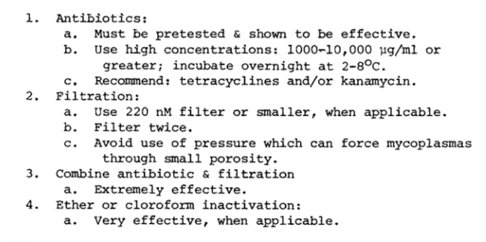
TABLE IX Procedures Used to Eliminate Mycoplasmas from Contaminated Virus Pools
1. Antibiotics:
a. Must be pretested & shown to be effective.
b. Use high concentrations: 1000»~10,000 yg/ml or greater; incubate overnight at 2-8°C.
c. Recommend: tetracyclines and/or kanamycin.
2. Filtration:
a. Use 220 nM filter or smaller, when applicable.
b. Filter twice.
c. Avoid use of pressure which can force mycoplasmas through small porosity.
3. Combine antibiotic & filtration a. Extremely effective.
4. Ether or cloroform inactivation:
a. Very effective, when applicable.
1. Eliminating Mycoplasma from Contaminated Virus Pools In contrast, mycoplasmas can readily be eliminated from con- taminated virus pools by the combined use of pretested effective antibiotics and gentle filtration through a 220 nM filter twice.
In addition, because all mycoplasmas are very sensitive to ether and chloroform, these reagents are extremely effective when ap- plicable (Table I X ) .
G. Preventing Mycoplasma Contamination
Contamination can be effectively prevented by using good, basic quality control procedures. Mycoplasma-free cell cultures of early passage should be stored frozen in a large number of vials. During study, the cells are monitored, and when found con- taminated, discarded immediately. Preventive measures used suc- cessfully for reducing the risk of contamination are based on either eliminating the sources or on aborting the spread of con- tamination (Barile 1973afbl. For example, primary cell cultures are rarely contaminated and should be used whenever possible;
avoid mouth-pipetting and use rigid sterile procedures. Bovine
TABLE X P r e y e n t i y e M e a s u r e s f o r R e d u c i n g
t h e R i s k o f M y c o p l a s m a , C o n t a m i n a t i o n o f C e l l C u l t u r e s
1« Use primary cells when feasible.
2. Eliminate mouth pipetting.
3. Use rigid sterile procedures.
4. Treat commercial* bovine sera, trypsin, etc.
5. Filter the air servicing work-room.
6. Use laminar flow hood for work-area.
7. Decontaminate work-area daily.
8. Monitor and treat working-reagents.
9. Discard contaminated cells immediately.
10. Store mycoplasma-free cells for future use, 11. Good lucki i
sera and other reagents are treated by gentle filtration twice through a 220 nanometer filter and/or heat inactivated at 56°C for 45 minutes C S e e G . l ) . Laminar-flow hoods are used and the working area and rooms are decontaminated routinely (Table χ ) .
1. Pretesting and Pretreating Bovine Sera
Preventative measures have been used successfully to reduce the amount of mycoplasma contamination of cell cultures maintained in our laboratories. There has been no contamination with bovine mycoplasmas since we began pretesting and pretreating our sup- plies of commercial bovine sera as follows: several large lots of bovine sera are examined for mycoplasma contamination by the large volume broth culture procedure (Barile and Kern, 1971). If myco- plasmas do not grow, a large supply of the tested serum lot is purchased and stored frozen in 100 ml volumes. Cell cultures grown in medium with the serum lot are monitored weekly for myco- plasmas. If bovine mycoplasmas are isolated from these cell cul- tures, the lot of serum is replaced with, another pretested myco- plasmar»free lot. Every bottle of bovine serum is routinely pre~
treated by heat^inactivation at 56°C for 45 minutes and/or by filtration twice through, a 220 nM filter, r f heat^inactivation is
304 M I C H A E L F. B A R I L E
unsuitable for growth, of a particular cell culture, we recommend that the complete medium containing serum be filtered twice before use. Since high, pressure filtration can force the plastic myco-- plasma cells through the filter, high pressures should not be used. We have found that filtering complete medium containing 5 to 20% serum through a 220 nM filter can be accomplished without excess pressure, and that it can effectively remove contaminating mycoplasmas.
II. MYCOPLASMA-CELL-VIRUS INTERACTIONS
A. Mycoplasma-Cell Interactions
We are frequently asked: Can the investigator live with myco- plasma contamination, i.e., Does mycoplasma contamination affect the results of my study? The answer can be determined only ex- perimentally by examining the effect of the particular mycoplasma contaminant on the test system under study, and because myco- plasmas are known to affect cell cultures in many ways (Barile, 1973a), the investigator is obliged to make the effort and to rule out a mycoplasma effect. Mycoplasmas can produce cytopathic effects, inhibit lymphocyte transformation, cause chromosomal aberrations, mimic viruses, and either decrease or increase virus yields. Some mycoplasmas, especially the pathogens, can produce hemolysis, adsorb to red blood cells, as well as many other types of cells and hamagglutinate (Barile, 1973a). Some can alter the antigenicity of the cell membrane resulting in the development of autoantibodies to lung and brain tissues, as well as cold agglu- tinins during infection.
Β. Hemadsorption
Figure 2 illustrates hemadsorption of sheep red blood cells to colonies of Mycoplasma, pulmonis. In addition, sheep or guinea pig red blood cells can also adsorb to mycoplasmas in infected or
FIGURE 2 Illustrates hemadsorption of sheep red blood cells onto a colony of M. pulmonis,
contaminated cell cultures. The binding site is probably sialic acid because treating red blood cells with neuraminidase inhibits mycoplasma hemagglutination. Therefore, the investigator inter- ested in hemagglutinating agents must be cognizant of mycoplasma contamination.
C. Mycoplasma Attachment to Cell Membranes
A number of mycoplasmas have been seen closely associated with or attached to the plasma membranes of animal cells using either histologic staining, immunofluorescence or electron microscopy
CBarile, 1973aî.
M I C H A L E F. B A R I L E
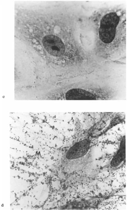
FIGURE 3 Illustrates attachment of M. hyorhinis to a primary rabbit kidney cell culture at low (b) and high (d) magnifications stained by the Giesma procedure. Note multiple mycoplasmas (M) per infected cell (3d). Compare mycoplasma infected (b and d) to mycoplasma-free (a and c) cell cultures.
308 M I C H A E L F. B A R I L E
FIGURE 4 Illustrates a positive homologous immunofluorescence reaction in a M. hominis contaminated HEp-2 cell culture.
1. Histologic Staining
Figure 3 illustrates attachment of Mycoplasma hyorhinis, a common contaminant, to a HEp-2 cell culture. The mycoplasmas ap- pear as pleomorphic bodies attached to the infected cell membrane.
Note the number of mycoplasmas per infected cell and the distor- tion of the cell morphology. Since mycoplasmas attach to mem- branes, ordinary histologic staining procedures provide one use- ful, simple, indirect procedure for detecting mycoplasmas in con- taminated cells (Barile, 1973a).
2. Immunofluorescence
Figure 4 illustrates a positive homologous immunofluorescent reaction with a mycoplasma contaminated HEp-2 cell culture. The fluorescent bodies appear very closely associated with the cell membrane. Rhodamine is used as a counterstain. We have shown that immunofluorescence is an extremely effective procedure for
DETECTION OF M.hvorhinis IN CELL CULTURE BY FLUORESCENT ANTIBODY AND
CULTURAL PROCEDURES
H 100
4 6 3 D A Y S O F I N C U B A T I O N
Ο -π Ο m r-
Γ "
C/)
23
5
Ο
FIGURE 5 Relative sensitivity for detecting mycoplasmas by comparing isolation and direct culture procedures with indirect fluorescent antibody procedures in M. hyorhinis contamination of cell cultures.
detecting mycoplasma in chronically infected or contaminated cell cultures (Barile et al., 1962; Barile and Del Giudice, 1972).
3. Sensitivity of Immunofluorescence
The relative sensitivity of the direct culture and the in- direct fluorescent antibody procedures for detecting the presence of mycoplasmas in contaminated cell cultures is shown in Fig. 5.
Cells were inoculated with small number of mycoplasmas and then tested daily by both procedures for 7 days. Whereas, very small numbers of mycoplasmas were isolated and detected by direct cul- ture procedures, the fluorescent antibody reactions were positive only when the cells became chronically infected with large num- bers, 7 logs/ml or greater, of mycoplasmas. In our studies, pri- mary isolation and direct culture procedures were shown to be more sensitive than indirect procedures for detecting mycoplasma con-- tamination. Because most contaminated cell cultures become chron- ically infected with, mycoplasmas, immunofluorescence is a useful
310 M I C H A E L F. B A R I L E
FIGURE 6 Illustrates a thin-section preparation of a M. pul- monis infected HeLa cell culture. Note multiple mycoplasmas (M) attached to tissue cell membrane.
indirect procedure and is effective for detecting contamination with the non-cultivable strains of Mycoplasma hyorhinis. These contaminants become cell-adapted, lose their ability to grow on artificial broth-agar medium and cannot be isolated using avail- able culture procedures CHopps et al., 1973).
4. Electron Microscopy
Figure 6 illustrates a thin-section preparation of a myco^
plasma contaminated HeLa cell culture. Occasionally, tfie electron microscopist is tha first to detect mycoplasma contamination.
Note the number of mycoplasmas per infected cell and their close
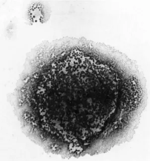
FIGURE 7 Illustrates the ultrastructure of M. pulmonis in in- fected HeLa cell culture. Note and compare the three-layered unit membranes (M) of the mycoplasma and HeLa cell.
association to the HeLa cell membrane. Some of the pathogenic species, such as M. pneumoniae, M. gallisepticum, and M. pulmonis, have we11-organized terminal apparati used for attachment to tis- sue cell membranes. For example. Mycoplasma pneumoniae has a terminal spike (Collier, 1972), which permits the pathogen to an- chor firmly to the infected tracheal cell membranes and to resist removal by the coughing and/or ciliary mechanisms. The ultras- structure of the mycoplasma cell is illustrated in Fig. 7, Unlike bacteria, mycoplasmas are devoid of cell w a l l and are enclosed by a typical threer-layered unit membrane. Accordingly, mycoplasmas are susceptible to the same physical and chemical stresses which are injurious for the membranes of cell cultures (Barile, 1973)
312 M I C H A E L F . B A R I L E
and thereby make elimination of mycoplasma contamination a chal- lenging problem. Moreover, it is not surprising then, that myco^
plasmas have now become very useful tools and a popular model for studying the chemical structure and function of membranes (Razin, 1975; Razin and Rottem, 1975].
D. Arginine Dehydrolase Activity
Another important property of mycoplasmas is their ability to break down arginine for energy. Schimke and Barile (1963) have shown that certain mycoplasmas catabolize arginine by a three- enzyme dehydrolase system with release of ATP. Since animal cells do not have this pathway, detection of arginine deiminase activity in cell cultures is a rapid chemical method for detecting myco- plasma contamination (Barile and Schimke, 1963).
The effect of arginine on the growth of the arginine-utilizing Mycoplasma arginini is shown in Fig. 8. Glucose and glutamine had no effect on growth, whereas the addition of arginine produced a marked stimulation of growth. Because mycoplasma contaminants require arginine for energy, they rapidly deplete arginine from the medium, depriving the cell culture of an essential amino acid, and producing profound effects on cell function and on virus prop- agation.
E. Covert Mycoplasma Contamination
Mycoplasma contamination may appear insidious and may go un- detected because: (1) even high titers, 1 07 to 1 08 logs/ml of mycoplasmas do not produce the overt turbid growth commonly asso- ciated with bacterial and fungal contamination; C 2 ) the initial cytopathic effects, and especially those produced by the arginine users, can be minimal and C31 mycoplasma contaminants compete for amino acids, sugars, pyrimidines and other nutrients causing nu^
tritional and "toxic" cellular changes,
HOURS OF INCUBATION
FIGURE 8 Illustrates the effect of arginine on the growth of M. arginini, an arginine-utilizing contaminant. The addition of arginine to the original broth (Δ) or added during culture (k) produced a marked stimulation of growth. The addition of glucose
(·) and glutamine (o) to broth had no effect.
These "toxic" changes can be reversed by changing the medium, providing the investigator with a false sense of security. To a large extent, the biochemical activity of the contaminant prede- termines the cytopathic effect (CPE). The CPE depends on whether the mycoplasma utilizes arginine or glucose for energy. Some mycoplasmas deplete arginine and affect cell function, but may not produce a destructive CPE.
F. Cytopathic Effects
A number of mycoplasmas can produce very severe CPE, charac- terized initially by the development of microcolonies followed by microlesions and small foci of necrosis (Barile, 1973al, As a result, many mycoplasmas, such, as H% gallisepticum (Fig. 91, pro- duce plaque formation, a property commonly associated with, virus- es. As the chronic mycoplasma infection or contamination pro- ceeds, the CPE becomes much, more generalized, and large areas of
314 M I C H A E L F. B A R I L E
FIGURE 9 Plaque formation in primary chick cell cultures produced by M. gallisepticum.
the monolayer are destroyed. Because the mycoplasma can generate large amounts of acid, the cells detach from the glass, producing a macroscopic V-shaped CPE within one to two weeks (Fig. 1 0 ) . Re- peated media changes and stronger buffering systems can delay or reduce the CPE and contamination can go undetected. On the other hand, the macroscopic CPE can be exploited as a means of detecting contamination by mycoplasma fermenters (Barile, 1973a). In brief, tenfold dilutions of the suspect "pour-off" or spent tissue cul- ture medium is inoculated into primary rabbit, monkey, or chick embryo cell cultures, incubated for 2 to 4 weeks without feeding and observed for macroscopic CPE (Fig. 1 0 ) . Uninoculated cell cultures are maintained in the same manner and serve as negative controls.
FIGURE 10 Illustrates the typical inverted V-shaped, macro- scopic cytopathic effect produced by mycoplasma fermenters. Note the progression of cell destruction in a M. hyorhinis-infected L-cell culture stained with one-tenth diluted Gentian Violet stain.
G. Effects of Mycoplasma Contamination
However insidious mycoplasma contamination may appear to the unsuspecting investigator, mycoplasma can produce severe micro- scopic and macroscopic CPE, including plaque formation. They can also affect cells in many other ways (Barile, 1973a). Some myco- plasma inhibit lymphocyte blastformation and some cause chromo-
somal aberrations. Mycoplasma deplete nutrients from medium and can affect or influence protein and/or nucleic acid synthesis.
Some mycoplasmas alter the antigenicity of cell membranes, causing the production of autoantibodies, and therefore, mycoplasma have been incriminated as possible causative candidates for various auto-immune diseases (Barile, 19651. In addition, certain myco- plasmas can either decrease or increase virus yields.
316 M I C H A E L F. B A R I L E
TABLE XX The Effect of Mycoplasma,s on
Stimulation of Human Lymphocytes to Phytohemagglutinin*
Lymphocytes Inoculated with Mycoplasmas
Total 3H-Thymidine Uptake cpm/culture (χ 103)
Species Arginine
Users None
Stimulant P H A None
Broth medium
8.7 11.1
331.6
Μ. ο rale M.arthritidis M. hominis
+ +
+ 0.55
1.5 2.1 18.7
M.pneumoniae
M. bovigenitalium
M. can is
— 22.5 396.2
407.0 498.3 M.hominis suppressed 3H thymidine uptake.
M.hominis and other arginine users suppressed P H A stimulation. The non-arginine utilizing, fermenting mycoplasmas had no effect, on PHA stimulation.
* Barile & Leventhal (1968)
H. Effect on Lymphocyte Blastformation
Certain mycoplasmas can inhibit lymphocyte blastformation.
In our studies, lymphocyte cultures were infected with either one of three fermenters or with one of three arginine users, in- cluding Mycoplasma orale, M. arthritidis and M. hominis (Table XI).
The data represent mean values from 10 patients and indicate that the arginine-using mycoplasmas markedly inhibited thymidine uptake of unstimulated cells and also inhibited the stimulatory effect of phytohemagglutinin (PHA). The mycoplasma fermenters, which do not utilize arginine for energy, had either no effect or a stimulatory effect. In subsequent studies, we examined the ef- fect of arginine depletion on the inhibition of PHA by M. hominis.
Whereas, Mt hominis inhibited PHA stimulation (Table X I I ) , feed- ing the cells with complete medium or supplementation with addi- tional arginine reversed the Mt hominis effect, On the other hand, arginine-^free medium did not reverse the effect (Barile and Leventhal, 19681,
TABLE XII The Effect of Arginine-Utilizing Mycoplasmas on Stimulation of
Human Lymphocytes to Phytohemaglutininf
After 3 Days, Cells Were Treated as Follows:
Total Tritiated Thymidine Uptake (day 5) cpm/Culture ( x 1 03) After 3 Days,
Cells Were Treated as Follows:
Mycoplasma- Free
M. hominis*
Infected
&
Control PHA PHA
M. hominis*
Infected
&
PHA
None 15.2 446.0 8.0
Replace with complete
Eagle's medium (MEM) 5.5 346.4 477.0 Replace with arginine-
Free EMEM
3.6 145.5 4.6
Add 10mM arginine 9.4 128.4 237.5
*M. arginine, M. orale & other arginine users showed similar effects.
Mycoplasma fermenters (non-arginine users) had no effect, t Barile & Levé thai (1968)
I. Effect of Mycoplasmas on Virus Yields
We have shown that certain mycoplasmas can influence virus propagation. The effect produced is dependent on the particular mycoplasma, virus and cell cultures used. Whereas some myco- plasmas have no detectable effect, others were shown to either in- crease or decrease virus yields. Several mechanisms have been established. For example, the arginine-using mycoplasmas decrease yields of arginine requiring DNA viruses by depleting arginine from the medium. On the other hand, some mycoplasmas increase the yields of viruses by inhibiting interferon induction and/or acti- vity (Barile, 1973a, Singer et al,, 1973).
1 , Decreased Vaccinia Virus Fields by Arginine Depletion Singer et al, 0-97QI have shown that arginine^utilizing myco- plasmas can decrease the titer of vaccinia virus (an arginine- requiring DNA virus! from Ι Ο1 to 1 02 logs (Table ΧΙΓΙΙ.
318 M I C H A E L F. B A R I L E
TABLE XIII Vaccinia, Virus Yields from Mycoplasma^Free & Mycoplasma Infected Cells with
and without Additional Argininet
Mycoplasma infected
Arginine added
H A cells
L o g10
differ from control
H E F cells
L o g10
differ from control
Control No 5.5 5.5
(none) Yes 5.8 + 0.3 5.8 + 0.3
M.arginini N o 3.3 -2.2 4.1 - 1.4
infected Yes 5.2 -0.3 5.2 -0.3
M.hyorhinis
infected No 5.5 to 5.5 ± 0
HA = human amnion cells; HEF = human epithelial cells Additional 0.4mM arginine v/v 0.1 mM arginine Data express mean values of several experiments t Singer etal 1972
Addition of arginine reverses the effect. Mycoplasma, hyorhinis Ca fermenter which does not require arginine for energy) had no effect.
2. Decreased Herpesvirus Yields by Arginine Depletion
Manischewitz et al. (1975) showed that the arginine-utilizing mycoplasmas can also decrease titers of Herpes simplex virus
(HSV), another arginine-requiring DNA virus (Fig. 1 1 ) . In these studies, the titers of HSV grown in cells contaminated with M.
arginini were 1 02 to 1 03 logs lower than titers obtained with mycoplasma-free cell cultures. M. hyorhinis, a fermenter, had no effect. Addition of arginine reversed the effect. We have sug- gested that the M. arginini effect may provide a simple procedure to establish, the arginine requirement for other DNA viruses
CBarile, 19731.
T H E EFFECT OF A N A R G I N I N E U T I L I Z I N G M Y C O P L A S M A * O N G R O W T H OF A N
TIME (hours)
* Other arginine-user showed same effect.
Non-arginine-using, (fermentors) had no effect.
# * S i m i l a r effects for other DNA arginine requiring viruses, Adenovirus, Vaccinia, SV-40.
Adding arginine to medium reversed the effect.
FIGURE 11 Illustrates the growth of Eerpes simplex virus in mycoplasma-free (·) and M. arginini (k) and M. hyorhinis (Ώ) in- fected Vero cell cultures. M. arginini, an arginine user, reduced HSV titers and the addition of arginine reversed the mycoplasma effect.
3. Reduction of Viral Plaques
Singer et al. C1972) showed that mycoplasmas can also signif- icantly reduce the morphology and number of Semliki Forest virus plaques. Manischewitz et al. (1975) showed that M. arginini can also reduce the number and size of HSV plaques. The reduction in number and size of HSV plaques was reversed by the addition of arginine. A. laidlawii, a non-arginine-utilizing mycoplasma, had no effect. To summarize, the arginine-utilizing mycoplasmas can reduce the number and plaque size of arginine-requiring DNA vi- ruses, and additional arginine can reverse the effect.
4. Increasing Virus Yields by Inhibiting Interferon Induction Singer et al. CL969aÎ have shown that mycoplasmas can also in^
crease virus yields, These studies were initiated because two of the same cultures maintained in parallel gave different titers of
320 M I C H A E L F. B A R I L E
TABLE Xiy Increased Semliki Forest Virus Yields from M, Hjyorhinis Infected Hamster Cellst
*Titer SFV L o9i q difference % Cells l o9 l 0 from control control
Control tCa) 6.4 Cb) 6.4 Cc) 6.4
M. hyorhinis (a) 7.3 (a) +0.9 (a) 790 infected Cb) 7.3 (b) +0.9 Cb) 790 (c) 7.1 (c) +0.7 Cc) 500
*PFU/0.1 ml.
fa, b , and c represent three separate experiments on three lots of cells.
+Singer et al., 1969a
Semliki Forest virus CSFV). Subsequent studies showed that the cell culture producing the higher titers was contaminated with Mycoplasma hyorhinis CTable XIV). Increased SFV titers were seen only when low input was used CTable X V ) . With low SFV input, a small number of cells are infected, leaving most of the cultured cells available to accept the interferon generated, and to develop resistance to viral infection. Thus, even though mycoplasmas in- hibit interferon induction, several cycles of virus multiplication are required to achieve amplification and to show a significant increase in virus yield. With high virus input, most or all of the cells are infected initially, and consequently the effect of mycoplasmas on interferon inhibition and on cell resistance cannot be demonstrated CSinger et al., 1969b).
5. Inhibiting Virus Induction of Interferon
Although, mycoplasmas do not directly^ induce interferon, they can inhibit interferon activity induced by either a virus, such as Vesicular Stomatitis Virus CVSVl, or by synthetic RNA copolymers, m the VSV studies, mycoplasmar-free and mycoplasma contaminated
T A B L E x y V S Y R e p l i c a t i o n i n C o n t r o l a n d M, Arginini I n f e c t e d H a m s t e r E m b r y o F i b r o b l a s t s *
Multiplicity of Infection 0.00003
Multiplicity of Infection 0.03
Hours Post
V S V TITER
(Log10/0.1 ml.) L o gl 0 Diff.
from Control
V S V TITER
(Logl0/0.1 ml.) L o g1 0 Diff.
from Control V S V
Inoc. Control Cultures
M. Arginini Inf. Cultures
L o gl 0 Diff.
from
Control Control Cultures
M. Arginini Inf. Cultures
L o g1 0 Diff.
from Control
4 0 0 0 2.2 2.2 0
6 0.9 0.9 0 4.4 4.3 -.1
8 1.8 2.1 + .3 5.2 5.4 + .2
12 3.3 3.9 + .6 6.4 6.5 + .1
24 5.9 7.1 + 1.2 N.D.* N.D. N.D.
* N.D. = not done t Singer eta/ 1969b
cells were inoculated with high input of VSV and then assayed one and two days later for both virus and interferon titers. Whereas mycoplasma did not affect the virus titers, they did inhibit in- duction of interferon reducing titers from a high of 1:256 to less than 1:4 (Table X V I ) . Additional arginine had no effect (Singer et al., 1969).
6. Inhibiting Poly I'C Induction of Interferon
Singer et al. (1969a) reported similar effects with the syn- thetic double-stranded Pöly I-C, a potent inducer of interferon.
The addition of Poly I»C stimulated interferon production and pro- duced cell resistance with a significant decrease in virus yields in the mycoplasma^free cells. On the other hand, the mycoplasmas in contaminated cells inhibited interferon production and thereby nullified the Poly I^C effect, resulting in titers similar to the control cells without Poly^ I«C CTable XVIIl.
322 M I C H A E L F. B A R I L E
TABLE XVI i n d u c t i o n o f I n t e r f e r o n b y S e m l i k i F o r e s t V i r u s i n M y c o p l a s m a , F r e e
a n d M y c o p l a s m a I n f e c t e d H a m s t e r C e l l s Days Following
Mycoplasma Infection
Cells*
*Titer SFV ( L o g1 0) After
Interferon Induction
Titer of Interferon
Induced
at bt a b
Control 7.6 7.3 1:128 1:64
1
M. arginini
Infected 7.4 7.3 1:8 1:8
M. hyorhinis
Infected 7.6 7.3 < 1:4 < 1:4
Control 7.3 7.5 1:256 1:64
2
M. arginini
Infected 7.5 7.7 1:8 1:4
M. hyorhinis
Infected 7.5 7.3 < 1:4 < 1:4
* PFU/0.1 ml. T a and b represent two separate experiments on two lots of cells, t From: Singer er. al, (1969)
TABLE XVII Induction of Viral Resistance by Poly I-Poly C in Mycoplasma-Free and Mycoplasma Infected Hamster
Cells Using Vesicular Stomatitis Virus
Cells Infected with Mycoplasmas
Poly I PolyC Added
VSV Titers L o g10
L o g10 differ from control
Control No 6.8
( my cop lasma - free) Yes 5.2 - 1.6
M.arginini- infected
No 7.1
M.arginini- infected
Yes 6.7 - 0 . 4
M. hyorhinis - infected No 7.0
M. hyorhinis - infected
Yes 6.9 - 0 . 1
* 100 μ/ml Poly I - Poly C with 300μ/ ml neomycin
TABLE .xyiir Sensitiyity of Mycoplasma Infected Cells to Exogenously Applied Interferont
Interferon titer
Cells a* b c
Control 1:32 1:32 1:32
M. arginini
infected 1:8 1:4 1:4
Af. hyorhinis
infected 1:4 1:4 1:4
*a, b, and c represent three different experiments on three separate cell lots,
fSinger et al., 1969a
7. Effect on Interferon Activity
Singer et al. (1969a) also showed that mycoplasmas can affect interferon activity. In these studies, a standard preparation of hamster interferon was assayed for activity in mycoplasma-free and mycoplasma-contaminated hamster cells (Table XVIII). The findings show that mycoplasmas decreased interferon activity 4- to 8-fold.
Therefore, mycoplasmas render cell cultures less sensitive to exogenously supplied interferon, and, consequently, contamination can affect the results of interferon assays.
Mycoplasma contamination could be used to advantage. Inhibi- tion of interferon production and activity by mycoplasmas may be used to enhance virus yields and may play a useful role in the detection of latent viruses.
324 M I C H A E L F. B A R I L E
III. ISOLATION AND DETECTION OF MYCOPLASMAS
A. Detecting Cell Culture Contamination
We have used four basic approaches for primary isolation and detection of mycoplasmas CBarile, 1973a, 1974), including: C D a standard culture procedure for routine specimens, cell cultures, biopsy tissues, throat cultures; (2) a semi-solid broth culture procedure for screening cell cultures for bacterial, fungal or mycoplasmal contamination; (3) the large specimen-broth culture procedure for isolation of mycoplasmas from contaminated commer- cial bovine sera; and (4) the use of primary and continuous cell cultures for the isolation and detection of fastidious, pathogenic mycoplasmas, and the cell-adapted strains of M. hyorhinis.
B. Direct Culture Procedures; Isolation of Mycoplasmas Our standard medium is the Edward-Hayflick formula supple- mented with arginine and dextrose as energy sources, phenol red as a dye indicator, thymic DNA and vitamins CBarile, 1974). Cell cultures are examined as follows: one ml or one-tenth ml of cell suspension is inoculated into broth or onto agar media, respec- tively. Agar plates are inoculated in duplicate: one is incu- bated aerobically, and the other in a 5 percent carbon dioxide-95 percent nitrogen atmosphere shown to be a superior environment for primary isolation of most mycoplasma contaminants CBarile et al.,
1962; Barile and Schimke, 1963). The cultures are incubated at 36°C for several weeks and examined periodically for growth of mycoplasmas. Detailed culture procedures are published elsewhere
CBarile, 1973a, 1974; Barile et al., 1973).
C. Efficacy of Culture Procedures
The pretesting of culture media and the standardization of culture procedures can influence successful isolation and growth of mycoplasmas CTable XIXI. Because the quality of the medium
T A B L E XIX S t a n d a r d i z a t i o n o f C u l t u r e M e d i u m
I. Basal medium; pretest each, batch II. Horse serum: pretest for toxicity
1. non^inactivate: M, pneumoniae 2. inactivate (56°, 3 0f) M, arginini III. Fresh yeast extract
1. 10% for M. pneumoniae 2. 5% for M. arginini IV. L-arginine^HCI
1. >0,5% toxic for M, hyosynoviae V. Thallium acetate (1:2000)
1. toxic for Ureaplasmas Φ some mycoplasmas VI. Agar: pretest for toxicity
1. use purified Ionagar #2 or Noble agar 2. use optimal gel strength
(too firm inhibits colony formation) VII. pH optimal:
1. Thermoplasmas f pH 2-3 2. Ureaplasmas, pH 6,0 3. Mycoplasmas pH 7.2^7.8
4. Achoeplasma laidlawii, pH 8,5
Medium components: pretest each lot of each component for toxicity & growth promotion
Completed medium: pretest each batch for efficacy, using small numbers of fastidious mycoplasmas
components can vary, each lot of each component should be pre- tested for its toxicity and for its growth-promoting properties.
For example, different lots of horse serum vary in their growth- promoting properties and in their toxicity. Thus, each lot must be pretested. In addition, whereas 10 percent fresh yeast extract
stimulates growth of Mycoplasma pneumoniae, it may be toxic for certain strains of M. arginini which prefer 5% yeast extract.
Likewise, whereas arginine (0.2%) stimulates growth, too much arginine (>0.5%) can inhibit growth of certain mycoplasmas
(Barile, 19741. The agar can also be toxic. Purified ionagar No, 2 C Q x o i d l or Noble agar ( P i f c o l are superior products and should be used, Too much, agar can also inhibit colony formation and growth* In summary, there is no standard medium, but rather each medium must be standardized and each batch, mist be pretested for
326 M I C H A E L F. BARILE
toxicity and for growth_^promoting properties to assure successful isolation and propagation of mycoplasmas.
D. Optimal Culture Procedure
The selection of the medium and culture procedure is dependent on the particular need and the particular mycoplasma under study.
For example, the Ureaplasmas, spiroplasmas, and especially the thermoplasmas Cwhich prefer to grow at 56°C and pH 2) have special requirements, media and growth conditions CTable X X ) . Another ex- ample of selecting the proper culture procedure is the use of the large specimen procedure for the isolation of mycoplasmas from contaminated bovine sera. This procedure is far superior to the standard agar culture procedure for this purpose. The best medi- um for primary isolation of the arginine-using mycoplasmas is semi-solid broth. However, the recommended medium for isolating mycoplasmas from contaminated cells is the standardized agar medi- um incubated in a 5% C 02 and 95% nitrogen atmosphere.
1. The Semi-Solid Broth Medium Procedure
The semi-solid broth medium procedure is a simple broth medi- um for screening cells for microbial contamination CBarile, 1973a). In brief, a small amount of agar CO.05%) is added to our standard broth medium dispersed in screw-capped tubes. The agar- in-broth provides an oxygen gradient and permits mycoplasmas to produce turbid growth, microcolony formation, and metabolic changes in pH and color of the culture medium, facilitating the detection of growth by visual examination. The arginine-utilizing mycoplasmas produce an alkaline shift and the fermenters an acid
shift in pH, This medium will also support the growth of many bacterial and fungal contaminants,.
T A B L E XX P r e f e r r e d M e d i u m f o r G r o w t h , o f M y c o p l a s m a s
1. Ureaplasmas; Hepes buffer, urea, acid p H C6f0I 2 . Spiroplasmas: Bove's medium, 29**32°C
3. Thermoplasmas ι acid p H C 2 , 0 )f 56°C 4. Acholeplasmas: no serum, pH 8,5 5. Arginine users: semisolid broth
6. Fermenters: agar medium, 5% C 02 in N2; broth for M. hyopneumoniae
7. "Non-cultivable" M. hyorhinis strains: cell cultures
8. Contaminated bovine serum: large specimen^broth culture procedure
2. Summary
In summary, (a) we have used culture procedures to isolate over 3000 mycoplasmas from contaminated cell cultures, commercial bovine sera, and biologic products, and also from various normal and infected tissues of man and animals; (b) the isolation and direct culture procedures are the most sensitive and the most ef- fective methods available for detecting mycoplasmas, and are the recommended procedures for examining cell cultures for mycoplasma contamination; and (c) because some strains of M. hyorhinis are non-cultivable (Hopps et al., 1973), an indirect procedure must be included in tests to detect mycoplasma contamination.
E. Principles of Indirect Procedures for Detecting Mycoplasma Contamination
The indirect procedures are reviewed elsewhere CBarile, 1973a). In brief, these procedures are based on the exploitation of several basic biological, biochemical, and antigenic properties of the mycoplasma contaminants: Cal because mycoplasmas attach to cell membranes and produce CPE, various staining procedures have been developed and used effectively to detect contamination, in^
eluding acridine orange, orcein, histologic stains, such as hema**
toxylin and eosin or Giesma, fluorescent antibody and specific DNA
328 M I C H A E L F. B A R I L E
binding stains,, auch. as bisbenzimidazole; Cbl some procedures are based on the presence of a particular enzymatic pathway in myco**
plasmas, but not animal cells, such as arginine deiminase and ura- cil phosphoribosyl transferase activity; Cc} other procedures are based on the presence of mycoplasma RNA or DNA in contaminated cell cultures.
F. Usefulness of Indirect Procedures
The usefulness of any particular procedure is dependent on at least three basic factors:
Ca) the sensitivity of the test: What is the minimal number of mycoplasmas capable of detection? The minimal number or order of
sensitivity in our experience is generally 7 logs/ml or greater of mycoplasmas;
Cb) the specificity or validity of the basic idea: Do all mycoplasma contaminants possess the basic property under examina- tion? For example, not all mycoplasma contaminants have arginine deiminase activity, and some do not have uracil phosphoribosyl transferase activity. Therefore, these indirect procedures and others have limitations which must be recognized. Moreover, some mycoplasmas attach poorly to cell membranes and are present mainly in the medium fluids. Consequently, these contaminants do not fix as efficiently as do the membrane-associated mycoplasmas in con- taminated cell cultures and may miss detection;
Cc) the presence of artifacts and other non-specific back- ground activity: Some transformed cell cultures contain excessive amounts of nuclear materials which can affect interpretation of results using DNA stains and autoradiography.
Accordingly, we prefer to inoculate cell culture specimens into an indicator primary cell culture and to use uninoculated cell cul-^
tures as negative controls, m summary, it is not enough to have a good basic idea. The basic mycoplasma property must be capable