85
© Springer Nature Switzerland AG 2019
K. Caliskan et al. (eds.), Noncompaction Cardiomyopathy, https://doi.org/10.1007/978-3-030-17720-1_6
A. Nemes (*)
2nd Department of Medicine and Cardiology Center, Faculty of Medicine, Albert Szent- Györgyi Clinical Center, University of Szeged, Szeged, Hungary
e-mail: nemes.attila@med.u-szeged.hu
6
Prevalence and Prevention of Thromboembolic Events
in Noncompaction Cardiomyopathy
Attila Nemes
Noncompaction Cardiomyopathy
NCCM is a rare cardiomyopathy with unknown origin which is frequently associ- ated with specific genetic abnormalities [1]. NCCM affects mostly the LV, and hypothesized to be resulting from the arrest of the normal compaction process of the myocardium during foetal development [2]. NCCM is characterized by prominent myocardial trabeculations and deep intertrabecular recesses in the LV cavity [3].
Due to its special phenotype, typical potential complications are heart failure, arrhythmias, and thromboembolic events.
Diagnosis of Noncompaction Cardiomyopathy
The diagnosis of NCCM is mostly based on echocardiography, although its limita- tions are widely known. For a comprehensive overview, we refer to Chap. 3.
Several echocardiographic criteria are used in the clinical practice, the most known criteria were created by Chin [4], Jenni [5], Stöllberger [6] and Belanger [7].
Although echocardiography is considered to be the gold-standard imaging technol- ogy in the diagnosis of NCCM, Diwadkar et al. found that echocardiography fails to detect NCCM morphology/hypertrabeculation in a significant number of a cohort of patients on cardiac magnetic resonance imaging (cMRI) in a recent study [8]. These findings suggest ambiguous results in the diagnosis of NCCM, making the real prevalence of NCCM more complicated.
Case 1: A Patient with Noncompaction Cardiomyopathy
A case of a 50-year-old female patient is presented. Muscular ventricular septal defect and persistent foramen ovale are present in her medical history with chronic heart failure originating from familiar NCCM. At the age of 40, the patient suffered a cerebrovascular accident with mild hemiparesis. Oral anticoagulation was pre- scribed along with optimization of her heart failure medication. Four years later, there was a suspected transient ischaemic attack. In 2015, a VDD-ICD was implanted for primary prevention for cardiac death; the intervention was complicated with right ventricular perforation, pericardial effusion and dislocation of the VDD lead.
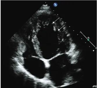
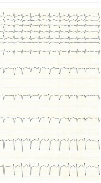
Recently, her functional class was in New York Heart Association functional grade I, routine two-dimensional Doppler echocardiography confirmed reduced LV sys- tolic function with ejection fraction of approximately 30–35% and grade 2 diastolic dysfunction (E/E′ proved to be 14.3) without significant valvular regurgitation or stenosis (Figs. 6.1 and 6.2). Wall motion analysis confirmed diffuse LV hypokinesis with inferior basal and midventricular akinesis (wall motion score index = 2.24), The right ventricular function proved to be normal. Electrocardiography (ECG) did not confirm significant arrhythmia (Fig. 6.3).
Fig. 6.1 Routine 2-dimensional echocardiography demonstrating
hypertrabecularization of the LV apex with intertrabecular recesses (see white arrows)
Fig. 6.2 Colour Doppler echocardiographic images demonstrating intertrabecular recesses (see white arrows)
Fig. 6.3 Routine electrocardiography demonstrating normofrequent sinus rhythm without signifi- cant arrhythmia or conductance disorder
Prevalence of Noncompaction Cardiomyopathy
The true prevalence of NCCM in the general population would be determined based on population-based echocardiographic screening. However, there are no population- based studies to determine the real prevalence of NCCM [1]. The first prospective and large echocardiography-based study to determine the real preva- lence of NCCM has just been published [1]. This prospective case-control study included all subjects who underwent a transthoracic echocardiogram in an aca- demic center within one-year [1]. From 10,857 echocardiograms performed, 26 cases of NCCM were detected giving an estimated prevalence of 0.24% for this cohort [1]. Most patients (77%) had ≤50% LV ejection fraction in higher ages.
When cases with dilated cardiomyopathies of different aetiologies were analysed, 6.8% of the cases showed NCCM with EF <50%. When only those patients with idiopathic dilated cardiomyopathy were included, the estimated prevalence of NCCM was 24%. When LV noncompaction was detected only incidentally in an otherwise healthy and asymptomatic population with normal ejection fraction, the prevalence was 0.05% [1]. In a recent study by Ivanov et al., a cohort of 700 patients referred for cMRI underwent diagnostic assessment for NCCM by four separate imaging criteria referenced by their authors as Petersen, Stacey, Jacquier, and Captur, with a NCCM prevalence of 39%, 23%, 25% and 3%, respectively [9].
In the childhood, large paediatric studies have found NCCM to be the most com- mon form of unclassified cardiomyopathies [10, 11].
Prevalence of Thromboembolic Events in Noncompaction Cardiomyopathy
There are conflicting results about the prevalence of the most feared NCCM- related complications, the thromboembolic events in NCCM, but these complica- tions seem to be not uncommon in NCCM. Cardiac emboli are theorized to result from thrombus formation within the intertrabecular recesses, but reduced sys- tolic function predispose to thrombosis as well. The higher risk of arrhythmias, including atrial fibrillation (AF) is also considerable source of the evolution of thromboembolism in NCCM [12]. The other important risk factors for thrombo- embolic events are previous thromboembolic events, hypertension, coagulopa- thy, etc. [13, 14]. According to the literature, stroke and embolism occur in at least 15% of patients with NCCM due to above mentioned reasons [14]. However, it should also be considered that in cases of NCCM with AF, thrombi may not only be derived from the intertrabecular spaces but also from the left atrium or left atrial appendage [15]. The overall thromboembolic annualized event rate is not different as compared to heart failure patients or subjects with other cardio- myopathies [16].
Anticoagulant Treatment for Stroke Prevention
Anticoagulant Treatment for Stroke Prevention in General Cardiac Patients
Oral anticoagulant therapy (OAC) prevents the majority of ischaemic strokes in patient with AF and is confirmed to prolong life [17]. Men with a CHA2DS2-VASc of 2 or more and women with a CHA2DS2-VASc score of 3 or more are suggested to benefit from OAC. Therefore, OAC should be considered from a CHA2DS2-VASc of 1 in men and 2 in women considering the potential adverse events, including bleeding risks and the patients’ preference. Vitamin K antagonists (VKA), the first anticoagulants used in patients with AF were confirmed to reduce the risk of stroke by two-thirds and to reduce mortality by 25% compared with control. VKA therapy is limited by the narrow therapeutic range of VKAs, the need for close monitoring and potential dose adjustments. Non-vitamin K type oral anticoagulants (NOACs), including the direct thrombin inhibitor dabigatran and the factor Xa inhibitors, apix- aban, edoxaban and rivaroxaban, are suitable alternative treatment options to VKAs for stroke prevention in patients with AF. NOACs have a predictable effect (onset and offset), no regular laboratory test is required to test the efficacy of NOACs.
NOACs are recommended in preference to VKAs or acetylsalicylic acid in AF patients with a previous stroke [17].
In patients with heart failure with reduced ejection fraction (HFrEF) or preserved EF (HFpEF) however, there is no evidence that the use of warfarin reduces mortality and morbidity as compared to placebo or acetylsalicylic acid [18]. Therefore, new studies with NOACs in patients with HFrEF are ongoing.
Anticoagulation Therapy for Stroke Prevention in Noncompaction Cardiomyopathy Patients
It is generally recommended to those presenting with antecedent of systemic embo- lism, presence of cardiac thrombus and AF and ventricular systolic dysfunction [19].
Unfortunately, due to its rarity, evidence-based recommendations for preventing thromboembolic events in isolated NCCM have not been established. It is still not clear under which conditions NCCM patients should be clearly anticoagulated for primary prophylaxis of stroke or embolism. According to the latest literature on how to use anticoagulants in LV noncompaction, the recommendations are the following:
• If NCCM is associated with AF, the usefulness of anticoagulants seem to be reasonable by reducing thromboembolic rate including vitamin K-antagonists (VKA) and new oral anticoagulants (NOAC) [15, 20].
• If NCCM is associated with a history of previous stroke and embolism, VKA or NOAC should be considered [15, 20].
• If NCCM is associated with systolic dysfunction and sinus rhythm, no significant overall difference could be demonstrated in the primary outcome between treat- ment with VKA and high dose acetylsalicylic acid [21]. Anticoagulant treatment in these patients without history of previous thromboembolism is still on debate.
Given the deep intertrabecular recesses and probably slurred local slow flows, historically the NCCM patients has been always considered as high-risk patients for thromboembolic events. Therefore, till we have more clear and specific evi- dence, we advise in general anticoagulation therapy in NCCM patients with sig- nificant LV dysfunction (i.e. empirically LVEF <40%) and/or heart failure.
To understand the potential indications of anticoagulation therapy in noncom- paction cardiomyopathy, here some cases are presented.
Case 2: A Patient with Noncompaction Cardiomyopathy with Atrial Fibrillation
A case of a 35-year-old male patient is presented. His medical history started with pal- pitations at the age of 32, when electrocardiography (ECG) showed sinus rhythm.
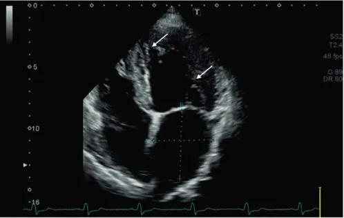
Routine transthoracic Doppler echocardiography revealed mildly enlarged LV and atria without significant valvular alterations. LV ejection fraction was 60%, grade 2 diastolic dysfunction could be confirmed with normal RV sizes and function. With Doppler echo- cardiography, deeply perfused intertrabecular recesses could be demonstrated suggest- ing NCCM (Fig. 6.4). The patients did not show any signs of heart failure. But Holter ECG showed paroxysmal atrial fibrillation (Fig. 6.5). Therefore, oral anticoagulation was initiated.
Fig. 6.4 Routine 2-dimensional echocardiography of a 35-year-old male demonstrating hypertra- becularization of the LV apex with intertrabecular recesses (see white arrows)
Case 3: A Patient with Noncompaction Cardiomyopathy with Systolic Dysfunction
A 58-year-old male presented with shortness of breath on exertion and atypical chest pain. His past medical history consisted of mild mitral regurgitation, hyperlip- idemia, and osteoporosis. His present symptoms started 1 month ago, with chest X-ray showing cardiomegaly. ECG showed sinus rhythm with normal heart rate (Fig. 6.6). Two-dimensional transthoracic echocardiography revealed signs of LV hypertrophy with diffuse myocardial lesion and reduced LV function (ejection frac- tion: 37%) could be demonstrated by. But, detailed echocardiographic evaluation of the thickened myocardial walls showed hypertrabecularization of the LV apex and
Fig. 6.5 Routine electrocardiography demonstrating high frequency atrial fibrillation
Fig. 6.6 Routine 2-dimensional echocardiography demonstrating hypertrabecularization of the LV apex with intertrabecular recesses
Fig. 6.7 Routine electrocardiography demonstrating normofrequent sinus rhythm without signifi- cant arrhythmia or conductance disorder
septum rather than LV hypertrophy. Colour Doppler echocardiography confirmed intertrabecular recesses, confirming the diagnosis of NCCM (Fig. 6.7). Given the significant LV dysfunction with a LVEF <40%, we prescribed empirically oral anti- coagulation along with ACE-inibitor and beta-blocker.
Conclusions
Thromboembolic events are relatively common in NCCM patients. Unfortunately, due to its rarity and relatively young disease entity, evidence-based recommenda- tions for preventing thromboembolic events in NCCM have not been established. In patients with atrial fibrillation or flutter oral anticoagulation including vitamin K antagonists (VKA) and new oral anticoagulants (NOAC), seem to be reasonable to reduce thromboembolic rate. In case of a history of previous stroke and thromboem- bolism, VKA or NOAC use is according the current clinical insights beyond any doubt. In NCCM patient with an systolic dysfunction, but sinus rhythm, overall benefit of preventive oral anticoagulation is yet to be demonstrated. Further research for evidence-based guidelines are highly needed.
References
1. Ronderos R, Avegliano G, Borelli E, Kuschnir P, Castro F, Sanchez G, Perea G, Corneli M, Zanier MM, Andres S, Aranda A, Conde D, Trivi M. Estimation of prevalence of the left ven- tricular noncompaction among adults. Am J Cardiol. 2016;118:901–5.
2. Udeoji DU, Philip KJ, Morrissey RP, Phan A, Schwarz ER. Left ventricular noncompaction cardiomyopathy: updated review. Ther Adv Cardiovasc Dis. 2013;7:260–73.
3. Ikeda U, Minamisawa M, Koyama J. Isolated left ventricular non-compaction cardiomyopathy in adults. J Cardiol. 2015;65:91–7.
4. Chin TK, Perloff JK, Williams RG, Jue K, Mohrmann R. Isolated noncompaction of left ven- tricular myocardium. A study of eight cases. Circulation. 1990;82:507–13.
5. Jenni R, Oechslin E, Schneider J, Attenhofer Jost C, Kaufmann PA. Echocardiographic and pathoanatomical characteristics of isolated left ventricular non-compaction: a step towards classification as a distinct cardiomyopathy. Heart. 2001;86(6):666–71.
6. Stöllberger C, Finsterer J. Left ventricular hypertrabeculation/noncompaction. J Am Soc Echocardiogr. 2004;17:91–100.
7. Belanger AR, Miller MA, Donthireddi UR, Najovits AJ, Goldman ME. New classification scheme of left ventricular noncompaction and correlation with ventricular performance. Am J Cardiol. 2008;102:92–6.
8. Diwadkar S, Nallamshetty L, Rojas C, Athienitis A, Declue C, Cox C, Patel A, Chae SH. Echocardiography fails to detect left ventricular noncompaction in a cohort of patients with noncompaction on cardiac magnetic resonance imaging. Clin Cardiol. 2017;40:364–9.
9. Ivanov A, Dabiesingh DS, Bhumireddy GP, Mohamed A, Asfour A, Briggs WM, Ho J, Khan SA, Grossman A, Klem I, Sacchi TJ, Heitner JF. Prevalence and prognostic significance of left ventricular noncompaction in patients referred for cardiac magnetic resonance imaging. Circ Cardiovasc Imaging. 2017;10:e006174.
10. Nugent AW, Daubeney PE, Chondros P, Carlin JB, Cheung M, Wilkinson LC, Davis AM, Kahler SG, Chow CW, Wilkinson JL, Weintraub RG, National Australian Childhood Cardiomyopathy Study. The epidemiology of childhood cardiomyopathy in Australia. N Engl J Med. 2003;348:1639–46.
11. Pignatelli RH, McMahon CJ, Dreyer WJ, Denfield SW, Price J, Belmont JW, Craigen WJ, Wu J, El Said H, Bezold LI, Clunie S, Fembach S, Bowles NE, Towbin JA. Clinical characteriza- tion of left ventricular noncompaction in children: a relatively common form of cardiomyopa- thy. Circulation. 2003;108:2672–8.
12. Miyake CY, Kim JJ. Arrhythmias in left ventricular noncompaction. Card Electrophysiol Clin.
2015;7:319–30.
13. Thavendiranathan P, Dahiya A, Phelan D, Desai MY, Tang WH. Isolated left ventricular non- compaction controversies in diagnostic criteria, adverse outcomes and management. Heart.
2013;99:681–9.
14. Stöllberger C, Blazek G, Dobias C, Hanafin A, Wegner C, Finsterer J. Frequency of stroke and embolism in left ventricular hypertrabeculation/noncompaction. Am J Cardiol.
2011;108:1021–3.
15. Finsterer J, Stöllberger C. Primary prophylactic anticoagulation is mandatory if noncompac- tion is associated with atrial fibrillation or heart failure. Int J Cardiol. 2015;184:268–9.
16. Rooms I, Dujardin K, De Sutter J. Non-compaction cardiomyopathy: a genetically and clini- cally heterogeneous disorder. Acta Cardiol. 2015;70:625–31.
17. Kirchhof P, Benussi S, Kotecha D, Ahlsson A, Atar D, Casadei B, Castella M, Diener HC, Heidbuchel H, Hendriks J, Hindricks G, Manolis AS, Oldgren J, Popescu BA, Schotten U, Van Putte B, Vardas P, Agewall S, Camm J, Baron Esquivias G, Budts W, Carerj S, Casselman F, Coca A, De Caterina R, Deftereos S, Dobrev D, Ferro JM, Filippatos G, Fitzsimons D, Gorenek B, Guenoun M, Hohnloser SH, Kolh P, Lip GY, Manolis A, McMurray J, Ponikowski P, Rosenhek R, Ruschitzka F, Savelieva I, Sharma S, Suwalski P, Tamargo JL, Taylor CJ, Van Gelder IC, Voors AA, Windecker S, Zamorano JL, Zeppenfeld K. 2016 ESC Guidelines for the management of atrial fibrillation developed in collaboration with EACTS. Eur Heart J.
2016;37:2893–962.
18. Ponikowski P, Voors AA, Anker SD, Bueno H, Cleland JG, Coats AJ, Falk V, González- Juanatey JR, Harjola VP, Jankowska EA, Jessup M, Linde C, Nihoyannopoulos P, Parissis JT, Pieske B, Riley JP, Rosano GM, Ruilope LM, Ruschitzka F, Rutten FH, van der Meer P, Members A/TF, Reviewers D. 2016 ESC Guidelines for the diagnosis and treatment of acute and chronic heart failure: The Task Force for the diagnosis and treatment of acute and chronic heart failure of the European Society of Cardiology (ESC). Developed with the special contri- bution of the Heart Failure Association (HFA) of the ESC. Eur J Heart Fail. 2016;18:891–975.
19. Tavares de Melo MD, de Araújo Filho JA, Parga Filho JR, de Lima CR, Mady C, Kalil-Filho R, Salemi VM. Noncompaction cardiomyopathy: a substrate for a thromboembolic event. BMC Cardiovasc Disord. 2015;15:7.
20. Erath JW, Hohnloser SH. Anticoagulation in atrial fibrillation: current evidence and guideline recommendations. Herz. 2018;43(1):2–10.
21. Homma S, Thompson JL, Pullicino PM, Levin B, Freudenberger RS, Teerlink JR, Ammon SE, Graham S, Sacco RL, Mann DL, Mohr JP, Massie BM, Labovitz AJ, Anker SD, Lok DJ, Ponikowski P, Estol CJ, Lip GY, Di Tullio MR, Sanford AR, Mejia V, Gabriel AP, del Valle ML, Buchsbaum R; WARCEF Investigators. Warfarin and aspirin in patients with heart failure and sinus rhythm. N Engl J Med. 2012;366:1859–69.